long-term follow-up of primary prophylactic implantable cardioverter
TRANSCRIPT

Clinical researchArrhythmia/electrophysiology
Long-term follow-up of primary prophylactic implantablecardioverter-defibrillator therapy in Brugada syndrome
Andrea Sarkozy1*, Tim Boussy1, Georgios Kourgiannides1, Gian-Battista Chierchia1,Sergio Richter1, Tom De Potter1, Peter Geelen1, Francis Wellens2,Marieke Dingena Spreeuwenberg3, and Pedro Brugada1
1Cardiovascular Research and Teaching Institute, OLV Hospital, Moorselbaan 164, 9300 Aalst, Belgium; 2Cardiovascular andThoracic Surgery Department, Aalst, Belgium; and 3Department of Clinical Epidemiology and Biostatistics, VU MedicalCenter, Amsterdam, Holland
Received 14 February 2006; revised 1 November 2006; accepted 30 November 2006; online publish-ahead-of-print 24 January 2007
Aims To analyse the follow-up data of implantable cardioverter-defibrillator (ICD) therapy in Brugadasyndrome (BS).Methods and results We conducted a retrospective, single centre study of 47 patients (mean age:44.5+ 15 years) with BS, who underwent primary prophylactic ICD implantation. All patients had baselinespontaneous (23 patients) or drug-induced (24 patients) coved type I ECG pattern. All patients were judgedto be at high risk because of syncope (26 patients) and/or a positive family history of sudden death (26patients). During a median follow-up of 47.5 months, seven patients had appropriate shocks. The presenceof spontaneous type I ECG and non-sustained ventricular tachyarrhythmia in the ICD datalog suggested atrend towards shorter appropriate shock-free survival by Kaplan–Meier analysis (P ¼ 0.037 andP ¼ 0.012, respectively). Seventeen patients received inappropriate shocks (IS); eight patients for sinustachycardia; six patients for new onset atrial arrhythmias; and five patients for noise oversensing. Inmulti-variable Cox-regression analysis, new onset atrial fibrillation (AF) and less than 50 years of age wereindependent predictors of significantly shorter IS-free survival (P ¼ 0.04 and P ¼ 0.036, respectively).Conclusion In high-risk patients with BS, primary prophylactic ICD therapy is an effective treatment. Inthis, young and otherwise healthy patient population, the IS rate is high.
KEYWORDSBrugada syndrome;
Sudden death;
ICD therapy
Introduction
Brugada syndrome (BS) is characterized by coved typeST-elevation in the right precordial leads (V1–V3) andincreased risk of sudden death in the absence of structuralheart disease.1 In the setting of BS, general agreementexists that patients presenting with aborted sudden deathhave a high risk of recurrent arrhythmic events and shouldreceive an implantable cardioverter-defibrillator (ICD).2
However, the risk stratification and the best treatmentapproach of patients with the coved type I ECG pattern butwithout previous cardiac arrest is controversial.3 In our pre-vious studies, 8% of this patient population suffered suddendeath or documented ventricular fibrillation (VF) during2 years of follow-up.4 Multivariable analysis identified ahistory of syncope and inducibility of sustained ventriculararrhythmia during electrophysiology (EP) study as predictorsof future sudden death or VF.4 Accordingly, our approach,published recently,3 favours the use of the combination ofclinical characteristics (syncope) and an EP study for riskstratification in this patient population.3 In high-risk patients,
who present with syncope and/or are inducible with EP study,we recommended the prophylactic implantation of an ICD forthe prevention of sudden death.3 Other groups have found amuch lower incidence of events and that inducibility at theEP study did not predict future cardiac events in patientswith BS and no previous cardiac arrest.5,6
Detailed data on cardioverter-defibrillator therapy in BS,with the exception of two small studies of secondary prophy-lactic ICD treatment has not been reported yet.7,8 Thedetailed follow-up of primary prophylactic ICD therapy haspreviously not been investigated. In addition, in this uniquelyyoung and active patient population, inappropriate shock (IS)rates and complications have not been reported yet.
We conducted a retrospective, single centre study tocollect information on appropriate and inappropriateshocks in the long-term follow-up of primary prophylacticICD therapy in BS.
Methods
Patient population
Since 1996, in our centre all patients diagnosed with BS are includedin a database and are followed in a prospective fashion. InNovember 2004, a separate database was initiated for patients
& The European Society of Cardiology 2007. All rights reserved. For Permissions, please e-mail: [email protected]
* Corresponding author. Tel: þ 32 53 72 4439; fax: þ 32 53 72 4185.E-mail address: [email protected]
European Heart Journal (2007) 28, 334–344doi:10.1093/eurheartj/ehl450
Dow
nloaded from https://academ
ic.oup.com/eurheartj/article/28/3/334/2887490 by guest on 14 February 2022

undergoing an ICD implantation with the diagnosis of BS. All patientsincluded until November 2004 in the general database were enteredretrospectively and after November 2004 prospectively in the ICDdatabase. The ICD database for the purpose of this study was lastassessed in October 2005. Patients were included in the currentstudy if they met all of the following inclusion criteria: (i) documen-tation of spontaneous or drug-induced �2 mm coved (type I)Brugada ECG pattern; (ii) no history of aborted sudden cardiacdeath; (iii) ICD implantation and/or follow-up (longer than 3months) in our centre; (iv) positive family history of sudden death(at an age �65 years) and/or history of syncope (at any age) and/or sustained ventricular arrhythmia induced during the EP study.Sixty consecutive patients were screened for inclusion in the analy-sis. Thirteen patients were excluded: three patients had only saddleback type ST-elevation documented; two patients had shorter than3 months follow-up, and eight patients had aborted sudden cardiacdeath prior to the ICD implantation. The remaining 47 patientswere included. The clinical data on 29 of these patients has beenpublished in previous studies.1,4
ECG definitions
BS was diagnosed, if a coved type �2 mm ST-elevation was docu-mented in �1 lead from V1 to V3 either spontaneously or afterclass I anti-arrhythmic drug (AAD) administration. Based on our pre-vious experience, if the ST-elevation was of the coved type, patientswith negative/isoelectric or positive T waves were included(Figure 1) and were classified as having type I ECG pattern. AnECG was classified as type II, if in the presence of �2 mm J pointelevation, .1 mm saddle back type ST-elevation with positive Twave was documented. An ECG was classified as type III, if �1 mmsaddle back or coved type ST-elevation was observed in the rightprecordial leads. The ECG was classified as normal, if the above-mentioned ST abnormalities were absent. In the case of eachpatient, attempts were made to collect and analyse the largestnumber of 12-lead ECGs possible. The collected ECGs were classifiedas baseline and follow-up ECGs depending on whether they weredocumented prior to or after the ICD implantation, respectively.The baseline ECG was classified as type I if prior to the ICDimplantation at least one ECG with coved type I ST-elevation wasdocumented. A patient was classified as having spontaneous type I
ECG, if either at baseline or during follow-up he has had at leastone documented coved type I ECG.
Class I anti-arrhythmic drug test
Class I anti-arrhythmic drug test was performed to unmask thediagnostic ECG pattern at the investigators preference for diag-nostic purposes. Most frequently, intravenous ajmaline 0.7 mg/kgadministered in 5 min, and less often flecainide 2 mg/kg or procai-namide 10 mg/kg given over a 10 min-period were used for thispurpose. The test was considered positive only if coved type I ECGwas documented (Figure 1).
Electrophysiological study
The EP study was performed at the investigators preference for riskstratification purposes. Our routine minimum ventricular arrhythmiainduction protocol included two basic cycle lengths and two extrasystoles, with a minimum coupling interval of 200 ms from onesite in the right ventricle. In ,10% of the patients, the EP studywas performed in other centres with three basic cycle lengthsand/or three extrasystoles and/or two different sites, with couplingintervals down to ventricular refractoriness. The result of theEP study was only considered positive if sustained ventriculararrhythmia (lasting .30 s, accompanied with syncope or requiringintervention for termination) was induced.
ICD implantation and follow-up
Single- or dual-chamber ICDs were implanted depending on theinvestigators preference. The decision was based on the presenceof a history of supraventricular arrhythmias and/or of concomitantpacing indication. The defibrillators were implanted betweenNovember 1991 and January 2005, with 46 of the 47 implants(98%) being performed after February 1996. Implantation wasperformed with a non-thoracotomy transvenous lead system in allpatients. Of the 47 patients, 44 (94%) underwent the implantationprocedure in our centre, performed by four cardiovascular surgeons.Of the 47 first implants, 36 (77%) were performed by one operatorhaving more than 10 years experience in arrhythmia surgery. Withthe exception of the first implant, from 1996, in each patient anattempt was made to introduce the shock lead through the cephalic
Figure 1 Examples of positive ajmaline test results. (A) The baseline 12-lead ECG shows right bundle branch block without significant ST-elevation. Followingintravenous ajmaline administration, coved type ST-elevation with negative Twaves in lead V1–V2 appears. (B) The baseline ECG in lead V2 shows saddle backtype II ST-elevation, which converts to coved type I ST-elevation following ajmaline administration. (C) Baseline type III ST-elevation in lead V1. Note the covedtype I ST-elevation after ajmaline administration in lead V1 with a positive T wave.
Long-term follow-up of primary prophylactic ICD therapy in BS 335
Dow
nloaded from https://academ
ic.oup.com/eurheartj/article/28/3/334/2887490 by guest on 14 February 2022

vein. With the exception of the first patient, all patients receivedthird generation defibrillators capable of recording and storingelectrocardiographic data at the time of episodes with shock. Theprogramming was left to the investigators preference. According toour routine protocol used since 1991, at the time of the implantation,in 41 of the 47 patients (87%), a single VF zone was programmed witha lower detection rate of 180 b.p.m. (19 patients) or between 190and 222 b.p.m. (22 patients). The pacing was programmed to aback-up rate of 35–50 b.p.m. in 45 of the 47 patients (96%).
Follow-up
The patients were followed in the outpatient clinic 1, 3, and 6months after implant, then every 6 months regularly, or in case ofsymptomatic shock therapy. At each visit, a 12-lead ECG wasalso recorded and analysed. The clinical data were collected andanalysed for all events with shocks. All available electrograms(EGM) of appropriate and inappropriate shocks were analysed byat least two investigators independently. In case of disagreement,the tracings were reanalysed until consensus was reached. Theshock was classified as appropriate, if corresponding to the clinicalsymptoms (presyncope, syncope) the analysis of the onset, stability,QRS morphology, and termination of the tachycardia with shocksuggested a ventricular origin. In the absence of electrocardio-graphic storage capacities of the defibrillator (one patient), theshock was called appropriate if it was preceded by syncope.
Statistical analysis
Continuous variables are expressed as mean+ SD or median (inter-quartile ranges). The Fisher’s exact tests were used to comparecategorical values. The two-sided unpaired Student’s t-test wasused to compare continuous variables. For each variable, univariateKaplan–Meier survival analysis was used to investigate for significantdifferences between groups in the time to the first event (appropri-ate or inappropriate shock or lead failure). Subsequently, the vari-ables which showed a P-value ,0.05 in Kaplan–Meier analysiswere entered in the multivariable Cox-regression analysis. To testthe proportional hazard assumption for each variable, an inter-action variable with time was created and included in theCox-regression analysis. Testing the significance of this variableindicated whether the proportional hazard assumption was reason-able. To test the linearity assumption, quadratic terms of continu-ous variables were entered in the Cox regression. A P-value ,0.05was considered significant for the Student’s t-test and for theCox-regression analysis. In case Fisher’s exact or Kaplan–Meieranalysis tests were performed, a P-value �0.01 was consideredsignificant to account for the inflation of the experiment-wisetype I error due to multiple testing.
Results
Patient characteristics
The baseline characteristics of the 47 patients are shown inTable 1. At the time of the implantation, the mean age was44.5+ 15 years (range from 6 to 67 years). At the time ofimplant, 29 patients (62%) were �50 years. The class I anti-arrhythmic drug test was performed in 41 patients (87%). Inthe remaining six patients, the baseline ECG showed obvioustype I coved ECG pattern, thus the test was felt to have noadded diagnostic value and was not performed. According toour protocol, an EP study was performed in all patients withthe exception of a 6-year old child, who developed recur-rent VF during the ajmaline test, requiring resuscitation.During the EP study, 38 of the 46 patients (83%) had induci-ble sustained ventricular arrhythmia. Eight patients (17%)were not inducible, but were judged to be at high risk on
the basis of other risk factors. Four of them had both apositive family history of sudden death and syncope. Twopatients had strongly positive family history of suddendeath and one patient had syncope.
ICD implantation
A dual-chamber ICD was implanted in 11 patients (23%) and asingle-chamber in the remaining 36 patients (77%). At firstimplant, only one patient (2%) had perioperative compli-cation in the form of pocket infection. One patient (2%),who underwent an abdominal pulse generator implantationin 1991, required a subcutaneous patch implantation owingto high defibrillator threshold (DFT).
Follow-up
The median follow-up was 47.5 months (4 years) with arange from 4.5 months to 13 years. Twenty-five percentageof the patients had �28 months and 25% �70 monthsfollow-up. Only one patient was lost to our follow-up after4.5 months, whereas all other patients presented for allthe regular scheduled follow-up visits. None of the patientsdied during follow-up. Twenty patients underwentaltogether 50 repeat surgical procedures. During these pro-cedures, only one patient had pneumothorax as periopera-tive complication. Interestingly, one patient, who had atthe initial implant normal DFT’s, had at the time of thebattery replacement high DFT’s, requiring subcutaneouspatch implantation.
Four patients had recurrent syncope without any episodein the ICD datalog during follow-up. Given the clinicalcircumstances, all four events were considered to be ofvasovagal in origin. Three of the four patients had syncopalepisodes prior to the ICD implant and none of them hadappropriate therapy. The patient, who had no previoussyncope, also had appropriate therapy for ventriculararrhythmia. However, the episode of syncope withouttherapy was preceded by vagal symptoms.
ECG findings
The analysis of a large number of 12-lead ECGs duringfollow-up showed that all 23 patients with baseline type I
Table 1 Baseline clinical characteristics of the 47 study patients
Clinical characteristics Number of patients (%)
Mean age (years) 44.5+ 15Male sex 35 (75%)Resting ECG
Type I 23 (49%)Type II 9 (19%)Type III 6 (13%)Normal 9 (19%)
Positive family history of sudden death 26 (55%)Syncope prior to ICD 26 (55%)Positive family history and syncope 14 (30%)EP study performed 46
Inducible (%) 38 (83%)AF prior to ICD 5 (11%)Cephalic/subclavian shock lead insertiona 21/24
aData available in 45 patients.
336 A. Sarkozy et al.
Dow
nloaded from https://academ
ic.oup.com/eurheartj/article/28/3/334/2887490 by guest on 14 February 2022

coved ECG had intermittent type I pattern and had atleast one non-coved ECG documented during follow-up.Meanwhile, five of the nine patients (56%) with baselinetype II ECG, and one of the six patients (17%) with baselinetype III ECG, had at least one documented type I coved ECGduring follow-up. However, in spite of the frequent ECGexaminations, none of the nine patients with baselinenormal ECG had type I coved ECG during follow-up.
Altogether 29 patients (62%) had spontaneous coved type IECG documented at least on one occasion, either prior orafter the ICD implantation.
Appropriate shocks
Seven patients had 17 episodes of appropriate shocks duringfollow-up (Figure 2A). The mean time to first appropriate
Figure 2 (A) Kaplan–Meier appropriate shock-free survival analysis in the study population. (B) Kaplan–Meier appropriate shock-free survival depending on thepresence of spontaneous type I ECG pattern.
Long-term follow-up of primary prophylactic ICD therapy in BS 337
Dow
nloaded from https://academ
ic.oup.com/eurheartj/article/28/3/334/2887490 by guest on 14 February 2022

therapy was 13 months (ranging from 3 days to 4 years). Allseven patients were male, three of them had a positivefamily history and three of them had an episode ofsyncope prior to the ICD implantation (one patient hadboth positive family history and an episode of syncope)(Table 2). However, two patients had neither familyhistory of sudden death nor syncope. All seven patientshad during the follow-up documented spontaneous covedtype I ECG. In three of them, the ajmaline test was not per-formed because of the obvious baseline type I ECG pattern.Spontaneous type I ECG, the absence of ajmaline test, andnon-sustained ventricular tachyarrhythmia (NSVT) in theICD datalog tended to occur more frequently among patientswho had appropriate shock (P ¼ 0.034, P ¼ 0.035, andP ¼ 0.018, respectively). In Kaplan–Meier analysis, the pre-sence of spontaneous type I ECG (P ¼ 0.037) and NSVT in theICD datalog (P ¼ 0.012) suggested a trend towards shorterappropriate shock survival, whereas the absence of class IAAD test (P ¼ 0.002) predicted significantly shorter appro-priate shock-free survival. Cox-regression analysis wasperformed with the presence of spontaneous type I ECG,NSVT in the ICD datalog, and the absence of class I AADtest as independent variables. However, in the Cox-regression analysis, none of these parameters was a signifi-cant predictor of appropriate shock-free survival.
EGM findings
In six patients, 14 intracardiac and/or shock electrogramswere available for analysis. Five episodes were due to VF infour patients and nine episodes were due to monomorphicventricular tachycardia/flutter in three patients (Figure 3).Six of the nine monomorphic ventricular arrhythmia occurredin two patients under quinidine treatment, either for recur-rent ventricular arrhythmia or for atrial fibrillation (AF).
Inappropriate shocks
Seventeen patients had IS during follow-up (Table 3,Figure 4A). On an average, 2.8 (range 1–8) episodesoccurred per patient. The time to the first IS was13.8+ 20 months (ranging from 1 day to 5.4 years). Inthree patients, the IS occurred in the VT zone, but in onlyone of the three patients had the VT zone lower detectionrate under 180 b.p.m. The remaining 14 patients receivedIS in the VF zone, with lower detection rates �180 b.p.m.The leading cause of IS was sinus tachycardia; eight
patients received IS in 17 episodes of sinus tachycardia.The average rate of sinus tachycardia was 184+ 11 b.p.m.The first IS occurred 4.5+ 5.2 months after the
implantation. The patients with IS due to sinus tachycardiatended to be younger than the rest of the population; meanage 36.4+8 vs. 46.2+ 15 years (P ¼ 0.08), respectively.
The second most frequent reason was supraventriculartachycardia (SVT) in six patients; atrial tachycardia in onepatient; atrial flutter in two patients; and AF in threepatients. Two of the six patients in this group had a dual-chamber device. However, all the shocks occurred inthe VF zone, with an average rate of 197+ 28 b.p.m.(Figure 5). All six patients developed the atrial arrhythmiafollowing the ICD implantation, and therefore were not onany medication at the time of the IS.
Five patients received IS for lead sensing malfunction(noise). One patient received IS for T wave oversensingand one patient for NSVT.
The corrective measures in the sinus tachycardia groupwere re-programming a single VF zone with a lower detec-tion rate from 195 to 200 b.p.m. in seven patients, and inone patient the programming of a fast VT zone between180 and 210 b.p.m. with onset criteria on and a VF zone.210 b.p.m. One patient received beta-blocker therapy inaddition. In the last mean 44.8 months of follow-up, noneof these patients had repeat shock for sinus tachycardia.In three patients of the SVT group, we initiated sotaloland in one patient bisoprolol therapy; two patients under-went isthmus ablation; one patient received an atrial lead.The lower VF zone detection rate was reprogrammed to200 b.p.m. in three patients and in one patient a secondfast VT zone (182–222 b.p.m.) with SVT enhancementcriteria on was also programmed. The mean shock-freefollow-up in these six patients following the correctivemeasures was 37.7 months. Interestingly, none of thepatients who have received sotalol or beta-blocker therapyhad any worsening of ventricular arrhythmias.
The patients with IS tended to be younger than thepatients without; mean age: 39.2+ 11 vs. 47.5+ 16 years(P ¼ 0.06), respectively. Among patients with IS, signifi-cantly more patients were less than 50 years, and signifi-cantly more of them had new onset AF during follow-up(P ¼ 0.006 and P, 0.001, respectively). In the univariateKaplan–Meier analysis, new onset AF and less than 50years of age predicted significantly shorter IS-free survival(P ¼ 0.0005 and P ¼ 0.01, respectively) (Figure 4B).Subsequently, new onset AF and less than 50 years of agewere entered in Cox-regression analysis. In Cox-regressionanalysis, new onset AF and less than 50 years of ageremained significant independent predictors of shorterIS-free survival (P ¼ 0.005 and P ¼ 0.036, respectively).
Lead-related complications
During the follow-up, six patients developed recurrent leadsensing malfunctions (noise/myopotential oversensing)requiring new lead implantation. Five of the six patientspresented with IS, and one with high impedance andpacing threshold. The malfunctioning lead was implantedthrough the cephalic vein in three and through the subcla-vian vein in three patients. The new lead was implantedon an average of 38 months (range 15–88 months) afterthe first implant. The patients with lead malfunction weresignificantly younger at the time of first implantation thanthe patients without; mean age: 32+ 6 vs. 46.4+ 14years (P ¼ 0.02), respectively. Univariate Kaplan–Meier
Table 2 Clinical characteristics of the sevenpatients who had appropriate shocks
Age at implant (years) 50+ 13Male sex 7 patientsBaseline type I 5 patientsAjmaline test not performed 3 patientsSpontaneous type I ECG 7 patientsSyncope 3 patientsPositive family history 3 patientsInducible at EP study 7 patientsNSVT 4 patients
338 A. Sarkozy et al.
Dow
nloaded from https://academ
ic.oup.com/eurheartj/article/28/3/334/2887490 by guest on 14 February 2022

Figure 3 Example of initiation of monomorphic VT in a 64-year-old male patient taking quinidine therapy. The top tracings show the ventricular and the bottom the shock electrograms. (A) Notice the baseline AF with fastventricular rates and the short run of NSVT. This VTwas terminated with the first shock (not shown). (B) Two minutes later another VT episode with a different morphology was initiated. This VTwas also successfully termi-nated by the first shock, which also converted the patient to sinus rhythm (not shown).
Long-termfollow
-upof
prim
aryprop
hylacticICDtherap
yin
BS
339
Dow
nloaded from https://academ
ic.oup.com/eurheartj/article/28/3/334/2887490 by guest on 14 February 2022

analysis revealed a trend towards shorter lead failure-freesurvival in patients less than 50 years of age (P ¼ 0.047).
Discussion
Appropriate shocks
Event ratesDuring a median 4 years follow-up, seven patients experi-enced appropriate shocks for potentially life-threateningventricular arrhythmias after implantation of an ICD forprimary prevention in BS. This discharge rate is comparableto the ones reported in other primary prophylactic ICD trialsof patients with structural or primary electrical diseases. Inhypertrophic cardiomyopathy, annual appropriate dischargerates between 3.3 and 5% have been reported in well-documented large studies.9,10 In the long QT syndrome,the data over primary prophylactic ICD implantation areless clear, as the largest group reported up to date includedonly 10 patients.11–13 Annual appropriate shock ratesfrom 1.9 to 21.5% have been reported in these smallstudies.11–13 In the primary prophylactic SCD-HeFT trial,which included patients with structural heart disease, theannual appropriate discharge rate was 5.1%.14 However,there are major differences between the patient populationwith BS (and other primary electrical diseases) and theSCD-HeFT patient population. The two most importantones are the young age of our patients and the absence ofstructural heart disease. As the mean age of our patientpopulation is 44 years, and because there are no other com-peting risk factors, the life expectancy of our patients witheffectively treated ventricular arrhythmias would be morethan 30 years. If the annual event rate following the implan-tation remains constant in time, this means that all patientswould receive a potentially life-saving appropriate therapyduring their lifetime. Although one of the main difficultiesin BS is to predict the timing of the transition from asympto-matic to symptomatic disease, available data indicates thatit is reasonable to hypothesize a life-long risk of suddendeath in BS.4–6 However, until further long-term follow-upstudies prove this hypothesis, it is desirable to try toimprove our risk stratification strategies to help us identify
the individuals at risk of sudden death, without decreasingthe specificity and losing young patients due to suddendeath.
Predictors of appropriate shocksOur previous studies identified the presence of a baselineabnormal ECG, syncope, and inducibility during the EPstudy as predictors of future arrhythmic events.4 Wheninterpreting the results of the present study, similar to pre-vious studies, we should emphasize that given the low eventrates in a relatively small study population, it is very difficultto draw any definitive conclusions regarding predictors offuture events.
In this study, likely due to the small patient populationand low event rates, we could not confirm the previouslywell established role of syncope as a predictor of futurearrhythmia events.3–6 Interestingly, four patients had recur-rent, most likely vasovagal syncope during follow-up(although the under-detection of spontaneously terminatingventricular arrhythmias cannot be excluded). This findingindicates that some patients had vasovagal syncope priorto the ICD implantation. In these patients, the presence ofsyncope likely falsely suggests an increased risk of suddendeath. However, unfortunately, in this young patient popu-lation, it is not always easy to differentiate if the origin ofthe syncope is benign vasovagal or a potentially fatal, spon-taneously terminating polymorphic ventricular tachycardia.Similarly, inducibility, during the EP study, was also not apredictor of appropriate shocks. However, it should beremembered that from our general BS patient population,all patients who were inducible and has had no previouscardiac arrest were included in the present study, as theyall received an ICD. In the meantime, some patients whowere not inducible but had other high-risk factors such assyncope and/or strongly positive family history were alsoincluded and served as the non-inducible control group.Thus, in our data analysis we compared the induciblepatients with a specific biased control group of non-inducible but otherwise high-risk patient population in thepresence of low follow-up event rates.
The results of the present study suggest a possible role forthe presence of the spontaneous coved type I ECG patternin the prediction of malignant ventricular arrhythmias.However, likely due to our small sample size and lowevent rate, in multivariable analysis, neither the presenceof spontaneous type I ECG nor the absence of a class AADtest remained an independent predictor of future arrhyth-mic events. Our ICD study population was unique, as theregular follow-up allowed the analysis of a large numberof ECGs over a long time period in each patient. Based onthese frequent ECG examinations, we describe for the firsttime, that all patients with spontaneous coved type I ECGhave non-diagnostic ECGs during follow-up. In the mean-time, patients who have baseline repeatedly normal ECGsdo not show conversion to spontaneous type I ECG duringfollow-up. This group did not have appropriate shocks inthe first 5 years after ICD implantation.
In the present study, we also describe a potentially newrisk factor, the presence of NSVT. However, it should beremembered that the NSVT episodes saved in the ICDdatalog, are not identical to the traditional definition ofNSVT. The NSVT episodes in our trial met the rate andinitial detection criteria in the VF zone, but terminated
Table 3 Clinical characteristics of patients whohad inappropriate shocks or other complications
Inappropriate shocks 17 patientsAge at implant (years) 39.2+ 11Male sex 13 patientsSingle-chamber ICD 14 patientsDual-chamber ICD 3 patientsIS for sinus tachycardia 8 patientsIS for SVT 6 patientsIS for noise oversensing 5 patientsIS for NSVT 1 patientIS for Twave oversensing 1 patient
Lead-related complications 6 patientsAge at implant (years) 31.7+ 6Male sex 6 patients
Other ICD complications 4 patientsPulse generator migration 1 patientHigh DFT 2 patientsPneumothorax 1 patient
340 A. Sarkozy et al.
Dow
nloaded from https://academ
ic.oup.com/eurheartj/article/28/3/334/2887490 by guest on 14 February 2022

during the duration criteria or more frequently in the char-ging period. Thus, the non-sustained episodes were fast( �180 b.p.m.) and either long lasting (usually more thaneight consecutive beats) or repetitive (runs of minimumfive beats in the VF zone in close proximity). Furthermore,in multivariable analysis, in the presence of low eventrates, NSVT was no longer a significant predictor. Furtherstudies are needed to clarify the role of NSVT episodes inthe ICD datalog in predicting future arrhythmic events.
Trigger of eventsSurprisingly, and in contrast to a previous secondary prophy-lactic ICD population of five patients,7 in the present study,three of the six patients with available electrograms hadmonomorphic ventricular arrhythmia. Two of these patients
were taking quinidine, one of them for recurrent ventriculararrhythmias and the other patient for AF. The first patienthad VF prior to and monomorphic arrhythmia after theinitiation of quinidine therapy. These findings suggest, first,that patients might have recurrent arrhythmias on quinidinetreatment; secondly, that the anti-arrhythmic medicationsmight influence the arrhythmia presentation and convertpolymorphic to monomorphic ventricular arrhythmias.
Inappropriate shocks
One of the main findings of the present study is the veryhigh IS rate. Previously, no data has been published over ISrates in BS.
Figure 4 (A) Kaplan–Meier IS-free survival analysis in the study population. (B) Kaplan–Meier analysis of IS-free survival depending on age at the time of the ICDimplant.
Long-term follow-up of primary prophylactic ICD therapy in BS 341
Dow
nloaded from https://academ
ic.oup.com/eurheartj/article/28/3/334/2887490 by guest on 14 February 2022

Figure 5 Examples of IS for atrial arrhythmias with fast ventricular rates. On both ICD tracings the top tracings show the atrial, the middle the ventricular, and the bottom tracing the shock electrograms. (A) Initiation of AF,with ventricular rate at 238 b.p.m. during jogging in a 49-year-old male. AF has not been documented in this patient prior to the ICD implantation. (B) Initiation of 1:1 atrial tachycardia with a ventricular rate of 240 b.p.m.in a 32-year-old male patient.
342A.Sarkozy
etal.
Dow
nloaded from https://academ
ic.oup.com/eurheartj/article/28/3/334/2887490 by guest on 14 February 2022

Given the young age and active lifestyle of our patients,many of them engaging in regular sport activities, it is notsurprising that the leading cause of IS was sinus tachycardiaat rates .180 b.p.m. In addition, patients with BS, in con-trast with the long QT and hypertrophic cardiomyopathyICD populations are less likely to receive beta-blockers dueto the previously described worsening effect of thesedrugs on the ST-elevation pattern.2 In most cases of IS forsinus tachycardia, the reprogramming of higher lower detec-tion rates was safe and prevented recurrent shocks.The second most frequent reason for IS was atrial arrhyth-
mia with fast ventricular rates. It is well documented thatpatients with BS have a higher risk of atrial arrhythmias.15,16
In our study population, 12 patients were diagnosed withatrial arrhythmia. Interestingly, seven patients developednew atrial arrhythmia during follow-up. Given the absenceof rate control medications, six of these patients presentedwith IS. The combination of the reprogramming of higherlower detection rates, initiation of anti-arrhythmic treat-ment (medical or catheter ablative), and/or upgrade to adual-chamber system prevented in most cases the recur-rence of IS. Interestingly, sotalol or other beta-blockertherapy did not worsen ventricular arrhythmias in anypatient in our study population.Accordingly, based on the results of this study, we have
changed our routine programming protocol. Currently, inpatients younger than 50 years, we program a single VFzone with a lower detection rate �200 b.p.m. In thepresence of increased risk of fast atrial arrhythmias,anti-arrhythmic drug therapy, or documented ventriculartachycardia, the programming of a fast VT zone between180 and up to 220 b.p.m. with SVT enhancement criteria(in single-chamber devices the onset criteria) is a reasonablealternative.Another important reason for IS was the oversensing of
noise in five patients. During our follow-up, altogether sixpatients required a new lead implantation because of thesensing or pacing malfunction of the lead. Manifest fracturewas diagnosed in two patients, the remaining patients hadlikely insulation break or undiagnosed lead fracture. Thepatients with lead-related problems were younger andwere all males, suggesting that the age and the active life-style of the patients was an important aetiological factor.
Limitations
The major limitations of our study are the small number ofpatients, the low event rates, and its retrospectivenature. Furthermore, the follow-up of 4.5 years, in patientswith life-long risk of arrhythmias, should still be consideredshort. Accordingly, all of our results, especially regardingthe predictors of appropriate events should be interpretedwith great caution. In addition, the classification of appro-priate and inappropriate therapies in single-chamberdevices is sometimes difficult. This might have lead to theunderestimation of appropriate shock rates. Finally, it canbe argued that our initial routine lower VF zone detectionrate programming of 180 b.p.m. was rather conservativeand does not mirror the current clinical practice. However,it should be noted that many of the patients were implantedbetween 1992 and 2000, when little was known about thearrhythmia characteristics of patients with BS, especiallyin the presence of anti-arrhythmic drug therapy.
Conclusions
In high-risk patients with BS without previous cardiac arrest,ICD therapy is an effective (seven patients need to beimplanted with an ICD to treat one potentially lethalarrhythmia) and safe treatment. In this young and otherwisehealthy patient population, the IS rate is very high. Theinappropriate shocks are frequently due to sinus tachycardiaor new onset atrial arrhythmias with fast ventricular rates.Careful programming of the device is necessary to avoidthese complications, especially in young (,50 years ofage) patients. Lead-related problems are frequent in thispatient population, likely also due to the young age andactive lifestyle of the patients.
Acknowledgements
We thank Professor Walter Paulus and Dirk Van De Ghoor (GuidantInc.) for their support and all the physicians who provided follow-updata on our patients.
Conflict of interest: none declared.
References
1. Brugada P, Brugada J. Right bundle branch block, persistent ST segmentelevation and sudden cardiac death: a distinct clinical and electrocardio-graphic syndrome. J Am Coll Cardiol 1992;20:1391–1396.
2. Antzelevitch C, Brugada P, Borggrefe M, Brugada J, Brugada R, Corrado D,Gussak I, LeMarec H, Nademanee K, Perez Riera AR, Shimizu W,Schulze-Bahr E, Tan H, Wilde A. Brugada syndrome. Report of thesecond consensus conference. Circulation 2005;111:659–670.
3. Brugada P, Priori SG. Controversies in cardiovascular medicine: shouldpatientswith an asymptomatic Brugada electrocardiogramundergo pharma-cological and electrophysiological testing. Circulation 2005;112:279–292.
4. Brugada J, Brugada R, Brugada P. Determinants of sudden cardiac deathin individuals with the electrocardiographic pattern of Brugada syndromeand no previous cardiac arrest. Circulation 2003;108:3092–3096.
5. Eckardt L, Probst V, Smits JP, Bahr ES, Wolpert C, Schimpf R, Wichter T,Boisseau P, Heinecke A, Breithardt G, Borggrefe M, LeMarec H, Bocker D,Wilde AA. Long-term prognosis of individuals with right precordialST-segment-elevation Brugada syndrome. Circulation 2005;111:257–263.
6. Priori S, Napolitano C, Gasparini M, Pappone C, Della Bella P, Giordano U,Bloise R, Giustetto C, De Nardis R, Grillo M, Ronchetti E, Faggiano G,Nastoli J. Natural history of Brugada syndrome. Insights for risk stratifica-tion and management. Circulation 2002;105:1342–1347.
7. Kakishita M, Kurita T, Matsuo K, Taguchi A, Suyama K, Shimizu W,Aihara N, Kamakura S, Yamamoto F, Kobayashi J, Kosakai Y, OheT. Mode of onset of ventricular fibrillation in patients with Brugadasyndrome detected by implantable cardioverter defibrillator therapy.J Am Coll Cardiol 2003;36:1646–1653.
8. Matsuo K, Kurita T, Inagaki M, Kakishita M, Aihara N, Shimizu W, TaguchiA, Suyama K, Kamakura S, Shimomura K. The circadian pattern ofthe development of ventricular fibrillation in patients with Brugadasyndrome. Eur Heart J 1999;20:465–470.
9. Maron BJ, Shen WK, Link MS, Epstein AE, Almquist AK, Daubert JP,Bardy GH, Favale S, Rea RF, Boriani G, Estes NA III, Spirito P. Efficacy ofimplantable cardioverter-defibrillators for the prevention of suddendeath in patients with hypertrophic cardiomyopathy. N Engl J Med2000;342: 365–373.
10. Begley DA, Mohiddin SA, Tripodi D, Winkler JB, Fananapazir L. Efficacy ofimplantable cardioverter defibrillator therapy for primary and secondaryprevention of sudden cardiac death in hypertrophic cardiomyopathy.PACE 2003;26:1887–1896.
11. Groh WJ, Silka MJ, Oliver RP, Halperin BD, McAnulty JH, Kron J. Useof implantable cardioverter-defibrillators in the congenital long QTsyndrome. Am J Cardiol 1996;78:703–706.
12. Goel AK, Berger S, Pelech A, Dhala A. Implantable cardioverter defibril-lator therapy in children with long QT syndrome. Pediatr Cardiol2004;25:370–378.
13. Monnig G, Kobe J, Loher A, Eckardt L, Wedekind H, Scheld HH,Haverkamp W, Milberg P, Breithardt G, Shulze-Bahr E, Bocker D.
Long-term follow-up of primary prophylactic ICD therapy in BS 343
Dow
nloaded from https://academ
ic.oup.com/eurheartj/article/28/3/334/2887490 by guest on 14 February 2022
Implantable cardioverter-defibrillator therapy in patients with congenitallong-QTsyndrome: a long-term follow-up. Heart Rhythm 2005;2:497–504.
14. Bardy GH, Lee KL, Mark DB, Poole JE, Packer DL, Boineau R, Domanski M,Troutman C, Anderson J, Johnson G, McNulty SE, Clapp-Channing N,Davidson-Ray LD, Fraulo ES, Fishbein DP, Luceri RM, Ip JH; SuddenCardiac Death in Heart Failure (SCD-HeFT) Investigators. Amiodarone oran implantable cardioverter-defibrillator for congestive heart failure. NEngl J Med 2005;352:225–237.
15. Morita H, Kusano-Fukushima K, Nagase S, Fujimoto Y, Hisamatsu K,Fujio H, Haraoka K Kobayashi M, Morita KH, Nakamura K, Emori T,Matsubara H, Hina K, Kita T, Fukatani M, Ohe T. Atrial fibrillation andatrial vulnerability in patients with Brugada syndrome. J Am CollCardiol 2002;40:1437–1444.
16. Bordachar P, Reuter S, Garrigue S, Cai X, Hocini M, Jais P, Haissaguerre M,Clementy J. Incidence, clinical implications and prognosis of atrialarrhythmias in Brugada syndrome. Eur Heart J 2004;25:879–884.
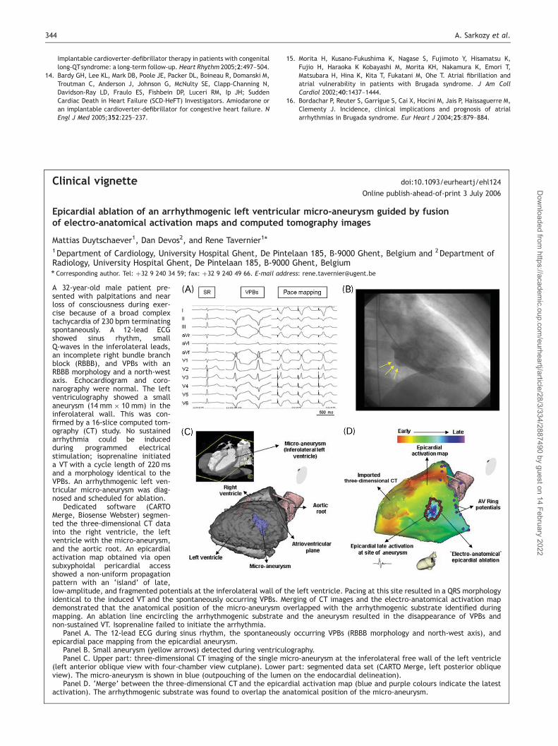
Clinical vignette doi:10.1093/eurheartj/ehl124
Online publish-ahead-of-print 3 July 2006
Epicardial ablation of an arrhythmogenic left ventricular micro-aneurysm guided by fusionof electro-anatomical activation maps and computed tomography images
Mattias Duytschaever1, Dan Devos2, and Rene Tavernier1*1Department of Cardiology, University Hospital Ghent, De Pintelaan 185, B-9000 Ghent, Belgium and 2Department ofRadiology, University Hospital Ghent, De Pintelaan 185, B-9000 Ghent, Belgium* Corresponding author. Tel: þ32 9 240 34 59; fax: þ32 9 240 49 66. E-mail address: [email protected]
A 32-year-old male patient pre-sented with palpitations and nearloss of consciousness during exer-cise because of a broad complextachycardia of 230 bpm terminatingspontaneously. A 12-lead ECGshowed sinus rhythm, smallQ-waves in the inferolateral leads,an incomplete right bundle branchblock (RBBB), and VPBs with anRBBB morphology and a north-westaxis. Echocardiogram and coro-narography were normal. The leftventriculography showed a smallaneurysm (14 mm � 10 mm) in theinferolateral wall. This was con-firmed by a 16-slice computed tom-ography (CT) study. No sustainedarrhythmia could be inducedduring programmed electricalstimulation; isoprenaline initiateda VT with a cycle length of 220 msand a morphology identical to theVPBs. An arrhythmogenic left ven-tricular micro-aneurysm was diag-nosed and scheduled for ablation.
Dedicated software (CARTOMerge, Biosense Webster) segmen-ted the three-dimensional CT datainto the right ventricle, the leftventricle with the micro-aneurysm,and the aortic root. An epicardialactivation map obtained via opensubxyphoidal pericardial accessshowed a non-uniform propagationpattern with an ‘island’ of late,low-amplitude, and fragmented potentials at the inferolateral wall of the left ventricle. Pacing at this site resulted in a QRS morphologyidentical to the induced VT and the spontaneously occurring VPBs. Merging of CT images and the electro-anatomical activation mapdemonstrated that the anatomical position of the micro-aneurysm overlapped with the arrhythmogenic substrate identified duringmapping. An ablation line encircling the arrhythmogenic substrate and the aneurysm resulted in the disappearance of VPBs andnon-sustained VT. Isoprenaline failed to initiate the arrhythmia.
Panel A. The 12-lead ECG during sinus rhythm, the spontaneously occurring VPBs (RBBB morphology and north-west axis), andepicardial pace mapping from the epicardial aneurysm.
Panel B. Small aneurysm (yellow arrows) detected during ventriculography.Panel C. Upper part: three-dimensional CT imaging of the single micro-aneurysm at the inferolateral free wall of the left ventricle
(left anterior oblique view with four-chamber view cutplane). Lower part: segmented data set (CARTO Merge, left posterior obliqueview). The micro-aneurysm is shown in blue (outpouching of the lumen on the endocardial delineation).
Panel D. ‘Merge’ between the three-dimensional CT and the epicardial activation map (blue and purple colours indicate the latestactivation). The arrhythmogenic substrate was found to overlap the anatomical position of the micro-aneurysm.
344 A. Sarkozy et al.
Dow
nloaded from https://academ
ic.oup.com/eurheartj/article/28/3/334/2887490 by guest on 14 February 2022