lopez_and_pereira 2004 larval development of thamnocephalus
DESCRIPTION
Se describe en desarrollo larval del anostraceo Thamnocephalus venezuelensis y se describen las caracteristicas biométricas de sus huevos de resistencia dentro del escenario de potencial uso en acuicultura o acuarofiliaTRANSCRIPT
7/17/2019 Lopez_and_Pereira 2004 Larval Development of Thamnocephalus
http://slidepdf.com/reader/full/lopezandpereira-2004-larval-development-of-thamnocephalus 1/10
Nauplius 12(1), 11-20, 2004
N a u p l i u
s
11
Larval development and biometry of cysts in Thamnocephalus
venezuelensis Belk & Pereira, 1982 (Anostraca)
López, B.1, 2 and Pereira, G.1
1 Instituto de Zoología Tropical, Universidad Central de Venezuela, Apartado 47058, Caracas 1041-A / (2) Centro de
Ecología, Instituto Venezolano de Investigaciones Científicas, Apartado 21827, Caracas 1020-A, Venezuela. e-mail:
[email protected] Beatriz López, Centro de Ecología, Instituto Venezolano de Investigaciones Científicas, Apartado 21827, Caracas 1020-A,
Venezuela. e-mail: [email protected]. Corresponding author
Abstract
The species Thamnocephalus venezuelensis is a native Venezuelan fairy shrimp that inhabits temporary ponds in semiarid zone in the north-western of the country. The complete larval development under
laboratory conditions is described and illustrated; additionally the biometry of resting eggs (cysts) is
characterized. Untreated and decapsulated cysts showed a mean diameter of 355 µm and 313 µm
respectively. The first larval stage is a nauplius typical of the group that presented a mean length of 510
µm and 0.98 mg/egg of mean weight. These values have been the largest found in anostracan species
until present. Larval development started with nauplii that gradually go through successive molts to
reach the adult size. Females start producing eggs near twentieth day of development. Fairy shrimps
have been considered for use as live food in larval fish rearing; therefore this information is of outstanding
importance due to the potential value of T. venezuelensis for practical use in aquaculture or another practical
purpose.Key words: larval development, Thamnocephalus, Anostraca, fairy shrimps, resting eggs, biometry.
Introduction
The species of cosmopolitan genus Artemia (Anostraca), widely known as brine shrimps, represent
a suitable food for many group of organisms of the animal kingdom, from invertebrates to fishes
(Sorgeloos, 1980). As a consequence, an increasing interest in mass culturing of freshwater anostracan
(fairy shrimps) for applied purpose took place in the last decades. Several fairy shrimps species have been
used for different practical purpose, for instance: The species Thamnocephalus platyurus Packard, 1877 and
Streptocephalus sudanicus Daday de Dées, 1910 were used as test organisms in aquatic toxicology (Van
Sprang and Janssen, 1997; Lahr et al. 2001). The species Streptocephalus macrourus Daday, 1908 S. proboscideus
(Frauenfeld), 1873 and S . dichotomus Baird, 1860 were employed as filter feeding stage in waste reclamation
systems (Mitchell, 1991; Ali and Brendonck, 1995; Munuswamy et al . 1997). Several species of the genera
Chirocephalus , Branchipus , Branchinella and Tanymastix, have been tested as live food in freshwater aquaculture
in Italia (Mura, 1992), also the species S. proboscideus and S. dichotomus in India (Munuswamy et al. 1992;
Prasath et al. 1994).
The fairy shrimp Thamnocephalus venezuelensis Belk and Pereira 1982, is the only species of this genus
found in South America (Moore and Young, 1964); there is another record of this genus for Argentina,
but the species has not been properly described yet (Cohen et al. 1999).
Thamnocephalus venezuelensis inhabits freshwater bodies of temporal nature in the semiarid region of
Venezuela that includes Zulia, Falcón and Lara states in the northwest of the country. The taxonomicdescription is all we known about T. venezuelensis , remaining unknown other biological and ecological
aspects. Therefore, the purpose of this paper is to describe the complete larval development of T.
venezuelensis and characterize the biometry of the resting eggs (cysts), due mainly to its potential value as
live food in aquaculture.
7/17/2019 Lopez_and_Pereira 2004 Larval Development of Thamnocephalus
http://slidepdf.com/reader/full/lopezandpereira-2004-larval-development-of-thamnocephalus 2/10
López, B. and Pereira, G. “Larval development in anostraca”
N a u p l i u s
12
Materials and Methods
The samples were collected in temporary ponds in the area of Cerro Blanco (10º 8’ 26”N; 69º 33’
53”W) in Lara state. T. venezuelensis is found typically in highly turbid pools devoid of any floating or
submerged vegetation, accompanied by another anostracan species, Dendrocephalus spartaenovae Margalef 1961 (unpublished data).
Adult females were collected with a hand-net of 1 mm mesh sieve during the rain season in 1996,
and carried to the laboratory for egg collection and storage. The cysts were incubated during two weeks
in water from the natural habitat; afterwards they were cleaned removing the debris by filtration and
washing them with distilled water; and dried to constant weight at 25°C and stored in darkness.
For biometric data the mean diameter of 250 randomly selected cysts were measured, in both
hydrated and decapsulated cysts. Decapsulation was performed according to Bulkowski and Meade (1986).
The following aspects were considered: outer diameter of hydrated cysts (1 hour in distilled water),
decapsulated diameter, chorion thickness and instar I naupliar length. Chorion thickness was determined
by the difference between outer and decapsulated diameter. Cysts and nauplii were observed andphotographed using Scanning Electron Micrography (SEM). The mean weight was estimated by weighting
five samples of 100 randomly selected cysts.
For hatching, a sample of 600 cysts was placed in a 1000 ml beaker with artificial freshwater,
prepared following Peltiers and Weber (1985) methodology. The culture medium was previously aerated
until saturation, under constant light (4000 Lux) and room temperature (25±2°C). After hatching, a
sample of 10 nauplii was collected every hour for 12 hours, and then every 6 hours for the subsequent 5
days; finally, every 12 hours for the next 21 days. All samples were stored in formalin 5%. The description
of the larval development is based in changes that occur through time following the indications given by
Bernice (1972) and Pereira and González (1994). Each larva was measured (total length) with an ocular
micrometer. Drawings of whole larvae were made with the aid of a drawing tube mounted on a binocular
microscope. The larvae of fairy shrimps were kept under artificial light and fed daily with a mixture of
the algae Selenastrum capricornutum (Printz) and Scenedesmus sp. (Chlorophyceae) grown in the laboratory
according to Wayne-Nichols (1975). The water was aerated by means of an electric pump and replaced
every two days.
Results
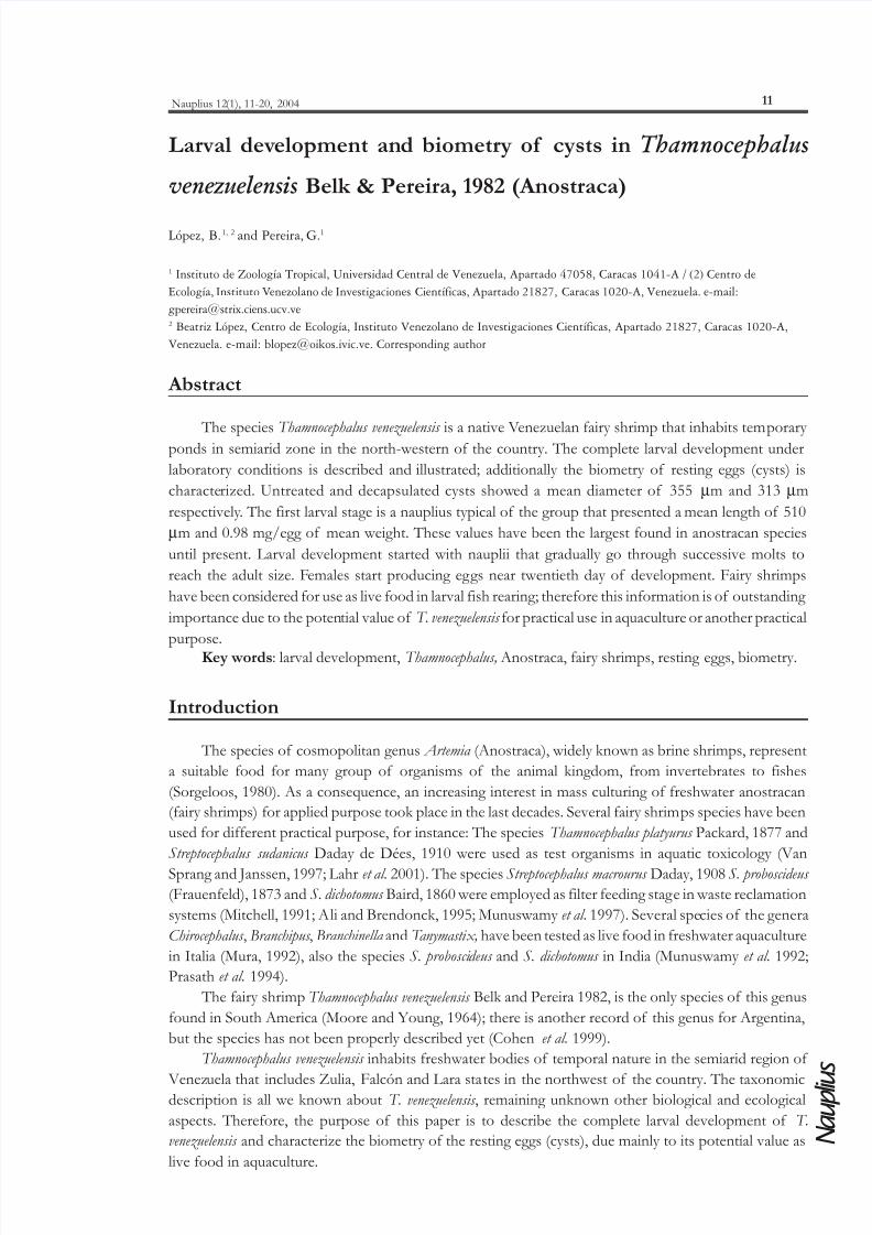
The eggs have a polyhedral shape showing a surface of rough aspect, ornamented with borders
that form polygonal surfaces (Figure 1A). Results of the biometrical characteristics for the cysts and
nauplii are shown in the Table I. The cysts present a mean diameter (hydrated) of 355 µm and a mean
weight (desiccated) of 0.98 mg/egg. Hatching occurs 12 hours after incubation in two phases: Pre-eclosion and eclosion. During the pre-eclosion the egg is hydrated until the eggshell is broken, while the
nauplius remains in the eclosion membrane. Subsequently, the nauplius breaks the membrane of eclosion
and starts the free swim.
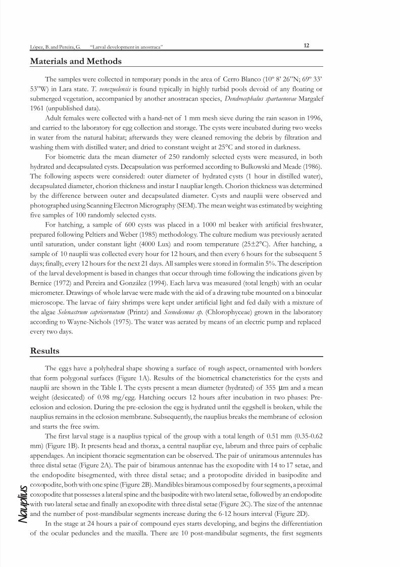
The first larval stage is a nauplius typical of the group with a total length of 0.51 mm (0.35-0.62
mm) (Figure 1B). It presents head and thorax, a central naupliar eye, labrum and three pairs of cephalic
appendages. An incipient thoracic segmentation can be observed. The pair of uniramous antennules has
three distal setae (Figure 2A). The pair of biramous antennae has the exopodite with 14 to 17 setae, and
the endopodite bisegmented, with three distal setae; and a protopodite divided in basipodite and
coxopodite, both with one spine (Figure 2B). Mandibles biramous composed by four segments, a proximal
coxopodite that possesses a lateral spine and the basipodite with two lateral setae, followed by an endopodite with two lateral setae and finally an exopodite with three distal setae (Figure 2C). The size of the antennae
and the number of post-mandibular segments increase during the 6-12 hours interval (Figure 2D).
In the stage at 24 hours a pair of compound eyes starts developing, and begins the differentiation
of the ocular peduncles and the maxilla. There are 10 post-mandibular segments, the first segments
7/17/2019 Lopez_and_Pereira 2004 Larval Development of Thamnocephalus
http://slidepdf.com/reader/full/lopezandpereira-2004-larval-development-of-thamnocephalus 3/10
Nauplius 12(1), 11-20, 2004
N a u p l i u
s
13
present exopodite and endopodite. There are two pairs of caudal setae at the posterior end at each side
of the anus. The mean total length of the larvae in this stage is 1.2 mm (1.1-1.2 mm) (Figure 2E).
The stage, 48 hours after hatching, presents pedunculated eyes and the maxillas II and I are more
differentiated. The antennae II are directed anteriorly. There are eleven thoracic segments; of those the
first six have associated appendages. The differentiation of abdominal segments begins. There are between9-11 caudal setae at each side of the anus. The mean total length of this larval stage is 1.23 mm (1.2-1.3
mm) (Figure 2F).
Figure 1: Scanning electron microscopy micrographs of an egg and a nauplius of T. venezuelensis . A. Complete egg; B. Nauplius at zerohours after hatching when the larva begins the free swim.
Table I: Biometrical characteristics of cysts and nauplii of T. venezuelensis.
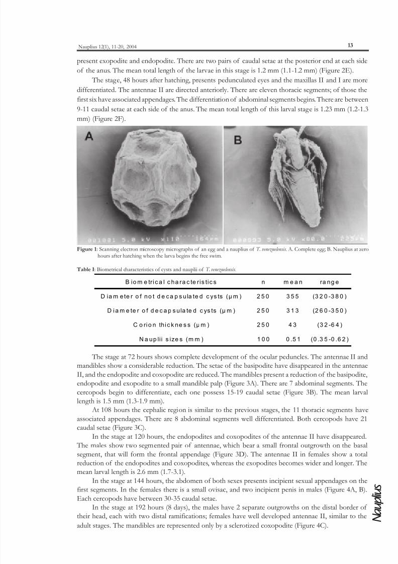
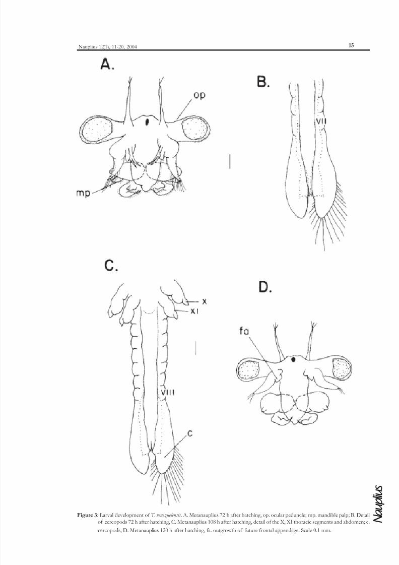
The stage at 72 hours shows complete development of the ocular peduncles. The antennae II and
mandibles show a considerable reduction. The setae of the basipodite have disappeared in the antennae
II, and the endopodite and coxopodite are reduced. The mandibles present a reduction of the basipodite,
endopodite and exopodite to a small mandible palp (Figure 3A). There are 7 abdominal segments. The
cercopods begin to differentiate, each one possess 15-19 caudal setae (Figure 3B). The mean larvallength is 1.5 mm (1.3-1.9 mm).
At 108 hours the cephalic region is similar to the previous stages, the 11 thoracic segments have
associated appendages. There are 8 abdominal segments well differentiated. Both cercopods have 21
caudal setae (Figure 3C).
In the stage at 120 hours, the endopodites and coxopodites of the antennae II have disappeared.
The males show two segmented pair of antennae, which bear a small frontal outgrowth on the basal
segment, that will form the frontal appendage (Figure 3D). The antennae II in females show a total
reduction of the endopodites and coxopodites, whereas the exopodites becomes wider and longer. The
mean larval length is 2.6 mm (1.7-3.1).
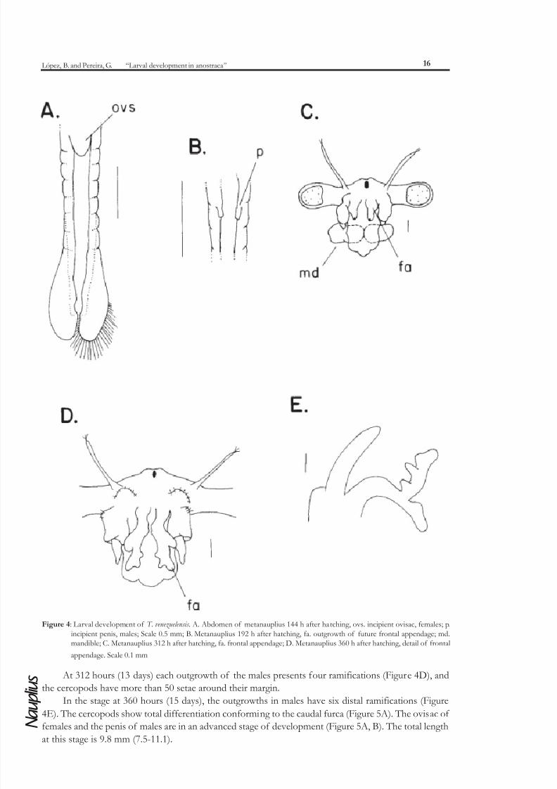
In the stage at 144 hours, the abdomen of both sexes presents incipient sexual appendages on the
first segments. In the females there is a small ovisac, and two incipient penis in males (Figure 4A, B).
Each cercopods have between 30-35 caudal setae.
In the stage at 192 hours (8 days), the males have 2 separate outgrowths on the distal border of
their head, each with two distal ramifications; females have well developed antennae II, similar to the
adult stages. The mandibles are represented only by a sclerotized coxopodite (Figure 4C).
B io m e tr i c a l c ha ra c te ris tic s n m e a n ra ng e
D ia m e te r o f no t d e ca p s u la te d c ys ts (µ m ) 2 5 0 3 5 5 (3 2 0 -3 8 0 )
D ia m e te r o f d e c a p s ula te d c ys ts (µ m ) 2 5 0 3 1 3 (2 6 0 -3 5 0 )
C o rio n th i c k ne s s (µ m ) 2 5 0 4 3 (3 2 -6 4 )
N a up li i s ize s (m m ) 1 0 0 0 .5 1 (0 .3 5 -0 .6 2 )
7/17/2019 Lopez_and_Pereira 2004 Larval Development of Thamnocephalus
http://slidepdf.com/reader/full/lopezandpereira-2004-larval-development-of-thamnocephalus 4/10
López, B. and Pereira, G. “Larval development in anostraca”
N a u p l i u s
14
Figure 2: Larval development of T. venezuelensis . A. First instar nauplius, a1. antennula; a2. antenna; B. Detail of antenna II, ex. exopodite;
en. endopodite; bs. basipodite; cx. coxopodite; C. Detail of mandible; D. Metanauplius 12 h after hatching, lb. labrum; E.Metanauplius 24 h after hatching, ce. compound eyes; mx. maxillas; F. Metanauplius 48 h after hatching, op. ocular penducle;
mx1. first maxilla; mx2. second maxilla. Scale 0.1 mm.
7/17/2019 Lopez_and_Pereira 2004 Larval Development of Thamnocephalus
http://slidepdf.com/reader/full/lopezandpereira-2004-larval-development-of-thamnocephalus 5/10
Nauplius 12(1), 11-20, 2004
N a u p l i u
s
15
Figure 3: Larval development of T. venezuelensis . A. Metanauplius 72 h after hatching, op. ocular peduncle; mp. mandible palp; B. Detail
of cercopods 72 h after hatching, C. Metanauplius 108 h after hatching, detail of the X, XI thoracic segments and abdomen; c.
cercopods; D. Metanauplius 120 h after hatching, fa. outgrowth of future frontal appendage. Scale 0.1 mm.
7/17/2019 Lopez_and_Pereira 2004 Larval Development of Thamnocephalus
http://slidepdf.com/reader/full/lopezandpereira-2004-larval-development-of-thamnocephalus 6/10
López, B. and Pereira, G. “Larval development in anostraca”
N a u p l i u s
16
Figure 4: Larval development of T. venezuelensis . A. Abdomen of metanauplius 144 h after hatching, ovs. incipient ovisac, females; p.
incipient penis, males; Scale 0.5 mm; B. Metanauplius 192 h after hatching, fa. outgrowth of future frontal appendage; md.mandible; C. Metanauplius 312 h after hatching, fa. frontal appendage; D. Metanauplius 360 h after hatching, detail of frontal
appendage. Scale 0.1 mm
At 312 hours (13 days) each outgrowth of the males presents four ramifications (Figure 4D), and
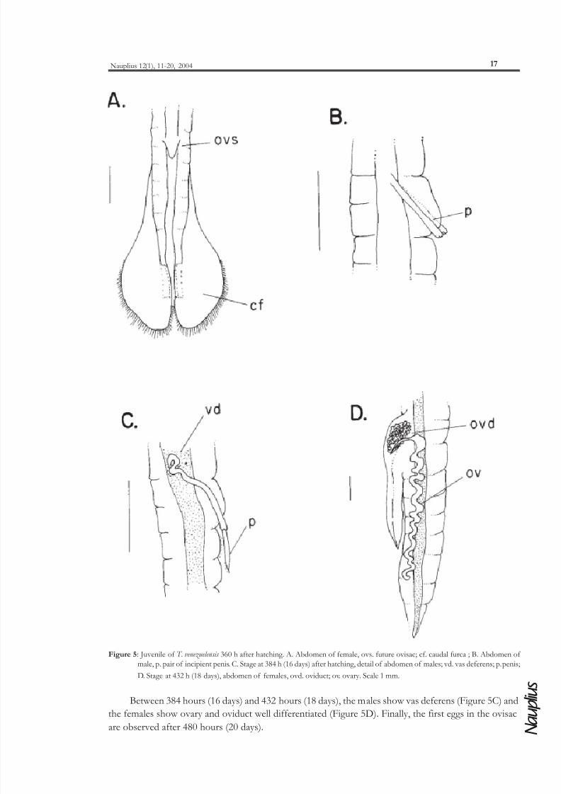
the cercopods have more than 50 setae around their margin.In the stage at 360 hours (15 days), the outgrowths in males have six distal ramifications (Figure
4E). The cercopods show total differentiation conforming to the caudal furca (Figure 5A). The ovisac of
females and the penis of males are in an advanced stage of development (Figure 5A, B). The total length
at this stage is 9.8 mm (7.5-11.1).
7/17/2019 Lopez_and_Pereira 2004 Larval Development of Thamnocephalus
http://slidepdf.com/reader/full/lopezandpereira-2004-larval-development-of-thamnocephalus 7/10
Nauplius 12(1), 11-20, 2004
N a u p l i u
s
17
Figure 5: Juvenile of T. venezuelensis 360 h after hatching. A. Abdomen of female, ovs. future ovisac; cf. caudal furca ; B. Abdomen of
male, p. pair of incipient penis. C. Stage at 384 h (16 days) after hatching, detail of abdomen of males; vd. vas deferens; p. penis;
D. Stage at 432 h (18 days), abdomen of females, ovd. oviduct; ov. ovary. Scale 1 mm.
Between 384 hours (16 days) and 432 hours (18 days), the males show vas deferens (Figure 5C) and
the females show ovary and oviduct well differentiated (Figure 5D). Finally, the first eggs in the ovisac
are observed after 480 hours (20 days).
7/17/2019 Lopez_and_Pereira 2004 Larval Development of Thamnocephalus
http://slidepdf.com/reader/full/lopezandpereira-2004-larval-development-of-thamnocephalus 8/10
López, B. and Pereira, G. “Larval development in anostraca”
N a u p l i u s
18
Discussion
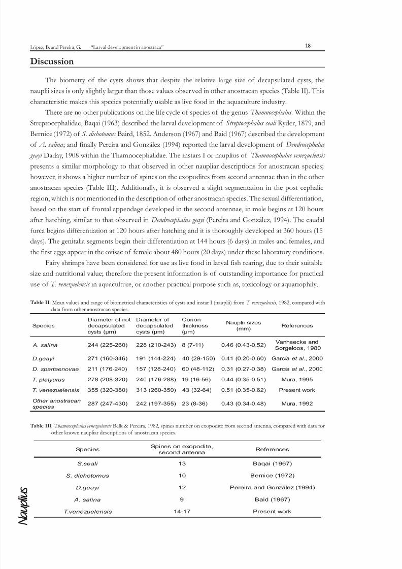
The biometry of the cysts shows that despite the relative large size of decapsulated cysts, the
nauplii sizes is only slightly larger than those values observed in other anostracan species (Table II). This
characteristic makes this species potentially usable as live food in the aquaculture industry.
There are no other publications on the life cycle of species of the genus Thamnocephalus. Within the
Streptocephalidae, Baqai (1963) described the larval development of Streptocephalus seali Ryder, 1879, and
Bernice (1972) of S. dichotomus Baird, 1852. Anderson (1967) and Baid (1967) described the development
of A. salina ; and finally Pereira and González (1994) reported the larval development of Dendrocephalus
geayi Daday, 1908 within the Thamnocephalidae. The instars I or nauplius of Thamnocephalus venezuelensis
presents a similar morphology to that observed in other naupliar descriptions for anostracan species;
however, it shows a higher number of spines on the exopodites from second antennae than in the other
anostracan species (Table III). Additionally, it is observed a slight segmentation in the post cephalic
region, which is not mentioned in the description of other anostracan species. The sexual differentiation,based on the start of frontal appendage developed in the second antennae, in male begins at 120 hours
after hatching, similar to that observed in Dendrocephalus geayi (Pereira and González, 1994). The caudal
furca begins differentiation at 120 hours after hatching and it is thoroughly developed at 360 hours (15
days). The genitalia segments begin their differentiation at 144 hours (6 days) in males and females, and
the first eggs appear in the ovisac of female about 480 hours (20 days) under these laboratory conditions.
Fairy shrimps have been considered for use as live food in larval fish rearing, due to their suitable
size and nutritional value; therefore the present information is of outstanding importance for practical
use of T. venezuelensis in aquaculture, or another practical purpose such as, toxicology or aquariophily.
Table II: Mean values and range of biometrical characteristics of cysts and instar I (nauplii) from T. venezuelensis , 1982, compared withdata from other anostracan species.
Table III: Thamnocephalus venezuelensis Belk & Pereira, 1982 , spines number on exopodite from second antenna, compared with data forother known naupliar descriptions of anostracan species.
seicepS
tonf or etemaiD
detaluspaced
)mµ(stsyc
f or etemaiD
detaluspaced
)mµ(stsyc
noir oC
ssenkciht
)mµ(
sezisiilpuaN
)mm( secner ef eR
ani l as. A )062-522(442 )342-012(822 )11-7(8 )25.0-34.0(64.0 dnaekceahnaV
0891,soolegr oS
i y aeg .D )643-061(172 )422-441(191 )051-92(04 )06.0-02.0(14.0 aícr aG .l at e 0002,
eav oneat r a ps.D )042-671(112 )042-821(751 )211-84(06 )83.0-72.0(13.0 aícr aG .l at e 0002,
sur uy t al p.T )023-802(872 )882-671(042 )65-61(91 )15.0-53.0(44.0 5991,ar uM
si snel euz enev .T )083-023(553 )053-062(313 )46-23(34 )26.0-53.0(15.0 kr owtneser P
nac ar t sonar eht O
sei c e ps )034-742(782 )553-791(242 )63-8(32 )84.0-43.0(34.0 2991,ar uM
seicepS ,etidopoxenosenipS
annetnadnoces secner ef eR
i l aes.S 31 )7691(iaqaB
sumot ohc i d .S 01 )2791(ecinr eB
i y aeg .D 21 )4991(zeláznoGdnaar ier eP
ani l as. A 9 )7691(diaB
si snel euz enev .T 71-41 kr owtneser P
7/17/2019 Lopez_and_Pereira 2004 Larval Development of Thamnocephalus
http://slidepdf.com/reader/full/lopezandpereira-2004-larval-development-of-thamnocephalus 9/10
Nauplius 12(1), 11-20, 2004
N a u p l i u
s
19
Acknowledgements
Funding for this research was provided by Consejo de Desarrollo Científico y Humanístico de la
Universidad Central de Venezuela (CDCH-UCV), through the projects 03.31.3895.97 and 03.31.2763.98.
The results presented in this work are part of the Master Science Thesis of Beatriz López carried out atthe Universidad Central de Venezuela, Facultad de Ciencias, Instituto de Zoología Tropical (IZT). The
authors gratefully acknowledge Dr. Carlos Carmona-Suárez for his comments that improved the
manuscript.
References
Ali, A. J. and Brendonck, L. 1995. Evaluation of agro-industrial wastes as diets for culture of the fairy shrimp Streptocephalus proboscideus (Franuenfeld, 1873) (Crustacea: Branchiopoda: Anostraca).Hydrobiologia, 298: 167-173.
Anderson, D. T. 1967. Larval development and segment formation in the branchiopod crustacean Limnadia
stanleyana King (Conchostraca) and Artemia salina (L)(Anostraca). Australian Journal of Zoology, 15:47-91.
Baid, I. C. 1967. On the development of Artemia salina (Crustacea: Anostraca). Journal of Bombay NaturalHistory Society, 64: 432-439.
Baqai, I. U.1963. Studies on the postembryonic development of the fairy shrimp Streptocephalus seali Ryder. Tulane Studies in Zoology, 10: 1-120
Belk, D. and Pereira, G. 1982. Thamnocephalus venezuelensis , new species (Anostraca, Thamnocephalidae)first report of Thamnocephalus in South America. Journal of Crustacean Biology, 2(2): 223-226.
Bernice, R. 1972. Hatching and post-embryonic development of Streptocephalus dichotomus (Baird) (Crustacea: Anostraca). Hydrobiologia, 40: 251-278.
Bulkowski, L. and Meade, J. W. 1986. Chemical decapsulation of fairy shrimp Streptocephalus seali Ryder(Anostraca). Crustaceana, 51 (2): 212-215.
Cohen, R. G.; Vernet, S.; Corbella, C. and Michelutti, P. 1999. About the presence of halophilic anostracansof the Thamnocephalus genus from Salinas Grandes at NW of Cordoba (Argentina). p. 5-7 In LosOceános y sus Organismos. Physis Sección A. Vol. 57.
García, J.; Marcano, S. and Pereira, G. 2000. Aspectos de la eclosión de los quistes de dos especies deDendrocephalus (Crustacea: Anostraca: Thamnocephalidae) de uso potencial como alimento vivo.Hydrobiologia, 48(1): 145-149.
Lahr, J.; Badji, A.; Marquenie, S.; Schuiling, E.; Ndour, K. B.; Diallo, A. O. and Everts, J. W. 2001. Acutetoxicity of locust insecticides to two indigenous invertebrates from Sahelian temporary ponds.Ecotoxicology and Environmental Safety, 48(1): 66-75.
Mitchell, S. A. 1991. The growth rate and growth efficiency of Streptocephalus macrourus (Crustacea, Anostraca)cultured on micro algae. Hydrobiologia, 212: 1-10.
Moore, W. G. and Young, J. B. 1964. Fairy shrimps of the genus Thamnocephalus (Branchiopoda, Anostraca)in the United States and Mexico. South-western Naturalist, 9: 68-77.
Munuswamy, N.; Mertens, J.; De Walsche and Dumont, H. J. 1992. Lipid classes and fatty acid profiles incrytobiotic cyst of Streptocephalus dichotomus and Streptocephalus proboscideus (Crustacea: Anostraca).Hydrobiologia, 231: 65-68.
Munuswamy, N.; Nazar, A. K. A.; Velu, C. S. and Dumont, H. J. 1997. Culturing the fairy shrimpStreptocephalus dichotomus Baird using livestock waste: a reclamation study. Hydrobiologia, 358(0): 199-203.
Mura, G. 1992. Preliminary testing of Anostraca from Italy for use in freshwater fish culture. Hydrobiologia,241: 185-194.
Mura, G. 1995. Biometrics and fatty acid composition of the resting eggs of Thamnocephalus platyurus (Anostraca) in view of an eventual use as fish feed. Crustaceana, 68 (5): 629-635.
Peltier, W. H. and Weber, C. I. 1985. Methods for measuring the acute toxicity of effluents to freshwaterand marine organisms. Environmental Protection Agency, Cincinnati. U.S.A. 216 p.
Pereira, G. and González, M. 1994. Larval development and population biology of Dendrocephalus geayi Daday, 1908 (Anostraca) in temporary ponds from Venezuela. Crustaceana, 66(2): 163-177.
Prasath, E. B.; Munuswamy, N. and Nazar, A. K. A. 1994. Preliminary studies on the suitability of a fairy shrimp Streptocephalus dichotomus as live food in aquaculture. Journal of the World Aquaculture Society,25(2): 204-207.
7/17/2019 Lopez_and_Pereira 2004 Larval Development of Thamnocephalus
http://slidepdf.com/reader/full/lopezandpereira-2004-larval-development-of-thamnocephalus 10/10
López, B. and Pereira, G. “Larval development in anostraca”
N a u p l i u s
20
Sorgeloos, P. 1980. The use of brine shrimp Artemia in aquaculture. p. 277-290 In Persoone, G., Sorgeloos,P., Roels, O. and Jaspers, E. Ed.s. The Brine Shrimp Artemia . Vol. 3. Ecology, culturing, use in aquaculture.Universa Press, Wetteren, Belgium.
Van Sprang, P. A. and Janssen, C. R. 1997. Identification and confirmation of ammonia toxicity incontaminated sediments using a modified toxicity identification evaluation approach. Environmental
Toxicology and Chemistry, 16(12): 2501-2507. Vanhaecke, P. and Sorgeloos, P. 1980. International study on Artemia . The biometrics of Artemia strains
from different geographical origins. p. 393-405. In Persoone, G., Sorgeloos, P., Roels, O. and Jaspers,E. eds. The Brine Shrimp Artemia . Vol. 3. Ecology, culturing, use in aquaculture. Universa Press, Wetteren,Belgium.
Wayne-Nichols, H. 1975. Growth media-freshwater. p. 7-24. In Stein, J. Ed. Handbook of phycologicalmethods. Culture methods and growth measurements. Cambridge University Press, London, UnitedKingdom.
Received: 10th Jul 2004 Accepted: 11th Dec 2004