loss of ip3 receptor function in neuropeptide secreting neurons leads to obesity in adult drosophila
TRANSCRIPT
RESEARCH ARTICLE Open Access
Loss of IP3 receptor function in neuropeptidesecreting neurons leads to obesity in adultDrosophilaManivannan Subramanian1,2†, Siddharth Jayakumar1,3†, Shlesha Richhariya1 and Gaiti Hasan1*
Abstract
Background: Intracellular calcium signaling regulates a variety of cellular and physiological processes. The inositol1,4,5 trisphosphate receptor (IP3R) is a ligand gated calcium channel present on the membranes of endoplasmicreticular stores. In previous work we have shown that Drosophila mutants for the IP3R (itprku) become unnaturallyobese as adults with excessive storage of lipids on a normal diet. While the phenotype manifests in cells of the fatbody, genetic studies suggest dysregulation of a neurohormonal axis.
Results: We show that knockdown of the IP3R, either in all neurons or in peptidergic neurons alone, mimics knownitpr mutant phenotypes. The peptidergic neuron domain includes, but is not restricted to, the medialneurosecretory cells as well as the stomatogastric nervous system. Conversely, expression of an itpr+ cDNA in thesame set of peptidergic neurons rescues metabolic defects of itprku mutants. Transcript levels of a gene encoding agastric lipase CG5932 (magro), which is known to regulate triacylglyceride storage, can be regulated by itprknockdown and over-expression in peptidergic neurons. Thus, the focus of observed itpr mutant phenotypes ofstarvation resistance, increased body weight, elevated lipid storage and hyperphagia derive primarily frompeptidergic neurons.
Conclusions: The present study shows that itpr function in peptidergic neurons is not only necessary but alsosufficient for maintaining normal lipid metabolism in Drosophila. Our results suggest that intracellular calciumsignaling in peptidergic neurons affects lipid metabolism by both cell autonomous and non-autonomousmechanisms.
Keywords: Calcium, Lipid homeostasis, Hyperphagia, Magro
BackgroundCalcium is a key signaling molecule in multi-cellular or-ganisms that regulates a variety of cellular processes [1,2].The IP3R (Inositol 1,4,5 trisphosphate Receptor) is a ligandgated calcium channel present on the membranes ofendoplasmic reticular (ER) stores. It mediates the releaseof ER calcium upon binding of its cognate ligand IP3. InDrosophila there is a single gene, itpr, for the IP3R whichis 60% homologous to mammalian IP3R1 [3]. Previousstudies have shown that expression of the Drosophila IP3Ris widespread in all tissues and cell types examined [4,5].
However, depending on their allelic strength, itpr mutantsexhibit relatively specific metabolic and neuronal pheno-types. Hetero-allelic combinations of strong itpr mutantsexhibit metabolic defects, altered feeding and transcrip-tional changes in metabolic gene pathways during larvalstages [6,7]. itprka1091 and itprug3 are point mutations inthe modulatory domain (Gly 1891 Ser), and in the ligandbinding (Ser 224 Phe) domain respectively of the IP3R.These mutants are lethal as homozygotes, while theirhetero-allelic combination (itprka1091/ug3 or itprku) is adultviable [8]. Recently, we demonstrated the presence ofmetabolic changes in itprku adult animals leading to star-vation resistance, increased body weight, elevated TAGsand hyperphagia [9].In mammals, disorders like type 2 diabetes, coronary
heart disease, respiratory complications and osteoarthritis
* Correspondence: [email protected]†Equal contributors1National Centre for Biological Sciences, Tata Institute of FundamentalResearch, Bangalore 560065, IndiaFull list of author information is available at the end of the article
© 2013 Subramanian et al.; licensee BioMed Central Ltd. This is an Open Access article distributed under the terms of theCreative Commons Attribution License (http://creativecommons.org/licenses/by/2.0), which permits unrestricted use,distribution, and reproduction in any medium, provided the original work is properly cited. The Creative Commons PublicDomain Dedication waiver (http://creativecommons.org/publicdomain/zero/1.0/) applies to the data made available in thisarticle, unless otherwise stated.
Subramanian et al. BMC Neuroscience 2013, 14:157http://www.biomedcentral.com/1471-2202/14/157

are a result of altered fat metabolism [10]. The complexityof these diseases arises in part from regulation of fatmetabolism through the interaction of signaling path-ways involving multiple tissues and organs. Geneticstudies in model organisms help understand aspects ofthis complexity. In Drosophila, fat metabolism is es-sential for maintaining energy homeostasis. Nutrientfat in the form of Triacylglycerides (TAGs) is brokendown to fatty acids in the mid-gut, absorbed and re-synthesized as TAGs in the fat bodies [11]. Perturba-tions in fat metabolism can lead to changes in TAGlevels and consequent obesity [11,12]. Lipids stored infat body cells are utilized under stress conditions andthe storage and mobilization of lipids to target tissuesis tightly regulated, based on energy requirements.This requires communication between the gut, fatbody cells and oenocytes, the cells analogous to themammalian liver in Drosophila [13]. Furthermore, sig-nals from the brain coordinate feeding behavior as wellas the utilization of stored TAGs, finally affecting thebody weight of an organism [14-16]. Based on theobese and hyperphagic phenotypes of itprku it appearsthat calcium release by the IP3R helps maintain thisaxis of lipid metabolism and feeding in Drosophila.Here we show that peptidergic neurons are an import-ant focus of IP3R function in the context of metaboliccontrol.
ResultsThe IP3R affects Drosophila metabolism through itsfunction in peptidergic neuronsTo identify the tissue focus of itpr mutant phenotypesan itpr RNAi strain (dsitpr) was used to specificallyknockdown the IP3R in all neurons and in the fat body.Pan-neuronal knockdown of the IP3R lead to a signifi-cant level of starvation resistance. In contrast, animalswith knock down of the IP3R in the fat body exhibitedthe same extent of viability post starvation as controlanimals (Figure 1A). Obesity, starvation resistance andhyperphagia in itprka1091/ug3 mutants can be rescued byexpression of an itpr+ cDNA in a subset of peptidergicneurons that secrete the insulin-like peptides (Dilps)amongst other neuropeptides [9,17,18]. These cells aremarked by the Dilp2GAL4 strain. To test for necessity ofthe IP3R in Dilp neurons, dsitpr was driven by Dilp2GAL4.Surprisingly, these animals did not exhibit any starvationresistance (Figure 1A). Consequently, we tested animalswith knockdown of itpr by the dimm GAL4 that expressesin a larger subset of exclusively peptidergic neurons (in-cluding the Dilp neurons) [18]. This resulted in animalswith a significant level of starvation resistance when com-pared with controls (Figure 1A).In order to test if lack of starvation resistance with IP3R
knockdown in Dilp2 neurons, arose due to insufficientknockdown, we expressed a GFP tagged IP3R transgene
Figure 1 itpr levels in peptidergic neurons are critical for the starvation resistance phenotype. (A) Flies with knock down of itpr usingpan-neuronal and dimm drivers show starvation resistance as compared to the wild type whereas itpr knock down in either Dilp2 neurons or fatbodies do not show any starvation resistance. (B) Flies with knock down of itpr in Dilp2 neurons and dimm neurons using UASitpr-GFP showeddecrease in GFP levels when compared to their controls without knockdown, cell bodies shown by arrow heads. Scale bar is 50 μm.(C) Over-expression of itpr+ in both pan-neuronal and peptidergic domains rescues starvation resistance observed in itprku.
Subramanian et al. BMC Neuroscience 2013, 14:157 Page 2 of 8http://www.biomedcentral.com/1471-2202/14/157
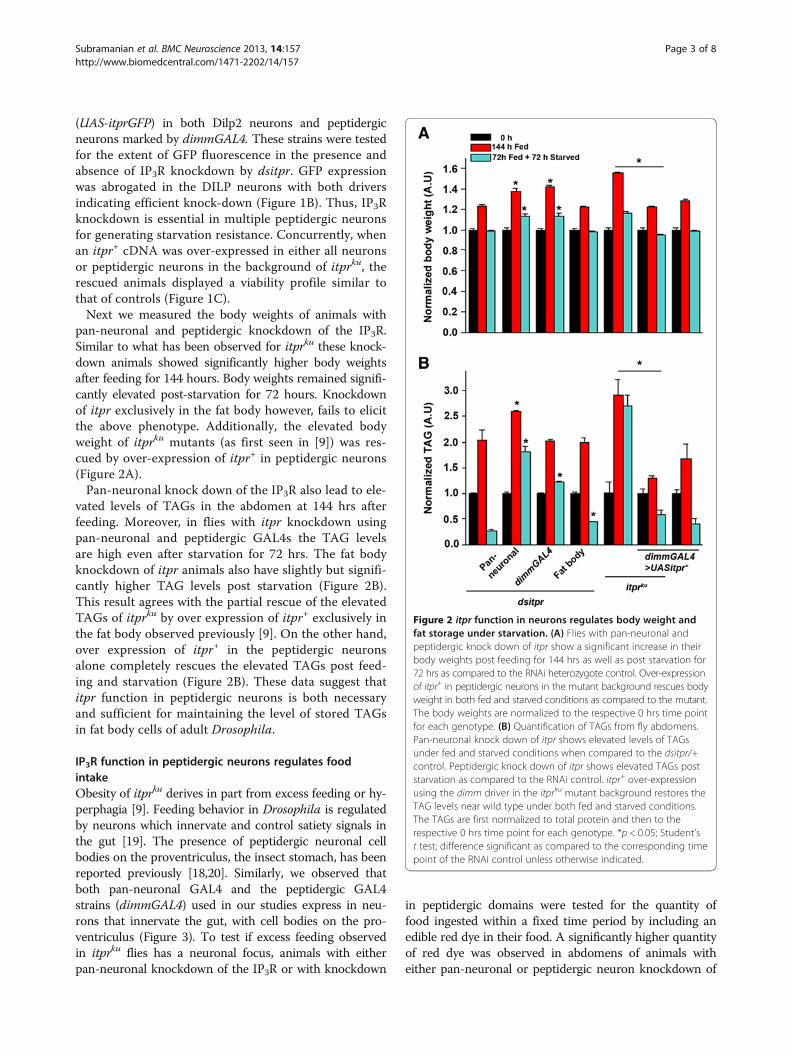
(UAS-itprGFP) in both Dilp2 neurons and peptidergicneurons marked by dimmGAL4. These strains were testedfor the extent of GFP fluorescence in the presence andabsence of IP3R knockdown by dsitpr. GFP expressionwas abrogated in the DILP neurons with both driversindicating efficient knock-down (Figure 1B). Thus, IP3Rknockdown is essential in multiple peptidergic neuronsfor generating starvation resistance. Concurrently, whenan itpr+ cDNA was over-expressed in either all neuronsor peptidergic neurons in the background of itprku, therescued animals displayed a viability profile similar tothat of controls (Figure 1C).Next we measured the body weights of animals with
pan-neuronal and peptidergic knockdown of the IP3R.Similar to what has been observed for itprku these knock-down animals showed significantly higher body weightsafter feeding for 144 hours. Body weights remained signifi-cantly elevated post-starvation for 72 hours. Knockdownof itpr exclusively in the fat body however, fails to elicitthe above phenotype. Additionally, the elevated bodyweight of itprku mutants (as first seen in [9]) was res-cued by over-expression of itpr+ in peptidergic neurons(Figure 2A).Pan-neuronal knock down of the IP3R also lead to ele-
vated levels of TAGs in the abdomen at 144 hrs afterfeeding. Moreover, in flies with itpr knockdown usingpan-neuronal and peptidergic GAL4s the TAG levelsare high even after starvation for 72 hrs. The fat bodyknockdown of itpr animals also have slightly but signifi-cantly higher TAG levels post starvation (Figure 2B).This result agrees with the partial rescue of the elevatedTAGs of itprku by over expression of itpr+ exclusively inthe fat body observed previously [9]. On the other hand,over expression of itpr+ in the peptidergic neuronsalone completely rescues the elevated TAGs post feed-ing and starvation (Figure 2B). These data suggest thatitpr function in peptidergic neurons is both necessaryand sufficient for maintaining the level of stored TAGsin fat body cells of adult Drosophila.
IP3R function in peptidergic neurons regulates foodintakeObesity of itprku derives in part from excess feeding or hy-perphagia [9]. Feeding behavior in Drosophila is regulatedby neurons which innervate and control satiety signals inthe gut [19]. The presence of peptidergic neuronal cellbodies on the proventriculus, the insect stomach, has beenreported previously [18,20]. Similarly, we observed thatboth pan-neuronal GAL4 and the peptidergic GAL4strains (dimmGAL4) used in our studies express in neu-rons that innervate the gut, with cell bodies on the pro-ventriculus (Figure 3). To test if excess feeding observedin itprku flies has a neuronal focus, animals with eitherpan-neuronal knockdown of the IP3R or with knockdown
in peptidergic domains were tested for the quantity offood ingested within a fixed time period by including anedible red dye in their food. A significantly higher quantityof red dye was observed in abdomens of animals witheither pan-neuronal or peptidergic neuron knockdown of
Figure 2 itpr function in neurons regulates body weight andfat storage under starvation. (A) Flies with pan-neuronal andpeptidergic knock down of itpr show a significant increase in theirbody weights post feeding for 144 hrs as well as post starvation for72 hrs as compared to the RNAi heterozygote control. Over-expressionof itpr+ in peptidergic neurons in the mutant background rescues bodyweight in both fed and starved conditions as compared to the mutant.The body weights are normalized to the respective 0 hrs time pointfor each genotype. (B) Quantification of TAGs from fly abdomens.Pan-neuronal knock down of itpr shows elevated levels of TAGsunder fed and starved conditions when compared to the dsitpr/+control. Peptidergic knock down of itpr shows elevated TAGs poststarvation as compared to the RNAi control. itpr+ over-expressionusing the dimm driver in the itprku mutant background restores theTAG levels near wild type under both fed and starved conditions.The TAGs are first normalized to total protein and then to therespective 0 hrs time point for each genotype. *p < 0.05; Student’st test; difference significant as compared to the corresponding timepoint of the RNAi control unless otherwise indicated.
Subramanian et al. BMC Neuroscience 2013, 14:157 Page 3 of 8http://www.biomedcentral.com/1471-2202/14/157

the IP3R (Figure 4A, B). Oil red O staining demonstratedelevated TAGs in the guts of flies with IP3R knockdownindicating greater ingestion and digestion of lipids(Figure 4C). Next we tested if expression of itpr+ inpeptidergic neurons rescued the excess feeding observedin itprku [9]. itpr+ expression in peptidergic neurons ofitprku rescued hyperphagia as evident from the signifi-cantly reduced level of red dye in the abdomens of rescuedanimals compared with control animals (Figure 5A, B).
IP3R function in peptidergic neurons affects transcriptionof a gut lipaseFor obese and hyperphagic animals, excess storage ofTAGs is likely to be accompanied by altered levels oflipid metabolizing enzymes in the gut. Magro, encodedby CG5932, is the Drosophila homolog of mammaliangastric lipase, LipA and is expressed in the anterior partof the gut and released into the intestine to digestTAGs. Levels of Magro have been shown to modulateTAG levels as knocking down Magro shows reductionin TAG levels [21]. itprku mutants have elevated levelsof TAGs as well as elevated levels of magro [9]. Sincepeptidergic knockdown of itpr phenocopies the itprku
mutant, transcript levels of the gastric lipase magrowere measured from isolated guts of appropriate geno-types (Figure 5C). magro expression in guts from flieswith knockdown of the IP3R in peptidergic neuronsappears significantly elevated. Moreover, expression ofitpr+ in peptidergic neurons of itprku animals could res-cue the elevated levels of magro transcripts (Figure 5C).Thus, IP3R mediated signals in peptidergic neurons appearto regulate magro transcription in the gut in a manner thatis not yet understood.
Discussion and conclusionsInsulin-like peptides (ILPs), which are secreted by a subsetof the medial neurosecretory cells in the brain (Figure 1),regulate lipid homeostasis in the fat body cells of adultDrosophila [22,23]. The obese phenotype observed inadult itpr mutants suggested a role for IP3 mediated cal-cium signaling in modulating ILP release and secretion.However, significant differences were observed betweenthe phenotypes of itpr mutant animals rescued by ex-pression of an itpr+ cDNA as compared with rescue byover-expression of Drosophila ILP2, suggesting thatIP3R mutants affect a broader axis of neurohormonalcontrol than the one defined by insulin signaling [9].IP3-mediated calcium signaling and its modulation ofthe neurohormonal axis, leading to obesity, have nowbeen investigated in greater detail. The ILP secretingmedial neurosecretory cells also secrete a number ofother neuropeptides, which regulate stress and metabol-ism in Drosophila [24,25]. While it is possible that theseneuropeptides, in addition to the ILPs, contribute to theitpr mutant phenotype of obesity this seems unlikely be-cause knockdown of the IP3R in neurons which secreteILPs and additional neuropeptides (the DILP neurons)had no effect on starvation resistance and obesity in itprmutants. The peptidergic neurons, defined by dimmGAL4,include the DILP neurons plus other neurosecretorycells in the brain [18]. Because knockdown of the IP3Rin peptidergic neurons phenocopied the itpr mutant,and restoring itpr function specifically in peptidergicneurons rescued mutant phenotypes, we conclude thatIP3R function and Ca2+ release affects lipid metabolismprimarily through regulation of neuropeptide secretion.The composite phenotypes of hyperphagia and obesityin IP3R mutants and peptidergic knockdown animalssuggest a role for these neuropeptides in regulation offeeding and TAG storage and utilisation in the fatbody.Our data support the idea that feeding and lipid mobilisa-tion are regulated by non-overlapping sets of neurosecre-tory cells, possibly comprising the mNSCs which regulateTAG utilisation in the fatbody, and other neuropeptidesecreting cells such as the neurons of the stomatogastricsystem, which regulate satiety and feeding. Moreover, itis likely that feedback mechanisms exist between thesetwo groups of neurons such that knockdown of the IP3Rin one set can be compensated by the other, as in thecase of knockdown in the DILP neurons. A recent studyrevealed the role of drosulfakinin (DSK), a neuropeptideexpressed in the DILP neurons in feeding and satiety[25]. The target cells of DSK are not known and in thelight of our observations, we hypothesize that thesemight be peptidergic neurons of the stomatogastric ner-vous system. Indeed, axonal projections from the mNSCshave been shown to target the proventriculus and midgutin both larval and adult animals [19]. Thus, when IP3R
Figure 3 Peptidergic neurons express in the proventriculus.Neuronal cell bodies were observed on the larval proventriculuswhen mCD8-GFP was driven with both, the pan-neuronal (top) andpeptidergic (bottom) drivers. Scale bar is 50 μm.
Subramanian et al. BMC Neuroscience 2013, 14:157 Page 4 of 8http://www.biomedcentral.com/1471-2202/14/157

knockdown affects both sets of neurons, as in dimmGAL4knockdown and in itpr mutants, it is likely that such feed-back mechanisms are abrogated. As a consequence despitethe existence of sufficient TAG stores in the fatbody theanimals continue to feed excessively, leading to a furtherincrease in TAG deposits and obesity.The altered neurohormonal axis created by knockdown
of itpr in peptidergic neurons, and in itprku mutants, leadsto non-cell autonomous effects such as up-regulation ofthe gastric lipase CG5932 (magro) which has been previ-ously reported to be expressed mainly in the proventricu-lus and then delivered to the intestinal lumen [11]. Ourdata support the idea that magro transcription is regulatedby neuropeptides released from the stomatogastric ner-vous system (SNS). Stimuli received by the SNS are likelyto derive from elevated feeding and body TAG levels. Asimilar neurohormonal gut brain axis is known to exist inmammals in which the vagus nerve which innervates thegastro-intestinal tract plays a role in regulating feeding, sa-tiety and nutrient absorption [15,26] and responds to theorexin neuropeptides [27]. A better understanding of sig-naling mechanisms that regulate interactions betweenthese axes is likely to help in devising new therapeuticmeasures for human obesity.
MethodsDrosophila strainsitpr RNAi experiments utilized the UAS-dsitpr strain(1063R-2) obtained from the National Institute of Genetics,Kyoto, Japan. The UAS-itprGFP strain used was fromSrikanth et al., 2006 [28]. itprka1091/ug3 (itprku) is a heteroal-lelic combination of single point mutants in the itpr genethat were generated in an EMS (ethyl methane sulfonate)screen [8]. The embryonic wild-type itpr cDNA (UASitpr+)[5] was used for rescue experiments. UASmCD8-GFP (II)used was obtained from the Bloomington Stock Centre,Bloomington, IN. GAL4 strains used were a fat bodyGAL4,c729GAL4 [29], a Dilp2 neuronal GAL4,Dilp2GAL4[30], a pan-neuronal GAL4, elavC155GAL4 [31] and a pepti-dergic neuron GAL4, dimmGAL4 [32].
Starvation assaysFlies were grown on normal food (80 g of corn flour, 20 gof D-Glucose, 40 g of sucrose, 8 g of agar and 15 g of yeast
Figure 4 Knockdown of itpr in neurons shows increasedfeeding. (A) Flies with pan-neuronal and peptidergic itpr knockdownshow increased feeding as compared to the RNAi control as observedin their abdomen when fed colored food. (B) Spectrophotometricquantification of the red dye in fly abdomen (*p < 0.05; Student’s t test;difference significant as compared to the RNAi control). (C) Oil Red Ostaining for TAGs in the gut also shows increased accumulation ofTAGs in the guts of flies with pan-neuronal and peptidergic knockdown of itpr as compared to the RNAi control.
Subramanian et al. BMC Neuroscience 2013, 14:157 Page 5 of 8http://www.biomedcentral.com/1471-2202/14/157

extract in a total volume of 1 litre). They were aged on thesame food for 3 days and the starvation assay was carriedout as described in [9].
ImmunohistochemistryAdult brains of the specified genotypes were dissected andfixed in 4% paraformaldehyde. A rabbit anti-GFP primaryantibody (1:10,000; #A6455, Molecular Probes, Eugene,USA) was used with a rabbit Alexa Fluor 488 secondary(1:400; #A1108) to probe for levels of UASitprGFP+. Thesamples were mounted in 60% glycerol and confocal im-ages were acquired using an Olympus FV1000 ConfocalMicroscope and viewed using FV10-ASW 3.0 viewer(Olympus Corporation, Japan).
Oil red staining of neutral lipidsFor Oil Red O staining, guts were fixed in PBS containing4% paraformaldehyde. They were incubated for 20 to30 min in 0.1% Oil Red O (Sigma, St. Louis, USA), washedin PBS and mounted in 60% glycerol. Photographs wereobtained on an Olympus BX60 Microscope with an Evolu-tion VF camera (Media Cybernetics, Bethesda, USA).
TAG assayTAGs were estimated with a Triglyceride reagent kit(GPO-ESPAS, Ranbaxy Diagnostic Limited, India) in ho-mogenates from Drosophila abdomens and were normal-ized to protein levels as described in [9]. Total protein wasestimated from the same homogenate using a BCA kitfrom Sigma-Aldrich.
Quantification of feeding assayFreshly eclosed flies were collected and aged for 3 days onnormal food, starved for 24 hours and transferred intovials containing 1.2% red dye (Chromotrope FB, Sigma),1% agar and 100 mM sucrose. They were allowed to feedin a dark chamber for 2 hours post which the intake of reddye was monitored from abdominal lysates of three inde-pendent batches, as described in [9].
RNA isolation and qRT-PCRTotal RNA was isolated from ~5 guts of the indicatedgenotypes dissected in PBS for each sample. RNA
Figure 5 Over-expression of itpr+ in peptidergic neuronsrescues feeding defect of itprku mutant. (A) Over-expression ofitpr+ in peptidergic neurons rescues the feeding defects of itprku
mutants as seen by the amount of red dye in their abdomen.(B) Spectrophotometric quantification of the red dye in fly abdomen.(C) Quantitative real-time PCR analysis of the transcript levels of gastriclipase CG5932 magro from gut tissue of peptidergic knock down of itpris similar to that observed in itprku. Over-expression of itpr+ in thepeptidergic neurons in the mutant background restores its levels tonear wild type. *p < 0.05; Student’s t test; difference significant ascompared to the wild type unless otherwise indicated.
Subramanian et al. BMC Neuroscience 2013, 14:157 Page 6 of 8http://www.biomedcentral.com/1471-2202/14/157

isolation was performed using TRIzol reagent (Invitrogen,Life technologies, Carlsbad, CA, USA) following manufac-turer’s instructions. RNA was dissolved in nuclease freewater and quantified using a NanoDrop machine (Thermoscientific, Wilmington, DE, USA), and the integritywas checked on a 1.5% TAE gel. Approximately 500 ngof RNA was used for cDNA preparation by ReverseTranscription as described in [7]. qPCR was performedon an ABI 7500 fast machine operated with ABI 7500software using MESA GREEN qPCR Master MIX Plusfor SYBR Assay I dTTP (Eurogentec, Belgium). EachqPCR experiment was repeated three times with inde-pendently isolated RNA samples at 1:10 dilution. rp49 wasused as the internal control. A melt curve was performedafter the assay to check for specificity of the reaction. Thefold change of gene expression in the mutant relative towild-type was determined by the comparative DDCtmethod [33]. In this method the fold change is 2-DDCt
where DDCt = (Ct (target gene) –Ct (rp49))mutant - (Ct(target gene) – Ct (rp49))Wild type.
Sequences of the primer used are as follows:Rp49: F- 5′-CGGATCGATATGCTAAGCTGT-3′ R- 5′-
GCGCTTGTTCGATCCGTA-3′CG5932: F-5′-GCAGCACGGATTGTTCAGTAA-3′ R-5′-
CTGTTCAGCGAGATGATGATG-3′
Statistical analysisComputations of means, standard error of the mean(SEM) and students t-tests were performed using Origin7.5. All error bars in all figures represent SEM.
Competing interestsThe authors declare that they have no competing interests.
Authors’ contributionsMS conducted the starvation and body weight experiments, TAG assay forthe rescue, feeding assays and assembled the figures; SJ carried out theconfocal imaging, feeding assays and helped in drafting of the manuscript;SR performed the transcript analysis, analyzed the data and drafted themanuscript; GH conceived the experiments, interpreted the results andcritically revised the paper. All authors read and approved the finalmanuscript.
AcknowledgmentsWe thank Suman Kumar Metya for performing the TAG assay in dsitprknockdown flies and Afrin Jahan for help with some starvation assays. Thiswork was supported by grants from the Dept. of Biotechnology and Dept. ofScience and Technology, Govt. of India to GH. MS was supported by anICMR fellowship, SJ by a CSIR fellowship and SR by grant from NCBS, TIFR.
Author details1National Centre for Biological Sciences, Tata Institute of FundamentalResearch, Bangalore 560065, India. 2Mysore University, Mysore 570006, India.3Manipal University, Manipal 576104, India.
Received: 23 September 2013 Accepted: 5 December 2013Published: 18 December 2013
References1. Cai X: Unicellular Ca2+ signaling ‘toolkit’ at the origin of metazoa.
Mol Biol Evol 2008, 25(7):1357–1361.
2. Berridge MJ: Inositol trisphosphate and calcium signalling. Nature 1993,361(6410):315–325.
3. Hasan G, Rosbash M: Drosophila homologs of two mammalianintracellular Ca(2+)-release channels: identification and expressionpatterns of the inositol 1,4,5-triphosphate and the ryanodine receptorgenes. Development 1992, 116(4):967–975.
4. Raghu P, Hasan G: The inositol 1,4,5-triphosphate receptor expression inDrosophila suggests a role for IP3 signalling in muscle development andadult chemosensory functions. Dev Biol 1995, 171(2):564–577.
5. Venkatesh K, Siddhartha G, Joshi R, Patel S, Hasan G: Interactions betweenthe inositol 1,4,5-trisphosphate and cyclic AMP signaling pathwaysregulate larval molting in Drosophila. Genetics 2001, 158(1):309–318.
6. Agrawal N, Padmanabhan N, Hasan G: Inositol 1,4,5- trisphosphatereceptor function in Drosophila insulin producing cells. PLoS One 2009,4(8):e6652.
7. Kumar S, Dey D, Hasan G: Patterns of gene expression in Drosophila InsP3receptor mutant larvae reveal a role for InsP3 signaling in carbohydrateand energy metabolism. PLoS One 2011, 6(8):e24105.
8. Joshi R, Venkatesh K, Srinivas R, Nair S, Hasan G: Genetic dissection of itprgene function reveals a vital requirement in aminergic cells ofDrosophila larvae. Genetics 2004, 166(1):225–236.
9. Subramanian M, Metya SK, Sadaf S, Kumar S, Schwudke D, Hasan G: Alteredlipid homeostasis in Drosophila InsP3 receptor mutants leads to obesityand hyperphagia. Dis Model Mech 2013, 6(3):734–744.
10. Kopelman PG: Obesity as a medical problem. Nature 2000,404(6778):635–643.
11. Sieber MH, Thummel CS: The DHR96 nuclear receptor controlstriacylglycerol homeostasis in Drosophila. Cell Metab 2009, 10(6):481–490.
12. Gronke S, Muller G, Hirsch J, Fellert S, Andreou A, Haase T, Jackle H,Kuhnlein RP: Dual lipolytic control of body fat storage and mobilizationin Drosophila. PLoS Biol 2007, 5(6):e137.
13. Gutierrez E, Wiggins D, Fielding B, Gould AP: Specialized hepatocyte-likecells regulate Drosophila lipid metabolism. Nature 2007,445(7125):275–280.
14. Sandoval D, Cota D, Seeley RJ: The integrative role of CNS fuel-sensingmechanisms in energy balance and glucose regulation. Annu Rev Physiol2008, 70:513–535.
15. Strader AD, Woods SC: Gastrointestinal hormones and food intake.Gastroenterology 2005, 128(1):175–191.
16. Al-Anzi B, Sapin V, Waters C, Zinn K, Wyman RJ, Benzer S: Obesity-blockingneurons in Drosophila. Neuron 2009, 63(3):329–341.
17. Nassel DR: Neuropeptides in the nervous system of Drosophila and otherinsects: multiple roles as neuromodulators and neurohormones.Prog Neurobiol 2002, 68(1):1–84.
18. Park D, Veenstra JA, Park JH, Taghert PH: Mapping peptidergic cells inDrosophila: where DIMM fits in. PLoS One 2008, 3(3):e1896.
19. Cognigni P, Bailey AP, Miguel-Aliaga I: Enteric neurons and systemicsignals couple nutritional and reproductive status with intestinalhomeostasis. Cell Metab 2011, 13(1):92–104.
20. Spiess R, Schoofs A, Heinzel HG: Anatomy of the stomatogastric nervoussystem associated with the foregut in Drosophila melanogaster andCalliphora vicina third instar larvae. J Morphol 2008, 269(3):272–282.
21. Sieber MH, Thummel CS: Coordination of triacylglycerol and cholesterolhomeostasis by DHR96 and the Drosophila LipA homolog magro.Cell Metab 2012, 15(1):122–127.
22. Broughton SJ, Piper MD, Ikeya T, Bass TM, Jacobson J, Driege Y, Martinez P,Hafen E, Withers DJ, Leevers SJ, et al: Longer lifespan, altered metabolism,and stress resistance in Drosophila from ablation of cells makinginsulin-like ligands. Proc Natl Acad Sci U S A 2005, 102(8):3105–3110.
23. Schlegel A, Stainier DY: Lessons from “lower” organisms: what worms,flies, and zebrafish can teach us about human energy metabolism.PLoS Genet 2007, 3(11):e199.
24. Kahsai L, Kapan N, Dircksen H, Winther AM, Nassel DR: Metabolic stressresponses in Drosophila are modulated by brain neurosecretory cellsthat produce multiple neuropeptides. PLoS One 2010, 5(7):e11480.
25. Soderberg JA, Carlsson MA, Nassel DR: Insulin-producing cells in thedrosophila brain also express satiety-inducing cholecystokinin-likepeptide, drosulfakinin. Front Endocrinol (Lausanne) 2012, 3:109.
26. Berthoud HR: The vagus nerve, food intake and obesity. Regul Pept 2008,149(1–3):15–25.
27. Kirchgessner AL: Orexins in the brain-gut axis. Endocr Rev 2002, 23(1):1–15.
Subramanian et al. BMC Neuroscience 2013, 14:157 Page 7 of 8http://www.biomedcentral.com/1471-2202/14/157

28. Srikanth S, Banerjee S, Hasan G: Ectopic expression of a DrosophilaInsP(3)R channel mutant has dominant-negative effects in vivo. CellCalcium 2006, 39(2):187–196.
29. Petersen UM, Kadalayil L, Rehorn KP, Hoshizaki DK, Reuter R, Engstrom Y:Serpent regulates Drosophila immunity genes in the larval fat bodythrough an essential GATA motif. EMBO J 1999, 18(14):4013–4022.
30. Rulifson EJ, Kim SK, Nusse R: Ablation of insulin-producing neurons in flies:growth and diabetic phenotypes. Science 2002, 296(5570):1118–1120.
31. Lin DM, Goodman CS: Ectopic and increased expression of Fasciclin IIalters motoneuron growth cone guidance. Neuron 1994, 13(3):507–523.
32. Hewes RS, Park D, Gauthier SA, Schaefer AM, Taghert PH: The bHLH proteinDimmed controls neuroendocrine cell differentiation in Drosophila.Development 2003, 130(9):1771–1781.
33. Lorentzos P, Kaiser T, Kennerson ML, Nicholson GA: A rapid and definitivetest for Charcot-Marie-Tooth 1A and hereditary neuropathy with liabilityto pressure palsies using multiplexed real-time PCR. Genet Test 2003,7(2):135–138.
doi:10.1186/1471-2202-14-157Cite this article as: Subramanian et al.: Loss of IP3 receptor function inneuropeptide secreting neurons leads to obesity in adult Drosophila.BMC Neuroscience 2013 14:157.
Submit your next manuscript to BioMed Centraland take full advantage of:
• Convenient online submission
• Thorough peer review
• No space constraints or color figure charges
• Immediate publication on acceptance
• Inclusion in PubMed, CAS, Scopus and Google Scholar
• Research which is freely available for redistribution
Submit your manuscript at www.biomedcentral.com/submit
Subramanian et al. BMC Neuroscience 2013, 14:157 Page 8 of 8http://www.biomedcentral.com/1471-2202/14/157