loss of t-bet confers survival advantage to influenza bacterial ... j - er et al., 2018... ·...
TRANSCRIPT

Article
Loss of T-bet confers survival advantage toinfluenza–bacterial superinfectionJun Zhi Er1, Ricky Abdi Gunawan Koean2 & Jeak Ling Ding1,2,*
Abstract
The transcription factor, T-bet, regulates type 1 inflammatoryresponses against a range of infections. Here, we demonstrate apreviously unaddressed role of T-bet, to influenza virus and bacte-rial superinfection. Interestingly, we found that T-bet deficiencydid not adversely affect the efficacy of viral clearance or recoverycompared to wild-type hosts. Instead, increased infiltration ofneutrophils and production of Th17 cytokines (IL-17 and IL-22), inlungs of influenza virus-infected T-bet�/� mice, were correlatedwith survival advantage against subsequent infection by Strepto-coccus pneumoniae. Neutralization of IL-17, but not IL-22, inT-bet�/� mice increased pulmonary bacterial load, concomitantwith decreased neutrophil infiltration and reduced survival ofT-bet�/� mice. IL-17 production by CD8+, CD4+ and cd T cell typeswas identified to contribute to this protection against bacterialsuperinfection. We further showed that neutrophil depletion inT-bet�/� lungs increased pulmonary bacterial burden. Theseresults thus indicate that despite the loss of T-bet, immunedefences required for influenza viral clearance are fully functional,which in turn enhances protective type 17 immune responsesagainst lethal bacterial superinfections.
Keywords bacteria; influenza; Streptococcus pneumonia; T-bet
Subject Categories Immunology; Microbiology, Virology & Host Pathogen
Interaction
DOI 10.15252/embj.201899176 | Received 5 February 2018 | Revised 9
September 2018 | Accepted 12 September 2018
The EMBO Journal (2018) e99176
Introduction
Influenza viruses are respiratory intracellular pathogens that cause
significant morbidity and mortality in human populations. Although
vaccines and antiviral drugs have been developed against influenza
viruses, rapid evolution and host adaptation of the virus underlie its
constant threat for pandemic formation and debilitating disease
(Taubenberger & Kash, 2010), notably from recent H5N1 and H7N9
strains. In individuals infected by influenza viruses, the eventual
recovery depends on an efficient immune response spanning the
innate and adaptive immune systems (Braciale et al, 2012; Iwasaki
& Pillai, 2014), suggesting the robustness of host defences against
influenza viruses. Conversely, overactivation of immune responses
causes many influenza related deaths, indicating the need for proper
balance in immune regulation during infection (Taubenberger &
Morens, 2008; Newton et al, 2016). Hence, understanding and
targeting of host immune responses during influenza virus infec-
tions is an important facet in tackling influenza pneumonia.
Flu infections are frequently complicated by secondary bacterial
infections, significantly increasing the risk of severe pneumonia
(Metersky et al, 2012; Chertow & Memoli, 2013; McCullers, 2014).
In fact, bacterial complicated influenza infections accounted for
nearly all the deaths in the 1918 flu pandemic (Morens et al, 2008)
and up to 55% of deaths from the 2009 H1N1 pandemic (Rice et al,
2012; Centers for Disease Control and Prevention (CDC), 2009;
Mauad et al, 2010). Recent studies have identified interactions
between host, virus and bacteria as a main cause of heightened host
susceptibility (Sun & Metzger, 2008; Shahangian et al, 2009;
Ghoneim et al, 2013; Cao et al, 2014; Ellis et al, 2015). Based on
these insights, the identification and manipulation of factors central
to viral–bacterial lethality will formulate a viable treatment strategy
against post-influenza bacterial superinfection.
The transcription factor T-bet is a central regulator of type 1
immune responses. Its functions are hitherto known to be mediated
through the expression of cytokines IFNc and IL-12, chemokines
CCL3 and CCL4 as well as chemokine receptors such as CXCR3.
Furthermore, the expression of T-bet is involved in various cellular
functions—the development of CD4+ Th1 cells (Szabo et al, 2002);
suppression of Th2 and Th17 immunity (Hwang et al, 2005; Djuretic
et al, 2007; Lazarevic et al, 2011); maturation and cytolytic activity
of natural killer (NK) and CD8+ T cells (Sullivan et al, 2003;
Townsend et al, 2004); formation of memory immune cells
(Intlekofer et al, 2005, 2007; Joshi et al, 2007; Marshall et al, 2011;
Wang et al, 2012); and regulation of IgG class switching (Peng et al,
2002; Liu et al, 2003). As a result, T-bet is purportedly required for
immune defence against many classes of pathogens. Indeed, the
absence of T-bet has rendered hosts more susceptible to primary
infections by the parasite, Leishmania major (Szabo et al, 2002),
and bacterial species such as Mycobacterium tuberculosis (Sullivan
et al, 2005), Staphylococcus aureus (Hultgren et al, 2004),
Salmonella typhimurium (Ravindran et al, 2005) and Francisiella
1 NUS Graduate School for Integrative Sciences and Engineering, National University of Singapore, Singapore City, Singapore2 Department of Biological Sciences, Faculty of Science, National University of Singapore, Singapore City, Singapore
*Corresponding author. Tel: +65 6516 2776; Fax: +65 6779 248; E-mail: [email protected]
ª 2018 The Authors. Published under the terms of the CC BY NC ND 4.0 license The EMBO Journal e99176 | 2018 1 of 16
tularensis (Melillo et al, 2014). However, the role of T-bet in viral
immunity is less well defined. Earlier studies demonstrated its
requirement for control of Vaccina virus and herpesvirus infections
(Matsui et al, 2005; Svensson et al, 2005; Rubtsova et al, 2013),
while a recent study showed that loss of T-bet did not affect eventual
viral clearance from rhinovirus infection, but is instead involved in
suppression of viral-mediated allergic airway response (Glanville
et al, 2016). Hence, the regulatory functions and role of T-bet in viral
infections and coinfections of pathogens remain to be addressed.
In influenza virus infections, T-bet-mediated functions in CD8+
T, CD4+ T, Treg and B cells have been described (Mayer et al,
2008; Bedoya et al, 2013; Dolfi et al, 2013; Dutta et al, 2013; Hua
et al, 2013; Naradikian et al, 2016). Nevertheless, the outcome
from deficiency of T-bet on pathogenesis or survival on hosts
infected with influenza virus and subsequent bacterial complica-
tions have, to our knowledge, not been investigated. Given the
wide immunoregulatory effects of T-bet on type 1 immunity, we
unexpectedly found that T-bet-deficient mice could effectively clear
influenza viruses and recover from sub-lethal dose of infection.
Furthermore, the loss of T-bet led to increased neutrophil and eosi-
nophil infiltration in the lungs during influenza virus infection
with concomitant production of Th2 and Th17 cytokines. This
viral-induced inflammation was required for increased protection
against secondary respiratory infection by Streptococcus pneumo-
niae, as neutralization of IL-17 or neutrophil depletion led to
increased susceptibility to secondary bacterial infection. These
findings shed new perspectives on T-bet under influenza virus
infection and subsequent bacterial superinfections, with implica-
tions for modulation of T-bet as a strategy to improve survival
outcomes from post-influenza bacterial pneumonia.
Results
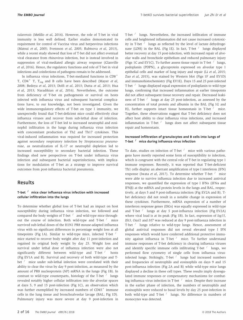
T-bet�/� mice clear influenza virus infection with increasedcellular infiltration into the lungs
To determine whether global loss of T-bet had an impact on host
susceptibility during influenza virus infection, we followed and
compared the body weights of T-bet�/� and wild-type mice through-
out the course of infection. Both wild-type and T-bet�/� mice
survived sub-lethal doses of the H1N1 PR8 mouse-adapted influenza
virus with no significant differences in percentage weight loss at all
timepoints (Fig 1A). Similar to wild-type mice, infected T-bet�/�
mice started to recover body weight after day 11 post-infection and
regained its original body weight by day 25. Weight loss and
survival under lethal dose of influenza infection were also not
significantly different between wild-type and T-bet�/� hosts
(Fig EV1A and B). Survival and recovery of both wild-type and T-
bet�/� mice under sub-lethal infection were correlated with their
ability to clear the virus by day 9 post-infection, as measured by the
amount of PR8 nucleoprotein (NP) mRNA in the lungs (Fig 1B). In
contrast to wild-type counterparts, histology of the T-bet�/� lungs
revealed notably higher cellular infiltration into the alveolar spaces
at days 5, 9 and 15 post-infection (Fig 1C), an observation which
was further exemplified by increased numbers of CD45+ immune
cells in the lung tissue and bronchoalveolar lavage (BAL; Fig 1D).
Pulmonary injury was more severe at day 9 post-infection in
T-bet�/� lungs. Nevertheless, the increased infiltration of immune
cells and heightened inflammation did not cause increased cytotoxic-
ity in T-bet�/� lungs as reflected by the level of lactate dehydroge-
nase (LDH) in the BAL (Fig 1E). In fact, T-bet�/� lungs displayed
better recovery at day 15 post-infection, with increased repair of alve-
olar walls and bronchiole epithelium and reduced pulmonary injury
(Figs 1C and EV1C). To further assess tissue repair in T-bet�/� lungs,
podoplanin (PDPN), a glycoprotein expressed on alveolar type 1
epithelial cells and marker of lung injury and repair (Li et al, 2015;
Zuo et al, 2015), was stained by Western blot (Figs 1F and EV1D)
and immunohistochemistry (Fig EV1E). Days 15 and 25 post-infected
T-bet�/� lungs displayed equal expression of podoplanin to wild-type
lungs, confirming that increased inflammation at earlier timepoints
did not affect subsequent tissue recovery and repair. Decreased leaki-
ness of T-bet�/� lungs at day 25 post-infection, as assessed by the
concentration of total protein and albumin in the BAL (Fig 1G and
H), further supports intact tissue homeostasis in T-bet�/� mice.
Together, these observations suggest that T-bet deficiency does not
affect host ability to clear influenza virus infections, and increased
inflammation in T-bet�/� lungs does not affect subsequent tissue
repair and homeostasis.
Increased infiltration of granulocytes and B cells into lungs ofT-bet�/� mice during influenza virus infection
To date, studies on infection of T-bet�/� mice with various patho-
gens have mostly reported some form of susceptibility to infection,
which is congruent with the central role of T-bet in regulating type 1
immune responses. Recently, it was reported that T-bet-deficient
Th1 cells display an aberrant amplification of type I interferon (IFN)
response (Iwata et al, 2017). To determine whether T-bet�/� mice
were able to survive influenza infection due to increased antiviral
responses, we quantified the expression of type I IFNs (IFNa and
IFNb) at the mRNA and protein levels in the lungs and BAL, respec-
tively, at days 5 and 9 post-influenza infection (Fig EV2A and B). T-
bet deficiency did not result in a notable change in expression of
these cytokines. Furthermore, mRNA expression of a number of
interferon response genes (ISGs) was equally expressed in wild-type
and T-bet�/� lungs at day 5 post-influenza infection (Fig EV2A)
where viral load is at its peak (Fig 1B). In fact, expression of Isg15,
Ifit3, Oasl1 and Irf7 was reduced at day 9 post-influenza infection in
T-bet�/� lungs relative to wild-type lungs. Hence, assessment of
global antiviral responses did not reveal elevated type I IFN
responses which would have conferred additional protective immu-
nity against influenza in T-bet�/� mice. To further understand
immune responses of T-bet deficiency in clearing influenza viruses
and identify specific immune cells infiltrating T-bet�/� lungs, we
performed flow cytometry of single cells from influenza virus-
infected lungs. Strikingly, T-bet�/� lungs had increased numbers
and frequencies of neutrophils and eosinophils on days 9 and 15
post-influenza infection (Fig 2A and B) while wild-type mice lungs
displayed a decline in these cell types. These results imply dysregu-
lated immune responses or compensatory mechanisms for combat-
ing influenza virus infection in T-bet�/� mice. Despite their increase
in the earlier phase of infection, the numbers of neutrophils and
eosinophils were reduced to basal levels by day 25 post-infection in
both wild-type and T-bet�/� lungs. No difference in numbers of
monocytes was detected.
2 of 16 The EMBO Journal e99176 | 2018 ª 2018 The Authors
The EMBO Journal T-betKO survives bacterial superinfection Jun Zhi Er et al
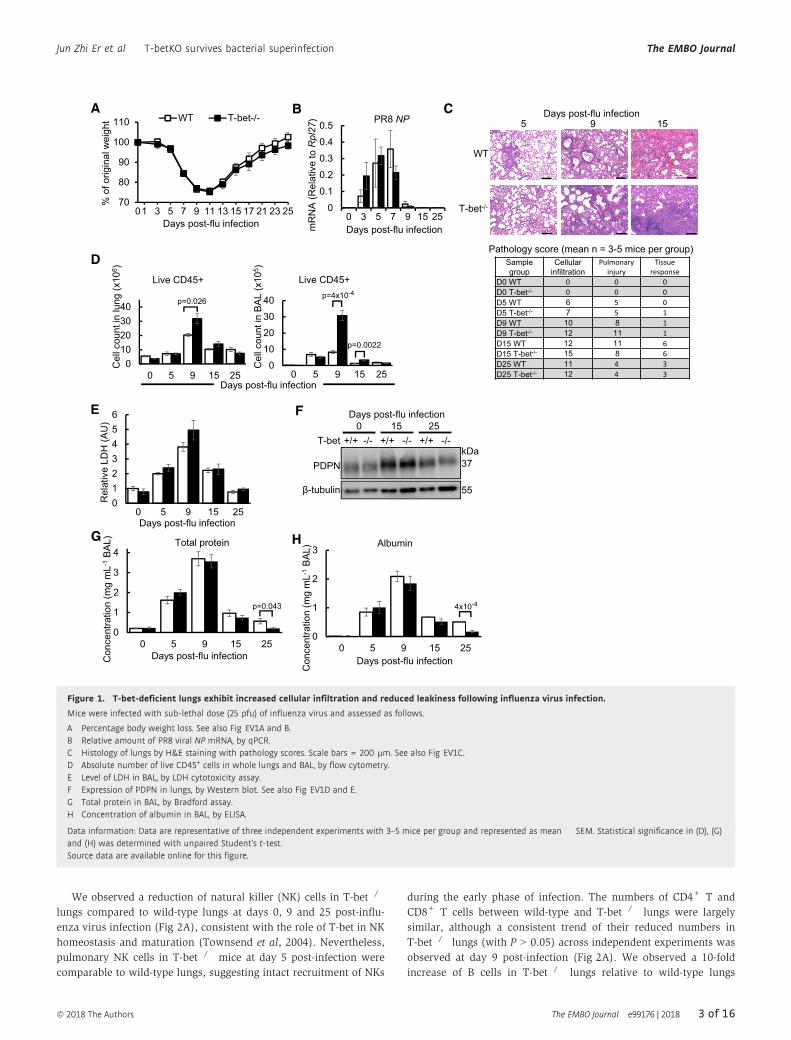
We observed a reduction of natural killer (NK) cells in T-bet�/�
lungs compared to wild-type lungs at days 0, 9 and 25 post-influ-
enza virus infection (Fig 2A), consistent with the role of T-bet in NK
homeostasis and maturation (Townsend et al, 2004). Nevertheless,
pulmonary NK cells in T-bet�/� mice at day 5 post-infection were
comparable to wild-type lungs, suggesting intact recruitment of NKs
during the early phase of infection. The numbers of CD4+ T and
CD8+ T cells between wild-type and T-bet�/� lungs were largely
similar, although a consistent trend of their reduced numbers in
T-bet�/� lungs (with P > 0.05) across independent experiments was
observed at day 9 post-infection (Fig 2A). We observed a 10-fold
increase of B cells in T-bet�/� lungs relative to wild-type lungs
0
1
2
3
4
0 5 9 15 25
Total protein
Days post-flu infectionCon
cent
ratio
n (m
g m
L-1
BA
L)
p=0.043
0123456
0 5 9 15 25
Rel
ativ
e LD
H (A
U)
Days post-flu infection
70
80
90
100
110
01 3 5 7 9 11 13 15 17 21 23 25
WT T-bet-/-%
of o
rigin
al w
eigh
t
Days post-flu infection
A B
D
G
0
1
2
3
0 5 9 15 25
Albumin
Days post-flu infection
Con
cent
ratio
n (m
g m
L-1
BA
L)
4x10⁻⁴
H
C
E F Days post-flu infection0 15 25
PDPN
β-tubulin
+/+ -/- +/+ -/- +/+ -/-kDa37
55
T-bet
Sample group
Cellular infiltration
Pulmonary injury
Tissue response
D0 WT 0 0 0D0 T-bet-/- 0 0 0D5 WT 6 5 0D5 T-bet-/- 7 5 1D9 WT 10 8 1D9 T-bet-/- 12 11 1D15 WT 12 11 6D15 T-bet-/- 15 8 6D25 WT 11 4 3D25 T-bet-/- 12 4 3
Pathology score (mean n = 3-5 mice per group)
Days post-flu infection5 9 15
WT
T-bet-/-0
0.1
0.2
0.3
0.4
0.5
0 3 5 7 9 15 25
PR8 NP
mR
NA
(Rel
ativ
e to
Rpl
27)
Days post-flu infection
010
2030
40
0 5 9 15 25
Live CD45+p=4x10⁻⁴
010203040
0 5 9 15 25
Live CD45+
p=0.026
Days post-flu infection
Cel
lcou
nt in
BA
L(x
105 )
Cel
lcou
nt in
lung
(x10
6 )
p=0.0022
Figure 1. T-bet-deficient lungs exhibit increased cellular infiltration and reduced leakiness following influenza virus infection.
Mice were infected with sub-lethal dose (25 pfu) of influenza virus and assessed as follows.
A Percentage body weight loss. See also Fig EV1A and B.B Relative amount of PR8 viral NP mRNA, by qPCR.C Histology of lungs by H&E staining with pathology scores. Scale bars = 200 lm. See also Fig EV1C.D Absolute number of live CD45+ cells in whole lungs and BAL, by flow cytometry.E Level of LDH in BAL, by LDH cytotoxicity assay.F Expression of PDPN in lungs, by Western blot. See also Fig EV1D and E.G Total protein in BAL, by Bradford assay.H Concentration of albumin in BAL, by ELISA.
Data information: Data are representative of three independent experiments with 3–5 mice per group and represented as mean � SEM. Statistical significance in (D), (G)and (H) was determined with unpaired Student’s t-test.Source data are available online for this figure.
ª 2018 The Authors The EMBO Journal e99176 | 2018 3 of 16
Jun Zhi Er et al T-betKO survives bacterial superinfection The EMBO Journal

specifically at day 15 post-influenza infection, suggesting a role for
T-bet in regulating proliferation or migration of B cells. Collectively,
the results demonstrate that absence of T-bet does not abrogate
innate or adaptive lymphocyte responses in the lungs.
Overall, analysis of immune responses revealed increased granu-
locytes and B cells in influenza-infected T-bet�/� lungs, while
concomitant intact type I IFN responses and recruitment of cytotoxic
and helper lymphocytes likely contributed to the effective clearance
of influenza virus.
T-bet deficiency confers survival advantage to post-influenzabacterial superinfection
Excessive pulmonary infiltration of neutrophils and eosinophils has
been observed in influenza virus-induced pneumonia and deaths
(Tumpey et al, 2005; Taubenberger & Morens, 2008; Jeon et al,
2010; Larranaga et al, 2016), which was taken to indicate lung
impairment. However, our results thus far did not suggest increased
susceptibility or damage to lung tissues despite heightened infiltra-
tion of neutrophils and eosinophils in T-bet�/� lungs. We then
asked how this increase in granulocytes in influenza-infected
T-bet�/� hosts might respond under bacterial superinfection, a
frequent complication following influenza infections.
To investigate responses of T-bet�/� mice to post-influenza
bacterial superinfection compared to wild-type hosts, mice were
infected with a sub-lethal dose of PR8 influenza virus, followed by
administration of a sub-lethal dose of S. pneumoniae serotype 19F,
at day 7 post-influenza infection (Fig 3A). Remarkably, T-bet�/�
mice displayed significantly enhanced survival following secondary
bacterial infection, compared to 85% lethality of wild-type mice
02468
101214
0 5 9 15 25
CD4+ T
p=0.07 p=0.05
0
20
40
60
0 5 9 15 25
CD8+ Tp=0.11
0306090
120150
0 5 9 15 25
NeutrophilWTT-bet-/- p<1x10⁻⁴
p=0.04
0
2
4
6
8
0 5 9 15 25
NK cell
p=0.0089
p=0.015 p=0.035
A
B
0
5
10
15
0 5 9 15 25
Monocyte
Days post-flu infection
Cel
lcou
nt in
lung
(x10
5 )
05
10152025
0 5 9 15 25
B cellp=0.037
0
5
10
15
20
0 5 9 15 25
Eosinophil
p=0.001
p=0.0067
6.79.2
3.04.0
2.22.3
12.012.9
CD45+ lung cells day 9 post-flu infection
WT
T-bet-/-
Neutrophil Eosinophil
Ly6G
CD11b CD11c
Sig
lecF
CD45+ lung cells day 15 post-flu infectionNeutrophil Eosinophil
Ly6G
CD11b CD11c
Sig
lecF
WT
T-bet-/-
Figure 2. Increased infiltration of neutrophils, eosinophils and B cells in T-bet-deficient lungs infected with influenza virus.
A Absolute cell counts from lungs of sub-lethally influenza virus-infected mice following analysis by flow cytometry. After gating out alveolar macrophages by forwardand side scatter, immune cells are defined as follows: neutrophil (Ly6G+CD11b+SiglecF�Ly6Cint), eosinophil (SiglecF+CD11c�/+F4/80�CD64�), monocyte(Ly6G�CD11b+MHCII�SiglecF�), natural killer cell (NK, B220�/intNK1.1+), B cell (B220hiNK1.1�), CD8+ T cell (TCRb+CD8+), CD4+ T cell (TCRb+CD4+). See also Fig EV2C and D.
B Flow cytometry plots of neutrophils and eosinophils in lungs at days 9 and 15 post-sub-lethal influenza virus infection. Numbers in plots indicate frequency of gatedimmune cells in each throughput.
Data information: Data are representative of three independent experiments with 3–4 mice per group and represented as mean � SEM. Statistical significance in (A)was determined with unpaired Student’s t-test. n.s., not significant.
4 of 16 The EMBO Journal e99176 | 2018 ª 2018 The Authors
The EMBO Journal T-betKO survives bacterial superinfection Jun Zhi Er et al

(Fig 3B). Furthermore, bacterial load in T-bet�/� lungs was lower
than wild-type lungs (Fig 3C). These survival advantages in T-bet�/�
mice were correlated to increased infiltration of neutrophils and
eosinophils in the lungs at days 1 and 3 of secondary bacterial infec-
tion (Fig 3D), suggesting that they could be involved in immune
defence. Compared to wild-type lungs, the number of NK cells tran-
siently increased in T-bet�/� lungs 1 day after secondary bacterial
infection, supporting enhanced trafficking or proliferation in
response to bacterial challenge, while CD8+ T cells were reduced at
day 3 post-infection. Similar to the viral infection only controls, B
cell numbers were increased in T-bet�/� lungs following secondary
bacterial infection, while CD4+ T cells are comparable to wild-type
lungs. Enhanced protection and immune cell responses against
secondary bacterial infection under T-bet deficiency were
reproduced with a more virulent S. pneumoniae serotype (S3;
Fig EV3B–D).
To determine whether mortality in wild-type mice was due to
viral-mediated suppression of antibacterial defences through T-bet,
naıve or influenza-infected mice were infected with equal doses of
bacteria. Wild-type mice infected with prior influenza virus suffered
heavier bacterial burden compared to infection with bacteria alone,
indicating lethality in viral–bacterial superinfection (Figs 3E and
EV3C). While T-bet�/� mice appeared to control bacteria more effi-
ciently than wild-type mice at 12 h post-bacteria-only infection
(Fig 3E), both mice strains were able to clear bacteria by 36 h post-
infection (Fig 3F), indicating suppression of antibacterial defences by
T-bet during influenza and bacterial superinfections. Moreover, the
numbers of neutrophils and eosinophils were significantly lower in
0
2
4
6
8
0 2.25 4.5WT T-bet-/- WT T-bet-/-1 3
Days post-secondarybacterial infection
log 1
0cf
u m
L-1
BA
L p=0.029 p=0.029
A B
D
C
Days post flu infection0 7
Influenza virus H1N1 PR8 (sub-lethal dose)
S. pneumoniae 19F (sub-lethal dose)
0
8 10
1 3-7
WT vs T-bet-/-
Days post-secondary bacterial infection
G
010203040506070
0 1 3
Neutrophilp<1x10⁻⁴
p=0.0042p=0.014
Cel
l cou
nt in
lung
(x10
5 )
0
5
10
15
20
25
0 1 3
B cellp=0.05
0102030405060
0 1 3
CD8+ Tp=3x10⁻⁴
0
3
6
9
12
15
0 1 3
CD4+ T
0
5
10
15
20
25
0 1 3
NK cellp=0.038
0
5
10
15
20
0 1 3
Eosinophil
p=0.011p=0.07
Days post-secondary bacterial infection
012345
-Flu +Flu
Eosinophil
n.s.
n.s.p=0.0014
0
5
10
15
-Flu +Flu
Neutrophil
p=0.043
n.s.n.s.
p=0.017
Cel
lcou
nt in
lung
(x10
5 )
0
25
50
75
100
0 2 4 6 8 10 12 14 16 18
WTT-bet-/-
p=6x10⁻⁴
S. pneumoniae 19F
% S
urvi
val
Days post-flu infection
0
0.5
1
0 2
log 1
0cf
u m
L-1
BA
L
36 h post-bacteria only infection
F
12 h post-bacterial infection
0
2
4
6
8
0 2 4
log 1
0cf
u m
L-1
BA
L
-Flu +Flu
p=0.064p=6x10⁻⁴
p=6x10⁻⁴n.s.
12 h post-bacterial infection
E
Figure 3. Improved resistance of T-bet-deficient mice against post-influenza bacterial superinfection.
A Model of post-influenza bacterial infection illustrating infection with sub-lethal dose of influenza virus (25 pfu) followed by sub-lethal dose of secondary bacterialchallenge (Streptococcus pneumoniae serotype 19F, 2 × 10⁵ cfu) 7 days later. Where appropriate, mice lungs were analysed at days 0, 1 and 3 post-secondarybacterial infection, corresponding to days 7, 8 and 10 post-influenza virus infection, respectively.
B–G Mice were superinfected as described in (A). (B) Survival of mice following bacterial superinfection. See also Fig EV3B. (C) Pneumococcal cfu in BAL of mice. See alsoFig EV3C. (D) Absolute immune cell counts in lungs, assessed by flow cytometry. See also Fig EV3D. (E) Pneumococcal cfu in BAL of mice 12 h after infection with2 × 105 cfu of S. pneumoniae serotype 19F alone (�Flu) or following influenza virus infection (+Flu). See also Fig EV3C. (F) Pneumococcal cfu in BAL 36 h afterinfection of naïve mice with 2 × 10⁵ cfu of S. pneumoniae serotype 19F alone. (G) Absolute neutrophil and eosinophil cell count in lungs infected as described in(E), assessed by flow cytometry.
Data information: Data from (B) are combined from two independent experiments, n = 13 mice per experimental group. Data from (C–G) are representative of at leasttwo independent experiments with 3–5 (C, D and G) or 5–7 (E and F) mice per experimental group. All data are represented as mean � SEM. Statistical significance wasdetermined by log-rank test in (B), unpaired Student’s t-test in (D) and (G) and Mann–Whitney U-test in (C) and (E). n.s., not significant.
ª 2018 The Authors The EMBO Journal e99176 | 2018 5 of 16
Jun Zhi Er et al T-betKO survives bacterial superinfection The EMBO Journal

the lungs of both bacteria-only infected wild-type and T-bet�/� mice,
compared to secondary infected T-bet�/� lungs (Fig 3G). Together,
these data demonstrate that enhanced inflammatory responses during
primary influenza infection in T-bet-deficient lungs contribute to
subsequent antibacterial defence and improved survival.
Increase in Th2 and Th17 cytokines in T-bet�/� lungsare correlated to enhanced infiltration of neutrophilsand eosinophils
To further understand the underlying immune mechanisms in T-
bet�/� mice during the inflammatory phase of influenza virus
infection, the BAL of days 5 and 9 post-infection was screened
for cytokines and chemokines using a protein bead array. Out of
24 analytes examined, significant differences in concentrations of
10 target proteins between wild-type and T-bet�/� lungs were
observed (Fig 4A). Strikingly, Th2 cytokines, IL-9 and IL-13, as
well as Th17 cytokines, IL-17 and IL-22, were significantly
elevated in T-bet�/� lungs (Figs 4A and EV4A), consistent with
the role of T-bet in repression of these immune responses
(Hwang et al, 2005; Villarino et al, 2010; Lazarevic et al, 2011).
Furthermore, IL-13 and IL-17 are known to drive pulmonary
recruitment of eosinophils and neutrophils, respectively (Pope
et al, 2001; Ye et al, 2001), which may explain the increased
cellular infiltration of these cell types into T-bet-deficient lungs
following infection. Secretion of pro-inflammatory cytokines, IL-6
and IL-18, is increased in T-bet-deficient lungs (Fig 4A), suggest-
ing possible compensatory inflammatory mechanisms perhaps
in view of reduced antiviral cytokine IL-28. Decrease in
concentrations of T-bet target protein, CCL3, was observed in
T-bet�/� lungs. IFNc-induced T cell chemoattractants like CXCL9
(MIG) and CXCL10 (IP-10) were also reduced, which may provide
an explanation for lowered numbers of pulmonary T cells in
T-bet�/� lungs (Figs 2A and 3D). Interestingly, no difference was
Days post-secondary bacterial infectionCon
cent
ratio
n (p
gm
L-1
BA
L)
0
50
100
150
200
0 1 3
IL-17p=0.035
p=0.0057
0
5
10
15
20
0 1 3
IL-22p=5x10⁻⁴
p=0.05p=0.08
Con
cent
ratio
n (p
gm
L-1
BA
L)
0
50
100
150
200
250
-Flu +Flu
IL-17
p<1x10⁻⁴p<1x10⁻⁴
0
20
40
60
-Flu +Flu
IL-22p=0.0017p=0.0084
12 h post-secondary bacterial infection
0
10
20
30
40
50
5 9
IL-17
WTT-bet-/-
p=0.06
p=0.02
0
250
500
750
1000
1250
5 9
IL-18p=9x10⁻⁴
0
150
300
450
600
750
5 9
IL-22
p=0.02
0
50
100
150
200
5 9
IL-9
p=0.002
0
25
50
75
100
5 9
IL-13p=0.002
0
2000
4000
6000
5 9
IL-6
p=0.02 p=2x10⁻⁵
0
100
200
300
400
500
5 9
IL-28p=0.01
0
30
60
90
120
150
5 9
CCL3
p=0.03
0
2000
4000
6000
5 9
CXCL9
p=8x10⁻⁴
0
500
1000
1500
2000
2500
5 9
CXCL10
p=0.09
Con
cent
ratio
n(p
gm
L-1
BA
L)
Days post-flu infection
A
B C
p=0.07
p=0.06
Figure 4. Increased IL-17 and IL-22 production in T-bet�/� lungs infected with influenza virus or superinfected with bacteria.
A Concentrations of cytokines or chemokines in the BAL of days 5 and 9 post-sub-lethal influenza virus infection as determined by protein bead array. See alsoFig EV4A and B.
B, C Mice were superinfected as described in Fig 3A. (B) Concentrations of IL-17 and IL-22 in BAL of mice, as determined by ELISA. See also Fig EV3E. (C) Concentrationsof IL-17 and IL-22 in BAL of mice 12 h after infection with 2 × 10⁵ cfu of Streptococcus pneumoniae serotype 19F alone (�Flu), or following superinfection (+Flu),assessed by ELISA.
Data information: Data in (A) are obtained from one independent experiment with four mice per group, and results of selected analytes IFNc, CXCL10, IL-6, IL-13 andIL-17 are reconfirmed in a separate independent experiment by a customized protein bead array. Data in (B) and (C) are representative of two independent experimentswith 3–4 mice per group. All data are represented as mean � SEM. Significance in (A–C) was determined by unpaired Student’s t-test. n.s., not significant.
6 of 16 The EMBO Journal e99176 | 2018 ª 2018 The Authors
The EMBO Journal T-betKO survives bacterial superinfection Jun Zhi Er et al

0
2
4
6
8
10
2.316.60
1 3Days post-secondary bacterial infection
log 1
0cf
u m
L-1
BA
Lp<1x10⁻⁴
p=0.0038
p=0.041
p=2x10⁻⁴
p=0.0013
p=7x10⁻⁴n.s.
n.s.
A
DC
0
0.4
0.8
1.2
1.6
2
1 3
Lcn2
1 3
n.s.
n.s.
Days post-secondary bacterial infection
ns
mR
NA
(Rel
ativ
e to
Rpl
27)
0
0.004
0.008
0.012
1 3
Reg3b
p=0.017
p=0.0047p=0.0057
n.s.
1 3
0
0.1
0.2
0.3
0.4
0.5
1 3
Reg3g
p<1x10⁻⁴p<2x10⁻⁴
p<2x10⁻⁴
p=0.03
1 3
0
2
4
6
8
Day 1
p=0.0033
0
20
40
60
Day 3p=1x10⁻⁴
Neu
troph
il co
unt i
n lu
ng (x
105 )
Post-secondary bacterial infection
0
1
2
3
4
0 3.75 7.5
log 1
0cf
u m
L-1
BA
L
1 3Days post-secondary
bacterial infection
p=0.018p=0.013
0
25
50
75
100
0 2 4 6 8 10 12 14 16 18
T-bet-/-
T-bet-/-+αIL-17
S. pneumoniae 19F
% S
urvi
val
Days post-flu infection
p=0.0014
B
En.s.
Figure 5. Increased immunity against bacterial superinfection in T-bet�/� mice is abrogated by neutralization of IL-17 or neutrophil depletion.
Mice were superinfected as described in Fig 3A.
A Bacterial load as determined by enumeration of cfu in BAL following administration of aIL-17 or aIL-22 blocking antibody. Control wild-type and T-bet�/� groupswere administered with IgG isotype control antibody.
B T-bet�/� mice were administered aIL-17 or IgG isotype control antibody every other day from day �2 post-secondary bacterial infection and monitored for survival.C Absolute neutrophil counts in the lungs following administration of aIL-17 or IgG isotype control antibody, as determined by flow cytometry.D Pneumococcal cfu in BAL following aLy6G antibody-mediated neutrophil depletion. Control T-bet�/� mice were administered with IgG isotype control antibody. See
also Fig EV5A.E mRNA expression of antimicrobial peptides in lungs following administration of aIL-17, IL-22 or IgG control antibody, as determined by qPCR.
Data information: Data in (A), (B) and (D) are combined from two independent experiments (n = 8–12 mice per group). Data in (C) and (E) are representative of twoindependent experiments with 3–4 in (C) or 4–6 in (E) mice per group. All data are represented as mean � SEM. Statistical significance was determined with Kruskal–Wallis test followed by Dunn’s multiple comparisons test in (A), log-rank test in (B), unpaired Student’s t-test in (C), Mann–Whitney U-test in (D) and one-way ANOVAfollowed by Tukey’s multiple comparisons test in (E). n.s., not significant. See also Fig EV5B and C.
ª 2018 The Authors The EMBO Journal e99176 | 2018 7 of 16
Jun Zhi Er et al T-betKO survives bacterial superinfection The EMBO Journal

observed in protein levels of the T-bet target—IFNc, as further
confirmed by ELISA (Fig EV4B). Nevertheless, reduced expression
of Ifng and Th1 cytokine Tnf mRNA was observed in T-bet�/�
lungs compared to wild-type (Fig EV4C), implying post-transcrip-
tional regulation of Th1 effector cytokines. Overall, these mecha-
nisms suggest a shift towards Th2 and Th17 immune responses
in the absence of T-bet during influenza virus infection, leading
to increased infiltration of granulocytes.
Th17 cytokines, IL-17 and IL-22, were previously shown to be
critical for defence against post-influenza secondary bacterial infec-
tion (Kudva et al, 2011). We confirmed the increased production of
these cytokines in the T-bet�/� lungs following secondary bacterial
infection (Figs 4B and EV3E). Infection with bacteria alone did not
induce significant production of IL-17 and IL-22 relative to superin-
fected T-bet�/� lungs (Fig 4C), indicating that prior virus infection is
required for an elevated immune response in an imminent secondary
bacterial infection. Hence, it is conceivable that the survival advan-
tage of T-bet�/� mice under post-influenza bacterial infection is due
to the induction of Th17-mediated antibacterial defences.
Neutralization of IL-17 and neutrophil depletion reduces immunedefences against post-influenza bacterial superinfection in T-bet-deficient mice
To specifically test whether IL-17 and IL-22 are responsible for the
survival advantage of influenza-infected T-bet�/� mice against
secondary bacterial infection, anti-IL-17 or anti-IL-22 monoclonal
antibodies were administered to neutralize the effects of these two
cytokines. Treatment with anti-IL-17 antibody, but not anti-IL-22 anti-
body resulted in increased bacterial load in T-bet�/� lungs following
post-influenza bacterial superinfection (Fig 5A). Correspondingly, the
survival of T-bet�/� mice against secondary S. pneumoniae infection
was also reduced following IL-17 depletion (Figs 5B and EV3F).
Concomitant with the role of IL-17 in the recruitment of neutrophils
(Ye et al, 2001; Miyamoto et al, 2003), neutralization of IL-17
reduced the number of infiltrating neutrophils to T-bet�/� lungs at
days 1 and 3 post-secondary bacterial infection (Fig 5C). Depletion of
neutrophils through administration of anti-Ly6G-specific antibody
(Fig EV5A) resulted in significantly heavier bacterial burden in T-
bet�/� lungs (Fig 5D), confirming direct neutrophil-mediated protec-
tion. Reduced resistance against secondary bacterial infection upon
IL-17 or neutrophil depletion did not appear to be a result of the
inability to clear prior influenza virus infection, as quantitation of
PR8 NP in the lungs did not show increased viral load upon antibody
administration (Fig EV5B and C).
Th17 responses are known to mediate antibacterial defences
through inducing antimicrobial peptides (Liang et al, 2006).
Compared to wild-type lungs, the expression of Reg3b and Reg3g
antimicrobial peptides, but not lipocalin 2 (Lcn2), was increased in
T-bet�/� lungs at day 1 post-secondary bacterial infection (Fig 5E).
Expression of Reg3g remained elevated in T-bet�/� lungs up to day
3 post-infection (Fig 5E). Neutralization of IL-17 and IL-22 reduced
expression of Reg3b and Reg3g in T-bet�/� lungs at day 1 post-
secondary bacterial infection. Therefore, it appears that IL-17- and
IL-22-mediated expression of antimicrobial peptides is involved only
in the early phase of post-influenza bacterial infection, while IL-17-
mediated neutrophil recruitment is a major factor conferring protec-
tion to T-bet�/� mice.
IL-17-producing CD8+, CD4+ and cd T cells contribute toprotection during post-influenza bacterial superinfection inT-bet-deficient mice
To determine the cell types responsible for type-17-mediated
immune protection, intracellular staining of IFNc and IL-17 on days
0, 1 and 3 post-secondary bacterial-infected lung lymphocytes was
performed. The data indicated an elevated frequency of IL-17-produ-
cing cells and a reduction in IFNc-producing cells in T-bet�/� lungs
(Fig 6A), suggesting a shift from type 1 to type 17 immune
responses due to the loss of T-bet, as also observed by others
(Lazarevic et al, 2011). Further assessment of individual cell types
revealed that up to 35% of total CD8+, CD4+ and cd T cells in T-
bet-deficient lungs accounted for more than 75% of all IL-17-produ-
cing cells (Fig 6B and C), while the proportion of IL-17-producing
NK cells was negligible. Amongst IL-17+ cells in T-bet�/� lungs, cdT cells form the majority in proportion and absolute numbers prior
to bacterial superinfection, while CD8+ T cells constitute the highest
frequency and numbers by day 3 post-secondary bacterial infection
(Fig 6C). Hence, we conclude that IL-17-protection in T-bet�/� lungs
against secondary bacterial infection is mediated through IL-17-
producing T cells, notably through CD8+ and cd T cells.
Taken together, our results support a mechanism of tripartite
host–viral–bacterial interaction whereby induction of IL-17 and IL-
22 in T-bet�/� hosts during primary influenza virus infection estab-
lishes an inflammatory environment characterized by elevated
neutrophils and antimicrobial peptide expression in the lungs. These
inflammatory defences are suppressed in wild-type hosts, but
primes T-bet-deficient hosts against lethality from subsequent bacte-
rial infections (Fig 7).
Discussion
Type 1 immune responses are essential for defence against all
classes of invading pathogens. As a central regulator of type 1
immunity, T-bet has been implicated in infections by pathogenic
bacteria and parasites affecting the lungs, skin, central nervous
system and gut (reviewed by Lazarevic & Glimcher, 2011), wherein
loss of function mostly resulted in greater pathogen burden. In viral
infections, loss of T-bet resulted in greater viral load (Matsui et al,
2005; Svensson et al, 2005; Rubtsova et al, 2013), attributable to
reduced NK and CD8+ T cell cytolytic activity and dysregulated B
cell responses. Beyond acute infections, T-bet is also involved in
secondary antiviral defences through mediating the formation of
memory B and T cells (Joshi et al, 2007; Marshall et al, 2011; Knox
et al, 2017). Nevertheless, a recent study found that the loss of
T-bet did not result in marked defects in the ability to clear acute
respiratory syncytial virus (RSV) infection despite reduced cellular
and humoral antiviral responses, but instead led to increased aller-
gic responses (Glanville et al, 2016). Similarly, our study showed
that infection with the influenza virus did not significantly affect
viral loads, tissue homeostasis or survival in T-bet-deficient hosts.
Strikingly, massive infiltration of neutrophils and eosinophils,
accompanied by production of Th2 and Th17 cytokines, was
observed in the inflammatory phase of influenza-infected T-bet-
deficient lungs. This altered inflammatory environment instead
provided immunity and survival advantage against subsequent
8 of 16 The EMBO Journal e99176 | 2018 ª 2018 The Authors
The EMBO Journal T-betKO survives bacterial superinfection Jun Zhi Er et al

80
NK1.1
0.04
1.78 0.4
8.8
0.03
0.986.4
12.8
0.08
3.09 6.4
5.0
0.04
0.163.0
36.9
1.1
7.58 0.6
7.2
WT
T-bet-/-
IFN
γ
IL-17
CD45+ lung cells Days post-secondary bacterial infection0 1 3
0
2.48 2.0
15.6
0
10
20
30
0 1 3
IFNγ+ cellsWTT-bet-/-
p=0.0082p=0.0016
p=0.07
Days post-secondary bacterial infection
% C
D45
+ ce
lls
0
2
4
6
8
0 1 3
IL-17+ cells
p=0.035p=0.035
p=2x10⁻⁴
0
20
40
60
WTT-bet-/-
0 1 3Days post-secondary bacterial infection
% IL
17+
cells
CD
3
CD8 CD4 γδTCR
Total
NK cell CD8+ T CD4+ T γδ T
A
B
C
IL17+
T-bet-/- CD45+ lung cells Day 3 post-secondary bacterial infection
0
2
4
6
8
0 1 3
NK celln.s.
n.s.
(x103)
IL17
+ ce
ll co
unt i
n lu
ng
0
2
4
6
8
0 1 3
CD8+ Tp=0.0043
p<1x10⁻⁴
p=0.05
(x105)
012345
0 1 3
CD4+ Tp=0.03
p=0.048 n.s.
(x105)
0
2
4
6
0 1 3
γδ Tp=0.0016
p=0.05n.s.
(x105)
Days post-secondary bacterial infection
10.813.6 35.3
0.3
Figure 6. CD8+, CD4+ and cd T cells are major sources of IL-17 in influenza and secondary bacterial superinfected T-bet�/� lungs.
Mice were superinfected as described in Fig 3A.
A Frequency of IFNc+ and IL-17+ immune cells following intracellular cytokine staining and flow cytometry.B Representative flow cytometry plots of total and IL-17+ lymphocyte subsets on day 3 post-secondary bacterial-infected lungs following intracellular cytokine staining.
Numbers in plots indicate the frequency of each IL-17+ cell subset over total numbers of respective cell types.C Frequency over total IL-17+ cells and absolute numbers of each IL-17 producing lymphocyte subset on days 0, 1 and 3 post-secondary bacterial superinfected lungs as
assessed by flow cytometry of intracellular cytokine stained cells.
Data information: Data are representative of at least two independent experiments with three mice per group. All data are represented as mean � SEM. Statisticalsignificance in (A) and (C) was determined by unpaired Student’s t-test.
ª 2018 The Authors The EMBO Journal e99176 | 2018 9 of 16
Jun Zhi Er et al T-betKO survives bacterial superinfection The EMBO Journal

bacterial superinfection, while neutralization of IL-17 or neutrophil
depletion in T-bet�/� mice decreased antibacterial immunity.
Hence, contrary to the existing view that T-bet is largely required
for resistance against primary virus or bacterial infections, our
results provide new evidence to the growing perspective that T-bet
loss need not eventually lead to greater susceptibility to infections.
T-bet transcription factor is expressed in a wide variety of
immune cells, including NK cells, NKT cells, B cells, CD4+ and
CD8+ T cells, dendritic cells and cd T cells (reviewed by Lazarevic
et al, 2013). Its mechanism of function is mainly through expression
of IFNc and mediating maturation, cytotoxicity or IgG class switch-
ing of these cells. All these immune responses are crucial against
viral infections, yet surprisingly, the loss of T-bet did not result in
marked deficiency in defence against the influenza virus. We show
that the viral load is comparable in T-bet-deficient lungs and wild-
type counterparts throughout the course of virus infection, suggest-
ing that the kinetics of viral clearance is not affected. Survival is also
indifferent under high virus doses between wild-type and T-bet�/�
mice. This can be explained by the largely intact immune responses
in T-bet�/� mice, with CD4+ T, CD8+ T and B cells still present in
comparable numbers. Although NK cells were reduced in the rest-
ing lung, their numbers were unaffected at the early phase of virus
infection, where they are known to be the predominant mediators of
cytotoxicity against viral infection (Leung & Ada, 1981; Stein-
Streilein & Guffee, 1986). These mechanistic insights suggest that
T-bet is dispensable for the clearance of influenza virus, or there
could be the presence of compensatory functions by other
regulatory factors. Eomes, a T-box transcription factor homologous
to T-bet and co-expressed in CD8+ T and NK cells, could be one
possible factor, as shown by earlier studies that demonstrated their
complementary roles against virus infections (Intlekofer et al, 2005,
2008). Increased neutrophils and eosinophils in T-bet�/� lungs may
also play compensatory roles to other immune cells following influ-
enza virus infection (Fujisawa, 2008; Tate et al, 2011; Hufford et al,
2012; Samarasinghe et al, 2017).
Further to cellular responses, antiviral gene expression and
production of type I and II IFNs in the lungs were also similar,
further supporting the robustness of immune responses in the
context of sub-lethal influenza infection. Our findings on T-bet-defi-
cient mice support results from a number of related Th1 knockout
studies that showed no defects in influenza resistance (Graham
et al, 1993; Nguyen et al, 2000; Price et al, 2000).
The presence of IFNc in the BAL in infected T-bet�/� lungs at
levels similar to that of wild-type levels is particularly intriguing,
considering the role of T-bet in the production of IFNc within
several cell types (Szabo et al, 2002; Lugo-Villarino et al, 2003;
Chen et al, 2007; Klose et al, 2013; Barros-Martins et al, 2016).
Correspondingly, we also observed a reduction in mRNA expression
of Ifng and frequency of IFNc-producing cells. Nevertheless, the
unaffected levels of total secreted IFNc in T-bet-deficient hosts
during inflammation, as observed by us and in other studies (Burrell
et al, 2008; Guo et al, 2009), suggest post-translational control or
expression based on specific immune environmental cues. This
additional level of control on the physiological amounts of IFNcwarrants further investigation and may have important implications
on aberrant type 1 responses.
Excessive neutrophils and eosinophils in the lungs following influ-
enza virus infection has been linked to exacerbated lung injury and
respiratory failure (Jeon et al, 2010; Narasaraju et al, 2011). In this
study, pulmonary injury was assessed by histology to be more severe
in day 9 post-influenza-infected T-bet�/� lungs, correlating with
significant increase in neutrophil and eosinophil numbers. Neverthe-
less, subsequent timepoint at day 15 post-influenza virus infection
showed reduced pulmonary injury and comparable efficacy in tissue
repair. Moreover, reduced lung leakiness at day 25 was observed in
T-bet�/� lungs, supporting the presence of mechanisms that might
ameliorate excessive inflammation or promote homeostasis. Previous
studies have shown IL-22 and IL-6 cytokines to be essential in
lung repair (Kumar et al, 2013; Pociask et al, 2013; Yang et al,
2017). While these two cytokines were observed to be elevated in
T-bet�/� lungs during day 9 post-influenza infection, it is currently
unclear whether this early increase in cytokines has contributed to
the tissue recovery at later timepoints. Overall, it appears that the
impact of neutrophilia and eosinophilia in T-bet�/� lungs is not
permanently detrimental following virus infection, and tissue home-
ostasis is not adversely affected with the loss of T-bet functions.
Influenza–bacterial superinfection is a common complication of
flu pandemic and a major cause of severe pneumonia and morbidity
in the human population (Metersky et al, 2012). One of the causes
of lethal synergism between influenza viruses and respiratory bacte-
rial pathogens involves suppression of immune responses to bacte-
ria by prior influenza virus infection (McNamee & Harmsen, 2006;
Sun & Metzger, 2008; Shahangian et al, 2009; Ghoneim et al, 2013;
Cao et al, 2014). Therefore, reactivating pulmonary immune
defences in influenza-infected lungs appears to be an outstanding
Neutrophil, (Eosinophil)
IL-17, IL-22, (IL-9, IL-13)
T-bet-/-
Secondary pneumococcus
RegIIIγ
Viral clearance,leakiness Host Survival
Bacterial load
αIL-17 /Neutrophil depletionRegIIIβ
PrimaryInfluenza virus
Figure 7. Summary of immune responses and effects of T-bet deficiencyfollowing influenza virus and bacterial superinfection.
T-bet-deficient lungs infected with sub-lethal dose of influenza virus upregulate
Th2 and Th17 responses, leading to increased pulmonary infiltration of
neutrophils and eosinophils. This altered inflammatory response did not
compromise efficiency in viral clearance or eventual tissue homeostasis, and in
fact reduced tissue leakiness following infection. Furthermore, the immune
environment resulting from influenza-infected T-bet-deficient lungs conferred
immunity and survival advantage to pneumococcal superinfected hosts.
Correspondingly, neutralization of IL-17 or neutrophil depletion by antibody
treatment in T-bet�/� hosts reduced pulmonary neutrophil infiltration or
antimicrobial peptide expression, reversing immunity against secondary
bacterial infection.
10 of 16 The EMBO Journal e99176 | 2018 ª 2018 The Authors
The EMBO Journal T-betKO survives bacterial superinfection Jun Zhi Er et al

opportunity against bacterial superinfection. The marked increase in
neutrophil infiltration as well as production of Th17 cytokines IL-17
and IL-22 in influenza-infected T-bet�/� lungs at a timepoint where
hosts are most susceptible to bacterial infection (Sun & Metzger,
2008; Chertow & Memoli, 2013) prompted us to examine antibacte-
rial defences of T-bet-deficient hosts during secondary infection.
Furthermore, previous studies have attributed the importance of
neutrophils and Th17 immune responses in this infection model
(McNamee & Harmsen, 2006; Shahangian et al, 2009; Kudva et al,
2011; Ivanov et al, 2013; Cao et al, 2014). We found that decreased
bacterial load in T-bet�/� superinfected lungs and increased survival
were attributable to IL-17-mediated neutrophil recruitment. While
IL-17 has also been shown to induce antibacterial defence through
upregulating antimicrobial peptides (Liang et al, 2006; Archer et al,
2016), our observation that there is no considerable increased
expression in T-bet�/� lungs at day 3 post-bacterial infection
suggests that this mechanism may only be applicable to early bacte-
rial load control. In addition, IL-22 neutralization did not abrogate
enhanced bacterial clearance in T-bet�/� mice. Hence, we propose
IL-17-mediated neutrophil recruitment as the major mechanism
conferring survival advantage to secondary infection in T-bet�/�
mice. As it is known that T-bet inhibits development of Th17 cells
(Villarino et al, 2010; Lazarevic et al, 2011), our results also suggest
suppressed Th17-mediated antibacterial immunity as an underlying
mechanism to lethal synergism between influenza and bacterial
superinfection in T-bet sufficient hosts. Inhibition of T-bet thus
confers protection against secondary bacterial infection through
reactivating Th17-mediated antibacterial defences.
Enhanced type 17 responses, due to the lack of T-bet, has been
described in a number of cell types, including CD8+ T cells (Burrell
et al, 2008; Intlekofer et al, 2008), CD4+ T cells (Rangachari et al,
2006; Yuan et al, 2008; Guo et al, 2009; Lazarevic et al, 2011) and
cd T cells (Barros-Martins et al, 2016). Of these, Lazarevic et al
described a mechanism wherein T-bet deficiency allowed for the
expression of Th17 transcription factor Rorct in Th1 cells, driving
naıve CD4+ T cells into Th17 differentiation instead of Th1 lineage.
We observed increased number of IL-17-producing immune cells
with a corresponding reduction in IFNc-producing cells in T-bet-
deficient infected lungs, suggesting that a similar mechanism of T-
bet-mediated Th17 suppression might be involved. While it has
been shown that cell intrinsic loss of IL-10 promotes CD4+ T cell
differentiation into the Th17 lineage with a corresponding reduction
in T-bet expression (McKinstry et al, 2009), we did not identify a
reciprocal reduction of IL-10 in BAL of infected T-bet-deficient lungs
in our cytokine screen eluding to elevated type 17 immunity.
Considering that a significant proportion of the collective type 17
immune response involves CD8+ and cd T cells and are protective
against pathogen infection, future studies might consider a system-
atic delineation of the molecular and cellular events underlying
T-bet-mediated type 17 immunity in these other cell types.
B cells were observed to be increased in T-bet�/� lungs at day 15
post-influenza infection and day 3 post-secondary bacterial infec-
tion. Current knowledge of T-bet function in B cells are limited to its
role in promoting IgG2a class switching leading to the control of
viral infections (Peng et al, 2002; Rubtsova et al, 2013; Barnett et al,
2016) and development of memory B cells (Wang et al, 2012; Knox
et al, 2017). Gene expression analysis also shows reduced tran-
scripts that are linked to proliferation and regulation of tissue
migration in T-bet-deficient B cells (Barnett et al, 2016). Hence, the
increase in B cells in T-bet-deficient lungs shown in this study may
be due to dysregulation in B cell trafficking or aberrant cell prolifera-
tion. However, such mechanisms need to be further tested, and the
physiological significance of increased T-bet-deficient B cells post-
infection remains to be determined.
In summary, our study demonstrates that T-bet is dispensable for
influenza virus clearance and loss of T-bet functions is not adverse
to disease outcomes. However, the increased Th17 immune
responses and pulmonary neutrophil recruitment in influenza-
infected T-bet�/� host proved to be beneficial through increased
antibacterial immunity. These results support the suppression of T-
bet as a potential treatment option against lethal bacterial superinfec-
tions, without affecting protection against primary influenza virus.
Materials and Methods
Mice and pathogens
Age- (8–13 weeks) and gender-matched C57BL/6 wild-type and T-
bet�/� mice (004648) from Jackson’s laboratory were used in all
experiments. Influenza virus used for infection is the mouse-
adapted H1N1 PR/8/34 (PR8) strain (ATCC, VR-95). Propagation
of viruses was performed in embryonated chicken eggs at 37°C
for 72 h. Allantoic fluid was harvested and viral titres were deter-
mined by standard plaque assay in Madin–Darby canine kidney
(MDCK) cells. Streptococcus pneumoniae serotypes 19F and S3
used for infection were originally clinical isolates from National
University Hospital, Singapore, provided by A/Prof Vincent Chow
(NUS Department of Microbiology and Immunology). Bacterial
stocks were cultured to mid-log phase in brain–heart infusion
broth (Sigma) supplemented with 5% foetal bovine serum
(Hyclone), at 37°C under anaerobic conditions. Bacteria counts
were determined by plating on trypticase soy agar with 5% sheep
blood (TSA II, Becton Dickinson) following serial dilution and
cultured for 18 h at 37°C under anaerobic conditions. All mice
experiments were performed under guidelines and protocols (R15-
1141, BR15-1142) approved by the NUS Institutional Animal Care
and Use Committee.
Infection
Administration of pathogens to mice was performed through the
intratracheal route. Specifically, mice anaesthetized with ketamine
and medetomidine cocktail were kept upright with the tongue held
out tightly to prevent swallowing of the inoculum. Influenza virus
diluted in 75 ll phosphate-buffered saline (PBS) or S. pneumoniae
in 25 ll of PBS was introduced into mouth to be breathed into lungs
directly through the trachea. Reversal of anaesthesia was achieved
by intraperitoneal injection of atipamezole hydrochloride. Pathogen
doses were empirically determined to cause almost no mortality
when given alone (Figs 1A and EV3A) and more than 50% mortality
in wild-type mice following post-influenza secondary bacterial infec-
tion (Figs 3B and EV3B). Weight and survival were monitored daily,
and mice with significant morbidity or more than 30% weight loss
were euthanized and considered having succumbed to infection.
Recovery in mice was assessed as having gained more than 0.3 g in
ª 2018 The Authors The EMBO Journal e99176 | 2018 11 of 16
Jun Zhi Er et al T-betKO survives bacterial superinfection The EMBO Journal

weight for two consecutive days and displaying improved clinical
characteristics.
In vivo cytokine neutralization
Cytokine neutralization was performed by administering a total of
100 lg of anti-IL-17A monoclonal antibody (BioXCell, Clone 17F3),
250 lg of anti-IL-22 monoclonal antibody (Genentech, Clone 8E11)
or equal amounts of mouse IgG1 isotype control antibody (Genen-
tech) per mouse on each dose. For depletion of IL-17 and IL-22 in
post-secondary bacterial superinfection, antibody treatments were
given every other day from 2 days before bacterial infection (or day
5 post-influenza virus infection). Treatments were administered
through both intranasal (20 ll) and intraperitoneal (200 ll) routes.However, after day 7 post-influenza virus infection where lungs are
severely inflamed, the full dose of antibody was administered
intraperitoneally in 200 ll volume.
Neutrophil depletion
For in vivo neutrophil depletion, 150 lg anti-Ly6G antibody (BioX-
Cell, Clone 1A8) or rat IgG2a isotype control antibody (BioXCell)
was administered in 100 ll volume intravenously by tail vein injec-
tion. Treatment was given from day 5 post-influenza infection and
every 24 h thereafter. Up to 70% depletion was confirmed by stain-
ing and enumeration of CD45+CD11b+Ly6Cmid cells, while binding
of blocking antibody to remaining neutrophils was observed by
negative staining to Ly6G antibody (Fig EV5A), as assessed by flow
cytometry.
Histology
Mouse left lung lobes were fixed in 4% paraformaldehyde (PFA) in
phosphate-buffered saline (PBS) at 4°C for 24–48 h. Following dehy-
dration and embedding in paraffin, 5 lm of lung sections was
excised onto glass slides. For assessing overall lung pathology,
haematoxylin and eosin (H&E) staining and pathological evaluation
were performed at the Advanced Molecular Pathology Laboratory,
IMCB, A*STAR, Singapore. Scoring was performed in a double-blind
manner from the assessment of 3–5 infected lungs per experimental
group. Pathology was based on assessment for inflammation,
pulmonary injury and tissue response, with specific criteria detailed
in Fig EV1C. Immunohistochemistry was performed by antigen
retrieval at 95°C in 10 mM citrate buffer, pH 6 for 20 min and subse-
quently stained with anti-podoplanin antibody (Rnd #AF3244), anti-
goat HRP secondary antibody (Sigma) and liquid DAB+ substrate kit
(GBI labs). Images were captured with Axio Observer Z1 microscope
(Carl Zeiss) and Axiocam 105 colour (Carl Zeiss) camera under 20×
magnification. Tile images were then stitched using the Zen 2 core
imaging software (Carl Zeiss).
Processing of lungs for single-cell suspensions
Lung lobes were cut into 0.5- to 1-mm pieces in Roswell Park
Memorial Institute media (RPMI 1640, Thermofisher Scientific)
supplemented with 0.5 mg/ml collagenase D (Roche) and 20 U/ml
Dnase I (Roche) and further dissociated using GentleMACS C tubes
(Miltenyi Biotec) through running of program Lung_01, followed by
incubation for 15 min at 37°C, and then the program Lung_02.
Single cells were filtered through 70-lm strainers, re-suspended in
30% Percoll (GE Healthcare) and centrifuged at 500 g (with brakes
off) for 10 min at 4°C to remove cell debris. Single-cell suspensions
were subjected to red blood cell lysis using ACK lysis buffer (Ther-
mofisher Scientific). Resultant cells were stained with trypan blue
(Gibco) and counted in a haemocytometer before flow cytometry
analysis.
Flow cytometry
Antibodies used for flow cytometry analysis were as follows:
CD45.2 (104), NK1.1 (PK136), CD11b (M1/70), CD11c (N418),
CD16/32 (93), Ly6C (HK1.4), F4/80 (BM8), MHCII (M5/114.15.2),
Ly6G (1A8), CD8a (53-6.7), B220 (RA3-6B2), TCRb (H57-597), CD4
(GK1.5), CD3e (145-2C11), IL-17A (eBio17B7) were from eBio-
science. Antibodies targeting CD24 (M1/69), cd TCR (GL3) and IFNc(XMG1.2) were from Biolegend. Antibody targeting SiglecF (E50-
2440) was from Becton Dickinson, while anti-CD64 (REA286) was
from Miltenyi Biotec. All surface antigen staining was performed in
1× PBS supplemented with 2% FBS on ice after staining with fixable
viability dye (eBioscience) and Fc receptor blocking. IC fixation
buffer (eBioscience) was used to fix cells after staining. For detec-
tion of intracellular cytokines, single-cell suspensions were incu-
bated in RPMI-1640 media (Gibco) supplemented with 10% FBS
(Hyclone) and GolgiPlug protein transport inhibitor (Becton Dickin-
son) in the absence (for unstimulated controls) or presence of
30 ng/ml Phorbol 12-Myristate 13-Acetate (Sigma) and 500 ng/ml
ionomycin (Thermofisher Scientific) for 5 h at 37°C. Following cell
surface staining, samples were fixed and permeabilized with FoxP3
transcription factor staining set (eBioscience) and stained with anti-
bodies against intracellular cytokines in permeabilization buffer.
Flow cytometry data were acquired on BD LSRFortessa with
compensation applied using stained compensation beads (eBio-
science). Analysis was performed on FlowJo V10 software (Tree
Star), and cells were gated using parallelly stained fluorescence
minus one (FMO) controls or unstimulated cells for intracellular
cytokine stains. Gating strategies are provided in Fig EV2C.
Bronchoalveolar lavage
Mice were euthanized with overdose of anaesthetic (ketamine and
medetomidine cocktail), and peripheral blood was drained by
cutting the posterior vena cava. Mouse lungs were perfused with
three separate washes of 1, 0.5 and 0.5 ml ice-cold PBS, respec-
tively, through a 23-G cannula inserted into the trachea. Cells in
BAL were pelleted by centrifugation at 800 g for 10 min at 4°C and
supernatant was collected for subsequent analysis.
Protein assays
Lactate dehydrogenase levels in BAL were assessed using LDH cyto-
toxicity assay kit (Thermofisher Scientific #88953). Total protein in
BAL was assessed using Bradford assay (Bio-Rad #5000006).
Concentration of albumin in BAL was determined by ELISA (Bethyl
Laboratories # E90-134_32). Concentrations of IL-17 and IL-22 were
determined by ELISA (Thermofisher Scientific IL-17 #88-7371, IL-22
#88-7422). Optical density readings were acquired through a
12 of 16 The EMBO Journal e99176 | 2018 ª 2018 The Authors
The EMBO Journal T-betKO survives bacterial superinfection Jun Zhi Er et al

microplate reader (Biotek). Concentrations of IFNa and IFNb were
determined by ProcartaPlex assay (Thermofisher Scientific IFNa#EPX01A-26027-901, IFNb #EPX01A-26044-901). Cytokine and
chemokine screening in BAL was performed using the mouse cyto-
kine 20-plex for Luminex platform (Thermofisher Scientific
#LMC0006M). Concentrations of IL-9, IL-18, IL-22 and IL-28 were
determined using ProcartaPlex Mix&match mouse panel (Ther-
mofisher Scientific #MXCE3XC). ProcartaPlex and protein bead
array data were acquired through the Magpix system and xPON-
TENT v3.1 software. All assays were carried out according to
respective manufacturer’s instructions.
Immunoblotting
Right superior lobe of mouse lung was homogenized in ice-cold
500 ll RIPA buffer (50 mM Tris pH 7.5, 0.5% sodium deoxycholate,
150 mM NaCl, 0.1% SDS, 1 mM EDTA, 1% NP-40) supplemented
with 1× protease inhibitor (Roche #05056489001) in a 2-ml Eppen-
dorf tube. The homogenate was clarified by centrifugation at
16,000 g for 10 min at 4°C, and supernatant was collected and quan-
titated using Bradford assay (Bio-Rad). To detect target proteins,
10 lg of total protein was loaded onto 12% SDS–PAGE gels, followed
by transfer to PVDF membrane using the Trans-Blot Turbo RTA
transfer kit (Bio-Rad, #170-4273) on the Trans-Blot Turbo Transfer
System (Bio-Rad) using the “Standard SD” program. Membranes
were blocked in 5% skimmed milk (Difco #232100) in TBST
(TBS + 0.05% Tween-20) for 1 h at room temperature and incubated
with anti-podoplanin antibody (Rnd systems, #AF3244) at 1:3,000
dilution in 5% milk in TBST at 4°C. Blots were washed three times
for 10 min each in TBST and incubated with rabbit anti-goat HRP
secondary antibody for 1 h at 1:3,000 dilution in 5% milk in TBST
for 1 h at room temperature. Subsequently, blots were washed three
times for 5 min each in TBST and once for 10 min in TBST. Detection
of secondary antibody was performed by incubation in WesternBright
ECL HRP substrate (Advansta #K-12045-D20) and imaged using
ImageQuant LAS 4000 Mini (GE Healthcare). For detection of loading
controls, membranes were stripped in Restore Western Blot stripping
buffer (Thermofisher Scientific #21059) and re-probed with anti-btubulin antibody (Cell Signaling Technologies, #2146).
RNA extraction and quantitative PCR
Right inferior lobe of mouse lungs was homogenized in 500 llTRIzol reagent (Thermofisher Scientific #15596026) in a 2-ml Eppen-
dorf tube and 100 ll of chloroform added with 15 s of vigorous shak-
ing. Sample was then incubated for 2 min at room temperature and
centrifuged at 12,000 g, 10 min at 4°C. The aqueous phase was then
transferred to a fresh tube, and 250 ll of isopropanol was added to
each sample, vortexed briefly and incubated at �80°C for 1 h or kept
overnight. Thereafter, RNA was precipitated by centrifugation at
18,000 g, 10 min, 4°C and supernatant was aspirated. To reduce
DNA contamination and further purify RNA, RNA pellet was re-
dissolved in 500 ll of TRIzol and the extracted using the procedures
as described above. RNA pellet from the second round of extraction
was washed in 500 ll of ice-cold 70% ethanol and pelleted at
8,000 g, 5 min, 4°C. RNA pellet was vacuum-dried and dissolved in
20 ml nuclease-free water. 2.5 lg of RNA was used for reverse tran-
scription using SuperScriptTM III First-Strand Synthesis System
(Thermofisher Scientific #18080051) according to manufacturer’s
instructions. The resulting cDNA was diluted 10 times in nuclease-
free water. Two microliter of cDNA was used for quantitation of the
following genes using the given primers listed in Appendix Table S1.
Quantitative PCR (qPCR) was performed using GoTaq qPCR Master
Mix (Promega #A6002) under the following parameters: Denatura-
tion at 95°C for 2 min, 45 cycles of amplification at 95°C for 10 s,
annealing at 60°C for 1 min. Primers used were verified to produce
single peaks under melting curve analyses. mRNA expression of
target genes was expressed relative to the expression of Rpl27.
Viral and bacterial load determination
For assessing viral loads, total RNA from homogenized mouse infe-
rior right lung lobes were extracted and reverse-transcribed to cDNA
(Thermofisher Scientific) as described above. H1N1 PR8 viral NP
mRNA was then quantified by qPCR. For assessing bacterial loads,
BAL fluid or lung homogenates (2 ml) were serially diluted with 1×
PBS and plated onto trypticase soy agar with 5% sheep blood (TSA
II, Becton Dickinson). The number of cfu was determined after
culturing for 18 h at 37°C under anaerobic conditions.
Statistics
All statistical analysis was performed using the Prism 7.04 software
(GraphPad). A P-value of < 0.05 was considered significant.
Expanded View for this article is available online.
AcknowledgementsThis work was supported by the National Medical Research Council (NMRC/
CBRG/0055/2013), Ministry of Education (MOE2013-T2-2-007 and R-154-000-
A76-114), Singapore. J.Z.E. is a graduate scholar of the NUS Graduate School of
Integrative Sciences and Engineering Scholarship. We fully acknowledge A/Prof
Vincent Chow’s lab (NUS Department of Microbiology and Immunology) and
Dr Li Liang (NTU School of Biological Sciences) for preparation of stock aliquots
of influenza and S. pneumoniae, as well as providing help and advice in setting
up of the infection model. We thank the NUHS flow cytometry laboratory unit
for their service and technical help.
Author contributionsJZE conceived, designed and performed experiments with intellectual input
from JLD; RAGK provided assistance to the performing of experiments and
analysis of data. JLD supervised and coordinated the study. JZE and JLD wrote
the manuscript with input from RAGK.
Conflict of interestThe authors declare that they have no conflict of interest.
References
Archer NK, Adappa ND, Palmer JN, Cohen NA, Harro JM, Lee SK, Miller LS,
Shirtliff ME (2016) Interleukin-17A (IL-17A) and IL-17F are critical for
antimicrobial peptide production and clearance of Staphylococcus aureus
nasal colonization. Infect Immun 84: 3575 – 3583
Barnett BE, Staupe RP, Odorizzi PM, Palko O, Tomov VT, Mahan AE, Gunn B,
Chen D, Paley MA, Alter G, Reiner SL, Lauer GM, Teijaro JR, Wherry EJ
ª 2018 The Authors The EMBO Journal e99176 | 2018 13 of 16
Jun Zhi Er et al T-betKO survives bacterial superinfection The EMBO Journal

(2016) Cutting edge: B cell-intrinsic T-bet expression is required to control
chronic viral infection. J Immunol 197: 1017 – 1022
Barros-Martins J, Schmolka N, Fontinha D, Pires de Miranda M, Simas JP,
Brok I, Ferreira C, Veldhoen M, Silva-Santos B, Serre K (2016) Effector cd T
cell differentiation relies on master but not auxiliary Th cell transcription
factors. J Immunol 196: 3642 – 3652
Bedoya F, Cheng G-S, Leibow A, Zakhary N, Weissler K, Garcia V, Aitken M,
Kropf E, Garlick DS, Wherry EJ, Erikson J, Caton AJ (2013) Viral antigen
induces differentiation of Foxp3+ natural regulatory T cells in influenza
virus-infected mice. J Immunol 190: 6115 – 6125
Braciale TJ, Sun J, Kim TS (2012) Regulating the adaptive immune response to
respiratory virus infection. Nat Rev Immunol 12: 295 – 305
Burrell BE, Csencsits K, Lu G, Grabauskiene S, Bishop DK (2008) CD8+ Th17
mediate costimulation blockade-resistant allograft rejection in T-bet-
deficient mice. J Immunol 181: 3906 – 3914
Cao J, Wang D, Xu F, Gong Y, Wang H, Song Z, Li D, Zhang H, Li D, Zhang L,
Xia Y, Xu H, Lai X, Lin S, Zhang X, Ren G, Dai Y, Yin Y (2014) Activation of
IL-27 signalling promotes development of postinfluenza pneumococcal
pneumonia. EMBO Mol Med 6: 120 – 140
Centers for Disease Control and Prevention (CDC) (2009) Bacterial
coinfections in lung tissue specimens from fatal cases of 2009 pandemic
influenza A (H1N1) - United States, May–August 2009. MMWR Morb
Mortal Wkly Rep 58: 1071 – 1074
Chen L, He W, Kim ST, Tao J, Gao Y, Chi H, Intlekofer AM, Harvey B,
Reiner SL, Yin Z, Flavell RA, Craft J (2007) Epigenetic and transcriptional
programs lead to default IFN-c production by cΔ T cells. J Immunol
178: 2730 – 2736
Chertow DS, Memoli MJ (2013) Bacterial coinfection in influenza: a grand
rounds review. JAMA 309: 275 – 282
Djuretic IM, Levanon D, Negreanu V, Groner Y, Rao A, Ansel KM (2007)
Transcription factors T-bet and Runx3 cooperate to activate Ifng and
silence Il4 in T helper type 1 cells. Nat Immunol 8: 145 – 153
Dolfi DV, Mansfield KD, Polley AM, Doyle SA, Freeman GJ, Pircher H, Schmader
KE, Wherry EJ (2013) Increased T-bet is associated with senescence of
influenza virus-specific CD8 T cells in aged humans. J Leukoc Biol 93:
825 – 836
Dutta A, Miaw S-C, Yu J-S, Chen T-C, Lin C-Y, Lin Y-C, Chang C-S, He Y-C,
Chuang S-H, Yen M-I, Huang C-T (2013) Altered T-bet dominance in IFN-
c-decoupled CD4+ T cells with attenuated cytokine storm and preserved
memory in influenza. J Immunol 190: 4205 – 4214
Ellis GT, Davidson S, Crotta S, Branzk N, Papayannopoulos V, Wack A (2015)
TRAIL+ monocytes and monocyte-related cells cause lung damage and
thereby increase susceptibility to influenza-Streptococcus pneumoniae
coinfection. EMBO Rep 16: 1203 – 1218
Fujisawa H (2008) Neutrophils play an essential role in cooperation
with antibody in both protection against and recovery from
pulmonary infection with influenza virus in mice. J Virol 82:
2772 – 2783
Ghoneim HE, Thomas PG, McCullers JA (2013) Depletion of alveolar
macrophages during influenza infection facilitates bacterial
superinfections. J Immunol 191: 1250 – 1259
Glanville N, Peel TJ, Schröder A, Aniscenko J, Walton RP, Finotto S, Johnston
SL (2016) Tbet deficiency causes T helper cell dependent airways
eosinophilia and mucus hypersecretion in response to rhinovirus infection.
PLoS Pathog 12: 1 – 16
Graham MB, Dalton DK, Giltinan D, Braciale VL, Stewart TA, Braciale TJ (1993)
Response to influenza infection in mice with a targeted disruption in the
interferon gamma gene. J Exp Med 178: 1725 – 1732
Guo S, Cobb D, Smeltz RB (2009) T-bet inhibits the in vivo differentiation of
parasite-specific CD4+ Th17 cells in a T cell-intrinsic manner. J Immunol
182: 6179 – 6186
Hua L, Yao S, Pham D, Jiang L, Wright J, Sawant D, Dent AL, Braciale TJ,
Kaplan MH, Sun J (2013) Cytokine-dependent induction of CD4+ T cells
with cytotoxic potential during influenza virus infection. J Virol 87:
11884 – 11893
Hufford MM, Richardson G, Zhou H, Manicassamy B, García-Sastre A, Enelow
RI, Braciale TJ (2012) Influenza-infected neutrophils within the infected
lungs act as antigen presenting cells for anti-viral CD8+ T cells. PLoS One
7: e46581
Hultgren OH, Verdrengh M, Tarkowski A (2004) T-box transcription-
factor-deficient mice display increased joint pathology and failure of
infection control during staphylococcal arthritis. Microbes Infect 6: 529 – 535
Hwang ES, Szabo SJ, Schwartzberg PL, Glimcher LH (2005) T helper cell fate
specified by kinase-mediated interaction of T-bet with GATA-3. Science
307: 430 – 433
Intlekofer AM, Takemoto N, Wherry EJ, Longworth SA, Northrup JT, Palanivel
VR, Mullen AC, Gasink CR, Kaech SM, Miller JD, Gapin L, Ryan K, Russ AP,
Lindsten T, Orange JS, Goldrath AW, Ahmed R, Reiner SL (2005) Effector
and memory CD8+ T cell fate coupled by T-bet and eomesodermin. Nat
Immunol 6: 1236 – 1244
Intlekofer AM, Takemoto N, Kao C, Banerjee A, Schambach F, Northrop JK,
Shen H, Wherry EJ, Reiner SL (2007) Requirement for T-bet in the aberrant
differentiation of unhelped memory CD8+ T cells. J Exp Med 204:
2015 – 2021
Intlekofer AM, Banerjee A, Takemoto N, Gordon SM, DeJong CS, Shin H,
Hunter CA, Wherry EJ, Lindsten T, Reiner SL (2008) Anomalous Type 17
response to viral infection by CD8+ T cells lacking T-bet and
eomesodermin. Science 321: 408 – 411
Ivanov S, Renneson J, Fontaine J, Barthelemy A, Paget C, Fernandez EM, Blanc
F, De Trez C, Van Maele L, Dumoutier L, Huerre M-R, Eberl G, Si-Tahar M,
Gosset P, Renauld JC, Sirard JC, Faveeuw C, Trottein F (2013) Interleukin-22
reduces lung inflammation during influenza A virus infection and protects
against secondary bacterial infection. J Virol 87: 6911 – 6924
Iwasaki A, Pillai PS (2014) Innate immunity to influenza virus infection. Nat
Rev Immunol 14: 315 – 328
Iwata S, Mikami Y, Sun H-W, Brooks SR, Jankovic D, Hirahara K, Onodera A,
Shih H-Y, Kawabe T, Jiang K, Nakayama T, Sher A, O’Shea JJ, Davis FP,
Kanno Y (2017) The transcription factor T-bet limits amplification of type I
IFN transcriptome and circuitry in T helper 1 cells. Immunity 46:
983 – 991.e4
Jeon EJ, Kim KH, Min KH (2010) Acute eosinophilic pneumonia associated
with 2009 influenza A (H1N1). Thorax 65: 268 – 270
Joshi NS, Cui W, Chandele A, Lee HK, Urso DR, Hagman J, Gapin L, Kaech SM
(2007) Inflammation directs memory precursor and short-lived effector
CD8+ T cell fates via the graded expression of T-bet transcription factor.
Immunity 27: 281 – 295
Klose CSN, Kiss EA, Schwierzeck V, Ebert K, Hoyler T, D’Hargues Y, Göppert N,
Croxford AL, Waisman A, Tanriver Y, Diefenbach A (2013) A T-bet gradient
controls the fate and function of CCR6-RORct+ innate lymphoid cells.
Nature 494: 261 – 265
Knox JJ, Buggert M, Kardava L, Seaton KE, Eller MA, Canaday DH, Robb ML,
Ostrowski MA, Deeks SG, Slifka MK, Tomaras GD, Moir S, Moody MA, Betts
MR (2017) T-bet+ B cells are induced by human viral infections and
dominate the HIV gp140 response. JCI Insight 2: 1 – 16
Kudva A, Scheller EV, Robinson KM, Crowe CR, Choi SM, Slight SR, Khader SA,
Dubin PJ, Enelow RI, Kolls JK, Alcorn JF (2011) Influenza A inhibits Th17-
14 of 16 The EMBO Journal e99176 | 2018 ª 2018 The Authors
The EMBO Journal T-betKO survives bacterial superinfection Jun Zhi Er et al

mediated host defense against bacterial pneumonia in mice. J Immunol
186: 1666 – 1674
Kumar P, Thakar MS, Ouyang W, Malarkannan S (2013) IL-22 from
conventional NK cells is epithelial regenerative and inflammation
protective during influenza infection. Mucosal Immunol 6: 69 – 82
Larrañaga JM, Marcos PJ, Pombo F, Otero-González I (2016) Acute
eosinophilic pneumonia as a complication of influenza A (H1N1)
pulmonary infection. Sarcoidosis Vasc Diffuse Lung Dis 33: 95 – 97
Lazarevic V, Chen X, Shim JH, Hwang ES, Jang E, Bolm AN, Oukka M, Kuchroo
VK, Glimcher LH (2011) T-bet represses TH 17 differentiation by preventing
Runx1-mediated activation of the gene encoding RORct. Nat Immunol 12:
96 – 104
Lazarevic V, Glimcher LH (2011) T-bet in disease. Nat Immunol 12: 597 – 606
Lazarevic V, Glimcher LH, Lord GM (2013) T-bet: a bridge between innate and
adaptive immunity. Nat Rev Immunol 13: 777 – 789
Leung KN, Ada GL (1981) Induction of natural killer cells during murine
influenza virus infection. Immunobiology 160: 352 – 366
Li L, Chong HC, Ng SY, Kwok KW, Teo Z, Tan EHP, Choo CC, Seet JE, Choi HW,
Buist ML, Chow VTK, Tan NS (2015) Angiopoietin-like 4 increases
pulmonary tissue leakiness and damage during influenza pneumonia. Cell
Rep 10: 654 – 663
Liang SC, Tan X-Y, Luxenberg DP, Karim R, Dunussi-Joannopoulos K, Collins
M, Fouser LA (2006) Interleukin (IL)-22 and IL-17 are coexpressed by Th17
cells and cooperatively enhance expression of antimicrobial peptides. J Exp
Med 203: 2271 – 2279
Liu N, Ohnishi N, Ni L, Akira S, Bacon KB (2003) CpG directly induces T-bet
expression and inhibits IgG1 and IgE switching in B cells. Nat Immunol 4:
687 – 693
Lugo-Villarino G, Maldonado-Lopez R, Possemato R, Penaranda C, Glimcher
LH (2003) T-bet is required for optimal production of IFN-c and antigen-
specific T cell activation by dendritic cells. Proc Natl Acad Sci USA 100:
7749 – 7754
Marshall HD, Chandele A, Jung YW, Meng H, Poholek AC, Parish IA,
Rutishauser R, Cui W, Kleinstein SH, Craft J, Kaech SM (2011)
Differential expression of Ly6C and T-bet distinguish effector and
memory Th1 CD4+ cell properties during viral infection. Immunity 35:
633 – 646
Matsui M, Moriya O, Yoshimoto T, Akatsuka T (2005) T-bet is required for
protection against vaccinia virus infection. J Virol 79: 12798 – 12806
Mauad T, Hajjar LA, Callegari GD, da Silva LFF, Schout D, Galas FRBG, Alves
VAF, Malheiros DMAC, Auler JOC, Ferreira AF, Borsato MRL, Bezerra SM,
Gutierrez PS, Caldini ETEG, Pasqualucci CA, Dolhnikoff M, Saldiva PHN
(2010) Lung pathology in fatal novel human influenza A (H1N1) infection.
Am J Respir Crit Care Med 181: 72 – 79
Mayer KD, Mohrs K, Reiley W, Wittmer S, Kohlmeier JE, Pearl JE, Cooper AM,
Johnson LL, Woodland DL, Mohrs M (2008) Cutting edge: T-bet and IL-27R
are critical for in vivo IFN-c production by CD8 T cells during infection.
J Immunol 180: 693 – 697
McCullers JA (2014) The co-pathogenesis of influenza viruses with bacteria in
the lung. Nat Rev Microbiol 12: 252 – 262
McKinstry KK, Strutt TM, Buck A, Curtis JD, Dibble JP, Huston G, Tighe M,
Hamada H, Sell S, Dutton RW, Swain SL (2009) IL-10 deficiency unleashes
an influenza-specific Th17 response and enhances survival against high-
dose challenge. J Immunol 182: 7353 – 7363
McNamee LA, Harmsen AG (2006) Both influenza-induced neutrophil
dysfunction and neutrophil-independent mechanisms contribute to
increased susceptibility to a secondary Streptococcus pneumoniae infection.
Infect Immun 74: 6707 – 6721
Melillo AA, Foreman O, Bosio CM, Elkins KL (2014) T-bet regulates immunity
to Francisella tularensis live vaccine strain infection, particularly in lungs.
Infect Immun 82: 1477 – 1490
Metersky ML, Masterton RG, Lode H, File TM, Babinchak T (2012)
Epidemiology, microbiology, and treatment considerations for bacterial
pneumonia complicating influenza. Int J Infect Dis 16: e321 – e331
Miyamoto M, Prause O, Sjostrand M, Laan M, Lotvall J, Linden A (2003)
Endogenous IL-17 as a mediator of neutrophil recruitment caused by
endotoxin exposure in mouse airways. J Immunol 170: 4665 – 4672
Morens DM, Taubenberger JK, Fauci AS (2008) Predominant role of bacterial
pneumonia as a cause of death in pandemic influenza: implications for
pandemic influenza preparedness. J Infect Dis 198: 962 – 970
Naradikian MS, Myles A, Beiting DP, Roberts KJ, Dawson L, Herati RS, Bengsch
B, Linderman SL, Stelekati E, Spolski R, Wherry EJ, Hunter C, Hensley SE,
Leonard WJ, Cancro MP (2016) Cutting edge: IL-4, IL-21, and IFN-c
interact to govern T-bet and CD11c expression in TLR-activated B cells. J
Immunol 197: 1023 – 1028
Narasaraju T, Yang E, Samy RP, Ng HH, Poh WP, Liew AA, Phoon MC, Van
Rooijen N, Chow VT (2011) Excessive neutrophils and neutrophil
extracellular traps contribute to acute lung injury of influenza
pneumonitis. Am J Pathol 179: 199 – 210
Newton AH, Cardani A, Braciale TJ (2016) The host immune response in
respiratory virus infection: balancing virus clearance and
immunopathology. Semin Immunopathol 38: 471 – 482
Nguyen HH, van Ginkel FW, Vu HL, Novak MJ, McGhee JR, Mestecky J (2000)
Gamma interferon is not required for mucosal cytotoxic T-lymphocyte
responses or heterosubtypic immunity to influenza A virus infection in
mice. J Virol 74: 5495 – 5501
Peng SL, Szabo SJ, Glimcher LH (2002) T-bet regulates IgG class switching
and pathogenic autoantibody production. Proc Natl Acad Sci USA 99:
5545 – 5550
Pociask DA, Scheller EV, Mandalapu S, McHugh KJ, Enelow RI, Fattman CL,
Kolls JK, Alcorn JF (2013) IL-22 is essential for lung epithelial repair
following influenza infection. Am J Pathol 182: 1286 – 1296
Pope SM, Brandt EB, Mishra A, Hogan SP, Zimmermann N, Rothenberg ME,
Pope SM, Hogan SP, Matthaei KI, Foster PS (2001) IL-13 induces eosinophil
recruitment into the lung by an IL-5- and eotaxin-dependent mechanism.
J Allergy Clin Immunol 108: 594 – 601
Price GE, Gaszewska-Mastarlarz A, Moskophidis D (2000) The role of alpha/
beta and gamma interferons in development of immunity to influenza A
virus in mice. J Virol 74: 3996 – 4003
Rangachari M, Mauermann N, Marty RR, Dirnhofer S, Kurrer MO, Komnenovic
V, Penninger JM, Eriksson U (2006) T-bet negatively regulates autoimmune
myocarditis by suppressing local production of interleukin 17. J Exp Med
203: 2009 – 2019
Ravindran R, Foley J, Stoklasek T, Glimcher LH, McSorley SJ (2005) Expression
of T-bet by CD4 T cells is essential for resistance to Salmonella infection. J
Immunol 175: 4603 – 4610
Rice TW, Rubinson L, Uyeki TM, Vaughn FL, John BB, Miller RR, Higgs E,
Randolph AG, Smoot BE, Thompson BT, NHLBI ARDS Network (2012)
Critical illness from 2009 pandemic influenza A virus and bacterial
coinfection in the United States. Crit Care Med 40: 1487 – 1498
Rubtsova K, Rubtsov AV, van Dyk LF, Kappler JW, Marrack P (2013) T-box
transcription factor T-bet, a key player in a unique type of B-cell
activation essential for effective viral clearance. Proc Natl Acad Sci USA
110: E3216 – E3224
Samarasinghe AE, Melo RCN, Duan S, LeMessurier KS, Liedmann S, Surman
SL, Lee JJ, Hurwitz JL, Thomas PG, McCullers JA (2017) Eosinophils promote
ª 2018 The Authors The EMBO Journal e99176 | 2018 15 of 16
Jun Zhi Er et al T-betKO survives bacterial superinfection The EMBO Journal

antiviral immunity in mice infected with influenza A virus. J Immunol 198:
3214 – 3226
Shahangian A, Chow EK, Tian X, Kang JR, Ghaffari A, Liu SY, Belperio JA,
Cheng G, Deng JC (2009) Type I IFNs mediate development of
postinfluenza bacterial pneumonia in mice. J Clin Invest 119: 1910 – 1920
Stein-Streilein J, Guffee J (1986) In vivo treatment of mice and hamsters with
antibodies to asialo GM1 increases morbidity and mortality to pulmonary
influenza infection. J Immunol 136: 1435 – 1441
Sullivan BM, Juedes A, Szabo SJ, von Herrath M, Glimcher LH (2003) Antigen-
driven effector CD8 T cell function regulated by T-bet. Proc Natl Acad Sci
USA 100: 15818 – 15823
Sullivan BM, Jobe O, Lazarevic V, Vasquez K, Bronson R, Glimcher LH, Kramnik
I (2005) Increased susceptibility of mice lacking T-bet to infection with
Mycobacterium tuberculosis correlates with increased IL-10 and decreased
IFN-c production. J Immunol 175: 4593 – 4602
Sun K, Metzger DW (2008) Inhibition of pulmonary antibacterial defense by
interferon-c during recovery from influenza infection. Nat Med 14:
558 – 564
Svensson A, Nordstrom I, Sun J-B, Eriksson K (2005) Protective immunity to
genital herpes simpex virus type 2 infection is mediated by T-bet. J
Immunol 174: 6266 – 6273
Szabo SJ, Sullivan BM, Stemmann C, Satoskar AR, Sleckman BP, Glimcher LH
(2002) Distinct effects of T-bet in TH1 lineage commitment and IFN-c
production in CD4 and CD8 T cells. Science 295: 338 – 343
Tate MD, Ioannidis LJ, Croker B, Brown LE, Brooks AG, Reading PC (2011) The
role of neutrophils during mild and severe influenza virus infections of
mice. PLoS One 6: 2 – 11
Taubenberger JK, Morens DM (2008) The pathology of influenza virus
infections. Annu Rev Pathol Mech Dis 3: 499 – 522
Taubenberger JK, Kash JC (2010) Influenza virus evolution, host adaptation,
and pandemic formation. Cell Host Microbe 7: 440 – 451
Townsend MJ, Weinmann AS, Matsuda JL, Salomon R, Farnham PJ, Biron
CA, Gapin L, Glimcher LH (2004) T-bet regulates the terminal
maturation and homeostasis of NK and Va14i NKT cells. Immunity 20:
477 – 494
Tumpey TM, Garcia-Sastre A, Taubenberger JK, Palese P, Swayne DE, Pantin-
Jackwood MJ, Schultz-Cherry S, Solorzano A, Van Rooijen N, Katz JM,
Basler CF (2005) Pathogenicity of influenza viruses with genes from the
1918 pandemic virus: functional roles of alveolar macrophages and
neutrophils in limiting virus replication and mortality in mice. J Virol 79:
14933 – 14944
Villarino AV, Gallo E, Abbas AK (2010) STAT1-activating cytokines limit Th17
responses through both T-bet-dependent and -independent mechanisms. J
Immunol 185: 6461 – 6471
Wang NS, McHeyzer-Williams LJ, Okitsu SL, Burris TP, Reiner SL,
McHeyzer-Williams MG (2012) Divergent transcriptional programming of
class-specific B cell memory by T-bet and RORa. Nat Immunol 13:
604 – 611
Yang M-L, Wang C-T, Yang S-J, Leu C-H, Chen S-H, Wu C-L, Shiau A-L (2017)
IL-6 ameliorates acute lung injury in influenza virus infection. Sci Rep 7:
43829
Ye P, Garvey P, Zhang P, Nelson S, Bagby G, Summer W (2001) Interleukin-17
and lung host defense against Klebsiella pneumoniae infection. Am J Respir
Cell Mol Biol 25: 335 – 340
Yuan X, Paez-Cortez J, Schmitt-Knosalla I, D’Addio F, Mfarrej B, Donnarumma
M, Habicht A, Clarkson MR, Iacomini J, Glimcher LH, Sayegh MH, Ansari MJ
(2008) A novel role of CD4 Th17 cells in mediating cardiac allograft
rejection and vasculopathy. J Exp Med 205: 3133 – 3144
Zuo W, Zhang T, Wu DZA, Guan SP, Liew AA, Yamamoto Y, Wang X, Lim SJ,
Vincent M, Lessard M, Crum CP, Xian W, McKeon F (2015) P63+ Krt5+
distal airway stem cells are essential for lung regeneration. Nature 517:
616 – 620
License: This is an open access article under the
terms of the Creative Commons Attribution-
NonCommercial-NoDerivs 4.0 License, which permits
use and distribution in any medium, provided the
original work is properly cited, the use is non-
commercial and no modifications or adaptations are
made.
16 of 16 The EMBO Journal e99176 | 2018 ª 2018 The Authors
The EMBO Journal T-betKO survives bacterial superinfection Jun Zhi Er et al