low back pain
TRANSCRIPT

an approach to a patient with low back pain


anatomy


Differential Diagnosis of Low Back Pain

REVISED LIST
I KNEW THERE’S A CATCH

PLAN OF ACTION

History
Physical examination

Site of pain
ISOLATED LOW BACK
PAIN
BACK PAIN WITH
LOWER LIMB PAIN

HISTORY
INFLAMMATORY LOW BACK PAIN(a) onset before age 45,
(b)Insidious onset, (c) improved by exercise,
(d) associated withmorning stiffness,
(e) at least 3 months duration(f) may have alternating buttock pain
MECHANICAL LOW BACK PAIN•worsen with activity, including
prolonged sitting and standing and bending forward, and improve with
recumbency initially•Patients with spinal stenosis
typically experience gluteal, thigh, & calf pain with standing, walking,
and lumbar extension. •Patients with Deg. causes to their
complaints typically have a variable severity with some good days and
some bad.

Patients with tumors that involve the structures of the lumbar spine may often experience worsening pain with recumbency and nocturnal exacerbation of pain
Patients with viscerogenic pain may have symptoms that are
worsened or exacerbated with meals or bowel movements,
abdominal tenderness, perimenstrual exacerbation, or a history of excess alcohol or nonsteroidal anti-inflammatory
drugs (NSAID) consumption. Patients may move around with
a forward flexed posture to avoid tension on the abdominal
muscles, trying to achieve a comfortable position

CHARACTER OF PAIN

Attempt to measure pain severity

Synovitis Past or present asymmetric arthritis or arthritis predominantly in the lower limbs
Family history Presence in first-degree or second-degree relatives of any of the following: (a) ankylosing spondylitis, (b) psoriasis, (c) acute uveitis, (d) reactive arthritis, (e)inflammatory bowel disease
Psoriasis Past or present psoriasis diagnosed by a doctor
IBD Past or present Crohn disease or ulcerative colitis diagnosed by a doctor and confirmed by radiographic examination or endoscopy
Alternating buttock pain Past or present pain alternating between the rightand left gluteal regions
Enthesopathy Past or present spontaneous pain or tenderness at examination at the site of the insertion of the Achilles tendon or plantar fascia
Acute diarrhoea Episode of diarrhoea occurring within 1 month before arthritis
Urethritis/cervicitis Non-gonococcal urethritis or cervicitis occurring within 1 month before arthritis
History s/o axial spondyloarthritides

If the back pain radiates into the lower extremities, suggesting pseudoclaudication(neurogenic claudication) secondary to spinal stenosis or sciatica (usually secondary to a herniated disk). Young adults are more likely to experience disk herniations, and elderly patients are more likely to have spinal stenosis. Sciatica results from nerve root compression and produces pain in a dermatomal (radicular) distribution, usually to the level of the foot or ankle. The pain is lancinating, shooting, and sharp in quality. It is frequently accompanied by numbness and tingling and may be accompanied by sensory and motor deficits. Sciatica due to disk herniation typically increases with cough, sneezing, or the Valsalva maneuver.
Sciatica should be differentiated from non-neurogenic sclerotomal pain. This pain can arise from pathology withinthe disk, facet joint, or lumbar paraspinal muscles and lig.
Like sciatica, sclerotomal pain is often referred into the lower extremities, but unlike sciatica, sclerotomal pain is nondermatomal in distribution, it is dull in quality, and the pain usually does not radiate below the knee or have associatedparesthesias. Most radiant pain is sclerotomal.
Bowel or bladder dysfunction should suggest the possibility of the cauda equina syndrome.
RADIATING PAIN

Nerve Root Pain• Associated w/ Radiculopathy
• Sciatica-herniated disk-foramenal or spinal stenosis-ligamentous hypertrophy-other space filling lesions: cysts, tumor, abscess-viral or immune inflammation-can occur w/ peripheral nerve involvement
• Spinal stenosis-neurogenic claudication (pseudo claudication)
1 or both legs-radiation to buttocks, thighs, lower legs-pain increase with extension (standing, walking)-pain decrease with flexion (sitting, stooping forward)

RED FLAG SIGNS


EXAMINATION OF SPINE
• The spine is viewed for curvatures and postural deformities. A list is present if the first thoracic vertebra is not centered over the sacrum. • Hyperlordosis or a flattened lumbosacral curve may be
identified, whereas marked kyphosis is noted best from the lateral position.
• The spinous processes and sacrum can be palpated and percussed or pressure applied to determine if there is any osseous injury.
• The ischial tuberosity can be palpated to determine if proximal hamstring tenderness or bursitis is present.
• The paraspinal muscles can be palpated for any areas of spasm, taut bands, or trigger points.
• In the supine position, • leg lengths should be measured to document discrepancies

Provocative tests for sacroiliac joints
FABERE test/ Patrick test
Sacral thrust & distraction test
Thigh thrust test
Gäenslen’s test

Patrick Test
Differentiation of hip pain from sacroiliac joint pain may be determined by the Patrick or “FABER” (flexion, abduction, external rotation) test.
A Patrick maneuver producing low back pain suggests sacroiliac joint pain but can be non-specific and seen with spondylolisthesis, spinal stenosis, facet syndrome, and acute discogenic pain due to annular tear.
A Patrick maneuver producing groin or anterior thigh discomfort suggests hip disease.

Gaenslen's test is performed with the patient supine (on the back). The hip joint is maximally flexed on one side and the opposite hip joint is extended.
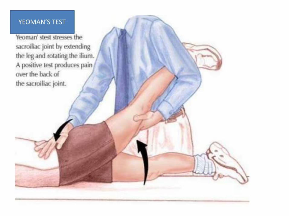
YEOMAN’S TEST

The patient lies supine with the hip and knee flexed where the thigh is at 90° to the table and slightly adducted. One of the examiners hands cups the sacrum and the other arm and hand wraps around the flexed knee. The pressure applied is directed dorsally along the line of the vertically oriented femur. The procedure is carried out on both sides. The presumed action is posterior shearing force to the SIJ of that side
THIGH THRUST TEST

COMPRESSION & DISTRACTION
PROVOCATION TESTS

HIP

• The hip joints should be examined for any decrease in range of motion because hip arthritis, which normally causes groin pain, may occasionally present as LBP
• Trochanteric bursitis with tenderness over the greater trochanter of the femur can be confused with LBP.
• The presence of more widespread tender points, especially in a female patient, suggests the possibility that LBP may be secondary to fibromyalgia.
FUNCTIONAL ASSESSMENT


FUNCTIONAL ASSESSMENT BY
INTERMALLEOLAR DISTANCE

• It has been shown that by using a combination of the distraction, thigh thrust, compression, sacral thrust, Gaenslen’s, and FABER tests, sacroiliac joint pathology is the likely pain generator when three or more of the tests are positive.

EXAMINATION FOR PT.s WITH RADIATING PAIN DOWN THE LIMB


ROOT TENSION SIGNS

Tension at dural sheath
starts from 20 deg. Elev.
BRAGGARD TEST
CROSSED STRAIGHT LEG RAISING TEST


Femoral leg stretch. In the femoral stretch test, the knee is flexed and lifted superiorly. Sharp pain generated in the anterior thigh is considered to constitute a positive test.

TEST INAPPROPRIATE RESPONSE*
Tenderness Superficial, nonanatomic tenderness to light touch
Simulation
Axial loading Vertical loading on a standing patient's skull produces low back pain
Rotation Passive rotation of shoulders and pelvis in same plane causes low back pain
Distraction Discrepancy between findings on sitting and supine straight leg raising tests
Regionaldisturbances
Weakness “Cogwheel” (give-way) weakness
Sensory Nondermatomal sensory loss
Overreaction Disproportionate facial expression, verbalization or tremor during examination
WADDELL'S TESTS FOR NONORGANIC PHYSICAL SIGNS
*—Three or more inappropriate responses suggest complicating psychosocial issues in patients with low back pain.

•DISTRACTION
•OVERREACTION
•REGIONAL DISTURBANCES
•SIMULATED TESTS
•TENDERNESS WHICH IS SUPERFICIAL ( to be differentiated from allodynia)

• McCombe and colleagues evaluated the reproducibility between three observers of physical signs used for back pain evaluation. The signs that were
• measurements of lordosis (by tape measure from the maximum kyphosis of the thoracic spine to that of the sacrum)
• flexion range (Schober test)
• determination of pain location on flexion and lateral bend
• straight-leg-raising test (pendulum goniometer measurement of the angle at which pain was first experienced and angle of maximum tolerance)
• determination of pain location in the thigh and legs
• sensory changes in the legs
• Nerve root tension signs were reliable if the location of pain was described. Reproducibility of bone tenderness over the sacroiliac joints, spinous processes, and iliac crests was greater than that associated with soft tissue structures.
• The diagnostic value of disturbed sensory and motor function was tested prospectively by Jensen in 52 patients with lumbar disc herniations confirmed at surgery.The positive predictive value of disturbed sensation in the L5 dermatome and weakness of foot dorsiflexion was 76% for herniation from the L4/5 lumbar disc. The positive predictive value of altered sensation in the S1 dermatome was only 50% adequately reproducible included the following:

IMAGING
I THINK U WERE TRYING TO SAY MRI

Without the Scout Image, it is going to be impossible for the layperson, as well as most general physicians, to discern which disc is which when viewing the all-important axial images (overhead view of the disc). It's like a roadmap that tells you which slice or current from the sagittal view (from the side) matches up with that same view in the axial plane.For example, the #10 slice in the Scout image to the left would match up with the #10 axial view, which in this case happens to run right through the bottom of the L 4/5 disc.

Gray Rami Communicantes
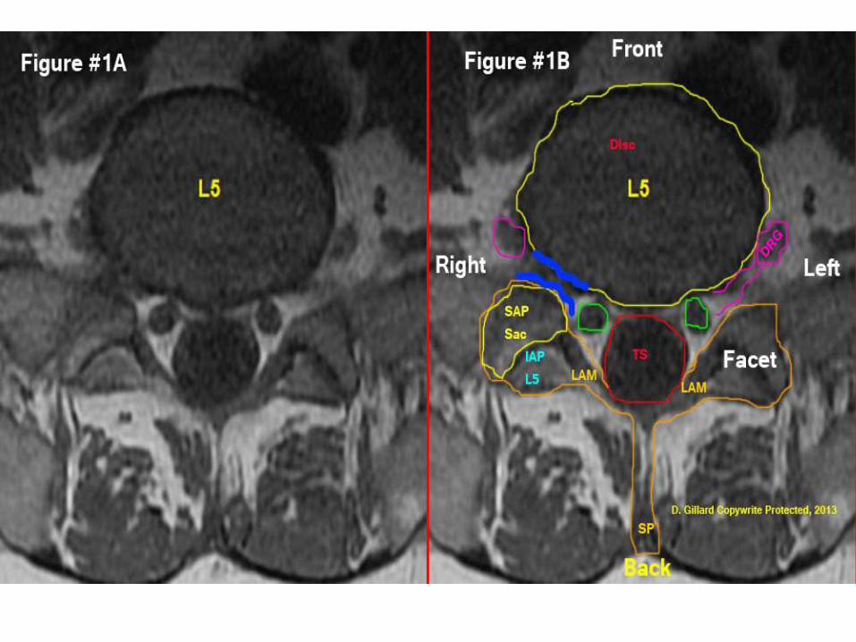

T2 axial mri cut at L4 disc level
demonstrates a T2 weightedaxial of a normal healthyL4 disc from a 45 year-old
-male. Now, because theT2 weighted images showwater content, we can seea distinct nucleus pulp
osus (light-colored centerof the disc) which is surr.by a darker annulus
fibrosis. we can also clearly see the nerve rootswithin the thecal sac.


Sagittal T1 image
This is a T1-weighted sagittal view of the sacrum. The black L5 disc can be seen b/w be L5 vertebra and S1 sacral segment

T2 weight MRI view of the lumbar spine from the side, or a Sagittal Image.First the basic structures: The discs, which are stationed between the vertebrae, should be a white color (hydration).
Note the 'blackness' (desiccation) of the L5 disc (disc between L5 and sacrum); this represents moderate degenerative disc disease. The PLL (tiny blue arrows) appears as a black vertically orientated line running down the posterior surface of the vertebral bodies and disc.
The thecal sac (red stars) is the 'super white' structure that fills the central spinal canal just behind the posterior vertebral bodies. This sac house the free-floating spinal nerve roots (cauda equina) and is made up of both motor and sensory nerve fiber.
The ligamentum Flavum (green star) courses between each of the vertebrae and adds stability to the spine. This structure can hypertrophy or thicken in some patients and help to cause the dreaded central canal stenosis.


This is a sagittal T2 waited MRI, which is a far lateral cut (way off to the edge). This demonstrates the very important neuroforamen and the exiting nerve roots (red arrows) within them.This is a very important slice and both sides should be carefully inspected to make sure no disk herniations have occurred here.Usually disk herniations do not go into this area (the neural foramen); however, when they do, not only can they cause severe sciatica, they can also be quite difficult to reach during discectomy.

Typical signs of inflammatory lesions in ankylosingspondylitis: (A) T1
pre-gadolinium sequence,(B) T1 postgadolinium seq.Thin arrows,
spondylitis anterior (shortarrows) and
posterior (long arrows). Bold arrow,spondylitis ant. Surr--ounding anerosion on the lower edge of the vertebralbody. Circle, inflammationin the
zygoapophyseal joint

spinal fusion (thin arrowshere in the dorsal part of thethoracic vertebrae)
is depicted better in the T1pregadolinium MRI sequence,spinal
inflammation (bold arrows)are only depicted eitherafter application of
gadolinium (B) or in the STIRsequence
(C).

. Coronal T1-weighted MR image
Sacral foramen
Sacroiliac joint

Definition of sacroiliitis highly suggestive of SpA (‘‘positive MRI‘‘) for application in the new ASAS classification criteria(Reproduced from Rudwaleit.)A. Types of findings required for definition of sacroiliitis by MRI• Active inflammatory lesions of the SI joints (reflecting active sacroiliitis) are required for the definition of ‘‘sacroiliitis on MRI’’ as one of the two imaging items in the ASAS classification criteria for axial SpA.• BME (STIR) or osteitis (T1 post-gadolinium)highly suggestive of SpA mustbe clearly present and located inthe typical anatomical areas (subchondral or periarticular bone marrow).• The sole presence of other active inflammatory lesionssuch as synovitis,enthesitis or capsulitis without concomitant BME/osteitis isnot sufficient for the definition of sacroiliitis on MRI.• Structural lesions such as fat deposition, sclerosis, erosions or bony ankylosis are likely to reflect previous inflammation. At this time, however, the consensus group felt that the sole presence of structural lesions without concomitant BME/osteitis does not suffice for thefulfilment of sacroiliitis on MRI in the ASAS classification criteria for axial SpA.B. Amount of signal required• If there is only one signal (lesion) per MRI slice suggesting active inflammation, the lesion should be present on at least two consecutive slices. If there is more than one signal (lesion) on a single slice, one slice may be sufficient.


•Capsulitis is comparable tosynovitis in terms of signal char.but these changes involve the
anterior andposterior capsule.Anteriorly, the joint capsulegradually continues into theperiosteum of the iliac and
sacral bones andthus corresponds to an enthesis.Capsulitis, therefore, may
extend far medially and laterallyinto the periosteum. Capsulitis
may be better detectable usingcontrast-enhanced T1-weightedfat-saturated images ascompared to STIR

Hyperintense signal onSTIR images and/or oncontrast-enhancedT1-weighted fat-saturtdimages at sites whereligaments and tendonsattach to bone, includingthe retroarticular space(interosseous ligaments)
. The signal may extendto bone marrow andsoft tissue. Enthesitis
may be better detectableusing contrast-enhancedT1-weighted fat-saturtdimages as compared toSTIR.
A. 1: Enthesitis (white arrow) of interosseous ligaments (contrast-enhanced T1-weighted fat-saturated images; coronal view). Also present: osteitis ofthe left iliac bone (black arrow). 2: Enthesitis (white arrows) of interosseous ligaments (contrast-enhanced T1-weighted fat-saturated images;axialview). Also present: osteitis of the left sacroiliac joint (black arrow).B. 1: Enthesitis (arrow) of interosseous ligaments (short tau inversion recovery(STIR)). 2: Enthesitis (arrow) of interosseous ligaments (STIR)


SURGEONS’ ASSISTANCE


GRAY ZONE
• PIRIFORMIS SYNDROME
• LUMBOSACRAL TRANSIENT VERTEBRA
• S.I. JOINT DYSFUNCTION
• BACK MOUSE
• EPIDURAL LIPOMATOSIS
• PREGNANCY

THAT’S ALL FOLKS