low cost mobile eeg for characterization of cortical ... cost mobile eeg for characterization of...
TRANSCRIPT
Low Cost Mobile EEG for Characterization of Cortical Auditory Responses
Bathiya Senevirathna1,2, Lauren Berman1, Nicola Bertoni4, Fabio Pareschi4, Mauro Mangia5, Riccardo Rovatti5, Gianluca Setti4, Jonathan Simon1,2,3, Pamela Abshire1,2
1Department of Electrical and Computer Engineering, 2Institute for Systems Research, 3Department of Biology, University of Maryland, College Park, MD, USA. Email: bsenevir, lauren16, jzsimon, [email protected]
4ENDIF, University of Ferrara, Italy. Email: nicola.bertoni@, fabio.pareschi, [email protected] 5DEI, University of Bologna, Italy. Email: mauro.mangia2, [email protected]
Abstract— We report a low cost mobile EEG system for characterizing cortical auditory responses. The system is built using commercial off-the-shelf components and each unit costs less than $200. It measures seven EEG channels plus one audio channel (envelope only), and communicates the data to external devices via Bluetooth. A novel implementation was pursued in order to support local signal compression using compressed sensing. At the same time, it provides a low cost solution that is useful for recording cortical auditory responses and extracting clinically relevant features of the waveform. This system has been designed with the eventual goal of long term monitoring of the brain activity of schizophrenic patients outside a clinical setting, in order to better understand auditory hallucinations and manage their ongoing treatment. In this preliminary study we obtained simultaneous audio and cortical recordings of evoked auditory responses from normal healthy subjects wearing the EEG for several hours in duration. We report evoked auditory responses for 2 Hz and 40 Hz click trains. We also report alpha wave responses, demonstrating stable and high quality recordings over a five hour period.
Keywords—EEG; auditory evoked potentials; low cost; schizophrenia
I. INTRODUCTION
Mobile, wireless electroencephalography (EEG) systems have become commonplace in recent years. These systems record electrical potentials from the surface of the scalp, which are generated by neural activity within the brain. EEG provides a noninvasive methodology to monitor brain activity, and wireless, wearable EEG systems provide the capability to monitor brain activity outside the laboratory, in normal everyday environments and during activities of daily living.
While there is great potential for using such systems in research, clinical and commercial applications, many technical challenges and barriers remain. These include the stability of recordings over time, low intrinsic signal quality, corruption by artifacts and noise, and cost. There are many existing commercial (Cognionics, Emotiv, NeuroMonitor, BioNomadix, BioSemi) and even open source (openBCI, openEEG) mobile EEG systems. Low-end commercial systems offer few recording channels and limited signal fidelity while more advanced systems offer clinical grade recording capability with many channels at a significantly increased cost. A single clinical grade system typically costs upwards of
$20,000, an expense that is prohibitive for many investigatory studies and limits the widespread adoption of mobile EEG systems.
Mental health is an important emerging application domain for mobile EEG systems. Schizophrenia is a brain disorder that is accompanied by auditory verbal hallucinations for more than 60-80% of patients [1]. These hallucinations arise from abnormal brain responses in the cortex. EEG has been shown to be useful in several studies that have characterized EEG recordings from schizophrenic patients during auditory hallucinations [2].
In this paper we report a low cost EEG system for characterizing cortical auditory responses. The eventual goal is to allow long term monitoring of schizophrenic patients outside a clinical setting, in order to better understand auditory hallucinations and manage their ongoing treatment. The system is built using commercial off-the-shelf components and measures seven EEG channels plus one audio channel (envelope only). It communicates the data to external devices via Bluetooth. This new system is a low cost solution that provides the EEG signal quality required for characterization of cortical auditory responses. Additionally, the system architecture has been designed to support future data compression via compressed sensing in order to reduce the data transmission bandwidth and preserve battery life for long term recording.
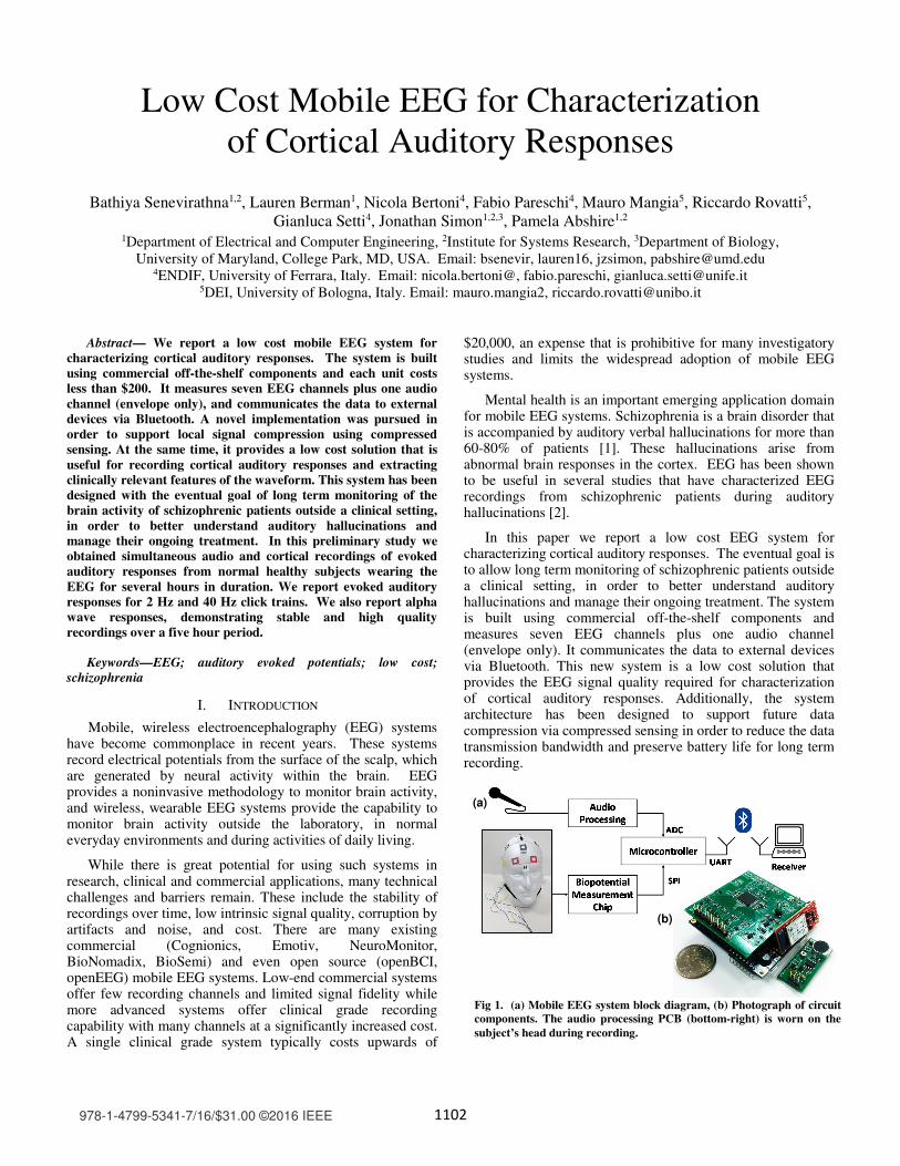
Fig 1. (a) Mobile EEG system block diagram, (b) Photograph of circuit components. The audio processing PCB (bottom-right) is worn on the subject’s head during recording.
(a)
(b)
978-1-4799-5341-7/16/$31.00 ©2016 IEEE
1102

II. SYSTEM IMPLEMENTATION
A block diagram of the system architecture is shown in Fig 1(a). An analog front end (AFE), consisting of a biopotential measurement chip and audio processing circuitry, acquires the EEG and audio signals. It sends the data to a microcontroller which then samples the audio envelope, performs desired signal processing on the EEG waveform, and sends the data to a Bluetooth module for transmission to a host computer.
A. System components
The device uses a commercial biopotential measurement chip, the TI ADS1299, which is designed for high precision recording of ECG and EEG signals. This chip consists of an eight channel, 24 bit recording system using a sigma-delta analog-to-digital converter (ADC). This ADC interfaces with a high performance microcontroller, and the recorded EEG signals are transmitted wirelessly to a base station using Bluetooth. This measurement chip has been used in many other EEG/ECG/EMG systems [3]–[6].
The current prototype device consists of a custom made PCB that houses the AFE chip along with ancillary power supply regulation circuitry. This board then connects to the microcontroller board (SAM G55 Xplained Pro, Atmel), which contains a SAM G55 microcontroller chip, and connects to the wireless transceiver (Bluetooth Mate Silver, SparkFun). The microcontroller communicates serially with both the ADC and wireless transceiver using SPI and UART, respectively. The ADC is configured to sample EEG signals from up to 8 channels at 250 SPS, with a gain of 12. The wireless transceiver is set to run at a 230.4 kBaud rate. All circuitry and components are battery powered using a high capacity 3.3 V (2500 mAh) lithium polymer battery. A DC-DC converter is used to provide the 5 V supply needed for the electronic components. The current prototype device consumes a maximum of 69 mA with all channels active.
The prototype additionally includes audio recording circuitry to allow for correlation of environmental sounds and EEG responses. Audio signals generally require Nyquist sampling rates above 30 kHz. Therefore to facilitate both audio and EEG sampling, and subsequent wireless transmission, the audio signal was processed to obtain its amplitude envelope. This was performed using standard precision peak rectifier and low pass filter circuitry. The prototype measures 6 x 6 x 3 cm3, and weighs 93 g. A photograph of the design is shown in Fig 1(b). The total cost of the device, including microcontroller, AFE components, and PCB manufacturing came to $180 with an additional $4 for each electrode used.
B. Data acquisition
A graphical user interface (GUI) was created in MATLAB to wirelessly communicate with the aEEG system. It allows the technician to enable/disable channels, change gain settings, adjust sampling rate, and see recordings in real time. The GUI is used to control a low-level dedicated serial port monitor (RealTerm: Serial Capture 2.0) which writes the incoming data stream into a binary file. This first ensures no loss of data if the wireless connection is interrupted, and second obviates the use
of high-level software such as MATLAB that is inefficient for meeting higher bandwidth (>6 channels) requirements.
1) Signal processing Raw time-domain signals recorded from the device were
first digitally bandpass filtered from 1 – 55 Hz using finite impulse response (FIR) filters, unless otherwise noted. The power spectral density (PSD) of signals was also computed in MATLAB using the Welch method with a window length of 512 samples and 500 sample overlap. In the following experiments, an auditory stimulus was presented to subjects and the resulting EEG activity was recorded. This type of correlation requires the need for precise temporal alignment of stimulus and corresponding response. Many commercially available EEG recording systems have methods of integrating external event triggers through parallel ports, serial ports, or binary TTL pulse [7]. For this system, time alignment is performed directly in software as the event triggers are part of the data stream itself. As described in Section II-A, an audio channel is obtained concurrently with the EEG data stream. Further details are discussed in Section III-B.
2) Experimental protocol Experiments were conducted to assess the quality of the
EEG system in response to a series of audio recordings played for subjects. In the first part of each experiment, subjects were kept in silence and instructed to keep their eyes open for 1 minute, followed by 1 minute with eyes closed. This test allows for evaluation of the alpha rhythm, a characteristic 8-12 Hz EEG signal that allows for simple validation of the signal quality [8]. In the second part of each experiment, a synthesized auditory click train was played for subjects at frequencies of 2 Hz and 40 Hz, for 2 minutes each. These types of low and high frequency auditory stimuli have been shown to allow for discrimination between healthy and schizophrenic subjects [9].
Standard gold-plated brass cup electrodes were used with electrode paste (Ten20 Paste, Weaver and Company). Signals were recorded simultaneously from positions FZ, CZ, PZ, and O2 of the standard 10/20 placement system [10]. Reference and ground (driven for common-mode cancellation) were placed at the left and right mastoid, respectively. Subjects were seated in a closed room and recording sessions for both parts of the experiment took place within 30 minutes, including electrode setup time. Additionally, extended testing periods of five hours were conducted to ascertain longer term signal quality. During these sessions, subjects kept their placed electrodes and were brought in intermittently for closed-environment testing.
III. RESULTS
A. Alpha wave activity
Fig 2 shows a 20 second sample EEG signal recorded from an electrode placed at the O2 position. The subject had their eyes open from the start of the test and closed their eyes at t = 8 seconds. After the subject’s eyes close, the recording shows the onset of alpha wave activity, indicated by the strong 10–12 Hz waves. This is also seen in the time-frequency plot of Fig 2. The top panel of Fig 3 shows the corresponding PSD of the
1103
same two signal segments. It confirms an increase of power in the 10-12 Hz band by 8 dB.
The effects of extended durations on the quality of recorded EEG signals were also studied. The bottom three panels of Fig 3 show the PSD plots of recordings taken from a single subject over the course of five hours. Panels (b), (c), and (d) show data recorded 30 min, 1.5 hours, and 5 hours after set-up of electrodes, respectively. It can be seen that there is no degradation of signal quality over this time period. In fact results for the first recording at t = 30 min appear to have the least alpha activity. This implies that the subject’s state at the time of recording is more influential in signal quality than degradation of electrodes. Indeed, it was noted that electrode-scalp interfacial impedance stabilized at roughly 1–1.5 hours after the wet electrodes were applied. However, further study is necessary to check the quality over longer (8+ hours) periods of time, and it is expected that the electrode paste eventually dries out [11]. Additionally, differences in the rate of change of electrode impedance can cause mismatches in the resulting signal attenuation between scalp and electrode. This means that comparative differences in signal amplitude between different channels may be the result of electrode mismatch rather than actual auditory evoked responses.
B. Click train activity
The presentation of an auditory stimulus to subjects can induce a corresponding response in normal EEG channels,
called auditory evoked potentials (AEPs) [12]. AEPs are a normal human response to sensory stimulation. Two click trains of 2 Hz and 40 Hz clicking frequency were played for subjects. The 2 Hz click train consists of a series of 240 audible clicks played over a period of 2 minutes. The data was divided into epochs corresponding to each audible click, with a duration of 480 ms. Responses were aligned according to the onset time of the stimuli, with correction for the delay introduced by the audio processing circuit and the microcontroller's ADC conversion time. The estimated response was obtained by averaging over repetitions in order to improve signal-to-noise (SNR) ratio [12]. Fig 4 shows the resulting AEP waveform on the CZ channel, for different number of repetitions (N) used in averaging.
One can observe a characteristic late-latency AEP that
ranges from the positive peak P1 around 50 ms, to the negative trough N2 around 225 ms. This AEP is a normal response to the auditory stimulus and originates from the auditory cortex [13]. Averaging over 200 repetitions produces a clear AEP, with similar results obtained by averaging over 100 and 150 windows. However as expected, averaging over smaller numbers of repetitions yields less improvement in the SNR, as is evident for the 50 window average.
The top panel of Fig 5 shows the PSD of the evoked auditory response to a 40 Hz click train at the CZ and O2 electrode positions. Recordings from this experiment were bandpass filtered from 3 – 55 Hz. A strong response at 40 Hz can be seen in the PSD at the CZ position, whereas most of the
Fig 2. Top: A sample 20 second EEG signal with transition from open to close eyes at t = 8 seconds, resulting in the onset of alpha wave generation, Bottom: Spectrogram of signal highlighting 12 Hz (color scale in units of dB/Hz)
Fig 3. PSD recordings with (solid) and without (dashed) eyes closed. Panel (a) corresponds to the recording shown in Fig 2. Panels (b)-(d) show extended duration recordings, with time t corresponding to time of recording, after initial electrode placement.
Fig 4. Example auditory response to 2 Hz click train on channel CZ. Improvement of SNR can be accomplished by increasing number of epochs used in averaging.
Fig 5. Top: Power Spectral Density of auditory response to 40 Hz click train on channels CZ (solid) and O2 (dashed). Bottom: Spectrogram of the CZ signal
P1
P2
N1
t = 0.5 hrs
N2
t = 1.5 hrs
t = 5.0 hrs
(a)
(b)
(c)
Time (s)
(d)
CZ O2
Mag
nitu
de (d
B)
1104

signal power is concentrated around 10 Hz for the O2 channel. This is indicative of the 40 Hz response being a cortical auditory response, rather than a measurement artifact. The bottom panel shows the time-frequency plot of the CZ channel recording. The onset and offset of the 40 Hz evoked response correlates with the start (12 s) and end (132 s) of the audible click train.
IV. CONCLUSION & FUTURE WORK
A design for a wireless, mobile, and low cost EEG recording system has been presented for use in cortical auditory studies. The system’s material cost of under $200 is lower than commercial and clinical grade systems that range from tens of hundreds to tens of thousands of dollars. Experiments were conducted to evaluate the recorded signal quality for detection of alpha rhythms and auditory evoked potentials. Preliminary results show a clear ability to detect AEPs in standard evoked auditory response studies.
Furthermore the system has the capability to support additional signal processing prior to transmission. In the future we intend to implement rakeness-CS methods for signal compression in order to reduce data transmission bandwidth [14], as described in a companion paper.
We anticipate the need for future improvements for use in real-world situations. The use of wet electrodes is not ideal for longer-term mobile applications due to the time and care required to properly affix the electrodes, and the change in electrode contact impedance over time. As such the development of dry electrodes has been of increasing interest, and dry electrode systems have been reported that make use of silicone rubber [15], active shielding [16], and flexible contacts [17].
We explored the use of sintered Ag/AgCl dry electrodes (TDE-200, Florida Research International), but were unable to obtain stable and reliable without application of an adhesive. Fig 6 shows EEG recordings made simultaneously from dry and wet electrodes, and highlights the issue of poor electrode-scalp contact. The raw time-domain data was high-pass filtered with a 3dB cutoff frequency of 2 Hz. A DC offset was added to separate the traces for plotting. The upper trace, corresponding to the dry electrode, shows significantly higher high frequency noise than the wet electrode. Signal quality is improved if the electrode is physically forced against the scalp but this may be impractical for real-world use without a purpose-built dry electrode cap [16], [17] or application of biocompatible adhesives [15].
ACKNOWLEDGMENT
We would like to thank the University of Maryland A. J. Clark School of Engineering’s "Engineering Systems for Brain Health Management" seed grant program for funding this work.
REFERENCES
[1] J. Graux, A. Bidet-Caulet, F. Bonnet-Brilhault, V. Camus, and N. Bruneau, “Hallucinations and negative symptoms differentially revealed by frontal and temporal responses to speech in schizophrenia,” Schizophr. Res., vol. 155, no. 1–3, pp. 39–44, 2014.
[2] R. van Lutterveld, I. E. C. Sommer, and J. M. Ford, “The neurophysiology of auditory hallucinations - A historical and contemporary review,” Front. Psychiatry, vol. 2, no. 28, pp. 1–7, 2011.
[3] C. Audette, C. Russomanno, W. Croft, R. Ceballos, and J. Murphy, “OpenBCI,” 2015. http://www.openbci.com.
[4] J. Ruelas, “EEG64 | A low cost, open source 64 channel EEG system,” 2015. http://www.eeg64.com/.
[5] J. A. Lovelace, T. S. Witt, and F. R. Beyette, “Bluetooth Enabled Electroencephalograph ( EEG ) Platform,” in Int. Midwest Symp. Circuits Syst, 2013, pp. 1172–1175.
[6] H. Converse, T. Ferraro, D. Jean, L. Jones, V. Mendhiratta, E. Naviasky, M. Par, T. Rimlinger, S. Southall, J. Sprenkle, and P. Abshire, “An EMG biofeedback device for video game use in forearm physiotherapy,” in Proc. IEEE Sensors, 2013, pp. 1–4.
[7] W. David Hairston, K. W. Whitaker, A. J. Ries, J. M. Vettel, J. Cortney Bradford, S. E. Kerick, and K. McDowell, “Usability of four commercially-oriented EEG systems,” J. Neural Eng., vol. 11, no. 4, 2014.
[8] R. I. Goldman, J. M. Stern, J. Engel, and M. S. Cohen, “Simultaneous EEG and fMRI of the alpha rhythm.,” Neuroreport, vol. 13, no. 18, pp. 2487–92, 2002.
[9] J. P. Hamm, C. S. Gilmore, N. A. M. Picchetti, S. R. Sponheim, and B. A. Clementz, “Abnormalities of neuronal oscillations and temporal integration to low- and high-frequency auditory stimulation in schizophrenia,” Biol. Psychiatry, vol. 69, no. 10, pp. 989–996, 2011.
[10] H. H. Jasper, “The ten twenty electrode system of the international federation,” Electroencephalogr. Clin. Neurophysiol., vol. 10, pp. 370–375, 1958.
[11] T. C. Ferree, P. Luu, G. S. Russell, and D. M. Tucker, “Scalp electrode impedance, infection risk, and EEG data quality,” Clin. Neurophysiol., vol. 112, no. 3, pp. 536–544, 2001.
[12] P. Kidmose, D. Looney, and D. P. Mandic, “Auditory evoked responses from Ear-EEG recordings,” Proc. Annu. Int. Conf. IEEE Eng. Med. Biol. Soc. EMBS, pp. 586–589, 2012.
[13] N. Kraus and T. Nicol, “Auditory Evoked Potentials,” Encycl. Neurosci., pp. 214–217, 2009.
[14] M. Mangia, R. Rovatti, and G. Setti, “Rakeness in the design of analog-to-information conversion of sparse and localized signals,” IEEE Trans. Circuits Syst. I, Reg. Papers., vol. 59, no. 5, pp. 1001–1014, 2012.
[15] G. Gargiulo, R. a. Calvo, P. Bifulco, M. Cesarelli, C. Jin, A. Mohamed, and A. van Schaik, “A new EEG recording system for passive dry electrodes,” Clin. Neurophysiol., vol. 121, no. 5, pp. 686–693, 2010.
[16] g.Tec Medical Instruments, “g.SAHARAelectrode - g.Electrodes,” 2015. http://www.gtec.at/Products/Electrodes-and-Sensors/g.Electrodes-Specs-Features.
[17] Cognionics Inc., “Flex Sensors (for hair) - Cognionics,” 2014. http://cognionics.com/images/Cognionics/spec_flex.pdf.
Fig 6. Comparison of recorded signal quality with dry and wet electrodes. Upper: dry electrode at CZ. Bottom: wet electrode at O2.
Ampl
itude
(V)
1105