low cost subatmospheric pressure therapy as an alternative
TRANSCRIPT

Rev Med (São Paulo). 2021 July-Aug;100(4):417-23.
417
doi: http://dx.doi.org/10.11606/issn.1679-9836.v100i4p417-423
Low cost subatmospheric pressure therapy as an alternative to temporary abdominal closure in a public hospital: case report
Terapia por pressão subatmosférica de baixo custo como alternativa para fechamento abdominal temporário em hospital público: relato de caso
Matheus Dantas Gomes Gonçalves1, Diego Laurentino Lima2, Raquel Nogueira Cordeiro3, Fabio Eduardo Revorêdo Rabelo Ferreira4, Carlos Esdras Almeida Moraes5,
Márcio Rogério Carneiro de Carvalho6
Gonçalves MDG, Lima DL, Cordeiro RN, Ferreira FERR, Moraes CEA, Carvalho MRC. Low cost subatmospheric pressure therapy as an alternative to temporary abdominal closure in a public hospital: case report / Terapia por pressão subatmosférica de baixo custo como alternativa para fechamento abdominal temporário em hospital público: relato de caso. Rev Med (São Paulo). 2021 July-Aug;100(4):417-23.
1. MD. Medical General Surgery Resident - Advanced Program, State public server Hospital – IAMSPE, São Paulo, SP. https://orcid.org/0000-0003-3469-1068. E-mail: [email protected].
2. MD. General Surgeon. Research fellow, Department of Surgery, Montefiore Medical Center, NYC, US. https://orcid.org/0000-0001-7383-1284. E-mail: [email protected].
3. MD. Physician and Researcher. Pernambuco Health College, Recife, PE. https://orcid.org/0000-0002-0238-8374. E-mail: [email protected]. MD. Department of General Surgery, Getulio Vargas Hospital, Recife, PE. https://orcid.org/0000-0003-2272-159X. E-mail: [email protected]. MD. Department of General Surgery, Getulio Vargas Hospital, Recife, PE. https://orcid.org/0000-0002-4712-2473. E-mail: [email protected]. MD. Department of General Surgery, Getulio Vargas Hospital, Recife, PE.https://orcid.org/0000-0002-2518-2707. E-mail: [email protected]: Diego Laurentino Lima. 1741 Seminole Avenue, The Bronx, New York - US. 10461-1807. E-mail: [email protected].
ABSTRACT: The use of sub atmospheric pressure through a vacuum dressing in patients with open abdomen is increasingly used for temporary abdominal closure. Vacuum curative can be used as a bridge between the onset of the initial lesion and surgery for definitive closure of the wound, in those cases in which primary fascial closure is not possible. The objective of this work is to present a practical proposal for temporary abdominal closure using vacuum curative with NPWT (modified Barker’s vacuum-packing technique - BVPT), as alternatives to the other techniques. The mechanism of action involves the control of exudate, reduction of perilesional interstitial edema, increased blood flow, stimulation of granulation tissue formation, and wound contraction. The curative presented low cost, easy application and using materials frequently available in most public hospitals. According to the literature, the modified BVPT presents good results and rates of complications similar to the commercial NPWT kits.
Keywords: Vacuum dressing; Temporary abdominal closure; Open abdomen; Modified Barker’s vacuum-packing technique.
RESUMO: A utilização da pressão subatmosférica através de curativo à vácuo em pacientes com abdome aberto é usada cada vez mais para o fechamento abdominal temporário. O curativo à vácuo pode ser utilizado como uma “ponte” entre o surgimento da lesão inicial e a cirurgia de fechamento definitivo da ferida, naqueles casos em que o fechamento fascial primário não é possível. O objetivo deste trabalho é apresentar uma proposta prática para o FAT utilizando curativo à vácuo (técnica de Barker modificada), como alternativas as demais técnicas. O mecanismo de ação envolve o controle do exsudato, redução do edema intersticial perilesional, aumento do fluxo sanguíneo, estímulo à formação de tecido de granulação e contração da ferida. O curativo apresentou baixos custos, fácil aplicação e utilizando materiais disponíveis com frequência na maioria dos hospitais públicos. De acordo com a literatura, a técnica de Barker modificada apresenta bons resultados e índices de complicações semelhantes aos kits de TPN comerciais.
Palavras-chave: Curativo à vácuo; Fechamento abdominal temporário; Abdome aberto; Técnica de Barker modificada.

418
Gonçalves MDG, et al. Low cost subatmospheric pressure therapy as an alternative to temporary abdominal
INTRODUCTION
Vacuum dressing is already widespread and has become an adjunct method in the treatment of
complex wounds, being used among different specialties, surgical or not. It consists of an active wound treatment that promotes its healing in a humid environment through controlled sub atmospheric pressure and applied locality1.
The dressing is composed of an interface material (foam or gauze), through which sub atmospheric pressure is applied and the exudate is removed. The interface material is put in contact with the wound bed to cover its entire length, including tunnels and cavities. This is covered by a transparent adhesive film, which totally isolates the wound from the external environment. A suction tube is connected to this system and to the exudate reservoir, which is adapted to a computerized device, in commercial kits, and it is possible to provide a sub atmospheric pressure in the wound bed1.
Patients with open abdomen have high morbidity and mortality rates, in addition to increased overall treatment costs (supplies and human resources) and longer length of stay. In these cases, there is a need for a temporary abdominal closure that provide for control of exudate, reduction of perilesional interstitial edema, increased blood flow, stimulation of granulation tissue formation, wound contraction, easy application and that are relatively inexpensive2.
The potential for economic and social impact of this situation has stimulated investment in dressings with new technologies. It is mandatory that the surgeon knows alternatives that can accelerate the process of repair of a wound, allowing the patient to be discharged earlier and return to their daily activities.
CASE REPORT
A 48-year-old male patient, paraplegic for 20 years and with ileostomy secondary to abdominal gunshot wound, admitted to the emergency room presenting parastomal hernia with recurrent semi occlusive symptoms. After failure of clinical treatment, the patient underwent laparotomy, which showed slender content with significant distension upstream of the segment present in parastomal hernia. Enterectomy of distal ileum was performed, with lateral-lateral anastomosis of ascending ileus-colon with linear stapler and loop colostomy with transverse colon. On the fifth postoperative day, there was dehiscence of the ascending ileus-colon anastomosis with a large amount of secretion with fecaloid aspect in all quadrants, and distal enterectomy, partial colectomy, ileostomy and mucous fistula were performed.
The patient underwent a new laparotomy with cavitary toilet and enterorrhaphy, in the 8th postoperative
day, due to fecal peritonitis by punctiform perforation in a small bowel loop at 80 cm from the Treitz angle. At the 12th postoperative day of the first surgery, the patient underwent a new laparotomy due to fecal peritonitis with a punctiform lesion in distal ileum approximately 2 cm from the fixation point of the previous ileostomy. Performed cavity toilet with remaking of Brooke’s ileostomy and peritoneostomy with the Bogota Bag technique. Three days after the last surgical procedure, due to accumulation of a large amount of intra-abdominal secretion, Bogota Bag was removed (Figure 1) and vacuum dressing was made (Figure 2), after a new cavity toilet. At this moment, an open abdomen with a wound contaminated with adhesions was evidenced, according to the classification of Björck et al.3.
Figure 1: Open abdomen at the beginning of the case (GRADE 2B) after Bogota Bag withdrawal
Figure 2: Making the first dressing
Vacuum dressing was performed in a modified model of the Barker technique. Used materials easily
419
Gonçalves MDG, et al. Low cost subatmospheric pressure therapy as an alternative to temporary abdominal
available in the public service hospital environment, such as plastic cover of laparoscopy, polyurethane sponges - asepsis brush, portvac drain 6.4mm, transparent adhesive plastic film polyurethane, mobile vacuum from operating room or present at the ward (Figure 3).
Figure 3: Materials used - sterile film (videolaparoscopic plastic cover), sterile surgical compresses, 6.4mm portvac drain, transparent polyurethane adhesive plastic film, mobile vacuum cleaner from room operating or wall fountain
At the end of the procedure, the abdomen was hermetically closed, and the drain was connected to the suction system, presenting retraction of the wound edges and beginning of cavitary drainage (Figure 4). The system was maintained at continuous sub atmospheric pressure of 20-30kpa (equivalent to negative 125mmhg).
The patient was referred to the surgery ward where the mobile vacuum system was connected to the hospital’s gas network system. It showed good drainage of cavity fluid and dressing change after 7 days, due to accumulation of secretions in the sponges and small leaks in the lower part of the dressing. At the time of the second exchange, the onset of tissue granulation has already been noticed. A new dressing was changed on the 12th day after the beginning of treatment, presenting again good aspect of wound edges and formation of thin tissue carapace over intestinal loops (Figure 5).
Figure 4: End of the first dressing, before and after connection to the vacuum system, respectively (D0)
Figure 5: Image of the second dressing before its removal and after removal, respectively (D 12
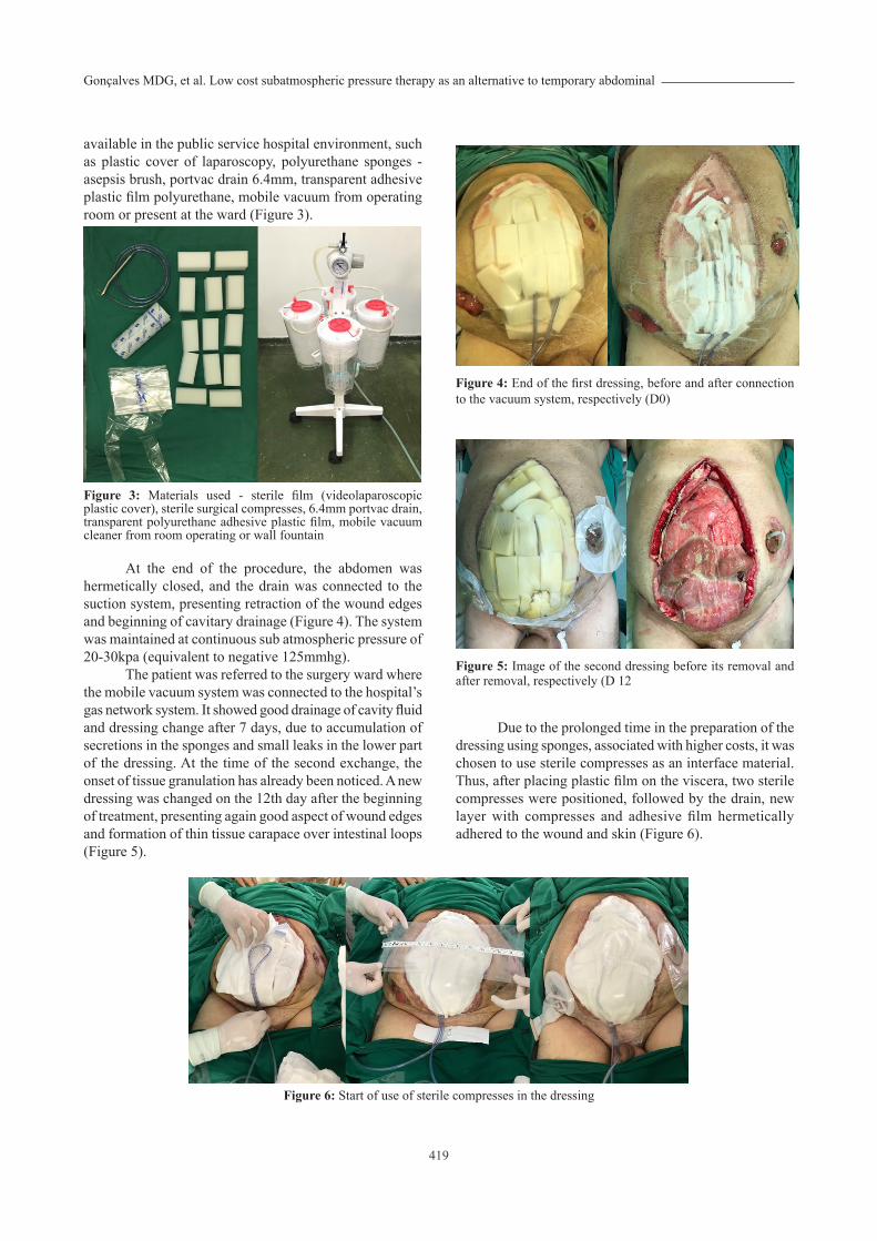
Due to the prolonged time in the preparation of the dressing using sponges, associated with higher costs, it was chosen to use sterile compresses as an interface material. Thus, after placing plastic film on the viscera, two sterile compresses were positioned, followed by the drain, new layer with compresses and adhesive film hermetically adhered to the wound and skin (Figure 6).
Figure 6: Start of use of sterile compresses in the dressing

420
Gonçalves MDG, et al. Low cost subatmospheric pressure therapy as an alternative to temporary abdominal
The use of compresses showed satisfactory results similar to that found with polyurethane sponges. Exchanges were performed at intervals of up to six days. After removal of the fourth dressing, a good aspect of the wound and the central block formation were noted, with a thicker tissue carapace. The surgical team opted at this time to approximate the aponeurosis with two Smead-Jones stitches with Prolene® 2 after placement of the initial compresses (Figure 7). To preserve the plastic film and prevent leaks, the knots of the approach points were made inside a disposable plastic syringe plunger, and, thus, there was no direct contact of its surface with the film.
Figure 7: Aspect after removal of the fourth dressing and preparation of the fifth dressing with approximation points (D17), respectively
The dressing changes continued in the intervals previously mentioned, presenting a gradual reduction of contamination and drainage of the cavity. After the removal of the sixth dressing, it was noticed the beginning of the approach of the edges of the wound and well-established central block. The surgery team opted to continue approximating the edges, now in the lower part of the abdomen (Figure 8).
Figure 8: Aspect after removal of the sixth dressing and preparation of the seventh dressing, maintaining the approximation points (D24), respectively.
The team performed three more dressing changes, and the wound showed improvement. Open abdomen closure with tissue formation covering the viscera and absence of secretions was evidenced, constituting a complex ventral hernia. The patient underwent vacuum dressing therapy for 38 days, totaling 10 exchanges and with joint outpatient follow-up between general surgery and the nursing team of the hospital’s curative committee (Figure 9).
Figure 9: Aspect after removal of the last dressing (D38) and wound after 18 days of discharge, respectively
DISCUSSION
The limited resources in Brazilian public health system and the growing number of complex wounds in our society have motivated the development of new methods for the application of sub atmospheric pressure under vacuum dressing. This demand to develop new types of vacuum dressings using sub atmospheric pressure is an attempt to provide the best treatments with cost reduction.
Although the modified vacuum dressing presents more expensive values and materials for its manufacture, compared to the treatment with the Bogota bag, there is benefit in its use. These values are compensated by saving nursing work hours by dressing changes, reduction of hospitalization time and ineffectiveness of previous treatments, since there is savings of several daily changes of secondary dressings (gauze, compresses, adhesives) drenched by exudate and effectiveness in treatment with sub atmospheric pressure1,4,7,14.
The vacuum dressing allows uniform distribution of sub atmospheric pressure in the wound bed. Its mechanisms of action involve biological (change in the conformation of the cytoskeletal cells, stimulation of granulation tissue formation, reduction of local inflammatory response) and physical (increased blood flow to the wound, reduction of edema and control of exudate, reduction of wound

421
Gonçalves MDG, et al. Low cost subatmospheric pressure therapy as an alternative to temporary abdominal
dimensions, clearance of bacterial load)1.Although Bruhin et al.7 recommended the continuous
use of sub atmospheric pressure with negative 80 mmHg, other studies claimed that higher continuous pressures are associated with better results in angiogenesis and approximation of wound edges5. When choosing the pressure level, a balance should be found between potential damage to the underlying organs (supporting lower pressures) and effective fluid removal (negative pressures of up to 120 mmHg leads to greater fluid drainage). There is no evidence on which blood pressure level is associated with the onset of complications.
Kamamoto5, in his doctoral thesis, proved that the use of barker’s technique modified for traumatic wounds obtained significantly statistical outcomes when compared with VAC® in the same pathologies, associated with great cost reduction, being the treatment of choice in the orthopedics service of the University of Sao Paulo.
Initially, in order to resemble polyurethane foam used as an interface material in VAC®, polyurethane sponges (component of the asepsis brush) were used as an interface material. This foam acts similarly in all commercial products and is able to compress under negative pressure, which can lead to a better preservation of the abdominal domain by mediating the constant medial traction of the abdominal wall, which is not possible with surgical compresses according to some studies4.
After performing a literature review, we chose to modify the dressing by replacing polyurethane sponges with sterile surgical compresses. This modification was done due to the prolonged time to pierce each sponge through the drain in the preparation of the dressing and in an attempt to further reduce costs. Thus, after the protection of the viscera, two sterile surgical compresses were placed along the fascial opening and a 6.4mm portvac drain was positioned on them with a fold on its own axis to obtain greater extension over the desired area and more uniform distribution. Then, the team applied another layer with two compresses, coated with plastic film adhered to the skin (3M, Curatec) and connection of the drain to the suction system.
For the suction system, a mobile vacuum from operating room was used that is connected to the hospital’s gas system, adjusted to a continuous sub atmospheric pressure of 20-30kpa (equivalent to negative 125mmhg). Another possibility is to connect the suction system of the dressing to the vacuum source available in the headboards of the beds in the wards, but it is not possible to maintain a continuous pressure accurately. Both systems make it possible to collect and drain the peritoneal liquid, and thus estimate losses with accuracy2.
Due to the nature of the pathology and surgical lesion, patients who are candidates for open abdomen may require multiple surgical interventions until adequate control and/or definitive resolution of the abdominal index
event is achieved. This may be associated with significant complications, including enteroatmospheric fistulas, loss of abdominal wall domain, and large hernias of the abdominal wall8.
Our dressing model was similar to several others in the literature that use subatmospheric pressure in the making of the modified Barker technique2. Despite its rusticity, dressing models with the modified Barker technique present similar rates of primary fascial closure, enteroatmospheric fistula and mortality when compared to other commercial dressings (VAC®, ABThera®).
Studies have demonstrated the need for early fascial closure (within 8 days) to significantly reduce complications of open abdomen (dysregulation of fluids and electrolyte balance, gastrointestinal fistula, adhesions, intra-abdominal infection, respiratory disorders)4. Among the main outcomes of all therapies used in open abdomen, primary fascial closure stands out. Barker et al.2 obtained the primary closure of open abdominal wounds in 68.4% of the patients after the use of their technique.
Using the modified Barker Technique in 74 patients over five years, Ozguc et al.9 obtained a rate of 45% in primary fascial closure and also an incisional hernia frequency of 50% in patients who underwent primary closure. None of them presented intestinal fistulas9. Montori et al.11 presented a fascial closure rate of 75.4% versus 93.8% (p = 0.10) when compared with ABThera® and Modified Barker Technique, respectively, in patients with intra-abdominal sepsis or abdominal trauma. However, the increase in primary fascial closure rate in the modified Barker technique group may present some bias due to the due to the addition of a retaining sequential fascial closure.
Among these techniques, this is the association of synthetic fabric (fixed in healthy aponeurosis) on the lining of the intestinal loops and its approximation to the midline after each dressing change, as performed in the work of Tolonen et al.12. In these cases, the mesh is sectioned in the midline, followed by the exchange of the protective material of the bowel loops, accompanied by approximation of the mesh with continuous suture and placement of the dressing in vacuum over the set. When using the traction techniques of the abdominal fascia combined with negative pressure therapy it is recommended to choose the technique that provide the best chance for primary fascial closure within the shortest time and minimal complications14
.There are several advantages of using commercially
available vacuum dressing kits, such as VAC®, ABThera®,
among others in temporary abdominal closure management. The procedure is well established, and all components are manufactured under a set of quality standards. However, due to lack of resources of public health in Brazil, it is essential to use and disseminate non-commercial vacuum dressings in temporary abdominal closure, in order to provide better treatments to patients with open abdomen. It is also necessary to avoid the use of obsolete techniques for

422
Gonçalves MDG, et al. Low cost subatmospheric pressure therapy as an alternative to temporary abdominal
these cases, such as the Bogotá bag, where it will not present drainage of the cavity, nor approach of the edges of the wound and will still cause aponeurosis damage, configuring greater difficulties in the primary fascial closure.
Despite the increased number of publications in the literature, the relation between the different techniques is a difficult task due to several methodological problems, related to the observational design of most studies with prominent heterogeneity of cases, outcome measures and possible confounding factors, in addition to publication biases.
An improvement tendency was observed in the primary fascial closure and a longer survival in patients with open abdomen were observed in those submitted to temporary abdominal closure with commercial or non-commercial vacuum dressing13,14. Although better results are observed with commercial kits, this fact does not prevent the use of the modified Barker technique in places of few financial resources as a great alternative to temporary abdominal closure10.
The dressing mentioned in this study cost around 72.00 reais (Brazilian currency) per exchange (Table 1). The final cost of the dressing is subjective to each institution, because there are large price variations in brands and materials, especially when they are destined to the Brazilian Unified Health System (SUS). Another factor that changes the cost by dressing replacement is different extents of abdominal failure. In this case report, it was initially used around 90 cm of polyurethane film to maintain the adhesion of the layers and cover evenly the entire wound until the healthy skin, about 5 cm of overlapping at each edge. With the improvement of the wound, we used about 60 cm per exchange. The other dressing materials did not present changes in quantity. In general, all materials are easily accessible in medium to large hospitals of the SUS.
Table 1. Supplies and prices for vacuum dressing exchange
Supplies/units R$
01 portvac suction drain 6.4mm 31,30
01 meter of sterile polyurethane film (Tegaderm 3M ®) 34,50
05 sterile surgical compresses 2,20
01 videolaparoscopy plastic cover 2,30
01 nylon suture 3-0 1,20
FINAL CONSIDERATIONS
In the current economic situation, with severe budget restrictions in public service hospitals, an affordable and rapidly applied temporary abdominal closure technique is effective. The Vacuum dressing should be part of the therapeutic arsenal of surgeons for the treatment of various types of complex wounds, including the treatment of patients with open abdomen.
The treatment of wounds with sub atmospheric pressure is a highly effective method. This type of therapy is increasing and its benefits should not be available only to a restricted group of patients capable of costing a high-cost therapy.
The materials used in the manufacture of the dressing, besides being available in most hospitals, have much lower cost when compared to commercial vacuum dressing devices on the market.
Therefore, seeking the best treatment for patients in public hospitals with finite resources, the importance of applying and spreading alternative techniques in temporary abdominal closure is justified, and disseminate the access to vacuum dressing.
Author’s participation: We inform for due purposes that the article was prepared jointly by the group of authors with the following degree of participation: Data collection (Matheus, Carlos Esdras, Diego, and Raquel). Organizational orientation and on the essence, argumentation, and relevance of the work: (Matheus, Fabio, Diego, and Raquel). Analysis, research of articles, reading and exclusion of research not pertinent to the involvement of the chosen theme: (Fabio, Marcio, Matheus, and Carlos Esdras). Reading and writing the content: (Diego, Matheus, and Raquel). Review of the text regarding integrity, veracity and the sources used: (Marcio, Fabio, Matheus, Diego, and Raquel). Thus, the group of authors certifies joint participation in the preparation of the article, hoping to contribute to the subject matter in question.
REFERENCES
1. Lima RVK, Coltro PS, Farina Junior JA. Negative pressure therapy for the treatment of complex wounds. Rev Col Bras Cir. 2007;44(1):81-93. doi: 10.1590/0100-69912017001001.
2. Barker DE, Green JM, Maxwell RA, Smith PW, Mejia VA, Dart BW, et al. Experience with vacuum-pack temporary abdominal wound closure in 258 trauma and general and vascular surgical patients. J Am Coll Surg. 2007;204(5):784-92. doi: 10.1016/j.jamcollsurg.2006.12.039.
3. Björck M, Bruhin A, Cheatham M, Hinck D, Kaplan M,
Manca G, et al. Classification-important step to improve management of patients with an open abdomen. World J Surg. 2009;33(6):1154-7. doi: 10.1007/s00268-009-9996-3.
4. Miller PR, Meredith JW, Johnson JC, Chang MC, et al. Prospective evaluation of vacuum-assisted fascial closure after open abdomen: planned ventral hernia rate is substantially reduced. Ann Surg. 2004;239(5):608-16. doi: 10.1097/01.sla.0000124291.09032.bf.
5. Fitzpatrick ER. Open abdomen in trauma and critical care. Crit Care Nurse. 2017;37(5):22-45. doi: 10.4037/ccn2017294.

423
Rev Med (São Paulo). 2021 July-Aug;100(4):417-23.
6. Kamamoto, F. Estudo comparativo entre o método USP de terapia por pressão negativa e o sistema VAC® no tratamento de feridas traumáticas [Tese]. São Paulo: Faculdade de Medicina da Universidade de São Paulo; 2016. Disponível em: http://www.teses.usp.br/teses/disponiveis/5/5140/tde-03052017-154110/.
7. Bruhin A, Ferreira F, Chariker M, Smith J, Runkel N, et al. Systematic review and evidence based recommendations for the use of Negative Pressure Wound Therapy in the open abdomen. Int J Surg. 2014;12(10):1105-14. doi: 10.1016/j.ijsu.2014.08.396.
8. Fernandéz, L. G. Management of the open abdomen: clinical recommendations for the trauma/acute care surgeon and general surgeon. Int Wound J. 2016;13:25-34. doi: 10.1111/iwj.12655.
9. Ozguc H, Paksoy E, Ozturk E. Temporary abdominal closure with the vacuum pack technique: a 5-year experience. Acta Chir Belgica. 2008;108(4):414-9. doi: 10.1080/00015458.2008.11680252.
10. Boele van Hensbroek P, Wind J, Dijkgraaf MG, Busch OR, Goslings JC. Temporary closure of the open abdomen: A systematic review on delayed primary fascial closure in patients with an open abdomen. World J Surg. 2009;33(2):199-207. doi: 10.1007/s00268-008-9867-3.
11. Montori G, Allievi N, Coccolini F, Solaini L, Campanati L, Ceresoli M, et al. Negative pressure wound therapy versus
modified barker vacuum pack as temporary abdominal closure technique for open abdomen management: a four-year experience. BMC Surg. 2017;17(1):1–6. doi: 10.1186/s12893-017-0281-3
12. Tolonen M, Mentula P, Sallinen V, Rasilainen S, Bäcklund M, Leppäniemi A. Open abdomen with vacuum-assisted wound closure and mesh-mediated fascial traction in patients with complicated diffuse secondary peritonitis: A single-center 8-year experience. J Trauma Acute Care Surg. 2017;82(6):1100-105. doi: 10.1097/TA.0000000000001452
13. Rasilainen S, Mentula P, Salminen P, Koivukangas V, Hyöty M, Mäntymäki LM, Pinta T, Haikonen J, Rintala J, Rantanen T, Strander T, Leppäniemi A. Superior primary fascial closure rate and lower mortality after open abdomen using negative pressure wound therapy with continuous fascial traction. J Trauma Acute Care Surg. 2020;89(6):1136-1142. doi: 10.1097/TA.0000000000002889. PMID: 32701909
14. Aguilar-Frasco J, Moctezuma-Velázquez P, Rodríguez-Quintero JH, Pastor-Sifuentes FU, Garcia-Ramos ES, Clemente-Gutierrez U, Morales-Maza J, Santes O, Hernández-Acevedo JD, Contreras-Jimenez E, Y Terán SM. Myths and realities in the management of the open abdomen with negative pressure systems. A case report and literature review. Int J Surg Case Rep. 2019;61:174-9. doi: 10.1016/j.ijscr.2019.07.047.
Submeted: 2018, October 14Accepted: 2021, July 02