luer activated device priming volume as a predictor of ...rymedtech.com/assets/files/bacterin - guy...
TRANSCRIPT
Introduction
The introduction of needleless connectors or luer activated devices (LAD) has been accompanied by an increase in catheter related bloodstream infections (CRBSI), which are among the most prominent of nosocomial infections.1,2
The effect of these LAD on CRBSI has been investigated and shown to be associated with biofilm development.3 However, little attention seems to have been given to the question of the priming volume and fluid pathway tortuosity of LAD.
The conventional disinfection protocol, swabbing the exterior surface of the LAD with 70% alcohol, has been shown to be ineffective, transmitting microorganisms into the fluid pathway in 67% of devices sampled.4 Microorganisms evade the barrier protection that the septum provides, colonize the interior of the LAD, multiply and form a biofilm.
Consequently, given an environment conducive to organism growth, the priming volume and its effect on the rate of such biofilm development is of importance, as it may lead to increased CRBSI rates in vivo.
Two experiments were designed wherein several commercially available LAD with significantly different priming volumes were subjected to heavy microbial colonization over 48 and 72 hour periods, and the resulting organism growth was quantitatively cultured every 24 hours.
Method
Twelve types of commercial needleless luer-activated connectors, each accessible by a blunt male-connector luer-lock syringe, were purchased from seven manufacturers and tested for biofilm adhesion in two separate assays in the following manner.
The test connectors were accessed with a sterile syringe containing phosphate buffered saline (PBS) and tryptic soy broth (TSB) media contaminated with approximately 105 colony-forming units (CFU) of S. epidermidis, which was injected at a rate of 0.007 mL/minute continuously for 48 and 72 hours respectively. The test connectors were then sampled every 24 hours for microbial colonization and cultured quantitatively.
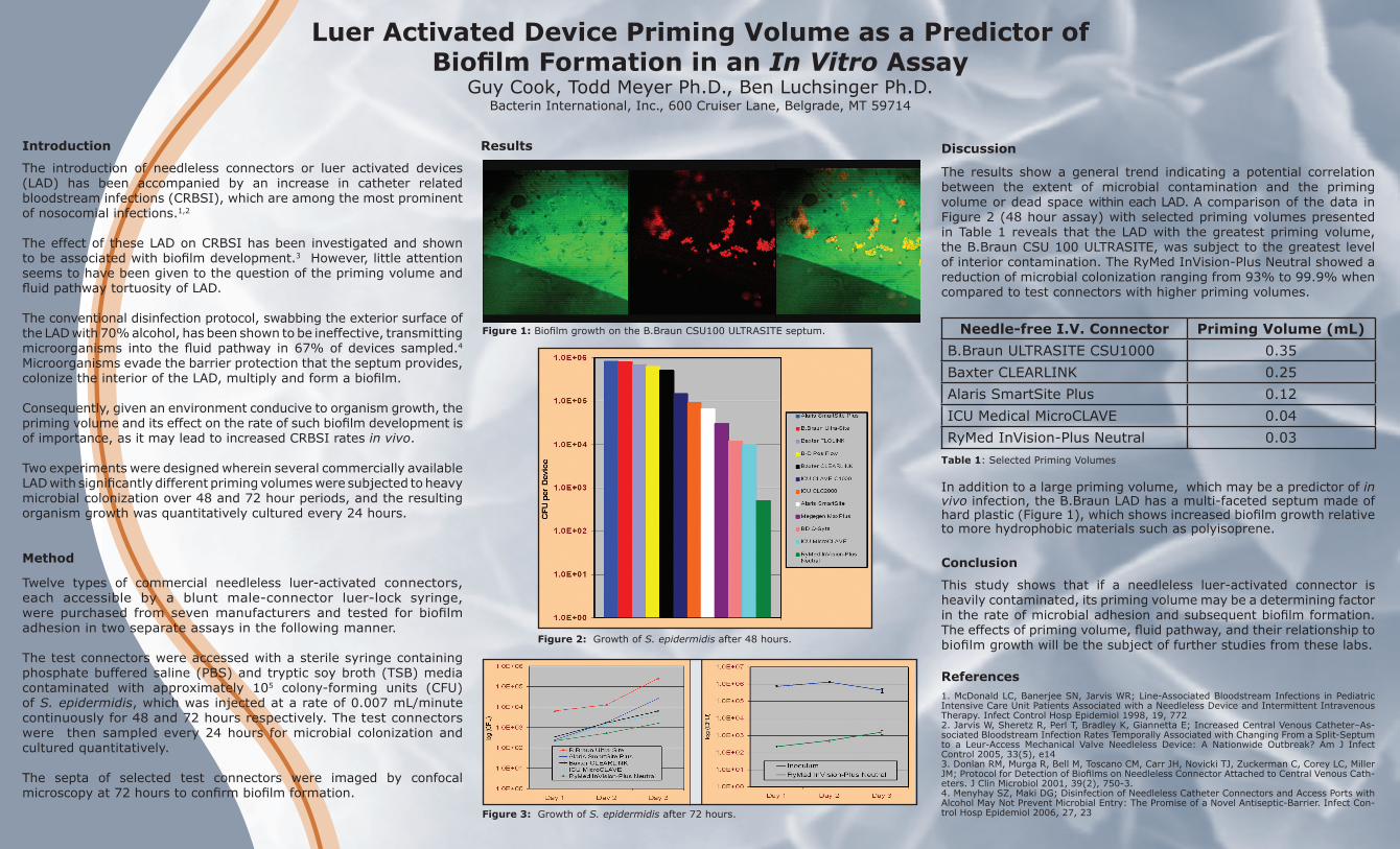
The septa of selected test connectors were imaged by confocal microscopy at 72 hours to confirm biofilm formation.
Figure 1: Biofilm growth on the B.Braun CSU100 ULTRASITE septum.
Figure 3: Growth of S. epidermidis after 72 hours.
Figure 2: Growth of S. epidermidis after 48 hours.
Luer Activated Device Priming Volume as a Predictor of Biofilm Formation in an In Vitro Assay
Guy Cook, Todd Meyer Ph.D., Ben Luchsinger Ph.D.Bacterin International, Inc., 600 Cruiser Lane, Belgrade, MT 59714
Results Discussion
The results show a general trend indicating a potential correlation between the extent of microbial contamination and the priming volume or dead space within each LAD. A comparison of the data in Figure 2 (48 hour assay) with selected priming volumes presented in Table 1 reveals that the LAD with the greatest priming volume, the B.Braun CSU 100 ULTRASITE, was subject to the greatest level of interior contamination. The RyMed InVision-Plus Neutral showed a reduction of microbial colonization ranging from 93% to 99.9% when compared to test connectors with higher priming volumes.
Needle-free I.V. Connector Priming Volume (mL)B.Braun ULTRASITE CSU1000 0.35Baxter CLEARLINK 0.25Alaris SmartSite Plus 0.12ICU Medical MicroCLAVE 0.04RyMed InVision-Plus Neutral 0.03
Table 1: Selected Priming Volumes
In addition to a large priming volume, which may be a predictor of in vivo infection, the B.Braun LAD has a multi-faceted septum made of hard plastic (Figure 1), which shows increased biofilm growth relative to more hydrophobic materials such as polyisoprene.
Conclusion
This study shows that if a needleless luer-activated connector is heavily contaminated, its priming volume may be a determining factor in the rate of microbial adhesion and subsequent biofilm formation. The effects of priming volume, fluid pathway, and their relationship to biofilm growth will be the subject of further studies from these labs.
References1. McDonald LC, Banerjee SN, Jarvis WR; Line-Associated Bloodstream Infections in Pediatric Intensive Care Unit Patients Associated with a Needleless Device and Intermittent Intravenous Therapy. Infect Control Hosp Epidemiol 1998, 19, 772 2. Jarvis W, Sheretz R, Perl T, Bradley K, Giannetta E; Increased Central Venous Catheter–As-sociated Bloodstream Infection Rates Temporally Associated with Changing From a Split-Septum to a Leur-Access Mechanical Valve Needleless Device: A Nationwide Outbreak? Am J Infect Control 2005, 33(5), e143. Donlan RM, Murga R, Bell M, Toscano CM, Carr JH, Novicki TJ, Zuckerman C, Corey LC, Miller JM; Protocol for Detection of Biofilms on Needleless Connector Attached to Central Venous Cath-eters. J Clin Microbiol 2001, 39(2), 750-3. 4. Menyhay SZ, Maki DG; Disinfection of Needleless Catheter Connectors and Access Ports with Alcohol May Not Prevent Microbial Entry: The Promise of a Novel Antiseptic-Barrier. Infect Con-trol Hosp Epidemiol 2006, 27, 23

About the Authors
Guy Cook is President, CEO, and CSO of the Company. Mr. Cook is considered an international expert in biofilm science and its application. He is published in the biofilm field and has been invited to speak at many prominent biofilm conferences, including the “Anti-Infective Materials Seminar” in Tokyo and the FDA-CDRH Antimicrobial Device Efficacy Testing Seminar. Prior to founding Bacterin in 1997, Mr. Cook started his career as a product specialist in the Image Analysis Department for Laboratory Equipment Company in Chicago. He later became President of Delta Re-sources in Crystal Lake, Illinois, which specialized in developing customized image analysis solutions for the academic community. In 1996 he moved to Montana and worked as a confocal microscopist for the Center for Biofilm Engineering at the Montana State University where he developed several proprietary testing models for the medical device industry. Mr. Cook attended Indiana University and received Bachelor of Science degrees in finance and economics.
Todd Meyer received his Ph.D. in organic chemistry from Montana State University and his Bachelors degree in chemistry from the University of California, Davis. His research involved organic synthesis in a variety of contexts and organometallic based molecular transformations promoted by cobalt, in combination with sterically and electronically tunable ligands. Currently, he is Senior Project Manager at Bacterin International, Inc., with responsibility for device-based projects.
Ben Luchsinger received his Ph.D. in physical chemistry from Montana State University and his Bachelors degree in chemistry from Luther College. His research, focused on the chemical interaction of human hemoglobin with nitric oxide and nitric oxide derivatives including how hemoglobin can generate nitric oxide. This work derived from original ideas that he developed. Currently, he is a Senior Scientist II and involved in a number of R&D projects at Bacterin International. Dr. Luchsinger has a number of highly referenced publications and two provisional patents.
5049A 4/2007
For corporate business or for more information, contact us at: Bacterin International • 600 Cruiser Lane • Belgrade, MT 59714
Phone: 406-388-0480 • Fax: 406-388-0422 w w w . b a c t e r i n . c o m