luis s. marsano, md - university of louisville
TRANSCRIPT

Principles of Electrocautery
Luis S. Marsano, MDProfessor of MedicineDivision of Gastroenterology and HepatologyUniversity of Louisville & Louisville VAMC2013

Electrocautery
• Radiofrequency Generator: – Generates high-frequency currents (100 K-Hz to 4 M-
Hz) which induces ionic vibration but no movement.– Ionic vibration generates intracellular heat but no
muscle/nerve depolarization.– Power settings are in Watts (amps x volts).
• Intracellular heat can cause: – boiling + explosion (CUT), and/or– dehydration (desiccation), and/or– fire sparks (fulguration).
} COAGULATION
Electrocautery
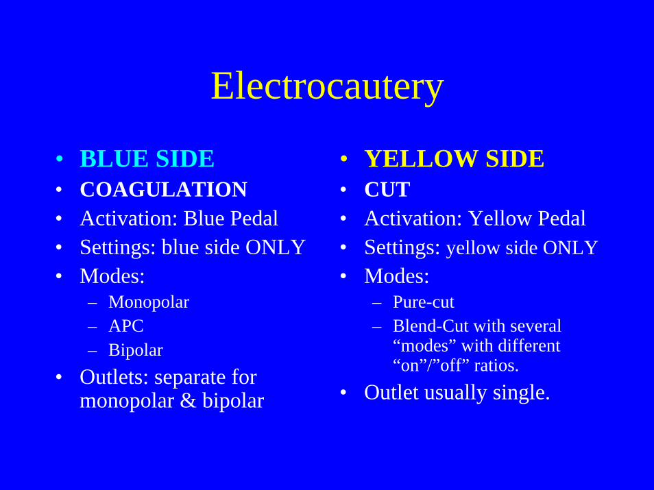
• BLUE SIDE• COAGULATION• Activation: Blue Pedal• Settings: blue side ONLY• Modes:
– Monopolar– APC – Bipolar
• Outlets: separate for monopolar & bipolar
• YELLOW SIDE• CUT• Activation: Yellow Pedal• Settings: yellow side ONLY• Modes:
– Pure-cut– Blend-Cut with several
“modes” with different “on”/”off” ratios.
• Outlet usually single.

Basic Physics Terminology• Voltage (volts): force that pushes the current (“Potential
Energy”). – More force = more destruction
• Resistance (ohm): quality of tissue that impedes flow of current. – More resistance = less current flow.– Resistance of skin > bone > fat > muscle > bowel wall
(326 ohms) > blood.• Intensity (amps): amount of electricity crossing an area (wire),
per second. • Current Density (amp/cm2): amount of current flowing through
a cross sectional area = Current Intensity(amps)/area(cm2)

Point to Remember
• The amount of Energy delivered to the “active” device (snare, hot forceps, sphincterotome, etc) is the same than the delivered to the “indifferent plate”, but the “current density” is very different due to the small “active” end compared with the large “indifferent plate”.

Basic Physics Terminology
• Generated heat: is proportional to the square of the current density: (Intensity/area)2 . – Small area of lesion/stalk causes disproportional
high heat.• Power output: Is given in Watts = amps x volts.
Voltage is constant, hence higher output increases the intensity of current (amps). – Higher output = higher current density = much
higher heat• Delivered Energy: Is given in Joules.
Energy (watts) x time (seconds)
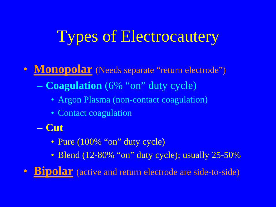
Types of Electrocautery
• Monopolar (Needs separate “return electrode”)
– Coagulation (6% “on” duty cycle)• Argon Plasma (non-contact coagulation)• Contact coagulation
– Cut• Pure (100% “on” duty cycle)• Blend (12-80% “on” duty cycle); usually 25-50%
• Bipolar (active and return electrode are side-to-side)

ERBE Electrocautery
BIPOLARCOAG
Indifferentelectrode
MONOPOLARCUT or COAG

Electrocautery Waveforms
COAGULATIONCUT
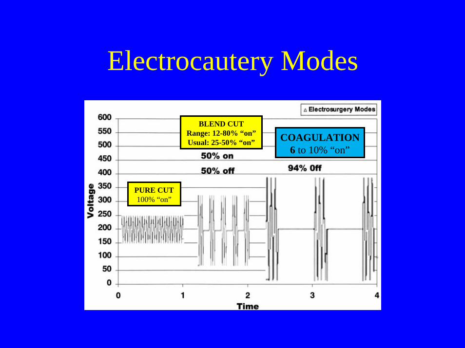
Electrocautery Modes
BLEND CUTRange: 12-80% “on”Usual: 25-50% “on”
PURE CUT100% “on”
COAGULATION6 to 10% “on”

Same energy (W): CUT vs COAGULATION “peak voltage”: Cut <<< Coagulation
CREST: [peak/root mean square] voltage
CUT
COAG
Usual CREST: Coag = 7-8, Pure-Cut < 2, Blend-Cut = 2.2-5
High CREST = longer cooling time/higher peak = more desiccation/fulguration & less cut

Same “peak voltage”: CUT vs COAGEnergy (W): CUT >>> COAGULATION
Physical Process for Electrocoagulation

Physical ProcessDesiccation (Coagulation)
• Slow heating of tissue in close contact, then fluid loss with bubbling, then steam release with cooling, then restarts, slow heating of tissue in close contact, then …
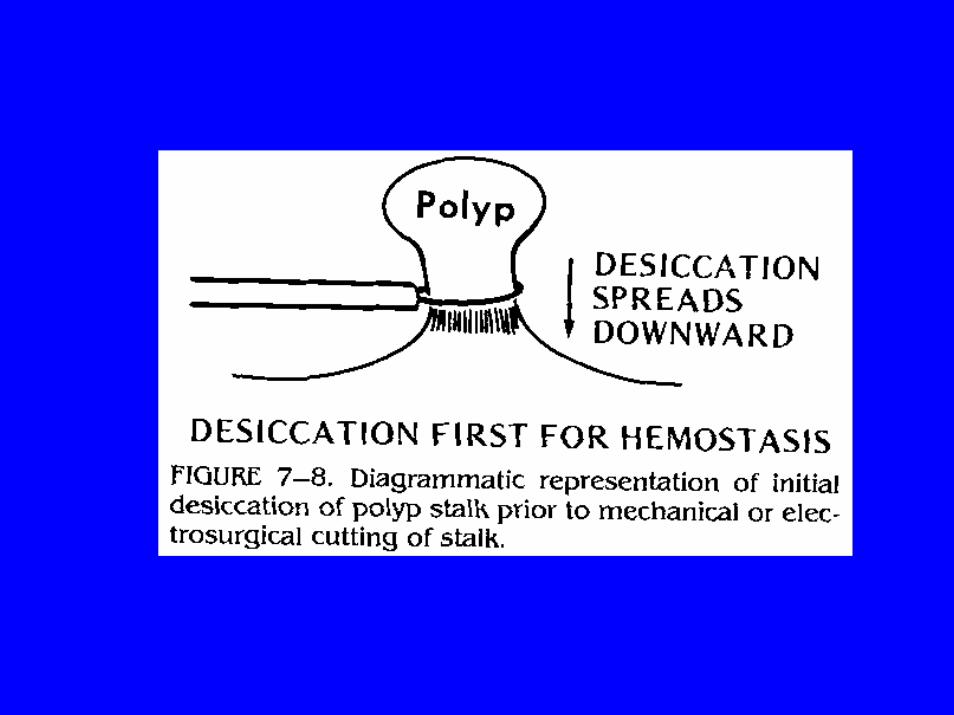
• Main Effect: Desiccation/Coagulation, with preemptive HEMOSTASIS.
• Best Instrument: – Microbipolar (no fulguration).
• Alternatives: – Monopolar coagulation @ 20-30 W, or – Blend-cut with high-CREST (3 or more) or low % duty @ 20-40W
• CONSIDERATIONS:– If setting is too low, may desiccate too deep; difficult to cut.– If too high and monopolar, may give deep fulguration.– Pressing on wall increases burn depth (pull in Hot Bx.)

Desiccation (Coagulation)
Desiccation needs long cooling time/high CREST & close contact

Physical ProcessFulguration
• Electrode not in contact with tissue (or insulated by desiccated tissue): ionization of surrounding air, then long spark with high current density, then superficial coagulation, then (if you continue) deep necrosis with black eschar.
• Effect: Tissue ablation.• Best instrument:
– Argon Plasma Coagulator @ 40-60 W. • Alternative:
– Monopolar coagulation, or – Blend-cut with high-CREST at high setting.
• High risk of transmural necrosis with prolonged burn (continuous “painting” in APC); use “saline pillow”.

Fulguration
Fulguration needs high “peak” voltage and very loose contact
Physical ProcessCutting
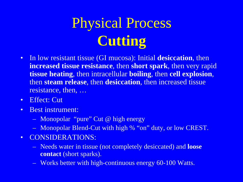
• In low resistant tissue (GI mucosa): Initial desiccation, then increased tissue resistance, then short spark, then very rapid tissue heating, then intracellular boiling, then cell explosion, then steam release, then desiccation, then increased tissue resistance, then, …
• Effect: Cut• Best instrument:
– Monopolar “pure” Cut @ high energy– Monopolar Blend-Cut with high % “on” duty, or low CREST.
• CONSIDERATIONS:– Needs water in tissue (not completely desiccated) and loose
contact (short sparks).– Works better with high-continuous energy 60-100 Watts.
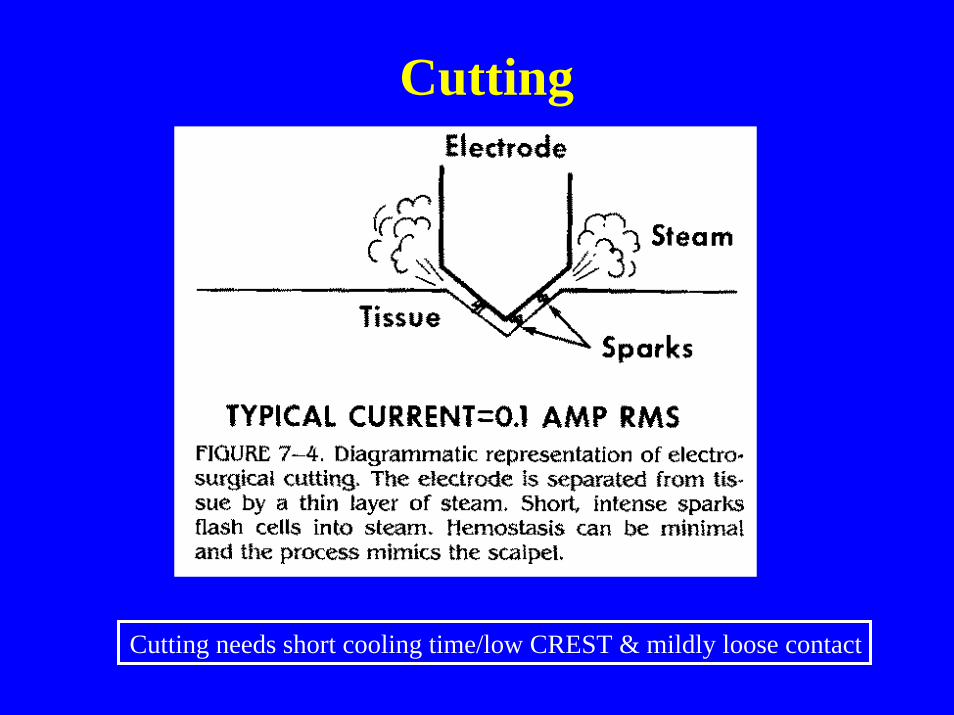
Cutting
Cutting needs short cooling time/low CREST & mildly loose contact

MONOPOLAR ELECTROCOAGULATION

Monopolar Electrocautery
• Has a large “Indifferent Plate” for electricity return and a small “active electrode”; – causes high current density and very high heat at active
electrode.
• CAUTIONS:– Causes deeper injury, hence is bad choice to control
active bleeding (high perforation risk except with non-contact technique like APC).
– There must be absence of flammable gases (bowel lavage) to avoid explosion.

Monopolar Electrocautery
• Indifferent plate should: – A) be near to site of active electrode, to decrease
resistance from other tissues, – B) have conductive gel to decrease skin resistance, – C) remain in complete contact all the time (dual plate w
monitoring circuit confirms contact) to maximize energy in active electrode.
• Examples: hot snare, hot biopsy, Argon Plasma Coagulator, sphincterotome, needle knife.

MONOPOLAR COAGULATION

Monopolar ModesContact Coagulation
• Very short “active” sinus wave (6-10% of cycle) with long cooling period (off, or inactive 90-94% of cycle).
• Long “cooling” facilitates desiccation, high “peak voltage”facilitates fulguration (CREST 7-8).
• Causes slow heating (excellent desiccation) followed with spark and superficial fulguration, after desiccation is complete.
– Deep fulguration can give transmural necrosis • Close contact gives more desiccation, but loose contact or
complete desiccation of surrounding tissue causes more fulguration/necrosis (keep continuous close contact).
• Complication: Post-polypectomy bleed after 2-8 days.

Argon Plasma Coagulation(Non-Contact Electrocoagulation)

Argon Plasma Coagulation• Argon gas flowing inside hollow catheter and ionized
by monopolar current in wire inside catheter. • Needs “indifferent plate” and absence of flammable
gases.• Causes coagulation at point of nearest conductive (non
desiccated) tissue: “around the corner” coagulation.• Distance of probe to tissue, and length of therapy:
– 2-8 mm (2-3 mm at lower setting) x 0.5-2 sec.• Depth of injury is decreased by “saline pillow” (from
86% to 21% deep).

Argon Plasma Coagulation
• Power: – For ablation, best setting is 70-90 W @ 1 LPM.– For hemostasis , best setting is 40-50 @ 0.5 LPM; Poor hemostasis
in active bleeding (dissipation).
• PRECAUTIONS:– Injury can be deep; give short bursts (tap) or continuous movement
“painting”.– Use lower setting if in contact with metal stents.– Avoid contact (gas injection and monopolar transmural burn),– Avoid fluid pool (energy dissipation),– Suction between treatments to decrease gas distention/perforation
risk.

Argon Plasma CoagulationSITE
Esoph/Stomach/ Rectum
Cecum & Rt. Colon
Tr. Colon to SigmSmall Bowel
FLOW 1.4 LPM 0.5 LPM 1-1.4 LPM
COAG A 60 WBlue Pedal
A 40 WBlue Pedal
Tap
A 50 WBlue Pedal
TIME to DEPTH
1 sec = 1.5 mm5 sec = 2 mm10 sec = 3 mm
1 sec = 1 mm8 sec = 2 mm
15 sec = 3 mm
1 sec = 1.5 mm7 sec = 2 mm12 sec = 3 mm

APC Settings per Clinical IndicationVargo J. Gastrointest Endosc 59(1):81-88; 2004
Energy FlowAblation polyp residue 40-65 W 0.8-2 LPMRadiation Proctopathy 40-60 W 0.6-3 LPMGAVE 40-100 W 2 LPMAngioectasia 40-60 W 2 LPMAblation of Barrett’s 30-90 W 0.5-2 LPMBleeding Ulcer 40-70 W 1.5-3 LPMPalliation GI Cancer 40-100 W 0.8-2.5 LPMPost variceal ablation 50-60 W 1.5-2 LPM

MONOPOLAR CUT
Monopolar ModesPure Cut
• Pure Cut: 100% on continuous sinusoidal wave without cooling-off period.
• Causes very rapid heating with cell explosion and formation of steam and sparks.
-Gives little coagulation (no desiccation)-Current has a very low “peak voltage”/”root of mean square voltage” ratio (low CREST < 2) -Not used in endoscopy, unless some coagulation was given first.-Complication: immediate bleed.

Monopolar ModesBlend-Cut
• Blend-Cut: Sinus wave duty cycle is “on” 25-50% of the time; allows for some cooling-off period (50-75% of time)– Gives less cell explosion (cut) than pure-cut, and moderate
desiccation + fulguration (coagulation) – Has higher CREST than pure-cut (2.2-5) – At same energy setting has higher peak voltage, hence can
fulgurate more.– Different levels of “blend” are set by the manufacturer.– In ERBE: Effect-1= minimal coag; Effect-4 = maximal coag;
ENDO-CUT uses Effect-3 (high coag alternating with blend-cut) – Responds only to “CUT setting”– Loose contact facilitates “blend” process.– Complication: Post-polypectomy bleed within 12 hours.

EndoCut

Point to Remember
• In general, at identical Power Setting (watts):– COAGULATION currents cause deeper tissue
injury than CUT (pure or blend) currents.– HOT FORCEPS cause deeper tissue injury than
HOT SNARES.

REMEMBERYellow + Blue IS NOT Green
• COAGULATION (Blue) unit is completely independent of CUT (Yellow) unit; – Power setting in COAG side does not affect the CUT
side.
• BLEND-CUT current is a feature of the CUT (Yellow) unit. – The degree of “blending” depends on the chosen mode
(Endo-Cut, vs blend-1, vs blend-2, etc.)

Endoscopic Electrocoagulation Techniques

Snare Polypectomy
• VARIABLES• Energy• Wave: coag vs blend-c• Stalk diameter• Wire tension (>1.5cm)• Wire diamet(.3-.4mm)
• PHASES• Desiccation• Cut: mechanical vs
electrosurg. vs mixed– Sequential– Combined

Snare Polypectomy
• Desiccation:– COAG @ 20-30 (25)W, or Blend Cut 2 @ 20-30 W, or
Endo-Cut-3@ 200 W; – Lower energy on Rt colon. Higher in thick stalk. – Saline “pillow” in sessile lesions. – Avoid:
• Too much desiccation: difficult to cut; transmural necrosis• Polyp contact with other wall: burn (shake it)• Fluid pool: loss of energy.

Snare Polypectomy
• Cutting:– A) Mechanical or Mixed: close snare with
constant mild-moderate pressure during or after COAGULATION.
– B) Electrosurgical: Close snare with very light hold during Blend-Cut or Endo-Cut.
• If snare gets stuck after excessive desiccation, change to pure-cut @ 100-150 W.(If tissue is too dry, will not cut)


Hot Biopsy
• COAG (Fulgurate-COAG) @ 25 W, or ERBE Soft-COAG @ 60 W– Tent-pull tissue right & left, close & away, with short
“tap” to coagulate all base; do not “overcook”; then pull to remove.
– Adequate for lesions up to 5 mm (cold snare is preferred); used in difficult-to-snare position.
• Residual polyp in Cold Bx = 30% of patients• Residual polyp in Hot Bx = 17% of patients• Residual polyp in cold snare = rare
– Saline “pillow” has little effect in decreasing thermal injury in Hot Biopsy (“tenting” is important).
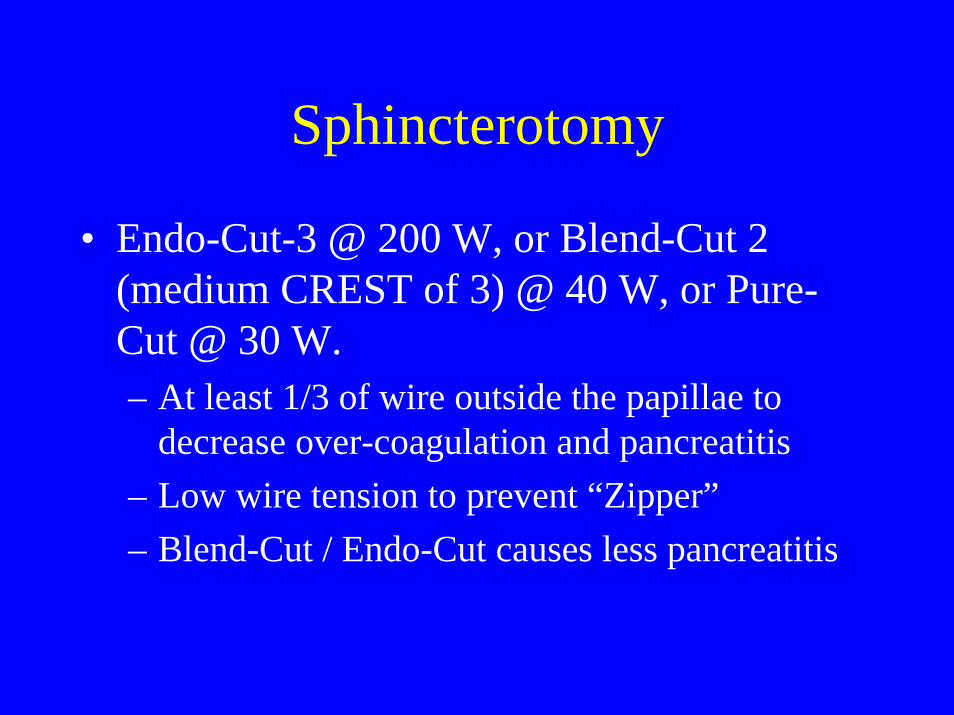
Sphincterotomy
• Endo-Cut-3 @ 200 W, or Blend-Cut 2 (medium CREST of 3) @ 40 W, or Pure-Cut @ 30 W.– At least 1/3 of wire outside the papillae to
decrease over-coagulation and pancreatitis– Low wire tension to prevent “Zipper”– Blend-Cut / Endo-Cut causes less pancreatitis
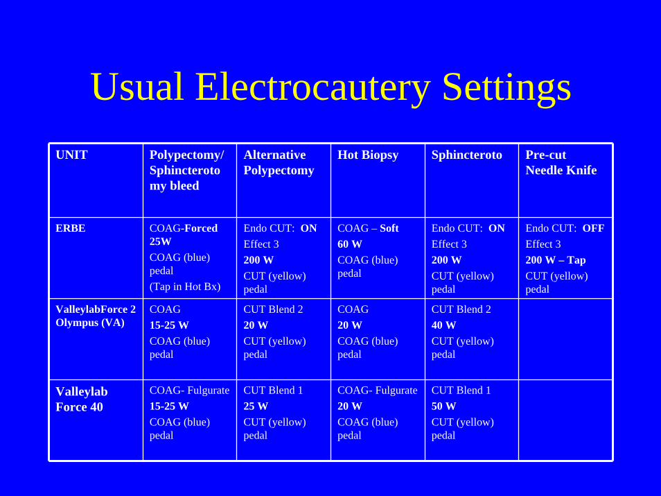
Usual Electrocautery SettingsUNIT Polypectomy/
Sphincterotomy bleed
Alternative Polypectomy
Hot Biopsy Sphincteroto Pre-cut Needle Knife
ERBE COAG-Forced25WCOAG (blue) pedal(Tap in Hot Bx)
Endo CUT: ONEffect 3200 WCUT (yellow) pedal
COAG – Soft60 WCOAG (blue) pedal
Endo CUT: ONEffect 3200 WCUT (yellow) pedal
Endo CUT: OFFEffect 3200 W – TapCUT (yellow) pedal
ValleylabForce 2 Olympus (VA)
COAG15-25 WCOAG (blue) pedal
CUT Blend 220 WCUT (yellow) pedal
COAG20 WCOAG (blue) pedal
CUT Blend 240 WCUT (yellow) pedal
Valleylab Force 40
COAG- Fulgurate15-25 WCOAG (blue) pedal
CUT Blend 125 WCUT (yellow) pedal
COAG- Fulgurate20 WCOAG (blue) pedal
CUT Blend 150 WCUT (yellow) pedal

BIPOLARELECTROCOAGULATION

Bipolar Electrocautery• Usually gives low-energy or “micro-bipolar”. Has two or
more small active electrodes very close to each other (active and return electrode)
• Does not use “indifferent plate”.• Risk of explosion with flammable gases (needs colon prep) • Less depth of injury. Saline pillow further decreases depth
of injury (very important in colon & small bowel). • Excellent desiccation and coagulation at low settings (15-
20 W). Excellent for hemostasis.• Example: BICAP, Gold-Probe.

BICAP Probe Tip
+-

Micro-Bipolar Mode
• Used only for hemostasis (BICAP and Gold-Probe); – Tumor-Probe use was very limited.
• Depth of burn is limited and “saline pillow” can decrease it further.
• Better effect with large 10-Fr probes (needs “Therapeutic-channel” (T) Scope)
• Settings, pressure applied, and length of time have been standardized.

BICAP in Upper Endoscopy(Inject “pillow” before SB burn)
Lesion Probe Pressure Energy Time/ Site
PUD/ Dieulafoy
10 Fr Very Firm 20 W 10-14 sec
M-W Tear
7-10 Fr Moderate 20 W 4 sec
Angiodysplasia
7-10 Fr Light 15 W 2 sec

BICAP in Colonoscopy(Inject “Pillow” before burn; tattoo after)
Lesion Probe Pressure Energy Time/ Site
Ulcer 10 Fr Moderate 20 W 2 sec
Stalk 10 Fr Moderate 20 W 2 sec
Diverticuli 10 Fr Moderate 20 W 2 sec
Cancer 10 Fr Moderate 20 W 2 sec
Angiodysplasia
7-10 Fr Light 15 W 1 sec

Complications Of Electrocautery

Complications Of Electrocautery
• Indifferent-Plate & EKG-Pad: burn• Fetal Stimulation• Capacitive Coupling Discharge: burn• Pacemaker: interference, burn, or device damage.• Implantable Cardioverter Defibrillator: trigger,
device damage, or asystole (dual system)• Deep Brain stimulator and Gastric stimulator:
“shock”, burn, or device damage.• Bowel Explosion

ComplicationsIndifferent-Plate & EKG-Pad Burn
Fetal Stimulation• Skin burn from Indifferent Plate (Return Electrode):
– If partial detachment causes small contact area, and high current density, giving a burn.
• EKG electrode pad burn: – If indifferent plate is separated, and EKG pads acts as “indifferent
plate”; EKG electrodes should be > 0.3 cm2 each• Fetal Stimulation:
– if electricity passes through the uterus to reach “indifferent-plate”• Prevention:
– Dual-Pad with “Return electrode Monitor” (REM) circuit, detects change in surface/resistance and “shuts-off” the unit.
– During pregnancy, indifferent-plate should not be placed causing the uterus to be between electrode & plate.

Dual Electrode Monitoring
IndependentPads

ComplicationsCapacitive Coupling Discharge
• Scope works as capacitor of currents induced by snare/hot-forceps; – important only with large scopes (Colon/ERCP).
• Scope can discharge current after contact with “frame-wires” (damaged external surface) or eye piece; can burn physician or inside of patient.
• Prevention: – S-cord connects “scope frame” to “indifferent plate”
and unloads charge. – Risk: if indifferent plate gets off, scope acts as
indifferent plate; should be used with “dual-pad”/REM.

ComplicationsPacemaker Interference
• Electrocautery generates electromagnetic fields of up to 60 V/m
• Pacemakers are inhibited with electromagnetic fields > 0.1 V/m.
• Permanent protective circuit may be damaged or need reprogramming if current is delivered close to pacemaker or its leads.
• With poorly grounded or non-isolated electrocautery, pacemaker can work as “indifferent plate” and cause myocardial burn or arrhythmia (use “dual-pads”).
RECOMMENDATIONPacemaker Interference
• RECOMMENDATION: – “Pacemaker-dependent” for rhythm or
hemodynamics (20%): use magnet• pacemaker should be placed on “continuous asynchronous
pacing” (VOO or DOO) with a “ring magnet”, only while only while electrocautery current is delivered. electrocautery current is delivered.
• after electrocoagulation, the pacemaker should be reactivated and assessed for normal function (telephonically).
– “No pacemaker-dependent”: magnet use is optional • used mostly for prolonged electrocautery use, like in APC for
GAVE or radiation proctitis.

ComplicationsImplantable Cardioverter Defibrillator
• Damage can occur as in case of pacemakers• Electrocautery waves can be interpreted as “R
waves” simulating arrhythmia or normal rhytm, and ICD will respond cardioverting, or inhibiting cardioversion or pacing.
• Most, but not all, ICDs respond to properly placed “magnet” by “suspension of tachycardia detection”, and/or “suspension of ICD therapy”, without affecting pacemaker function.

ComplicationsImplantable Cardioverter Defibrillator
• Patient with ICD who is “pacemaker dependent” will not have pacer changed to “asynchronous pacing” by the magnet: – risk of prolonged pacemaker inhibition by electrocautery, with
asystole.• Is difficult to ascertain if magnet is properly placed over ICD;
– change in position may affect inhibition.• Is difficult to know beforehand which effect magnet will have
on the ICD. – Example: ICDs made by “Guidant” may respond to magnet
by: • 1) Permanent disabling ICD therapy, • 2) Temporary disabling ICD therapy, or • 3) No change at all

RecommendationsImplanted Cardiac Defibrillators
(ASGE 2007)• Determine:
– 1) Cardiac device make/model/type, - 2) Indication for the device, – 3) Underlying cardiac rhythm, - 4) Degree of pacemaker-dependence.– 5) Frequency of VT and VF therapy - 6) Need for reprogramming for
Procedure.
• RECOMMENDATION: – 1) Consult a hearth rhythm specialist.
• Optimal solution is ICD interrogation & reprogramming of ICD detection/therapy, before and after the procedure.
– 2)If patient with ICD is “pacemaker-dependent” and device cannot be reprogrammed to “asynchronous mode”:
• strongly consider use of bipolar cautery.

RecommendationsImplanted Cardiac Defibrillators
(ASGE 2007)
• Monitor cardiac rhythm continuously.• Have available external defibrillator with
transcutaneous pacing capability.• Place “Indifferent Return Electrode” in thigh or
buttock (away from chest). Keep distance >/= 15 cm away from device.

RecommendationsImplanted Cardiac Defibrillators
(ASGE 2007)• Use “short & repetitive” bursts.• Consider use of Bipolar electrocautery in:
– Patient with ICD who is “pacemaker dependent”
– If working in distal esophagus– If working in proximal stomach– If working in splenic colonic flexure

ComplicationsNon-Cardiac Devices
• Deep Brain Stimulators (DBS): – paresthesias and “shock” with monopolar
electrocautery.• Gastric (Enterra) and DBS:
– 1) Temporary malfunction or reprogramming, – 2) Shock or “jolting” from induced currents, – 3) Electrode heating with tissue injury.
• Neurostimulators in spine, peripheral nerves, bladder or cochlea: – no complications if voltage reduced to zero and then
“shut off”.

RecommendationsNon-Cardiac Devices
(ASGE 2007)• Prefer non-cautery thermal probes or bipolar/multipolar
probes.• Use electrocautery at lowest effective power and for the
briefest time• Place return electrodes far away from the device generator
and wire-leads• Avoid electrocautery use near the device.• In patients with DBS & GES:
– consult with the specialist.• Neurostimulators other than DBS & GES:
– place voltage in zero, and turn-off the device.

ComplicationsBowel Explosion
• Poorly prep bowel gas is combustible: swallowed O2 + bacteria-produced H2 and methane.
• Do not use preps with poorly absorbed carbohydrates (mannitol, sorbitol, lactulose); clear liquid diet decreases H2.
• If prep is sub-optimal: – “exchange air” or – use CO2, or – avoid electrocautery (heater probe is OK)
Non-ElectrocauteryThermal Device

ThermocoagulationHeater Probe
• Can irrigate and tamponade + coagulate.• Can be applied “en face” or “tangential”.• 10 Fr probe is better, but needs “T-channel scope”.• Is not electrocautery (no sparks); safe in unprepared bowel.• Has an electrically heated coil inside a Teflon-covered
insulated cylinder; heats to 110 oC degrees.• Power setting in Joules (Watts x sec): HP unit decides time
needed to deliver the requested energy.• In ulcer; apply in 4 quadrants plus center.

Heater Probe in Upper Endoscopy(Inject “pillow” before SB burn)
Lesion Probe Pressure Energy Pulses/ Site
PUD/ Dieulafoy
10 Fr Very Firm 30 J 4
M-W Tear
7-10 Fr Moderate 20 J 3
Angiodysplasia
7-10 Fr Light 15 J 2

Heater Probe in Colonoscopy(Inject “Pillow” before burn; tattoo after)
Lesion Probe Pressure Energy Pulses/ Site
Ulcer 10 Fr Moderate 15 J 2
Stalk 10 Fr Moderate 15-20 J 2
Diverticuli 10 Fr Moderate 15 J 2
Cancer 10 Fr Moderate 20 J 2
Angiodysplasia
7-10 Fr Light 10 J 1