lumbar nerve root compression due to leakage of bone ... · der fluoroscopic guide. ... with use of...
TRANSCRIPT

Copyright © 2014 Korean Neurotraumatology Society 155
Introduction
The vertebral fractures is the most common complica-tions of osteoporosis.13) These fractures result in signifi-cant mortality and morbidity including prolonged and in-tractable pain.4,13) Percutaneous vertebroplasty, a therapeutic procedure for filling the collapsed vertebral body with polymethylmethacrylate, provides pain relief.2,4) In gener-al, percutaneous vertebroplasty is simple and safe if per-formed under continuing fluoroscopic control and technical precautions. Vertebroplasty has the potential risk of serious complications such as leakages of bone cement, cardiopul-monary complications, infection and the new fractures of the adjacent vertebrae.6,7) We present a case of extraspinal leakage after vertebroplasty at our hospital.
Case Report
A 73-year-old male was admitted to our hospital for low
back pain after slip down at one day earlier. After radiolog-ic evaluation, acute osteoporotic compression fracture of the L4 vertebra was found. The patient had experienced L1 vertebroplasty and T12-L2 screw fixation for L1 compres-sion fracture at another hospital before 2 years ago. Percu-taneous vertebroplasty for L4 vertebra was performed un-der fluoroscopic guide. After incision of the skin, an 11-guage vertebroplasty needle was placed percutaneously on the posterior part of the vertebral body via bilateral transpe-dicular approach. The needle was pushed through the cor-tex, situated the center of the pedicle as possible, and then directed into the vertebral body. The anterior third of the body was an ideal location for the needle placement. The contrast medium (Iohexol) was injected to estimate bone cement distribution and minimize bone cement leakage and intraoperative complication such as pulmonary thromboem-bolism. When a thin toothpaste consistency was achieved, the bone cement was injected into the vertebral body under continuous fluoroscopic control. The filling process was stopped immediately when leakage of bone cement was ob-served into lateral space of body. After the procedure, his back pain was improved immediately. But he complained severe pain which radiates down the right leg on standing position. The patient was unable to bear weight on his right leg after the procedure. Neurologic examination revealed
Lumbar Nerve Root Compression due to Leakage of Bone Cement after Vertebroplasty
Doo Soo Kim, MD, Se Youn Jang, MD, Min Ho Kong, MD, Kwan Young Song, MD, and Dong Soo Kang, MDDepartment of Neurosurgery, Seoul Medical Center, Seoul, Korea
We experienced a 73-year-old male with lumbar nerve root compression due to leakage of bone cement after vertebroplasty. He was underwent vertebroplasty for acute osteoporotic L4 compression fracture at our hospital. After vertebroplasty, his back pain was improved but right leg pain was newly developed. Lumbar computed tomography scanning showed that bone cements were leaked along the L4 nerve root. The leaked cements around L4 nerve root were removed carefully via paraspi-nal muscle-splitting approach. After operation, severe right leg radiating pain was improved. We recommend proper entry point, high viscosity of polymethylmethacrylate and constant monitoring can reduce complication. (Korean J Neurotrauma 2014;10(2):155-158)
KEY WORDS: Bone cements ㆍFractures compression ㆍOsteoporosis ㆍRadiculopathy ㆍVertebroplasty.
CASE REPORTKorean J Neurotrauma 2014;10(2):155-158
pISSN 2234-8999 / eISSN 2288-2243
http://dx.doi.org/10.13004/kjnt.2014.10.2.155
Received: September 11, 2014 / Revised: October 13, 2014Accepted: October 14, 2014Address for correspondence: Dong Soo Kang, MDDepartment of Neurosurgery, Seoul Medical Center, 156 Sinnae-ro, Jungnang-gu, Seoul 131-795, KoreaTel: +82-2-2276-8604, Fax: +82-2-2276-8537E-mail: [email protected]
online © ML Comm
156 Korean J Neurotrauma 2014;10(2):155-158
Lumbar Nerve Root Compression due to Bone Cement Leakage
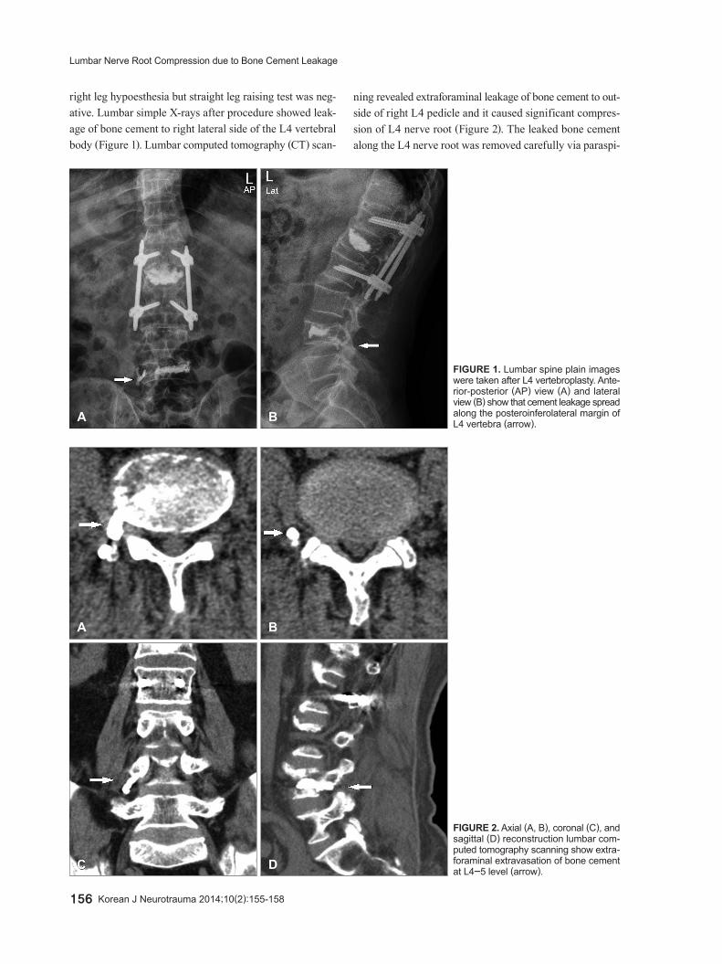
right leg hypoesthesia but straight leg raising test was neg-ative. Lumbar simple X-rays after procedure showed leak-age of bone cement to right lateral side of the L4 vertebral body (Figure 1). Lumbar computed tomography (CT) scan-
ning revealed extraforaminal leakage of bone cement to out-side of right L4 pedicle and it caused significant compres-sion of L4 nerve root (Figure 2). The leaked bone cement along the L4 nerve root was removed carefully via paraspi-
A B
FIGURE 1. Lumbar spine plain images were taken after L4 vertebroplasty. Ante-rior-posterior (AP) view (A) and lateral view (B) show that cement leakage spread along the posteroinferolateral margin of L4 vertebra (arrow).
A
C
B
D
FIGURE 2. Axial (A, B), coronal (C), and sagittal (D) reconstruction lumbar com-puted tomography scanning show extra-foraminal extravasation of bone cement at L4-5 level (arrow).
Doo Soo Kim, et al.
http://www.kjnt.org 157
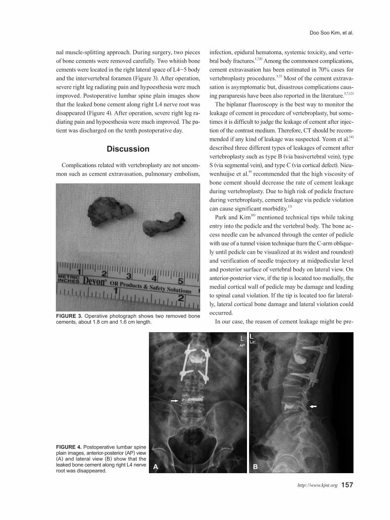
nal muscle-splitting approach. During surgery, two pieces of bone cements were removed carefully. Two whitish bone cements were located in the right lateral space of L4-5 body and the intervertebral foramen (Figure 3). After operation, severe right leg radiating pain and hypoesthesia were much improved. Postoperative lumbar spine plain images show that the leaked bone cement along right L4 nerve root was disappeared (Figure 4). After operation, severe right leg ra-diating pain and hypoesthesia were much improved. The pa-tient was discharged on the tenth postoperative day.
Discussion
Complications related with vertebroplasty are not uncom-mon such as cement extravasation, pulmonary embolism,
infection, epidural hematoma, systemic toxicity, and verte-bral body fractures.1,7,8) Among the commonest complications, cement extravasation has been estimated in 70% cases for vertebroplasty procedures.3,5) Most of the cement extrava-sation is asymptomatic but, disastrous complications caus-ing paraparesis have been also reported in the literature.5,7,12)
The biplanar fluoroscopy is the best way to monitor the leakage of cement in procedure of vertebroplasty, but some-times it is difficult to judge the leakage of cement after injec-tion of the contrast medium. Therefore, CT should be recom-mended if any kind of leakage was suspected. Yeom et al.14) described three different types of leakages of cement after vertebroplasty such as type B (via basivertebral vein), type S (via segmental vein), and type C (via cortical defect). Nieu-wenhuijse et al.9) recommended that the high viscosity of bone cement should decrease the rate of cement leakage during vertebroplasty. Due to high risk of pedicle fracture during vertebroplasty, cement leakage via pedicle violation can cause significant morbidity.11)
Park and Kim10) mentioned technical tips while taking entry into the pedicle and the vertebral body. The bone ac-cess needle can be advanced through the center of pedicle with use of a tunnel vision technique (turn the C-arm oblique-ly until pedicle can be visualized at its widest and roundest) and verification of needle trajectory at midpedicular level and posterior surface of vertebral body on lateral view. On anterior-posterior view, if the tip is located too medially, the medial cortical wall of pedicle may be damage and leading to spinal canal violation. If the tip is located too far lateral-ly, lateral cortical bone damage and lateral violation could occurred.
In our case, the reason of cement leakage might be pre-FIGURE 3. Operative photograph shows two removed bone cements, about 1.8 cm and 1.6 cm length.
FIGURE 4. Postoperative lumbar spine plain images, anterior-posterior (AP) view (A) and lateral view (B) show that the leaked bone cement along right L4 nerve root was disappeared. A B

158 Korean J Neurotrauma 2014;10(2):155-158
Lumbar Nerve Root Compression due to Bone Cement Leakage
sumed. The tip of needle located more laterally when needle tip is located in the pedicle and vertebral body. So pedicle and lateral end plate damages ware occurred.
Conclusion
We experienced a case of lumbar nerve root compression by leaked bone cement after vertebroplasty. Proper entry point of trocar, keeping trocar within pedicle and proper position of trocar between the pedicle and spinous process under biplanar fluoroscopy view would be help to prevent leakage of bone cement after vertebroplasty.
■ The authors have no financial conflicts of interest.
REFERENCES1) Chen JK, Lee HM, Shih JT, Hung ST. Combined extraforaminal
and intradiscal cement leakage following percutaneous vertebro-plasty. Spine (Phila Pa 1976) 32:E358-E362, 2007
2) Cho CH, Park JT, Yun JK, Moon SK. Comparative analysis be-tween male and female osteoporotic compression fractures in el-derly patients. Korean J Neurotrauma 9:131-134, 2013
3) Cotten A, Dewatre F, Cortet B, Assaker R, Leblond D, Duquesnoy B, et al. Percutaneous vertebroplasty for osteolytic metastases and myeloma: effects of the percentage of lesion filling and the leakage of methyl methacrylate at clinical follow-up. Radiology 200:525-530, 1996
4) Diamond TH, Champion B, Clark WA. Management of acute os-teoporotic vertebral fractures: a nonrandomized trial comparing percutaneous vertebroplasty with conservative therapy. Am J Med 114:257-265, 2003
5) Hadjipavlou AG, Tzermiadianos MN, Katonis PG, Szpalski M.
Percutaneous vertebroplasty and balloon kyphoplasty for the treat-ment of osteoporotic vertebral compression fractures and osteo-lytic tumours. J Bone Joint Surg Br 87:1595-1604, 2005
6) Harrington KD. Major neurological complications following per-cutaneous vertebroplasty with polymethylmethacrylate: a case re-port. J Bone Joint Surg Am 83-A:1070-1073, 2001
7) Lee BJ, Lee SR, Yoo TY. Paraplegia as a complication of percuta-neous vertebroplasty with polymethylmethacrylate: a case report. Spine (Phila Pa 1976) 27:E419-E422, 2002
8) Lin EP, Ekholm S, Hiwatashi A, Westesson PL. Vertebroplasty: ce-ment leakage into the disc increases the risk of new fracture of ad-jacent vertebral body. AJNR Am J Neuroradiol 25:175-180, 2004
9) Nieuwenhuijse MJ, Van Erkel AR, Dijkstra PD. Cement leakage in percutaneous vertebroplasty for osteoporotic vertebral compres-sion fractures: identification of risk factors. Spine J 11:839-848, 2011
10) Park SY, Kim YC. Vertebroplasty and kyphoplasty in Kim DH, Vaccaro AR, Dickman CA, Cho D, Lee S, Kim I (eds): Surgical anat-omy and techniques to the spine, ed 2. Philadelphia, PA: Elsevier Inc., pp697, 2013
11) Park SY, Modi HN, Suh SW, Hong JY, Noh W, Yang JH. Epidural cement leakage through pedicle violation after balloon kyphoplas-ty causing paraparesis in osteoporotic vertebral compression frac-tures - a report of two cases. J Orthop Surg Res 5:54, 2010
12) Tsai TT, Chen WJ, Lai PL, Chen LH, Niu CC, Fu TS, et al. Poly-methylmethacrylate cement dislodgment following percutaneous vertebroplasty: a case report. Spine (Phila Pa 1976) 28:E457-E460, 2003
13) Watts NB, Harris ST, Genant HK. Treatment of painful osteopo-rotic vertebral fractures with percutaneous vertebroplasty or ky-phoplasty. Osteoporos Int 12:429-437, 2001
14) Yeom JS, Kim WJ, Choy WS, Lee CK, Chang BS, Kang JW. Leak-age of cement in percutaneous transpedicular vertebroplasty for painful osteoporotic compression fractures. J Bone Joint Surg Br 85:83-89, 2003