lumbar spine sonoanatomy
TRANSCRIPT
LUMBAR SPINE SONOANATOMY
B. Ashrafnejad MD
Fellowship in Pain Medicine
INTRODUCTION
Spinal column
• As a rule, deep structures or spaces in the spine may only be reliably visualised ultrasonographically if “acoustic windows” are present (or created!) and used properly.
• Reliable visualisation of lumbar spine sonography is closely associated with BMI and/or individually highly different tissue properties that markedly influence echogenicity
Lumbar Spine Anatomy
Compared to the thoracic spine
Lumbar spine anatomy reveals that this part of the spine is more “open” to US examination
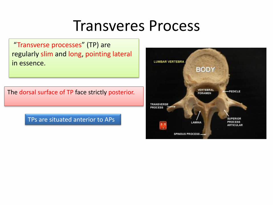
Transveres Process“Transverse processes” (TP) are regularly slim and long, pointing lateral in essence.
The dorsal surface of TP face strictly posterior.
TPs are situated anterior to APs
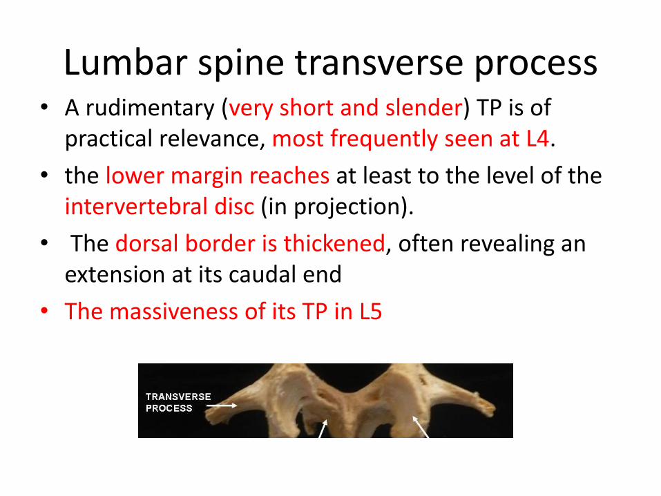
Lumbar spine transverse process • A rudimentary (very short and slender) TP is of
practical relevance, most frequently seen at L4.
• the lower margin reaches at least to the level of the intervertebral disc (in projection).
• The dorsal border is thickened, often revealing an extension at its caudal end
• The massiveness of its TP in L5
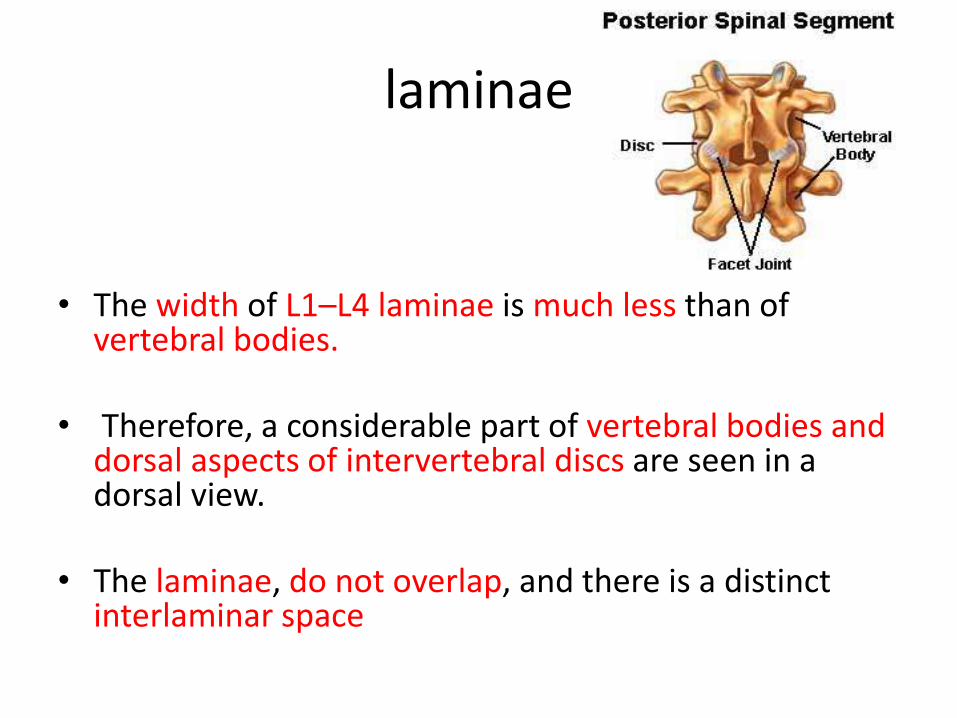
laminae
• The width of L1–L4 laminae is much less than of vertebral bodies.
• Therefore, a considerable part of vertebral bodies and dorsal aspects of intervertebral discs are seen in a dorsal view.
• The laminae, do not overlap, and there is a distinct interlaminar space
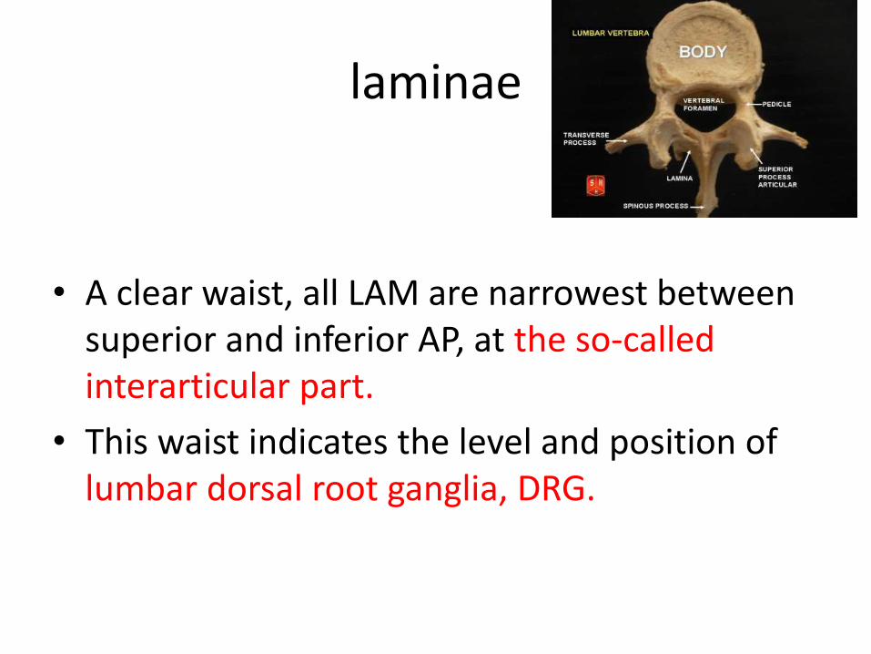
laminae
• A clear waist, all LAM are narrowest between superior and inferior AP, at the so-called interarticular part.
• This waist indicates the level and position of lumbar dorsal root ganglia, DRG.
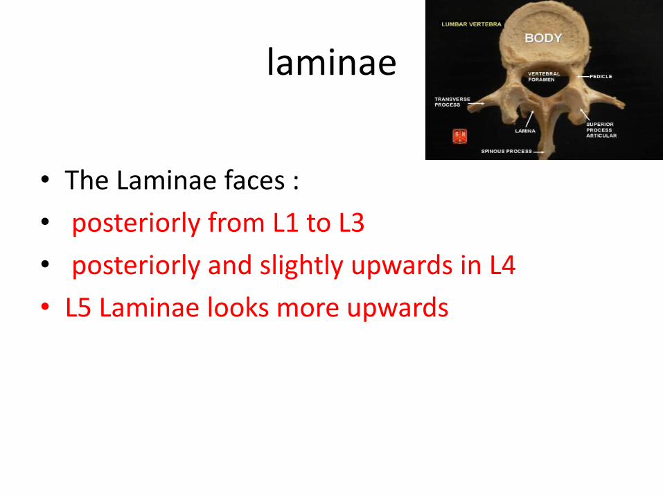
laminae
• The Laminae faces :
• posteriorly from L1 to L3
• posteriorly and slightly upwards in L4
• L5 Laminae looks more upwards
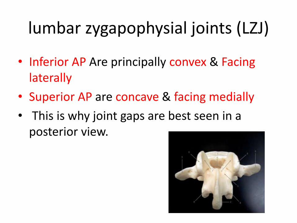
lumbar zygapophysial joints (LZJ)
• Inferior AP Are principally convex & Facinglaterally
• Superior AP are concave & facing medially
• This is why joint gaps are best seen in a posterior view.
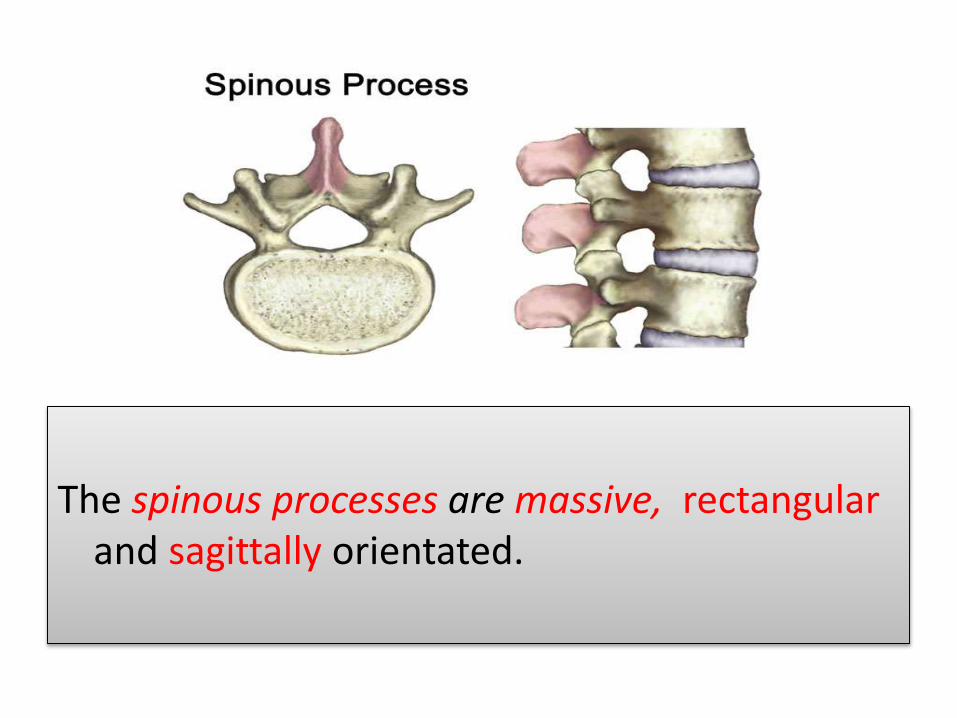
Spinous Process
The spinous processes are massive, rectangularand sagittally orientated.
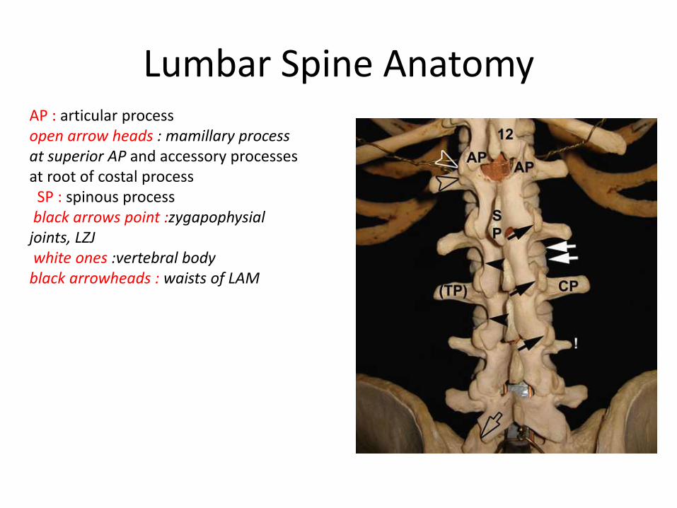
Lumbar Spine AnatomyAP : articular processopen arrow heads : mamillary process at superior AP and accessory processes at root of costal process
SP : spinous processblack arrows point :zygapophysial
joints, LZJwhite ones :vertebral body
black arrowheads : waists of LAM
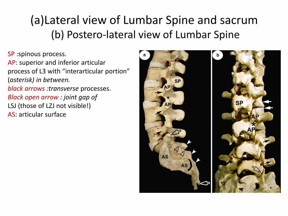
(a)Lateral view of Lumbar Spine and sacrum(b) Postero-lateral view of Lumbar Spine
SP :spinous process.AP: superior and inferior articularprocess of L3 with “interarticular portion” (asterisk) in between.black arrows :transverse processes.Black open arrow : joint gap ofLSJ (those of LZJ not visible!) AS: articular surface
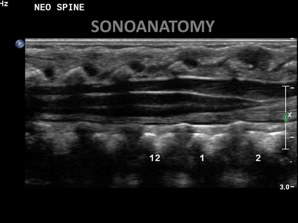
SONOANATOMY
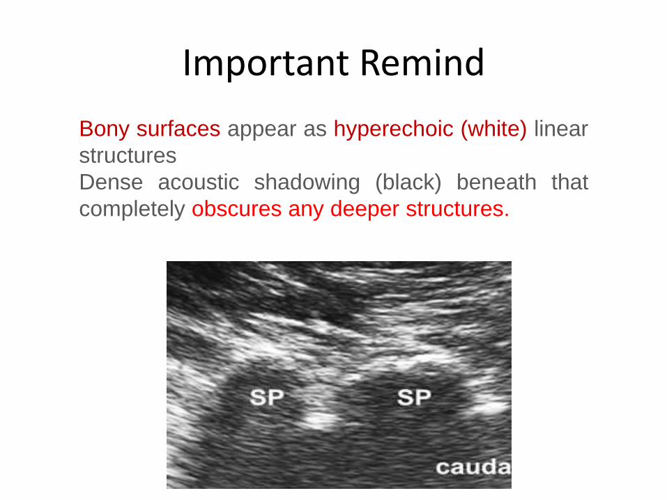
Important Remind
Bony surfaces appear as hyperechoic (white) linear
structures
Dense acoustic shadowing (black) beneath that
completely obscures any deeper structures.
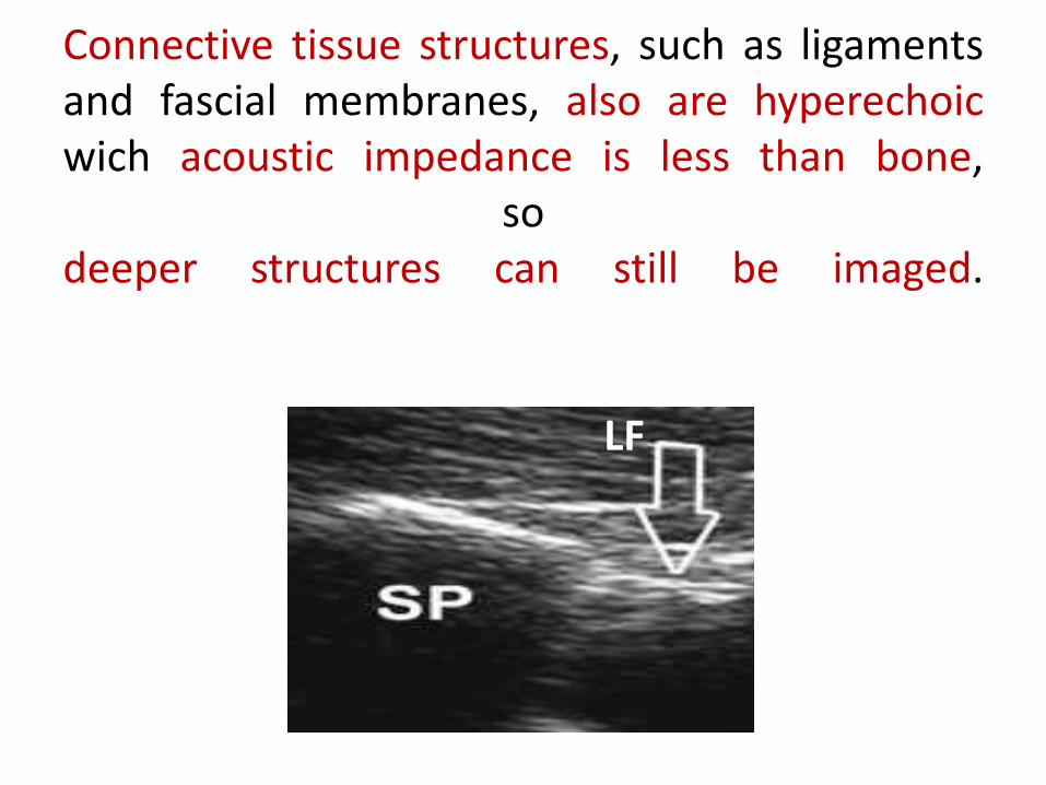
Connective tissue structures, such as ligamentsand fascial membranes, also are hyperechoicwich acoustic impedance is less than bone,
sodeeper structures can still be imaged.
LF
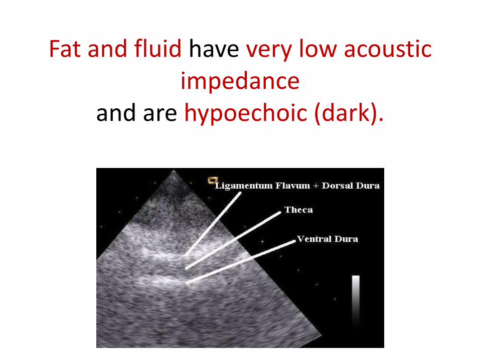
Fat and fluid have very low acoustic impedance
and are hypoechoic (dark).
preparation
• Position of choice: lateral decubitus or sitting position (prone)
• Probe of choice: curved-array, low-frequency(2–5 MHz) probe for wide field of view and deeper penetration
• 2 basic planes: sagittal, transverse
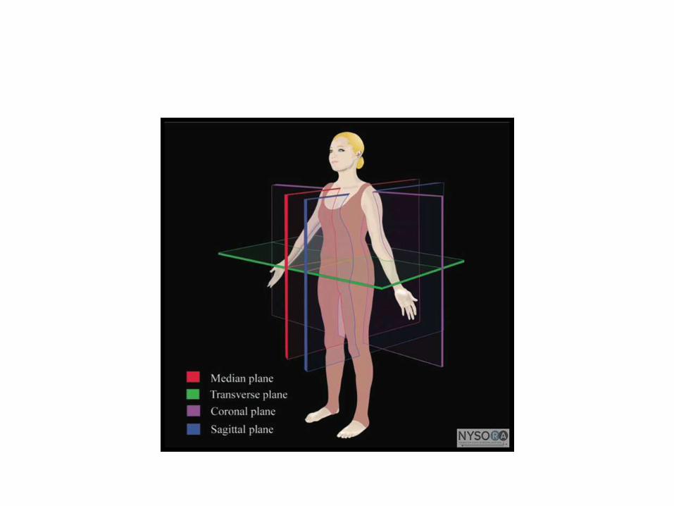
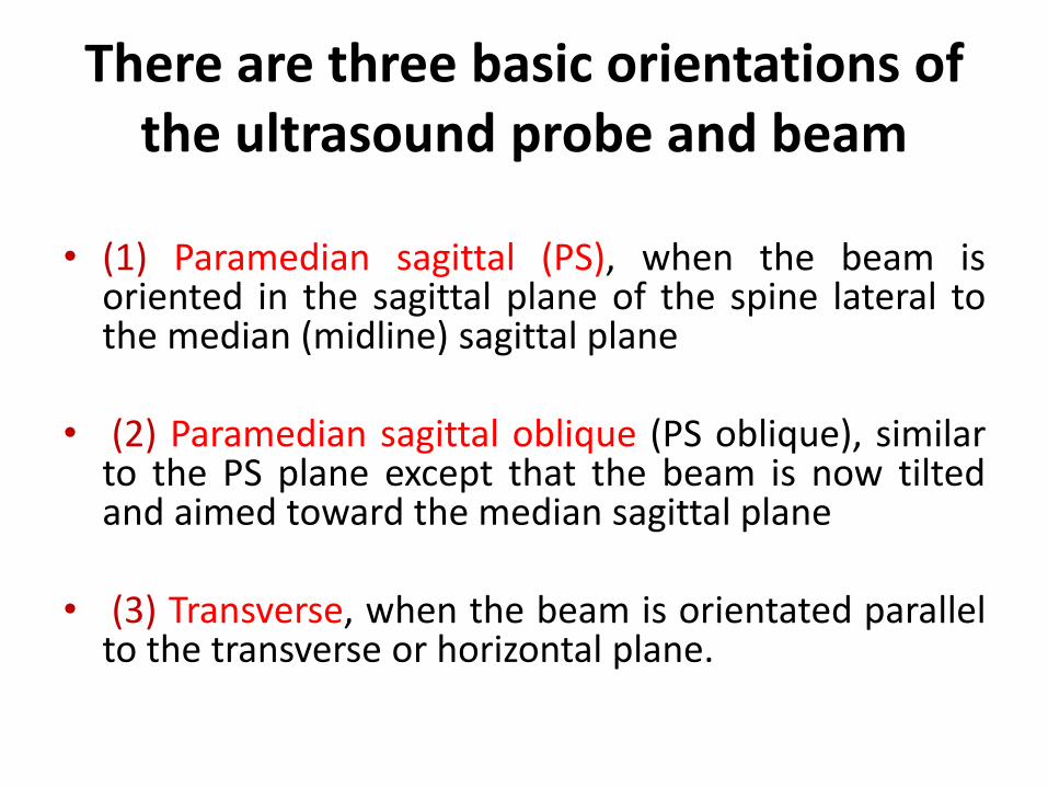
There are three basic orientations of the ultrasound probe and beam
• (1) Paramedian sagittal (PS), when the beam isoriented in the sagittal plane of the spine lateral tothe median (midline) sagittal plane
• (2) Paramedian sagittal oblique (PS oblique), similarto the PS plane except that the beam is now tiltedand aimed toward the median sagittal plane
• (3) Transverse, when the beam is orientated parallelto the transverse or horizontal plane.
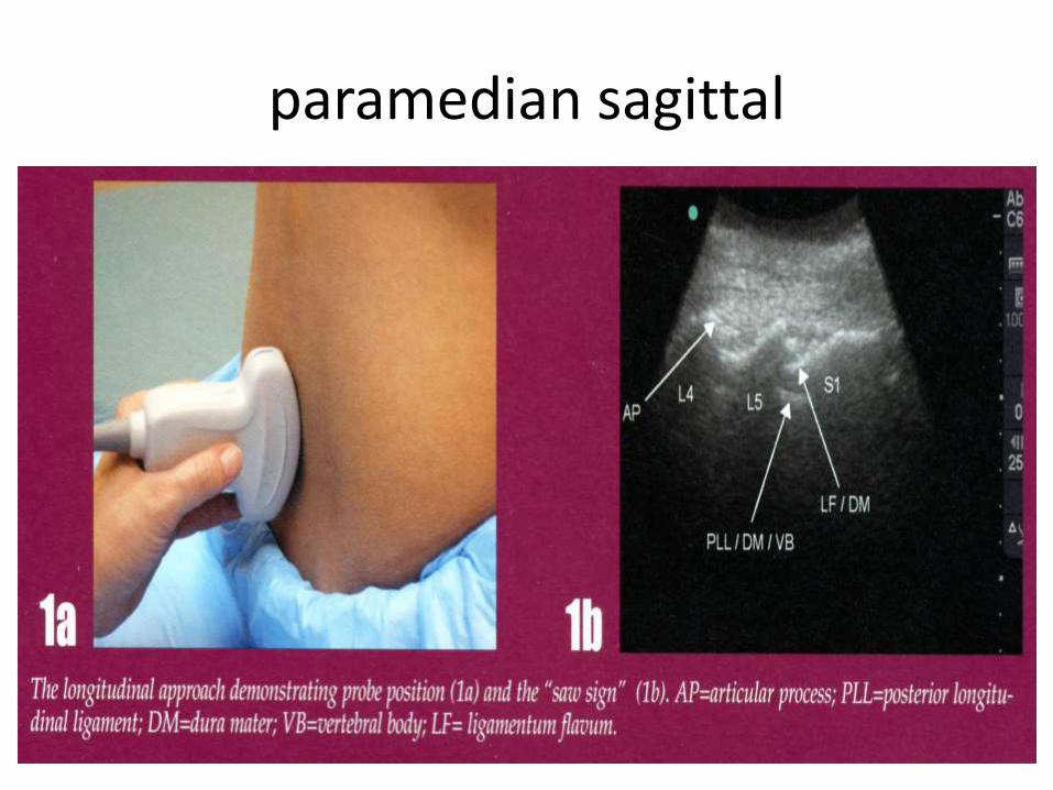
paramedian sagittal
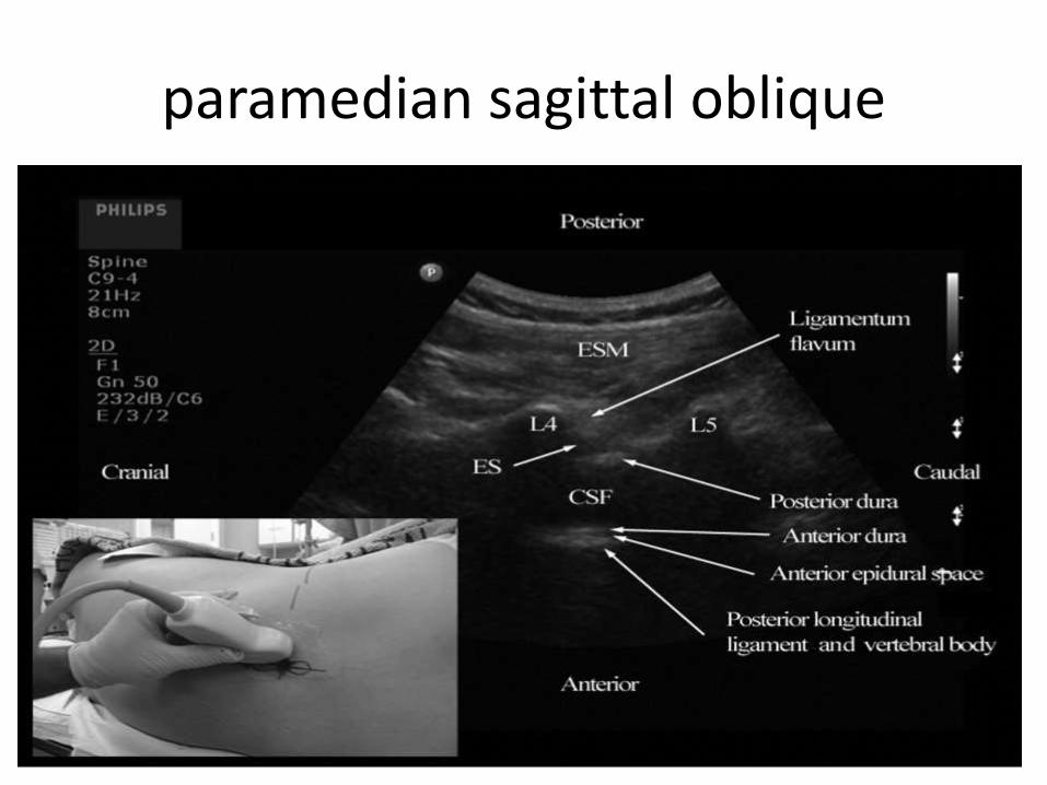
paramedian sagittal oblique
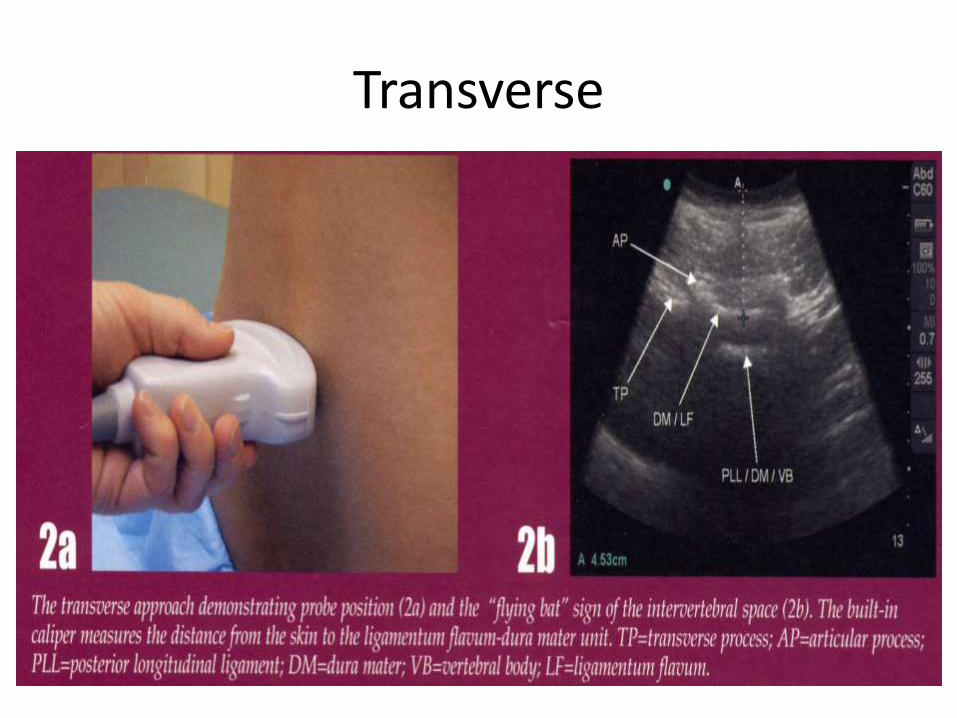
Transverse
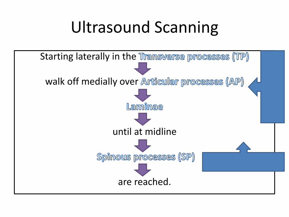
Ultrasound Scanning
Starting laterally in the
walk off medially over
until at midline
are reached.
Paramedian Sagital View
1
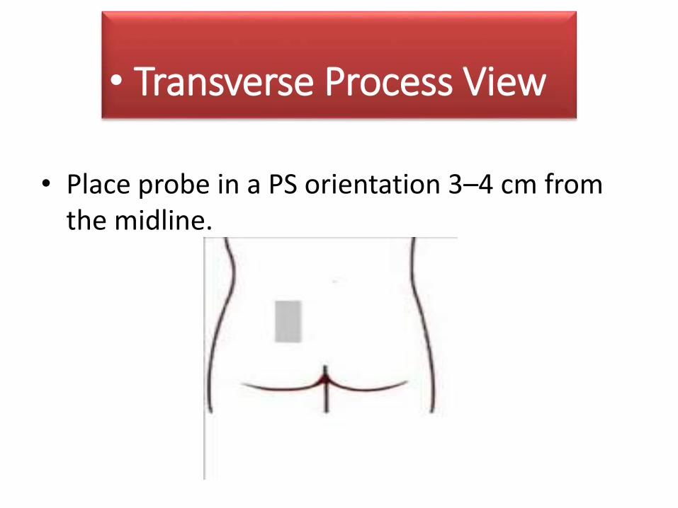
Transverse process view
• Place probe in a PS orientation 3–4 cm from the midline.
• Transverse Process View
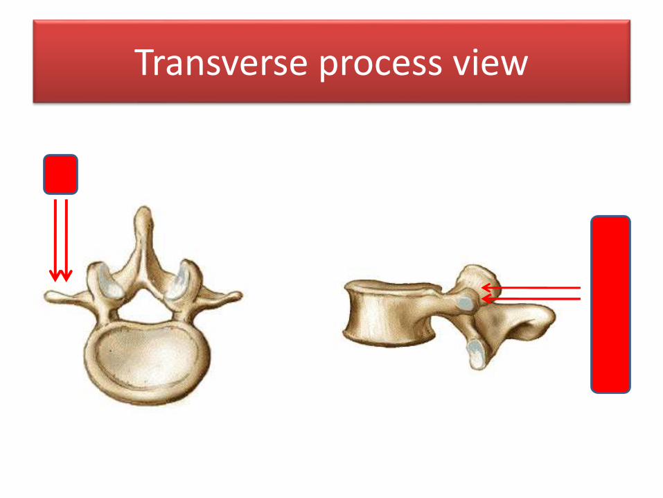
Transverse process view
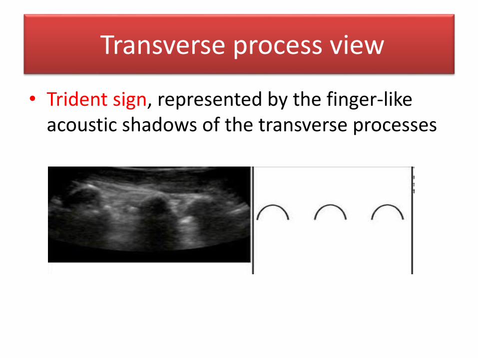
Transverse process view
• Trident sign, represented by the finger-like acoustic shadows of the transverse processes
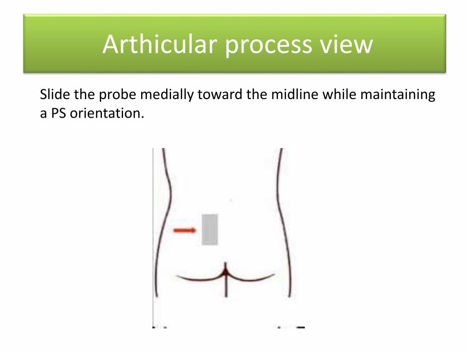
Slide the probe medially toward the midline while maintaining a PS orientation.
Arthicular process view
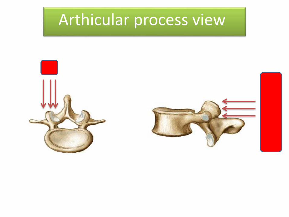
Articular Pricess ViewArthicular process view
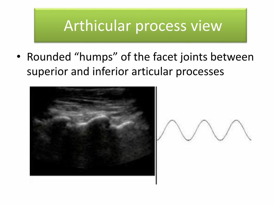
• Rounded “humps” of the facet joints between superior and inferior articular processes
Arthicular process view
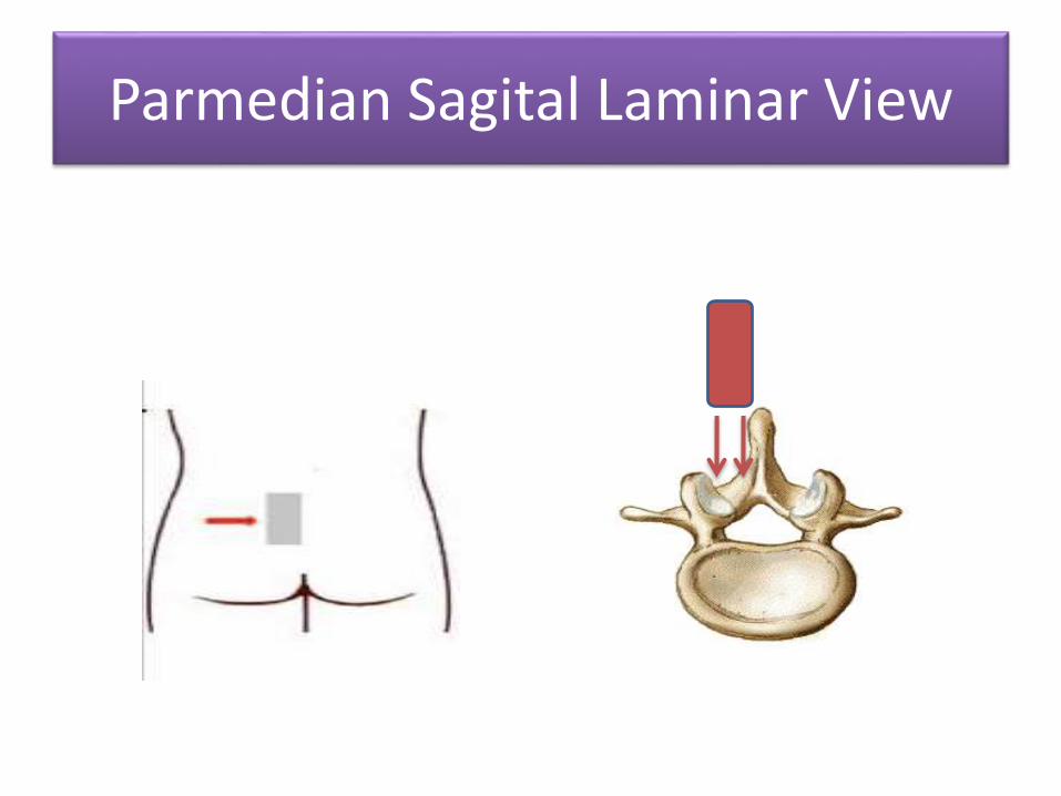
Parmedian Sagital Laminar View
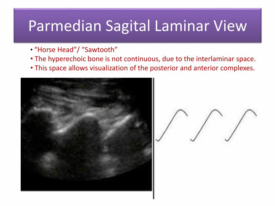
Parmedian Sagital Laminar View• “Horse Head”/ “Sawtooth”• The hyperechoic bone is not continuous, due to the interlaminar space.• This space allows visualization of the posterior and anterior complexes.
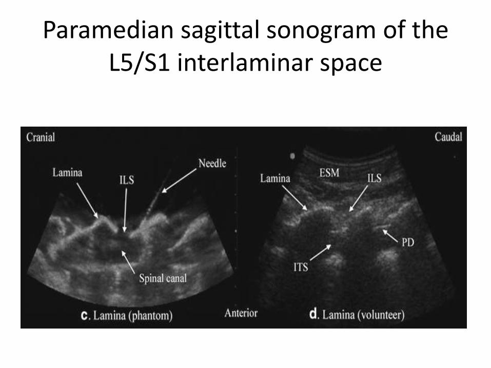
Paramedian sagittal sonogram of the L5/S1 interlaminar space
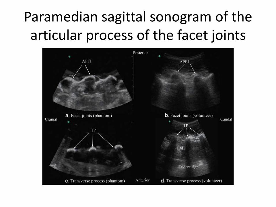
Paramedian sagittal sonogram of the articular process of the facet joints
Paramedian sagital oblique view
2
Paramedian sagital oblique view
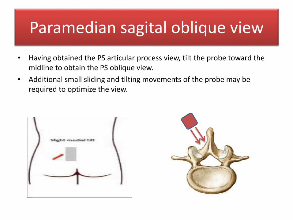
• Having obtained the PS articular process view, tilt the probe toward the midline to obtain the PS oblique view.
• Additional small sliding and tilting movements of the probe may be required to optimize the view.
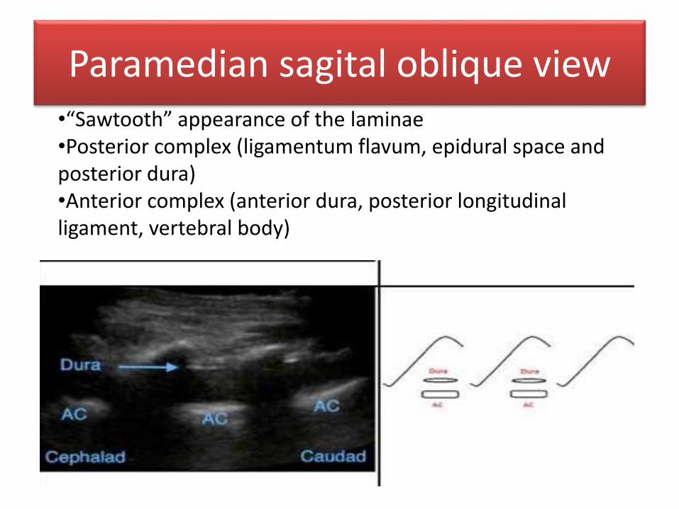
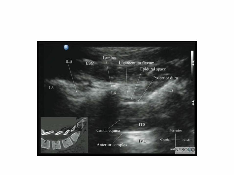
Paramedian sagital oblique view•“Sawtooth” appearance of the laminae•Posterior complex (ligamentum flavum, epidural space and posterior dura)•Anterior complex (anterior dura, posterior longitudinal ligament, vertebral body)
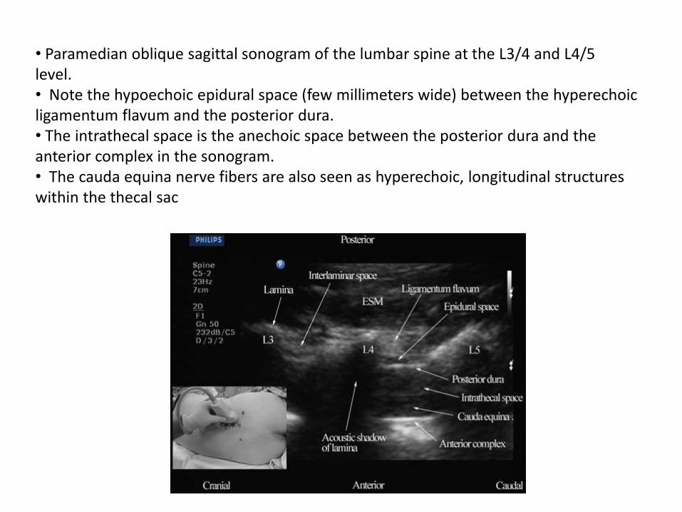
• Paramedian oblique sagittal sonogram of the lumbar spine at the L3/4 and L4/5level.• Note the hypoechoic epidural space (few millimeters wide) between the hyperechoicligamentum flavum and the posterior dura. • The intrathecal space is the anechoic space between the posterior dura and the anterior complex in the sonogram.• The cauda equina nerve fibers are also seen as hyperechoic, longitudinal structures within the thecal sac
Transverse View
4
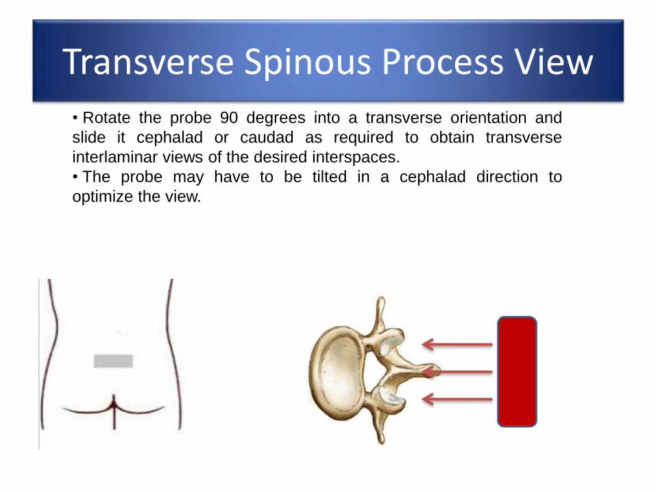
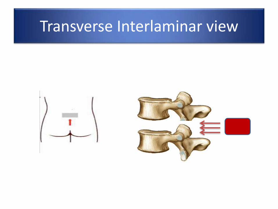
Transverse Spinous Process View• Rotate the probe 90 degrees into a transverse orientation and
slide it cephalad or caudad as required to obtain transverse
interlaminar views of the desired interspaces.
• The probe may have to be tilted in a cephalad direction to
optimize the view.
Transverse Spinous Process View
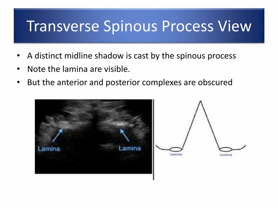
• A distinct midline shadow is cast by the spinous process
• Note the lamina are visible.
• But the anterior and posterior complexes are obscured
Transverse Spinous Process View
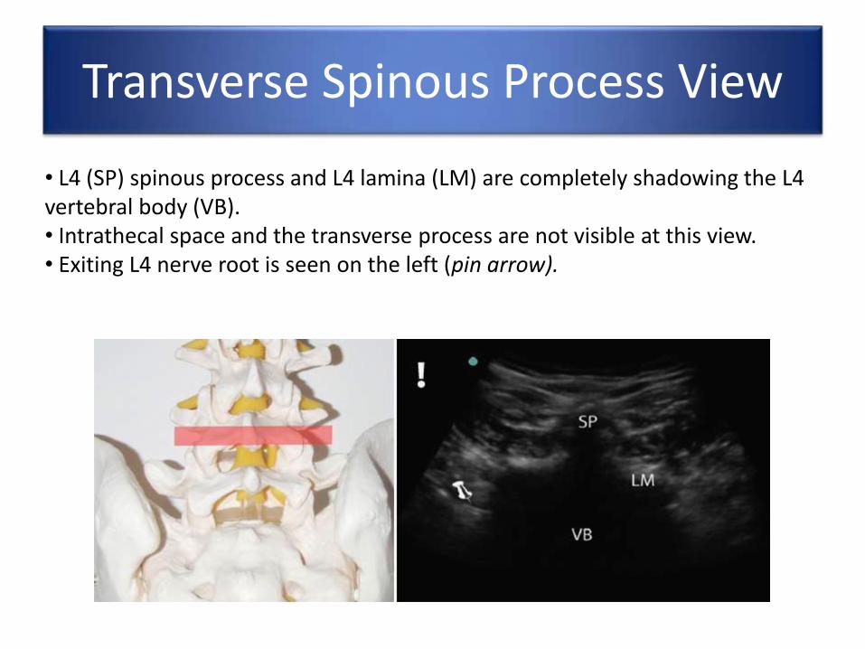
• L4 (SP) spinous process and L4 lamina (LM) are completely shadowing the L4 vertebral body (VB).• Intrathecal space and the transverse process are not visible at this view.• Exiting L4 nerve root is seen on the left (pin arrow).
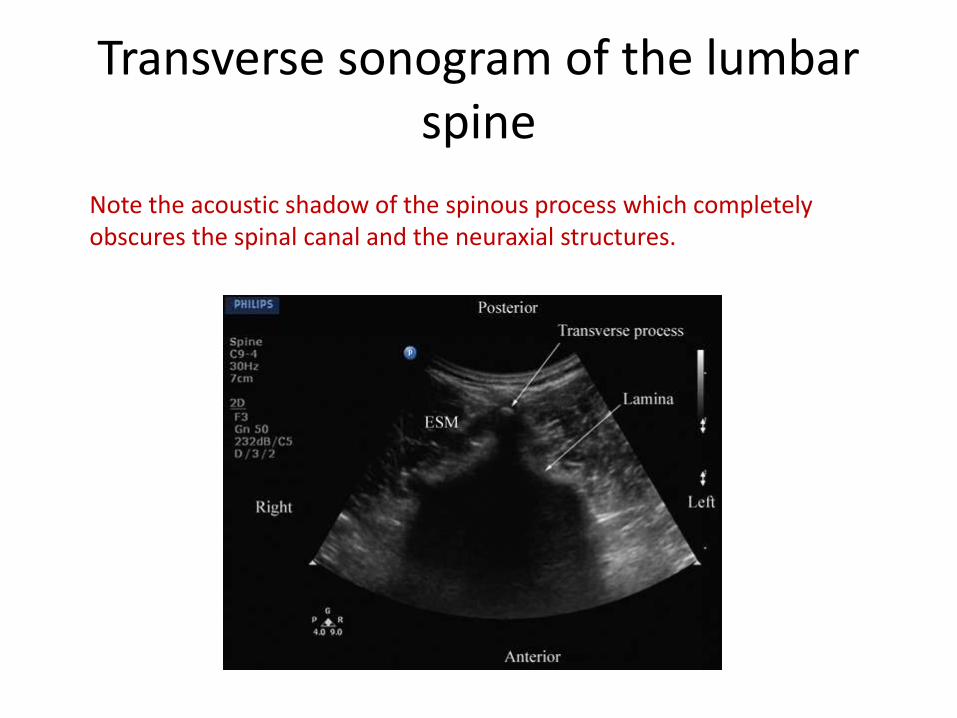
Transverse sonogram of the lumbar spine
Note the acoustic shadow of the spinous process which completely obscures the spinal canal and the neuraxial structures.
Transverse Interlaminar view
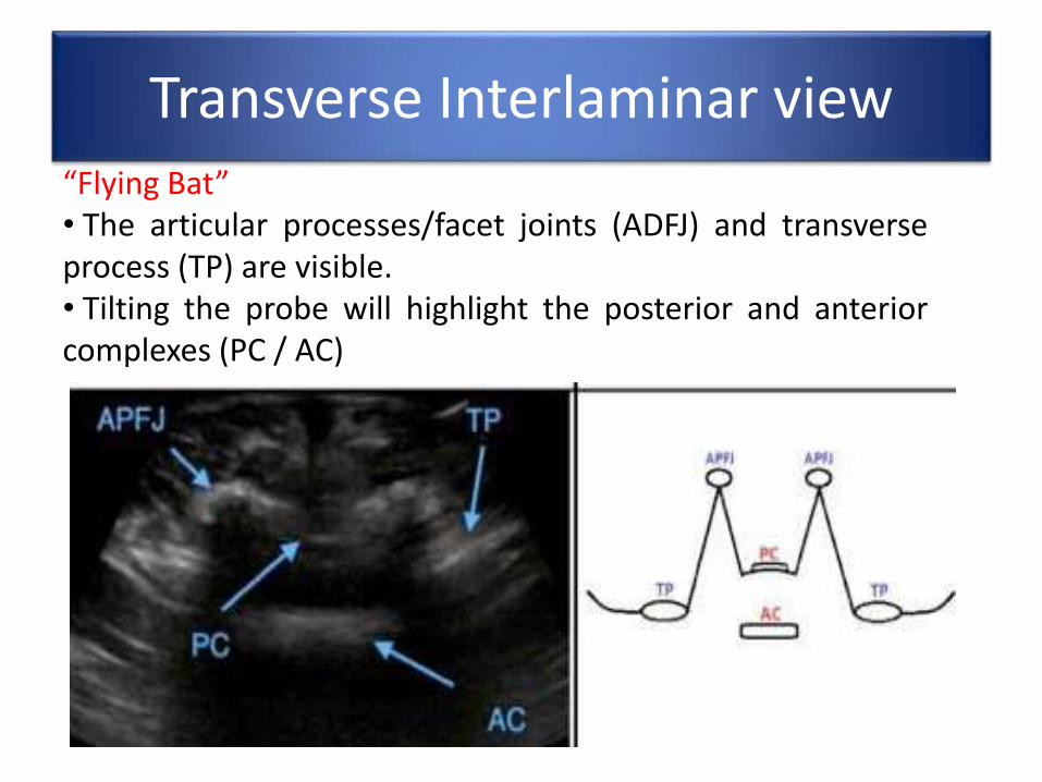
Transverse Interlaminar view“Flying Bat”• The articular processes/facet joints (ADFJ) and transverseprocess (TP) are visible.• Tilting the probe will highlight the posterior and anteriorcomplexes (PC / AC)
Transverse Interlaminar view
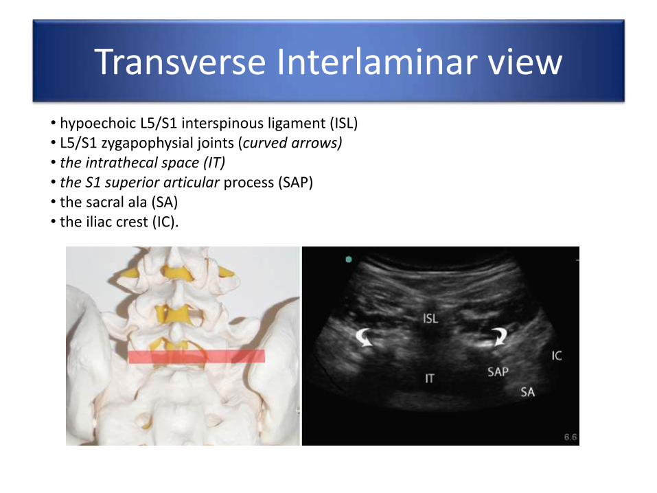
• hypoechoic L5/S1 interspinous ligament (ISL)• L5/S1 zygapophysial joints (curved arrows)• the intrathecal space (IT)• the S1 superior articular process (SAP)• the sacral ala (SA)• the iliac crest (IC).