lumbar spine surgery guideline
TRANSCRIPT
1
Washington State Department of Labor & Industries Surgical Guideline for Lumbar Spine—September 2021
Treatment Guideline for Lumbar Spine Surgery
TABLE OF CONTENTS
I. Review Criteria for Lumbar Spine Surgery ....................................................................... 3
A. Coverage Decisions Affecting This Guideline ....................................................................... 3
B. Lumbar Decompression Procedures .................................................................................... 5
C. Lumbar Fusion Procedures ................................................................................................ 11
II. Introduction ................................................................................................................. 20
A. Background and Prevalence .............................................................................................. 20
B. Pre-existing or Non-work-related Conditions .................................................................... 20
1. Nicotine Use ...................................................................................................... 20
III. Assessment .................................................................................................................. 21
A. History and Clinical Exam ................................................................................................... 21
B. Imaging ............................................................................................................................... 21
C. Opioids ............................................................................................................................... 22
D. Screening and Addressing Behavioral or Mental Health ................................................... 24
E. Preventing Complications .................................................................................................. 24
F. Measuring Functional Improvement ................................................................................. 25
IV. Non-operative Care ...................................................................................................... 25
V. Conditions and Surgical Procedures .............................................................................. 26
A. Lumbar Decompression Procedures .................................................................................. 26
1. Nerve Root Entrapment Due to Central/paracentral/foraminal/extra-foraminal
Herniated Nucleus Pulposus ................................................................................... 26
2. Recurrent Disc Hernation ........................................................................................ 27
3. Central Spinal Stenosis ............................................................................................ 28
4. Synovial Cyst ............................................................................................................ 29
5. Lateral Recess/Foraminal Stenosis .......................................................................... 30
6. Acute Cauda Equina Syndrome ............................................................................... 30
2
Washington State Department of Labor & Industries Surgical Guideline for Lumbar Spine—September 2021
B. Lumbar Fusion Procedures ................................................................................................ 32
1. Spondylolisthesis ..................................................................................................... 32
2. Prior Decompression at the Same Level ................................................................. 33
3. Pseudarthrosis, With or Without Hardware Failure ............................................... 33
4. Recurrent Disc Herniation ....................................................................................... 34
5. Foraminal Stenosis .................................................................................................. 35
6. Adjacent Segment Pathology .................................................................................. 36
C. Sacroiliac Joint Fusion ........................................................................................................ 36
D. Multidisciplinary Team Review of Lumbar Fusion Requests ............................................. 37
VI. Rehabilitation and Return to Work ............................................................................... 37
VII. Acknowledgements ...................................................................................................... 38
VIII. References ................................................................................................................... 40
3
Washington State Department of Labor & Industries Surgical Guideline for Lumbar Spine–September 2021
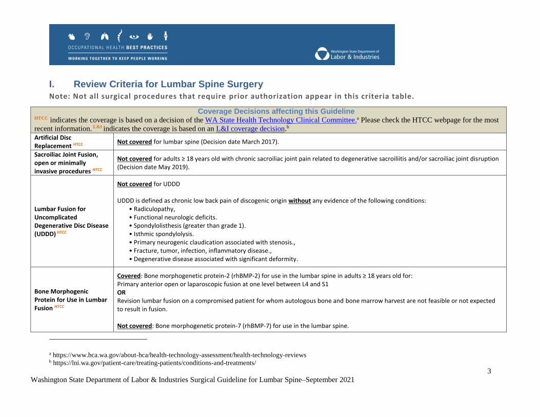
I. Review Criteria for Lumbar Spine Surgery
Note: Not all surgical procedures that require prior authorization appear in this criteria table.
Coverage Decisions affecting this Guideline HTCC indicates the coverage is based on a decision of the WA State Health Technology Clinical Committee.a Please check the HTCC webpage for the most
recent information. L&I indicates the coverage is based on an L&I coverage decision.b
Artificial Disc Replacement HTCC
Not covered for lumbar spine (Decision date March 2017).
Sacroiliac Joint Fusion, open or minimally invasive procedures HTCC
Not covered for adults ≥ 18 years old with chronic sacroiliac joint pain related to degenerative sacroiliitis and/or sacroiliac joint disruption (Decision date May 2019).
Lumbar Fusion for Uncomplicated Degenerative Disc Disease (UDDD) HTCC
Not covered for UDDD UDDD is defined as chronic low back pain of discogenic origin without any evidence of the following conditions:
• Radiculopathy, • Functional neurologic deficits. • Spondylolisthesis (greater than grade 1). • Isthmic spondylolysis. • Primary neurogenic claudication associated with stenosis., • Fracture, tumor, infection, inflammatory disease., • Degenerative disease associated with significant deformity.
Bone Morphogenic Protein for Use in Lumbar Fusion HTCC
o Covered: Bone morphogenetic protein-2 (rhBMP-2) for use in the lumbar spine in adults ≥ 18 years old for: o Primary anterior open or laparoscopic fusion at one level between L4 and S1 o OR o Revision lumbar fusion on a compromised patient for whom autologous bone and bone marrow harvest are not feasible or not expected
to result in fusion. o o Not covered: Bone morphogenetic protein-7 (rhBMP-7) for use in the lumbar spine.
a https://www.hca.wa.gov/about-hca/health-technology-assessment/health-technology-reviews b https://lni.wa.gov/patient-care/treating-patients/conditions-and-treatments/
4
Washington State Department of Labor & Industries Surgical Guideline for Lumbar Spine–September 2021
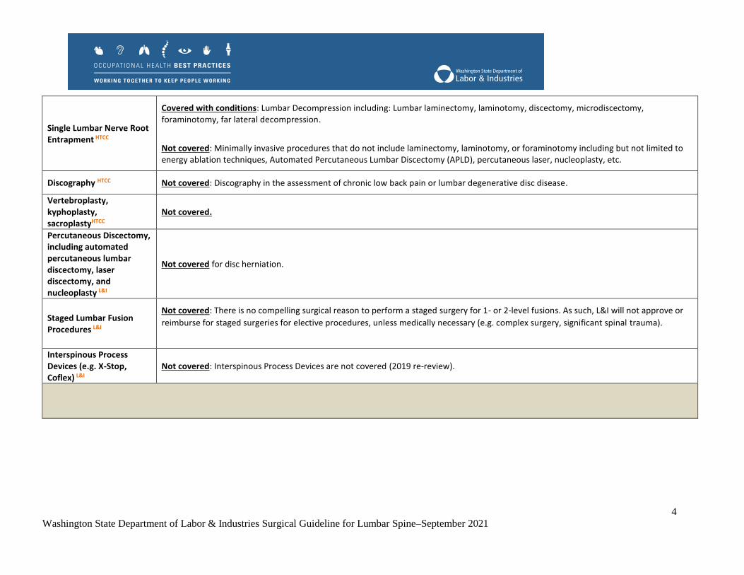
Single Lumbar Nerve Root Entrapment HTCC
Covered with conditions: Lumbar Decompression including: Lumbar laminectomy, laminotomy, discectomy, microdiscectomy, foraminotomy, far lateral decompression.
Not covered: Minimally invasive procedures that do not include laminectomy, laminotomy, or foraminotomy including but not limited to energy ablation techniques, Automated Percutaneous Lumbar Discectomy (APLD), percutaneous laser, nucleoplasty, etc.
Discography HTCC Not covered: Discography in the assessment of chronic low back pain or lumbar degenerative disc disease.
Vertebroplasty, kyphoplasty, sacroplastyHTCC
Not covered.
Percutaneous Discectomy, including automated percutaneous lumbar discectomy, laser discectomy, and nucleoplasty L&I
Not covered for disc herniation.
Staged Lumbar Fusion Procedures L&I
Not covered: There is no compelling surgical reason to perform a staged surgery for 1- or 2-level fusions. As such, L&I will not approve or
reimburse for staged surgeries for elective procedures, unless medically necessary (e.g. complex surgery, significant spinal trauma).
Interspinous Process Devices (e.g. X-Stop, Coflex) L&I
Not covered: Interspinous Process Devices are not covered (2019 re-review).
5
Washington State Department of Labor & Industries Surgical Guideline for Lumbar Spine–September 2021
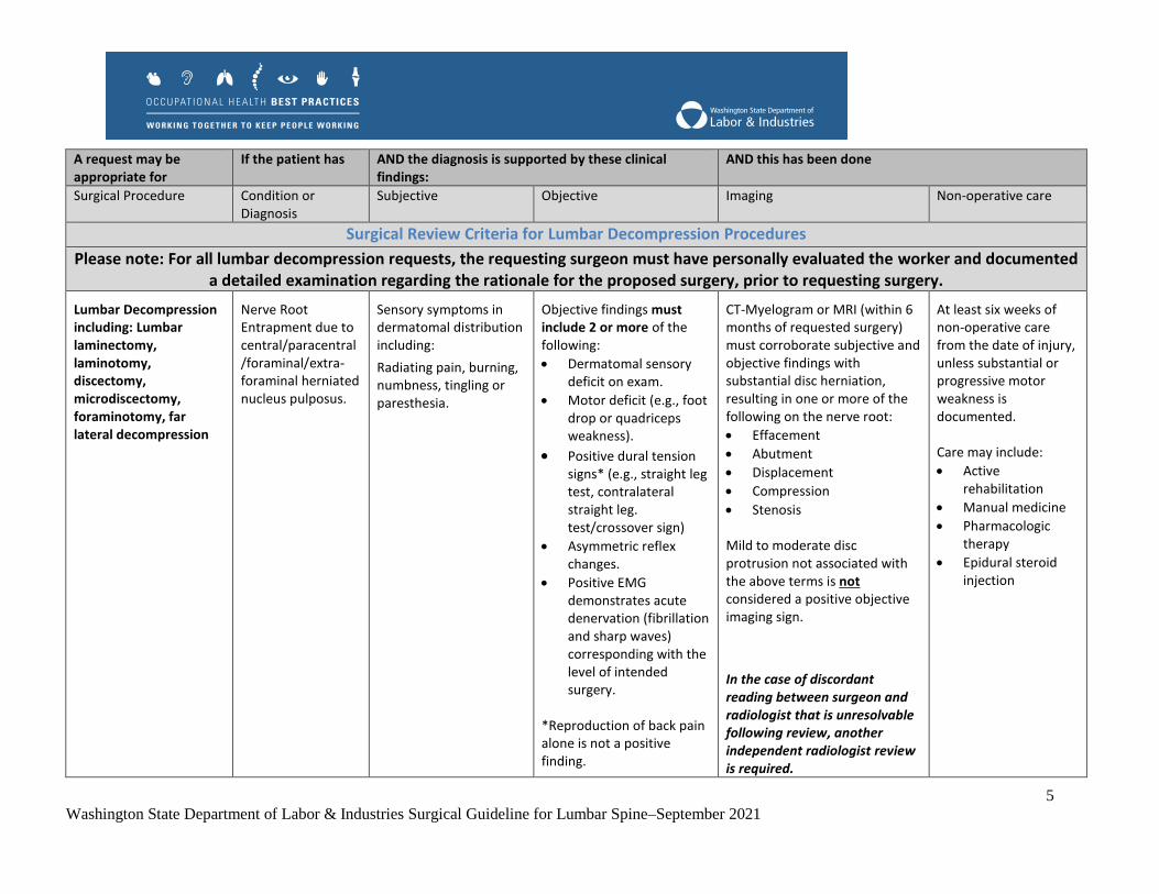
A request may be appropriate for
If the patient has AND the diagnosis is supported by these clinical findings:
AND this has been done
Surgical Procedure Condition or Diagnosis
Subjective Objective Imaging Non-operative care
Surgical Review Criteria for Lumbar Decompression Procedures
Please note: For all lumbar decompression requests, the requesting surgeon must have personally evaluated the worker and documented a detailed examination regarding the rationale for the proposed surgery, prior to requesting surgery.
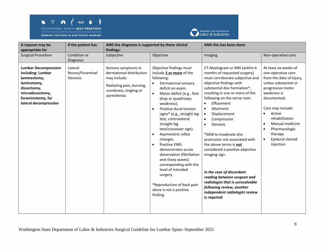
Lumbar Decompression including: Lumbar laminectomy, laminotomy, discectomy, microdiscectomy, foraminotomy, far lateral decompression
Nerve Root Entrapment due to central/paracentral/foraminal/extra-foraminal herniated nucleus pulposus.
Sensory symptoms in dermatomal distribution including:
Radiating pain, burning, numbness, tingling or paresthesia.
Objective findings must include 2 or more of the following:
Dermatomal sensory deficit on exam.
Motor deficit (e.g., foot drop or quadriceps weakness).
Positive dural tension signs* (e.g., straight leg test, contralateral straight leg. test/crossover sign)
Asymmetric reflex changes.
Positive EMG demonstrates acute denervation (fibrillation and sharp waves) corresponding with the level of intended surgery.
*Reproduction of back pain alone is not a positive finding.
CT-Myelogram or MRI (within 6 months of requested surgery) must corroborate subjective and objective findings with substantial disc herniation, resulting in one or more of the following on the nerve root:
Effacement
Abutment
Displacement
Compression
Stenosis Mild to moderate disc protrusion not associated with the above terms is not considered a positive objective imaging sign.
In the case of discordant reading between surgeon and radiologist that is unresolvable following review, another independent radiologist review is required.
At least six weeks of non-operative care from the date of injury, unless substantial or progressive motor weakness is documented. Care may include:
Active rehabilitation
Manual medicine
Pharmacologic therapy
Epidural steroid injection
6
Washington State Department of Labor & Industries Surgical Guideline for Lumbar Spine–September 2021
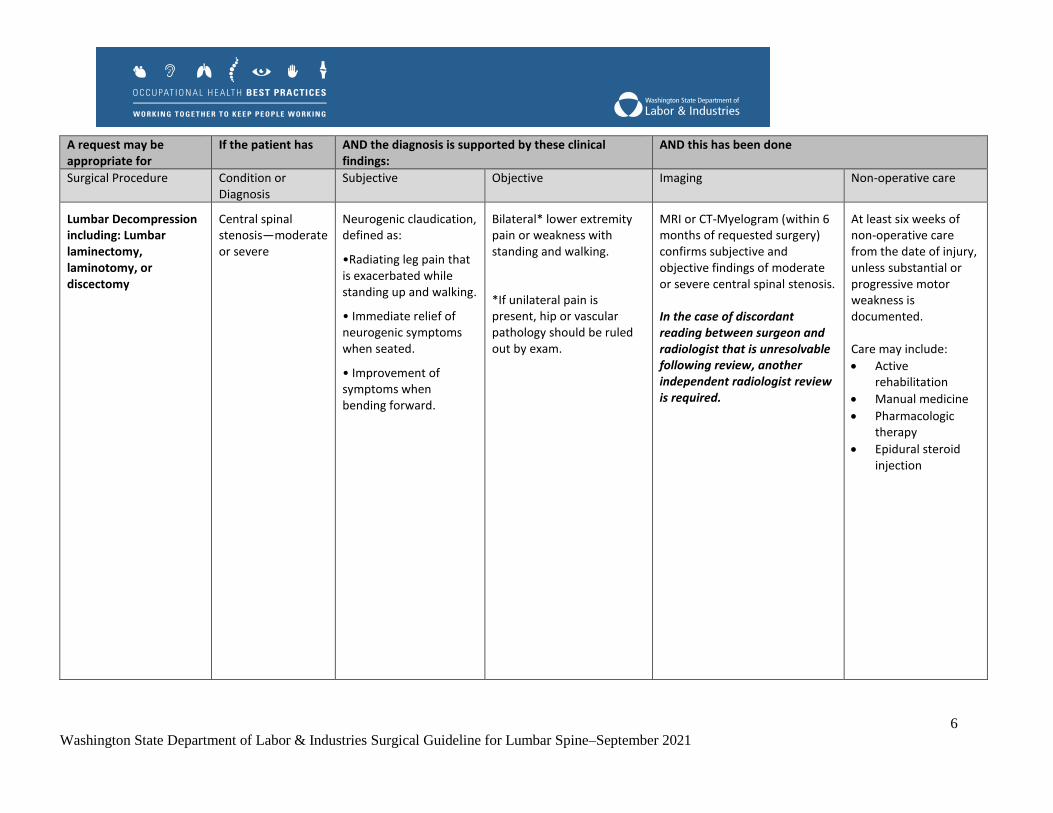
A request may be appropriate for
If the patient has AND the diagnosis is supported by these clinical findings:
AND this has been done
Surgical Procedure Condition or Diagnosis
Subjective Objective Imaging Non-operative care
Lumbar Decompression including: Lumbar laminectomy, laminotomy, or discectomy
Central spinal stenosis—moderate or severe
Neurogenic claudication, defined as:
•Radiating leg pain that is exacerbated while standing up and walking.
• Immediate relief of neurogenic symptoms when seated.
• Improvement of symptoms when bending forward.
Bilateral* lower extremity pain or weakness with standing and walking.
*If unilateral pain is present, hip or vascular pathology should be ruled out by exam.
MRI or CT-Myelogram (within 6 months of requested surgery) confirms subjective and objective findings of moderate or severe central spinal stenosis. In the case of discordant reading between surgeon and radiologist that is unresolvable following review, another independent radiologist review is required.
At least six weeks of non-operative care from the date of injury, unless substantial or progressive motor weakness is documented. Care may include:
Active rehabilitation
Manual medicine
Pharmacologic therapy
Epidural steroid injection
7
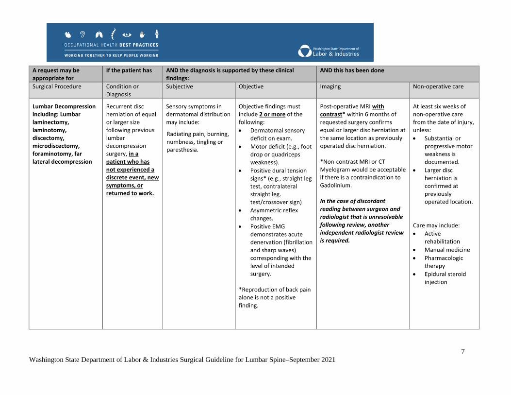
Washington State Department of Labor & Industries Surgical Guideline for Lumbar Spine–September 2021
A request may be appropriate for
If the patient has AND the diagnosis is supported by these clinical findings:
AND this has been done
Surgical Procedure Condition or Diagnosis
Subjective Objective Imaging Non-operative care
Lumbar Decompression including: Lumbar laminectomy, laminotomy, discectomy, microdiscectomy, foraminotomy, far lateral decompression
Recurrent disc herniation of equal or larger size following previous lumbar decompression surgery, in a patient who has not experienced a discrete event, new symptoms, or returned to work.
Sensory symptoms in dermatomal distribution may include:
Radiating pain, burning, numbness, tingling or paresthesia.
Objective findings must include 2 or more of the following:
Dermatomal sensory deficit on exam.
Motor deficit (e.g., foot drop or quadriceps weakness).
Positive dural tension signs* (e.g., straight leg test, contralateral straight leg. test/crossover sign)
Asymmetric reflex changes.
Positive EMG demonstrates acute denervation (fibrillation and sharp waves) corresponding with the level of intended surgery.
*Reproduction of back pain alone is not a positive finding.
Post-operative MRI with contrast* within 6 months of requested surgery confirms equal or larger disc herniation at the same location as previously operated disc herniation. *Non-contrast MRI or CT Myelogram would be acceptable if there is a contraindication to Gadolinium. In the case of discordant reading between surgeon and radiologist that is unresolvable following review, another independent radiologist review is required.
At least six weeks of non-operative care from the date of injury, unless:
Substantial or progressive motor weakness is documented.
Larger disc herniation is confirmed at previously operated location.
Care may include:
Active rehabilitation
Manual medicine
Pharmacologic therapy
Epidural steroid injection
8
Washington State Department of Labor & Industries Surgical Guideline for Lumbar Spine–September 2021
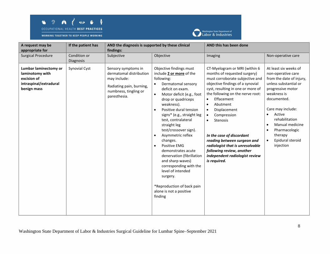
A request may be appropriate for
If the patient has AND the diagnosis is supported by these clinical findings:
AND this has been done
Surgical Procedure Condition or Diagnosis
Subjective Objective Imaging Non-operative care
Lumbar laminectomy or laminotomy with excision of intraspinal/extradural benign mass
Synovial Cyst Sensory symptoms in dermatomal distribution may include:
Radiating pain, burning, numbness, tingling or paresthesia.
Objective findings must include 2 or more of the following:
Dermatomal sensory deficit on exam.
Motor deficit (e.g., foot drop or quadriceps weakness).
Positive dural tension signs* (e.g., straight leg test, contralateral straight leg test/crossover sign).
Asymmetric reflex changes.
Positive EMG demonstrates acute denervation (fibrillation and sharp waves) corresponding with the level of intended surgery.
*Reproduction of back pain alone is not a positive finding
CT-Myelogram or MRI (within 6 months of requested surgery) must corroborate subjective and objective findings of a synovial cyst, resulting in one or more of the following on the nerve root:
Effacement
Abutment
Displacement
Compression
Stenosis In the case of discordant reading between surgeon and radiologist that is unresolvable following review, another independent radiologist review is required.
At least six weeks of non-operative care from the date of injury, unless substantial or progressive motor weakness is documented. Care may include:
Active rehabilitation
Manual medicine
Pharmacologic therapy
Epidural steroid injection
9
Washington State Department of Labor & Industries Surgical Guideline for Lumbar Spine–September 2021
A request may be appropriate for
If the patient has AND the diagnosis is supported by these clinical findings:
AND this has been done
Surgical Procedure Condition or Diagnosis
Subjective Objective Imaging Non-operative care
Lumbar Decompression including: Lumbar laminectomy, laminotomy, discectomy, microdiscectomy, foraminotomy, far lateral decompression
Lateral Recess/Foraminal Stenosis
Sensory symptoms in dermatomal distribution may include:
Radiating pain, burning, numbness, tingling or paresthesia.
Objective findings must include 2 or more of the following:
Dermatomal sensory deficit on exam.
Motor deficit (e.g., foot drop or quadriceps weakness).
Positive dural tension signs* (e.g., straight leg test, contralateral straight leg test/crossover sign).
Asymmetric reflex changes.
Positive EMG demonstrates acute denervation (fibrillation and sharp waves) corresponding with the level of intended surgery.
*Reproduction of back pain alone is not a positive finding.
CT-Myelogram or MRI (within 6 months of requested surgery) must corroborate subjective and objective findings with substantial disc herniation*, resulting in one or more of the following on the nerve root:
Effacement
Abutment
Displacement
Compression
Stenosis *Mild to moderate disc protrusion not associated with the above terms is not considered a positive objective imaging sign.
In the case of discordant reading between surgeon and radiologist that is unresolvable following review, another independent radiologist review is required.
At least six weeks of non-operative care from the date of injury, unless substantial or progressive motor weakness is documented. Care may include:
Active rehabilitation
Manual medicine
Pharmacologic therapy
Epidural steroid injection
10
Washington State Department of Labor & Industries Surgical Guideline for Lumbar Spine–September 2021
A request may be appropriate for
If the patient has AND the diagnosis is supported by these clinical findings:
AND this has been done
Surgical Procedure Condition or Diagnosis
Subjective Objective Imaging Non-operative care
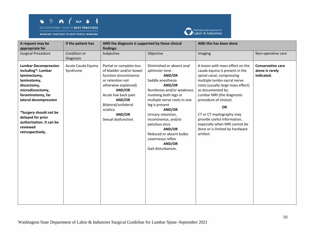
Lumbar Decompression including*: Lumbar laminectomy, laminotomy, discectomy, microdiscectomy, foraminotomy, far lateral decompression
*Surgery should not be delayed for prior authorization. It can be reviewed retrospectively.
Acute Cauda Equina Syndrome
Partial or complete loss of bladder and/or bowel function (incontinence or retention not otherwise explained)
AND/OR Acute low back pain
AND/OR Bilateral/unilateral sciatica
AND/OR Sexual dysfunction.
Diminished or absent anal sphincter tone
AND/OR Saddle anesthesia
AND/OR Numbness and/or weakness involving both legs or multiple nerve roots in one leg is present
AND/OR Urinary retention, incontinence, and/or patulous anus
AND/OR Reduced or absent bulbo cavernosus reflex
AND/OR Gait disturbances.
A lesion with mass effect on the cauda equina is present in the spinal canal, compressing multiple lumbo-sacral nerve roots (usually large mass effect) as documented by: Lumbar MRI (the diagnostic procedure of choice)
OR
CT or CT myelography may provide useful information, especially when MRI cannot be done or is limited by hardware artifact.
Conservative care alone is rarely indicated.
11
Washington State Department of Labor & Industries Surgical Guideline for Lumbar Spine–September 2021
A request may be appropriate for
If the patient has AND the diagnosis is supported by these clinical findings:
AND this has been done
Surgical Procedure Condition or Diagnosis
Subjective Objective Imaging Non-operative care
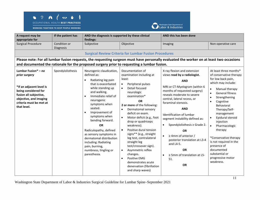
Surgical Review Criteria for Lumbar Fusion Procedures
Please note: For all lumbar fusion requests, the requesting surgeon must have personally evaluated the worker on at least two occasions and documented the rationale for the proposed surgery prior to requesting a lumbar fusion.
Lumbar Fusion* – no prior surgery
*If an adjacent level is being considered for fusion all subjective, objective, and imaging criteria must be met at that level.
Spondylolisthesis Neurogenic claudication, defined as:
Radiating leg pain that is exacerbated while standing up and walking.
Immediate relief of neurogenic symptoms when seated.
Improvement of symptoms when bending forward.
OR
Radiculopathy, defined as sensory symptoms in dermatomal distribution including: Radiating pain, burning, numbness, tingling or paresthesia.
Documentation of examination including at least:
Peripheral pulses
Detail focused neurologic examination*
OR 2 or more of the following:
Dermatomal sensory deficit on exam.
Motor deficit (e.g., foot drop or quadriceps weakness).
Positive dural tension signs** (e.g., straight leg test, contralateral straight leg test/crossover sign).
Asymmetric reflex changes.
Positive EMG demonstrates acute denervation (fibrillation and sharp waves)
X-ray flexion and extension views read by a radiologist.
AND
MRI or CT-Myelogram (within 6 months of requested surgery) reveals moderate to severe central, lateral recess, or foraminal stenosis.
AND
Identification of lumbar segment instability defined as:
Spondylolisthesis ≥ Grade 2.
OR
≥ 4mm of anterior / posterior translation at L3-4 and L4-5.
OR
≥ 5mm of translation at L5-S1.
OR
At least three months* of conservative therapy for low back pain, which may include:
Manual therapy
General fitness
Strengthening
Cognitive Behavioral Therapy/Self-management
Epidural steroid injection
Pharmacologic therapy
*Conservative therapy is not required in the presence of documented substantial or progressive motor weakness.
12
Washington State Department of Labor & Industries Surgical Guideline for Lumbar Spine–September 2021
A request may be appropriate for
If the patient has AND the diagnosis is supported by these clinical findings:
AND this has been done
Surgical Procedure Condition or Diagnosis
Subjective Objective Imaging Non-operative care
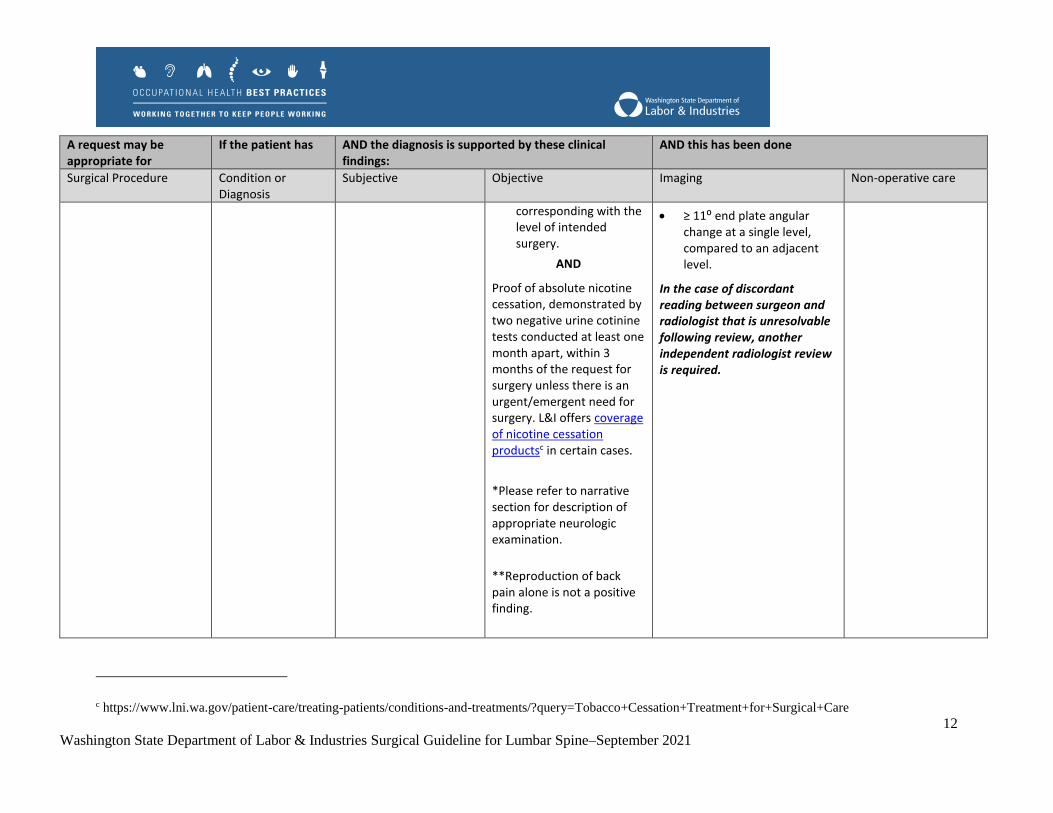
corresponding with the level of intended surgery.
AND
Proof of absolute nicotine cessation, demonstrated by two negative urine cotinine tests conducted at least one month apart, within 3 months of the request for surgery unless there is an urgent/emergent need for surgery. L&I offers coverage of nicotine cessation productsc in certain cases.
*Please refer to narrative section for description of appropriate neurologic examination.
**Reproduction of back pain alone is not a positive finding.
≥ 11⁰ end plate angular change at a single level, compared to an adjacent level.
In the case of discordant reading between surgeon and radiologist that is unresolvable following review, another independent radiologist review is required.
c https://www.lni.wa.gov/patient-care/treating-patients/conditions-and-treatments/?query=Tobacco+Cessation+Treatment+for+Surgical+Care
13
Washington State Department of Labor & Industries Surgical Guideline for Lumbar Spine–September 2021
A request may be appropriate for
If the patient has AND the diagnosis is supported by these clinical findings:
AND this has been done
Surgical Procedure Condition or Diagnosis
Subjective Objective Imaging Non-operative care
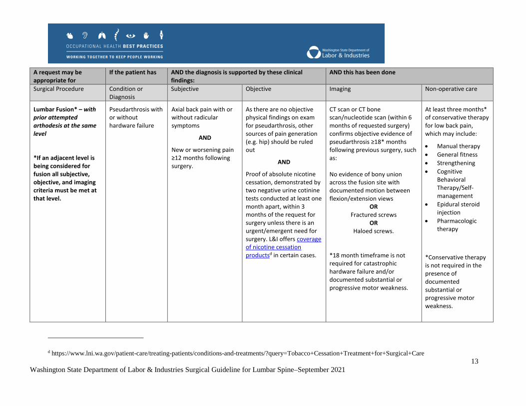
Lumbar Fusion* – with prior attempted arthodesis at the same level
*If an adjacent level is being considered for fusion all subjective, objective, and imaging criteria must be met at that level.
Pseudarthrosis with or without hardware failure
Axial back pain with or without radicular symptoms
AND
New or worsening pain ≥12 months following surgery.
As there are no objective physical findings on exam for pseudarthrosis, other sources of pain generation (e.g. hip) should be ruled out
AND
Proof of absolute nicotine cessation, demonstrated by two negative urine cotinine tests conducted at least one month apart, within 3 months of the request for surgery unless there is an urgent/emergent need for surgery. L&I offers coverage of nicotine cessation productsd in certain cases.
CT scan or CT bone scan/nucleotide scan (within 6 months of requested surgery) confirms objective evidence of pseudarthrosis ≥18* months following previous surgery, such as:
No evidence of bony union across the fusion site with documented motion between flexion/extension views
OR Fractured screws
OR Haloed screws.
*18 month timeframe is not required for catastrophic hardware failure and/or documented substantial or progressive motor weakness.
At least three months* of conservative therapy for low back pain, which may include:
Manual therapy
General fitness
Strengthening
Cognitive Behavioral Therapy/Self-management
Epidural steroid injection
Pharmacologic therapy
*Conservative therapy is not required in the presence of documented substantial or progressive motor weakness.
d https://www.lni.wa.gov/patient-care/treating-patients/conditions-and-treatments/?query=Tobacco+Cessation+Treatment+for+Surgical+Care
14
Washington State Department of Labor & Industries Surgical Guideline for Lumbar Spine–September 2021
A request may be appropriate for
If the patient has AND the diagnosis is supported by these clinical findings:
AND this has been done
Surgical Procedure Condition or Diagnosis
Subjective Objective Imaging Non-operative care
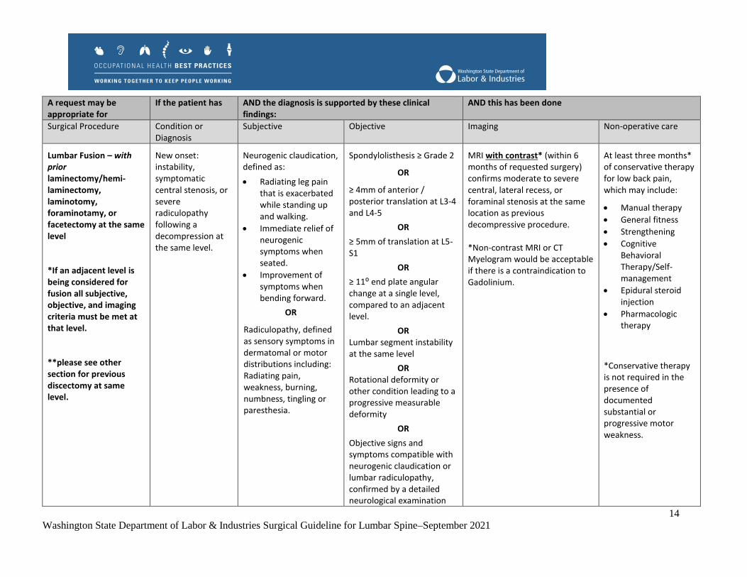
Lumbar Fusion – with prior laminectomy/hemi-laminectomy, laminotomy, foraminotamy, or facetectomy at the same level
*If an adjacent level is being considered for fusion all subjective, objective, and imaging criteria must be met at that level.
**please see other section for previous discectomy at same level.
New onset: instability, symptomatic central stenosis, or severe radiculopathy following a decompression at the same level.
Neurogenic claudication, defined as:
Radiating leg pain that is exacerbated while standing up and walking.
Immediate relief of neurogenic symptoms when seated.
Improvement of symptoms when bending forward.
OR
Radiculopathy, defined as sensory symptoms in dermatomal or motor distributions including: Radiating pain, weakness, burning, numbness, tingling or paresthesia.
Spondylolisthesis ≥ Grade 2
OR
≥ 4mm of anterior / posterior translation at L3-4 and L4-5
OR
≥ 5mm of translation at L5-S1
OR
≥ 11⁰ end plate angular change at a single level, compared to an adjacent level.
OR Lumbar segment instability at the same level
OR Rotational deformity or other condition leading to a progressive measurable deformity
OR
Objective signs and symptoms compatible with neurogenic claudication or lumbar radiculopathy, confirmed by a detailed neurological examination
MRI with contrast* (within 6 months of requested surgery) confirms moderate to severe central, lateral recess, or foraminal stenosis at the same location as previous decompressive procedure. *Non-contrast MRI or CT Myelogram would be acceptable if there is a contraindication to Gadolinium.
At least three months* of conservative therapy for low back pain, which may include:
Manual therapy
General fitness
Strengthening
Cognitive Behavioral Therapy/Self-management
Epidural steroid injection
Pharmacologic therapy
*Conservative therapy is not required in the presence of documented substantial or progressive motor weakness.
15
Washington State Department of Labor & Industries Surgical Guideline for Lumbar Spine–September 2021
A request may be appropriate for
If the patient has AND the diagnosis is supported by these clinical findings:
AND this has been done
Surgical Procedure Condition or Diagnosis
Subjective Objective Imaging Non-operative care
OR Positive EMG demonstrates acute denervation (fibrillation and sharp waves) corresponding with the level of intended surgery.
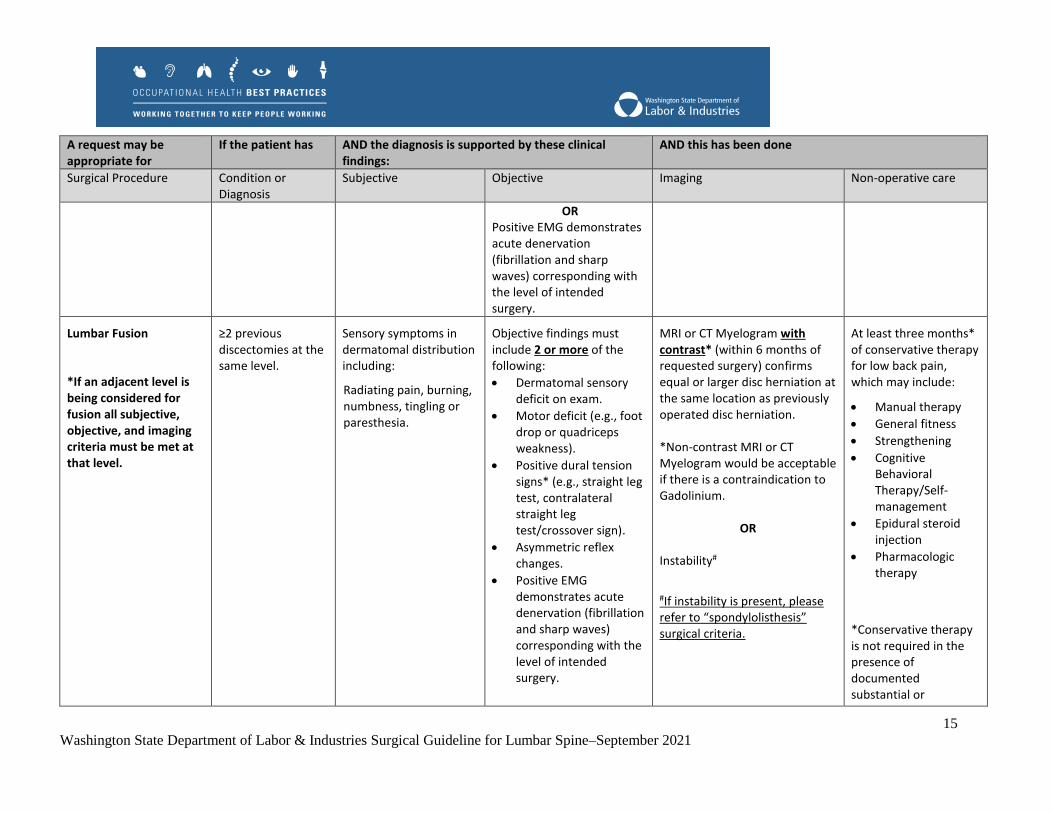
Lumbar Fusion
*If an adjacent level is being considered for fusion all subjective, objective, and imaging criteria must be met at that level.
≥2 previous discectomies at the same level.
Sensory symptoms in dermatomal distribution including:
Radiating pain, burning, numbness, tingling or paresthesia.
Objective findings must include 2 or more of the following:
Dermatomal sensory deficit on exam.
Motor deficit (e.g., foot drop or quadriceps weakness).
Positive dural tension signs* (e.g., straight leg test, contralateral straight leg test/crossover sign).
Asymmetric reflex changes.
Positive EMG demonstrates acute denervation (fibrillation and sharp waves) corresponding with the level of intended surgery.
MRI or CT Myelogram with contrast* (within 6 months of requested surgery) confirms equal or larger disc herniation at the same location as previously operated disc herniation. *Non-contrast MRI or CT Myelogram would be acceptable if there is a contraindication to Gadolinium.
OR Instability#
#If instability is present, please refer to “spondylolisthesis” surgical criteria.
At least three months* of conservative therapy for low back pain, which may include:
Manual therapy
General fitness
Strengthening
Cognitive Behavioral Therapy/Self-management
Epidural steroid injection
Pharmacologic therapy
*Conservative therapy is not required in the presence of documented substantial or
16
Washington State Department of Labor & Industries Surgical Guideline for Lumbar Spine–September 2021
A request may be appropriate for
If the patient has AND the diagnosis is supported by these clinical findings:
AND this has been done
Surgical Procedure Condition or Diagnosis
Subjective Objective Imaging Non-operative care
*Reproduction of back pain alone is not a positive finding.
progressive motor weakness.
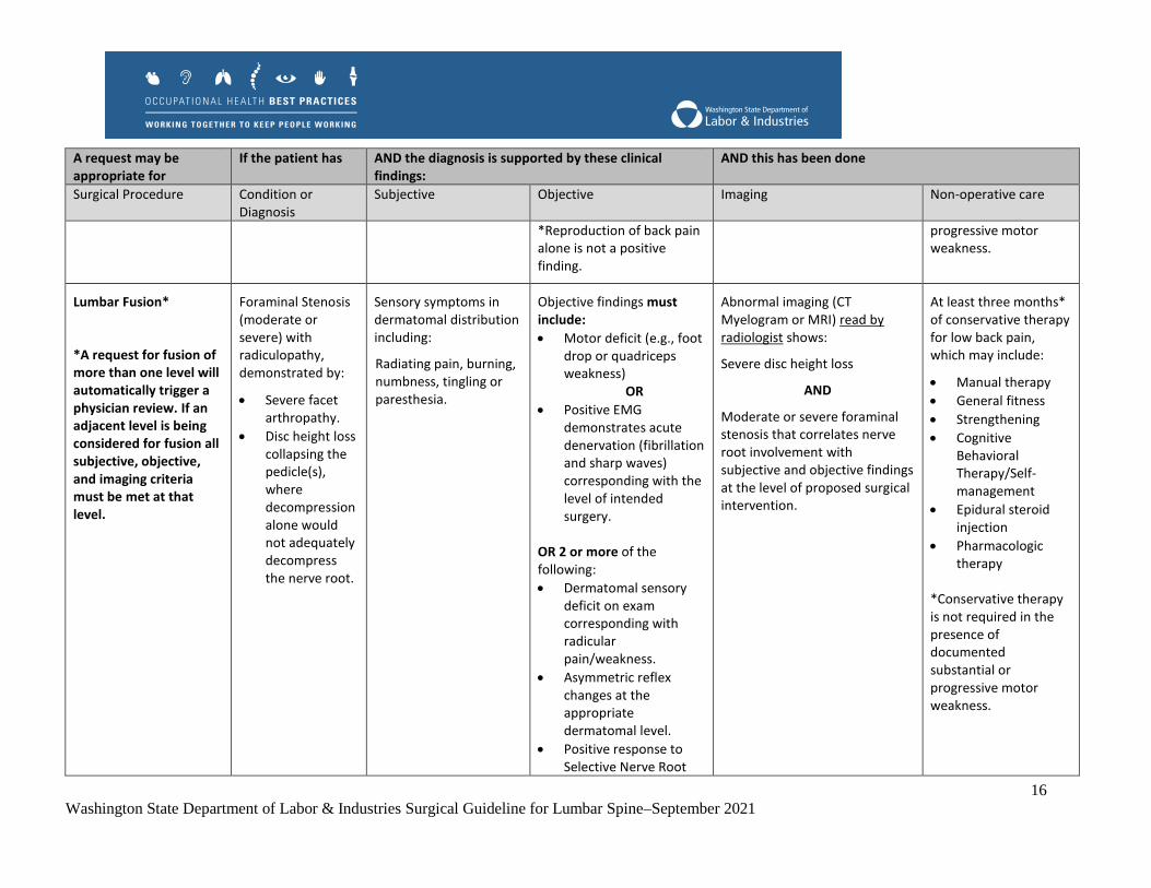
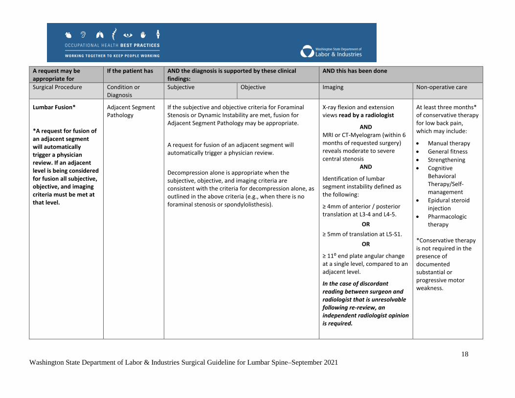
Lumbar Fusion*
*A request for fusion of more than one level will automatically trigger a physician review. If an adjacent level is being considered for fusion all subjective, objective, and imaging criteria must be met at that level.
Foraminal Stenosis (moderate or severe) with radiculopathy, demonstrated by:
Severe facet arthropathy.
Disc height loss collapsing the pedicle(s), where decompression alone would not adequately decompress the nerve root.
Sensory symptoms in dermatomal distribution including:
Radiating pain, burning, numbness, tingling or paresthesia.
Objective findings must include:
Motor deficit (e.g., foot drop or quadriceps weakness)
OR
Positive EMG demonstrates acute denervation (fibrillation and sharp waves) corresponding with the level of intended surgery.
OR 2 or more of the following:
Dermatomal sensory deficit on exam corresponding with radicular pain/weakness.
Asymmetric reflex changes at the appropriate dermatomal level.
Positive response to Selective Nerve Root
Abnormal imaging (CT Myelogram or MRI) read by radiologist shows:
Severe disc height loss
AND
Moderate or severe foraminal stenosis that correlates nerve root involvement with subjective and objective findings at the level of proposed surgical intervention.
At least three months* of conservative therapy for low back pain, which may include:
Manual therapy
General fitness
Strengthening
Cognitive Behavioral Therapy/Self-management
Epidural steroid injection
Pharmacologic therapy
*Conservative therapy is not required in the presence of documented substantial or progressive motor weakness.
17
Washington State Department of Labor & Industries Surgical Guideline for Lumbar Spine–September 2021
A request may be appropriate for
If the patient has AND the diagnosis is supported by these clinical findings:
AND this has been done
Surgical Procedure Condition or Diagnosis
Subjective Objective Imaging Non-operative care
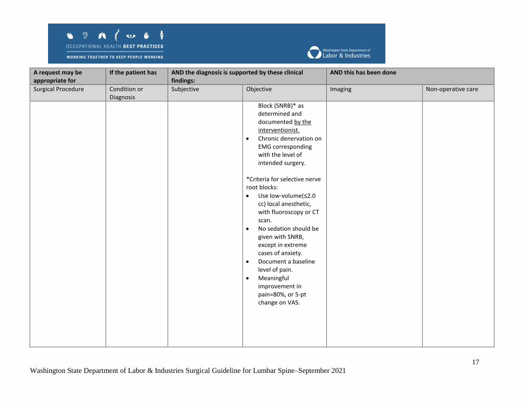
Block (SNRB)* as determined and documented by the interventionist.
Chronic denervation on EMG corresponding with the level of intended surgery.
*Criteria for selective nerve root blocks:
Use low-volume(≤2.0 cc) local anesthetic, with fluoroscopy or CT scan.
No sedation should be given with SNRB, except in extreme cases of anxiety.
Document a baseline level of pain.
Meaningful improvement in pain=80%, or 5-pt change on VAS.
18
Washington State Department of Labor & Industries Surgical Guideline for Lumbar Spine–September 2021
A request may be appropriate for
If the patient has AND the diagnosis is supported by these clinical findings:
AND this has been done
Surgical Procedure Condition or Diagnosis
Subjective Objective Imaging Non-operative care
Lumbar Fusion*
*A request for fusion of an adjacent segment will automatically trigger a physician review. If an adjacent level is being considered for fusion all subjective, objective, and imaging criteria must be met at that level.
Adjacent Segment Pathology
If the subjective and objective criteria for Foraminal Stenosis or Dynamic Instability are met, fusion for Adjacent Segment Pathology may be appropriate.
A request for fusion of an adjacent segment will automatically trigger a physician review.
Decompression alone is appropriate when the subjective, objective, and imaging criteria are consistent with the criteria for decompression alone, as outlined in the above criteria (e.g., when there is no foraminal stenosis or spondylolisthesis).
X-ray flexion and extension views read by a radiologist
AND MRI or CT-Myelogram (within 6 months of requested surgery) reveals moderate to severe central stenosis
AND
Identification of lumbar segment instability defined as the following:
≥ 4mm of anterior / posterior translation at L3-4 and L4-5.
OR
≥ 5mm of translation at L5-S1.
OR
≥ 11⁰ end plate angular change at a single level, compared to an adjacent level.
In the case of discordant reading between surgeon and radiologist that is unresolvable following re-review, an independent radiologist opinion is required.
At least three months* of conservative therapy for low back pain, which may include:
Manual therapy
General fitness
Strengthening
Cognitive Behavioral Therapy/Self-management
Epidural steroid injection
Pharmacologic therapy
*Conservative therapy is not required in the presence of documented substantial or progressive motor weakness.
19
Washington State Department of Labor & Industries Surgical Guideline for Lumbar Spine–September 2021
A request may be appropriate for
If the patient has AND the diagnosis is supported by these clinical findings:
AND this has been done
Surgical Procedure Condition or Diagnosis
Subjective Objective Imaging Non-operative care
Sacroiliac Joint Fusion
Note: A stepped approach to surgery and recovery must be in place prior to surgical approval, and must include all of the following components: 1. Post-surgical activation/reconditioning plan documented in the claim file by the surgeon. 2. Return to work/vocational rehabilitation plan documented by AP after review of surgeon’s activation plan. 3. Worker agreement to surgeon and AP plans.
A single documented traumatic inciting work related event that creates a force sufficient to cause Sacroiliac (SI) joint disruption or instability.
*SI joint fusion for chronic SI pain due to degenerative sacroiliitis and/or SI joint disruption is NOT covered.
Pain referrable to a Sacroiliac joint.
Diastasis of the pubis symphysis of at least 2.5 cm
OR
Asymmetric widening of the injured SI joint.
Failure of six or more months of non-operative care directed at successfully treating SI joint ligamentous instability.
20
Washington State Department of Labor & Industries Surgical Guideline for Lumbar Spine–September 2021
II. Introduction
A. Background and Prevalence
Low back injuries and low back pain (LBP) are some of the most common and costly occupational injury
claims.[1-5] L&I data for fiscal year 2020 alone identified 11,358 claims costing over $115 million in
lifetime incurred costs. LBP has been ranked as the fourth leading cause of disability worldwide behind
heart disease, cerebrovascular disease, and lower respiratory infection.[6] In the United States, LBP is the
leading cause of years lived with disability, ahead of depression, chronic obstructive pulmonary disease
(COPD), and other musculoskeletal disorders.[7] Past estimates of work-related injuries in the U.S. found
that spine and back injuries accounted for 17% of all work-related injuries.[1]
Conditions addressed in this guideline related to or experienced as low back pain, including stenosis and
spondylolisthesis, are highly prevalent in the general population and often seen in asymptomatic
individuals.[8-10] It is important to note that LBP is common and increases with age, and as such is not
always directly related to an injury or specific diagnosis.
B. Pre-existing or Non-work-related Conditions
1. Nicotine Use
Current nicotine usage has been associated with poor surgical outcomes in patients undergoing lumbar
spinal surgery, including increased risk of non-union/pseudarthrosis, worse clinical outcome scores, lower
return to work rates, and increased wound complications and risk of infection.[11-16]
In one study, non-smokers demonstrated significantly greater improvement in Oswestry Disability Index
(ODI) scores after decompression surgery than smokers, and non-smokers were also more likely to have
clinically meaningful improvement in their ODI scores at one year.[17] Smoking has also been shown to be
associated with recurrent disc herniation (Odds Ratio 1.99, 95% Confidence Interval 1.53-2.58) in a
recent systematic review.[18] Another study, Sanden et al., found similar results at 2 years, showing active
smokers who underwent decompression with or without fusion were more likely to be dissatisfied with
the results of their surgery, more likely to report increased analgesic use, and showed less improvement in
walking ability.[19] Further results as part of the larger Spine Patients Outcome Research Trial (SPORT)
found that smokers being treated for lumbar spinal stenosis were the only group in which operative
treatment did not perform better than non-operative care.[20]
In light of the negative outcomes related to smoking and nicotine usage in patients undergoing
decompression procedures, the department considers it best practice to abstain from nicotine for at least 4
weeks prior to surgery, as demonstrated by two negative urine cotinine tests during this time period.
Abstinence from nicotine is required for all fusion and repeat fusion procedures. This does not apply to
21
Washington State Department of Labor & Industries Surgical Guideline for Lumbar Spine–September 2021
progressive myelopathy or motor radiculopathy. Smoking cessation products may be covered in some
instances, for more information, review L&I’s tobacco cessation program.e
Continued post-operative smoking cessation is also highly recommended to reduce the potential for
complications such as infection or delayed fracture healing. While emphasis may be placed on
maintaining post-operative cessation, evidence demonstrates a recidivism rate greater than 60% at one
year in patients who underwent smoking cessation prior to lumbar fusion.[21]
III. Assessment
A. History and Clinical Exam
A thorough history and clinical examination are important in the proper diagnosis of any suspected
lumbar condition. Taking a history allows for proper differential diagnosis, as low back pain can be
caused by any number of mechanical or nonmechanical mechanisms. Further, proper clinical assessment
can help to identify where pain is originating from.
A clinical assessment may include:
Examination of peripheral pulses to differentiate between neurogenic and vascular claudication.
Hip range of motion testing to rule out hip pathology.
Detail-focused neurologic examinations including at least the following:
o Deep tendon reflexes (DTRs) of the upper and lower extremities.
o Strength testing of the major muscle groups of the upper and lower extremities.
o Testing of light touch and pain/temperature in the lower extremities—looking for
dermatomal sensory loss or distal symmetrical sensory loss.
Dural tension signs (e.g., straight leg test, contralateral straight leg test/crossover sign).
o Reproduction of back pain alone is not considered to be a positive finding.
Examination of potential reflex changes.
Electromyography.
B. Imaging
The recommended and required imaging procedures for lumbar spine surgeries are specified in the criteria
table, with further detail in individual sections below if necessary. Imaging studies such as radiographs
and advanced imaging such as magnetic resonance imaging (MRI) or computed tomography (CT), can be
used to aid in the diagnosis and clinical workup of lumbar conditions.
When considering imaging for low back pain, care should be taken to ensure that imaging is undertaken
only when a clear clinical indication exists. Generators of low back pain are often hard to pinpoint, and
MR imaging studies also tend to identify asymptomatic issues such as disc degeneration, annular tears, or
e https://www.lni.wa.gov/patient-care/treating-patients/conditions-and-
treatments/?query=Tobacco+Cessation+Treatment+for+Surgical+Care
22
Washington State Department of Labor & Industries Surgical Guideline for Lumbar Spine–September 2021
disc protrusion that may lead to unnecessary procedures.[22-24] Established guidelines suggest only
performing imaging studies in the presence of specific symptoms or neurologic deficits.[25-27]
Recent imaging studies should be used when making care-related decisions. L&I considers imaging
within 6 months prior to surgical requests as necessary to determine appropriate surgical decision-making.
In the case of a discordant reading between surgeon and radiologist that is unresolvable following re-
review, an independent radiologist opinion is required. L&I requires prior authorization for all MRIs;
please visit the Advanced Imaging Guidelinesf web page for complete information.
In considering imaging following previous surgeries, a contrast medium may be indicated. Use of
Gadolinium enhanced imaging has been successfully used to aid in differentiation of disc herniation and
peridural scarring.[28, 29] As such, contrast mediums may be required in certain imaging studies unless
otherwise contraindicated. Relative and absolute contraindications include[30]:
Relative contraindications:
o Certain medications, such as β-blockers, nonsteroidal anti-inflammatories (NSAIDs), and
interleukin-2.
Absolute Contraindications:
o Prior adverse reaction to contrast medium(s).
o History of asthma, allergies, heart disease, or underlying renal disease.
C. Opioids
Managing pain in workers who are undergoing surgery, especially those on chronic opioid therapy, can be
challenging and requires a coordinated treatment plan prior to surgery. Brat et al., examining a nationwide
insurance database of more than a million opioid naïve surgical patients, identified the duration of initial
opioid prescription after surgery as a risk factor for later opioid misuse (dependence, abuse, or overdose)
diagnoses.[31] Although only 0.6% of postoperative patients subsequently had such diagnoses, each
additional week of opioid therapy prescribed was associated with an adjusted 20% increase in hazard for
opioid misuse, with a total 44% increase in hazard if a refill was also prescribed. In addition, recent
studies have shown that patients are typically prescribed far more opioid pills than is necessary across a
broad variety of surgeries.[32-34] This overabundance of pills may result in diversion and other undesired
outcomes.
Although the majority of patients being considered for spine surgery have not been on opioids chronically
(≥3 months of opioids), those that have constitute a special class of patients because preoperative opioid
use is associated with worse outcomes following spine surgery. O’Donnell et al. reported that in a cohort
of lumbar discectomy patients from the Ohio workers’ compensation system, such preoperative opioid
use was negatively associated with return to work, and positively associated with failed back surgery
syndrome and much more frequent and prolonged post-operative opioid use.[35] In a prospective study in
f https://lni.wa.gov/patient-care/treating-patients/treatment-guidelines-and-resources/#advanced-imaging-guidelines
23
Washington State Department of Labor & Industries Surgical Guideline for Lumbar Spine–September 2021
Washington state among all payers, only opioid use, tobacco use, and being a workers’ compensation
patient were predictive of worse outcome from lumbar spine surgery.[36]
The Washington state Dr. Robert Bree Collaborative, after extensive review of relevant medical and
scientific literature, published specific recommendations regarding postoperative opioid useg across
several levels of surgical invasiveness.[37] If opioids are anticipated to be part of the postoperative pain
management plan, the goal for prescribing should be at the lowest effective dose for the shortest duration.
Preoperatively, the surgeon must:
Check the Prescription Drug Monitoring Program (PDMP) pre-operativelyh, and document in the
medical record all sources of ongoing prescriptions of all controlled substances, including daily
morphine equivalent doses of opioids. Care coordination is requiredi when the patient is on
combinations of opioids and sedatives, including benzodiazepines, carisoprodol, and
nonbenzodiazepine sedative-hypnotics (“Z drugs”).
Document a coordinated plan for managing surgical pain, including identifying the post-operative
prescriber, setting appropriate expectations for pain management, and evaluating for potential risk
for over-sedation/respiratory depression and difficult pain control.[38]
Refer for preoperative anesthesia or pain management consult for those patients who are
diagnosed with opioid use disorder/substance use disorder, on buprenorphine or chronic opioid
dose of 90 mg/day MED or more).
Document a recent, or conduct of, urine drug testing to identify undisclosed drug use and/or
abuse and verify compliance with treatment.
Postoperatively, the surgeon must:
Follow the Bree Collaborative recommendations for Prescribing Opioids for Postoperative Pain –
Supplemental Guidancej :
o Procedures with expected rapid recovery, such as meniscectomy, may be treated with
either a non-steroidal anti-inflammatory drug (NSAID) or a combination of NSAID and
acetaminophen. If opioids are warranted, prescribe 3 days or less (no more than 8-12
pills) of short acting opioids.
o For procedures with expected medium term recovery, such as discectomy or
laminectomy, use non-opioid analgesics and non-pharmacologic therapies as first line
therapy. If opioids are warranted, prescribe 7 days or less (no more than 42 pills) of short
acting opioids.
o For procedures with expected longer recovery times, such as lumbar fusion, use non-
opioid analgesics and non-pharmacologic therapies as first line therapy. If opioids are
warranted, prescribe 14 days or less of short acting opioids at the lowest effective dose.
o For patients on chronic opioid therapy, use non-opioid analgesics and non-pharmacologic
therapies as first line therapy and follow the above recommended opioid prescribing
g https://www.qualityhealth.org/bree/wp-content/uploads/sites/8/2018/09/Final-Supplemental-Bree-AMDG-Postop-
pain-091318-wcover.pdf h WAC 296-20-03035, 246-919-985, 246-918-935, 246-853-790 i WAC 246-919-970, 246-918-920, 246-853-775 j https://www.qualityhealth.org/bree/wp-content/uploads/sites/8/2018/09/Final-Supplemental-Bree-AMDG-Postop-
pain-091318-wcover.pdf

24
Washington State Department of Labor & Industries Surgical Guideline for Lumbar Spine–September 2021
durations, based on expected recovery time. Resume chronic opioid regimen if patient is
expected to continue postoperatively.
For the exceptional case that may warrant more opioids than the expected recovery period, re-
evaluate the patient to determine what is the delaying the normal course of recovery. Post-
surgical opioids should be tapered in all cases by no later than 6 weeks after surgery.
D. Screening and Addressing of Behavioral or Mental Health
While the presence of behavioral or mental health issues do not preclude a worker from surgery, evidence
has shown that the presence of such conditions may be associated with significantly poorer spinal
intervention outcomes postoperatively. Identified issues include lower satisfaction and worse outcomes,
increased perioperative complications, and continuance of narcotic use post-surgery.[39, 40]
The workers’ compensation population often demonstrates poor outcomes compared to other populations
undergoing similar interventions for spinal conditions, and it is especially important to address underlying
behavioral or mental health factors that may lead to worse surgical outcomes.[41] In one study of workers’
compensation patients undergoing lumbar fusion, a pre-operative diagnosis of depression was a negative
predictor of return to work, and at 3 years the depression group had an average excess work absence of
184 days compared to controls.[42] Another study, Wang et al., found that significant risk factors for sick
leave >90 days or disability pension following lumbar decompression included common mental disorders,
somatic comorbidity, and prescribed psychiatric medication.[43]
If comorbid behavioral or mental health issues are present, the surgeon should be aware of, work to
counsel, and manage patient expectations preoperatively. When properly identified, preoperative
treatment of depression may improve outcomes of operative intervention.[39] In addition, proper
identification and addressing of pre-operative pain expectations and psychosocial barriers can be
important in improving clinical outcomes.[44] Mancuso et al. identified that expectations about pain are an
independent variable in predicting pain improvement at 2-years post-lumbar surgery, patients who had
greater expectation of pain improvement following surgery had increased odds of reporting less pain
improvement (OR 1.4).[45] With these defined issues, screening tools for depression should be considered
for all fusion candidates, as studies show only a 28% sensitivity for surgeons to identify depression from
subjective rating alone.[39]
L&I has resources available for addressing behavioral health or mental health prior to surgery. Please
refer to lni.wa.gov/MLT or lni.wa.gov/mentalhealth for details.
E. Preventing Complications
Within workers’ compensation, the ultimate goal of any intervention is to enable the worker to recover
and return to work. When considering surgery, it is critical to conduct a thorough assessment of risk
factors and fitness for surgical intervention to evaluate the potential risks and benefits. In identifying
appropriate candidates for lumbar spine surgery, L&I recommends consideration of the following, based
in part on the work of the Robert Bree Collaborative, to increase the likelihood of positive outcomes[46]:
25
Washington State Department of Labor & Industries Surgical Guideline for Lumbar Spine–September 2021
1. Avoidance of smoking for a minimum of 4 weeks preoperatively with 6 to 8 weeks preferred.
2. Pre-operative plan for management of opioids, if patient has taken opioids for more than 3
months.
3. Screen for substance abuse; manage if screen is positive.
4. Hemoglobin A1c less than 8% in patients with diabetes.
5. Absence of an active, life-limiting condition that would likely cause death before recovery from
surgery.
6. Absence of severe disability from an unrelated condition that would severely limit the benefits of
surgery such as severe osteoporosis.
7. Absence of dementia that would interfere with recovery.
8. Screen for untreated depression or psychiatric disorders; manage if screen is positive.
9. Adequate nutritional status to ensure healing.
10. Sufficient liver function to ensure healing.
11. Body Mass Index (BMI) less than 40.
12. Complete a preoperative plan for postoperative return to function.
F. Measuring Functional Improvement
Consistent use of validated functional instruments can be imperative to providing proper care in the
treatment of lumbar spine conditions. Not only do these measures help guide appropriate interventions,
proper usage of functional instruments can have significant impact through identification of risk factors,
symptoms, and risk of developing ongoing comorbidities or disability.
The authors of this guideline recommend using the following validated tools for measuring pain and
functional improvement:
2-item Graded Chronic Pain Scale (GCPS)
Oswestry Disability Index (ODI)
Short Form 36 (SF-36)
Patient-Reported Outcomes Measurement Information System (PROMIS)
In general, a 30% or greater improvement in pain and/or function on validated functional scales is
considered meaningful improvement.[47] L&I has also produced a guideline on documenting functional
improvementk, including validated functional scales and information on proper application and
interpretation.
IV. Non-Operative Care
Non-operative care is the first line treatment for low back pain without the presence of substantial or
progressive motor weakness. The natural history of low back pain suggests the potential for recovery
without surgical intervention, and non-operative care has demonstrated effectiveness when compared to
k https://lni.wa.gov/patient-care/advisory-committees/_docs/2018DocFuncImprovfunctionalscales.pdf
26
Washington State Department of Labor & Industries Surgical Guideline for Lumbar Spine–September 2021
surgery across multiple conditions. A recent Cochrane review comparing surgical and conservative care
for lumbar stenosis found no clear benefits of surgery compared to non-operative care, and data from the
SPORT study have also demonstrated significant and sustained improvement in patients undergoing non-
operative care.[48-50]
There are no specific non-operative protocols or interventions that demonstrate superiority, with many
studies utilizing a combination of interventions such as manual therapy, manipulation, neuromobilization,
flexion/distraction, general fitness/strengthening, and Cognitive Behavioral Therapy or self-
management.[51, 52] L&I considers at least 6 weeks of non-operative care necessary prior to requests for
decompression, and depending on the condition or diagnosis 3-6 months of care prior to fusion
procedures.
V. Surgical Procedures
A. Lumbar Decompression Procedures
Lumbar decompression includes procedures such as lumbar laminectomy, laminotomy, discectomy,
microdiscectomy, foraminotomy, and far lateral decompression. These procedures are meant to increase
the amount of space in the spinal canal through removal of spinal elements irritating or compressing
neurovascular structures, thereby “decompressing” them, with the goal of reducing or relieving pain.
Significant evidence exists for the safety and efficacy of decompressive procedures in appropriately
selected patients. Lumbar decompression procedures may be covered for the following conditions when
appropriate subjective, objective, and imaging criteria have been met.
Care should be taken in consideration of surgery, as the natural history of acute low back pain is generally
positive, with symptom resolution often seen in the first month.[53, 54] Disc herniation, stenosis, and other
abnormal findings seen on imaging are often present in asymptomatic patients [26, 55-58], and other studies
have shown disc herniations may naturally resorb over time.[59-64] Additionally, receiving care under
workers’ compensation has been shown to be associated with increased risk of unsatisfactory surgical
outcomes.[41, 65]
1. Decompression for Nerve Root Entrapment due to
Central/paracentral/foraminal/extra-foraminal herniated nucleus
pulposus
Lumbar nerve root entrapment or compression causing injury of nerves within the spinal column, defined
as the condition radiculopathy, often presents as sensory symptoms (e.g. radiating pain, weakness,
numbness) in a dermatomal distribution. Nerve root entrapment is often caused by lumbar disc herniation,
identified by the displacement (protrusion or extrusion) of disc material (e.g. nucleus, cartilage, anular
tissue) beyond the margins of the intervertebral disc space.[66, 67] Other conditions, such as cancer,
fracture, or infection, are less common causes.[53]
27
Washington State Department of Labor & Industries Surgical Guideline for Lumbar Spine–September 2021
In cases without severe or substantially progressive motor weakness, a course of non-operative care of at
least six weeks including active rehabilitation, manual medicine, NSAIDs, or epidural steroid injections,
has been shown to be effective in treating low back pain.[54, 68, 69]
When considering moving to surgery, there is significant evidence of the long-term effectiveness of
decompression for the treatment of disc herniation and stenosis. SPORT (Spine Patient Outcomes
Research Trial), a long-term prospective study including both randomized and observational cohorts of
patients with lumbar disc herniation or stenosis, examined the long-term effectiveness of decompression
in relieving pain and improving outcomes. Results demonstrated significant and sustained improvement at
up to 8 years post surgery in both conditions, with superiority of surgical outcomes compared to non-
operative care, but findings were less positive in patients receiving workers’ compensation.[49, 50, 70-72]
Surgical decompression of lumbar nerve root entrapment is a covered procedure when MRI or CT-
Myelogram corroborates substantial disc herniation resulting in effacement, abutment, displacement, or
compression of the nerve root [27, 54, 73], and when two or more of the following objective symptoms are
present:
Dermatomal sensory deficit on exam.[74]
Motor deficit (e.g., foot drop or quadriceps weakness)[74, 75]
Positive dural tension signs (e.g., straight leg test, contralateral straight leg test/crossover sign).[54,
76, 77]
Asymmetric reflex changes.
Positive EMG demonstrates acute denervation (fibrillation and sharp waves).[78, 79].
2. Decompression for Recurrent Disc Herniation
While surgery is successful in treating most lumbar disc herniations, around 10% of operated patients
may experience symptomatic reherniation requiring repeat surgery.[18, 80-82] Though no specific timeline
can predict reherniation, numerous studies have demonstrated up to 50% of reherniations that require
reoperation occur within the first year following initial surgery.[80, 81, 83-85]
Available literature to identify risk factors for reherniation are somewhat limited; however, some recent
studies have identified lack of sensory or motor deficits, younger age, and higher baseline disability as
significant risk factors for reherniation.[80] Other significant risk factors for recurrent herniation identified
by a recent meta-analysis include diabetes, smoking status, and an initial classification of the herniation as
a disc protrusion (as opposed to extrusion of sequestration).[18]
When considering surgery for a recurrent disc herniation in a patient who has not experienced a discrete
event, new symptoms, or returned to workl, MRI or CT-Myelogram with a contrast medium must
l If a patient has been back to work for a minimum of 6 months and/or has experienced sustained improvement in
pain and function, refer to “Nerve Root Entrapment” for required information and surgical criteria.
28
Washington State Department of Labor & Industries Surgical Guideline for Lumbar Spine–September 2021
demonstrate a disc herniation of equal or larger size at the same location following previous lumbar
decompression surgery.
It is extremely important to determine the etiology of persistent pain after spinal surgery, as residual pain
following surgical intervention can be multifactorial, and repeat surgeries in the absence of new or
worsened pathology do occur. Repeat surgery should only be considered if recurrent or residual
herniation exists. Patients who undergo multiple spinal surgeries have a lower chance of successful pain
resolution, and are less likely to return to normal function following surgery.[86]
A correlation between residual low back pain after surgery and the presence of peridural scarring has been
established.[87] This scarring is considered normal following surgery, and is also common in
asymptomatic patients.[88, 89] Patients undergoing reoperation presenting with only scar tissue have
demonstrated poor outcomes.
To improve the identification of appropriate candidates for repeat surgical interventions, use of an
imaging contrast medium is required when requesting approval of a repeat decompression surgery, unless
otherwise contraindicated*. Studies have demonstrated the ability of Gadolinium enhanced imaging to aid
in differentiation of disc herniation and peridural scarring.[28, 29] L&I considers this distinction especially
important, as long-term studies have shown worse outcomes for workers’ compensation patients
undergoing surgery compared to patients with other forms of coverage.[41, 65]
In addition to imaging, two or more of the following objective symptoms must be present:
Dermatomal sensory deficit on exam.[74]
Motor deficit (e.g., foot drop or quadriceps weakness).[74, 75]
Positive dural tension signs (e.g., straight leg test, contralateral straight leg test/crossover sign).[54,
76, 77]
Asymmetric reflex changes.
Positive EMG demonstrates acute denervation (fibrillation and sharp waves).[28, 29, 78, 79]
*Identified relative and absolute contraindications to use of a contrast/Gadolinium medium include[30]:
Relative contraindications:
o Certain medications, such as β-blockers, nonsteroidal anti-inflammatories (NSAIDs), and
interleukin-2.
Absolute contraindications:
o Prior adverse reaction to contrast medium(s).
o History of asthma, allergies, heart disease, or underlying renal disease.
3. Decompression for Central Spinal Stenosis
Stenosis, or a narrowing of the spinal canal due to degeneration or growth of biological elements in the
spine, can also lead to nerve root entrapment or compression.[90] It is thought to be a normal process of
29
Washington State Department of Labor & Industries Surgical Guideline for Lumbar Spine–September 2021
aging and generally prevalent in normal populations, with one study of 938 participants aged 40-93
finding moderate spinal stenosis among 78% of the population, and severe spinal stenosis in 30%.[91]
While the prevalence of stenosis is high it is often asymptomatic and not requiring treatment, with the
same study finding only 17.5% of patients with severe spinal stenosis reporting any clinical symptoms.
As stenosis is thought to be highly prevalent without symptoms, non-operative care is considered an
appropriate first step. In cases without severe or substantially progressive motor weakness, a course of
non-operative care of at least 6 weeks including active rehabilitation, manual medicine, NSAIDs, or
epidural steroid injections, has been shown to be effective in treating low back pain.[54, 68, 69]
If non-operative care does not provide relief, surgical decompression of stenosis has been shown to be a
safe and effective procedure. In a large scale, long-term, multi-center trial, patients who underwent
decompression for stenosis had greater improvement on the Oswestry Disability Index than similar
patients undergoing only non-operative care.[20, 92] Patients in this study were considered to have stenosis
based on the presence of neurogenic claudication or radicular pain for at least twelve weeks, and a
confirmatory cross-sectional imaging study demonstrating stenosis at one or more levels. Other
systematic reviews have also highlighted the effectiveness of decompression in treating lumbar stenosis,
showing its effectiveness to be comparable to, and less invasive than, lumbar fusion.[93-95]
As always, care should be taken when setting expectations for surgery. Results from the SPORT study
found an 18% reoperation rate in patients who underwent surgical treatment for lumbar stenosis
(N=417).[96] Of this population, 52% of reoperations were for persistent stenosis or progressive
spondylolisthesis.
Based on the demonstrated effectiveness, decompression for central spinal stenosis is a covered procedure
when neurogenic claudication is present (if neurogenic claudication is suspected, at minimum the pulse of
the patient should be checked), the patient experiences bilateral lower extremity pain or weakness when
standing or walking, and when MRI or CT-Myelogram confirms subjective and objective findings of
moderate or severe central spinal stenosis.
4. Decompression for Synovial Cyst
Lumbar synovial cysts are generally rare, often benign in nature, and their pathogenesis is thought to be
associated with disruption or degeneration of facet joints in the lumbar spine.[97] Their presence has been
shown to contribute to painful spondylolisthesis and radiculopathy.[98, 99]
There is limited evidence for conservative treatment of synovial cysts. One smaller study found durable
symptom relief at 6 months in 10 of 30 patients who received steroid injections in the facet joints.[100]
Surgical excision of lumbar synovial cysts has been shown to be effective, with several studies
demonstrating resolution of symptoms and sustained improvement at short to long-term follow-up.[98, 101,
102]
30
Washington State Department of Labor & Industries Surgical Guideline for Lumbar Spine–September 2021
Decompression of a synovial cyst is a covered procedure when CT-Myelogram or MRI corroborate
subjective and objective findings of a synovial cyst that results in one or more of the following on the
nerve root: effacement, abutment, displacement, or compression (stenosis). In addition to imaging, two or
more of the following objective symptoms must be present:
Dermatomal sensory deficit on exam.[74]
Motor deficit (e.g., foot drop or quadriceps weakness).[74, 75]
Positive dural tension signs (e.g., straight leg test, contralateral straight leg test/crossover sign).[54,
76, 77]
Asymmetric reflex changes.
Positive EMG demonstrates acute denervation (fibrillation and sharp waves).[28, 29, 78, 79]
5. Decompression for Lateral Recess/Foraminal Stenosis
Lumbar foraminal stenosis (caused by compression of the spinal nerves as they exit the spinal canal
through the foraminae) and lateral recess stenosis (caused by compression of the spinal nerves in the
lateral recess of the spinal canal) are common causes of symptomatic lumbar radiculopathy that can often
be overlooked due to the location of the narrowing at the bony exit of the nerve root.[103]
Non-operative treatment is recommended initially for the treatment of foraminal stenosis, with studies
demonstrating the effectiveness of active rehabilitation, manual medicine, NSAIDs, and epidural steroid
injections.[104] Various surgical techniques have shown benefit in treating foraminal stenosis.[105-109]
Decompression for lateral recess/foraminal stenosis is a covered procedure when CT-Myelogram or MRI
corroborate subjective and objective findings with substantial disc herniation that results in one or more
of the following on the nerve root: effacement, abutment, displacement, or compression (stenosis). Mild
to moderate disc protrusion not associated with the previous terms is not considered a positive objective
imaging sign.
In addition to imaging, two or more of the following objective symptoms must be present:
Dermatomal sensory deficit on exam.[74]
Motor deficit (e.g., foot drop or quadriceps weakness).[74, 75]
Positive dural tension signs (e.g., straight leg test, contralateral straight leg test/crossover sign).[54,
76, 77]
Asymmetric reflex changes.
Positive EMG demonstrates acute denervation (fibrillation and sharp waves)[28, 29, 78, 79].
6. Decompression for Acute Cauda Equina Syndrome
Acute cauda equina syndrome (CES) is a rare, compressive disorder of the lumbosacral nerve roots below
the tip of the conus medullaris. Only a small number of patients who present with low back pain will have
CES, with an estimated prevalence of 0.04%.[110] It is characterized by multiple lumbo-sacral sensory-
motor deficits which may have disabling long term consequences, and it requires immediate surgical
31
Washington State Department of Labor & Industries Surgical Guideline for Lumbar Spine–September 2021
attention.[111] Due to the emergent nature of CES, controlled studies are not feasible and the literature is
limited to case series, case studies and narrative reviews.
CES has been reported to result from the following work- and non-work-related conditions:[112, 113]
Disc herniation (most common cause; most often central herniation).
Trauma (e.g. gunshot wound, vertebral fracture).
Infection (e.g. discitis, vertebral osteomyelitis, epidural abscess).
Degenerative conditions (e.g. degenerative spondylolisthesis, spinal stenosis).
Metastatic or primary tumor (with or without pathologic fracture).
Post-surgical complications (e.g. epidural hematoma, fat graft, durotomy, use of Gelfoam).
Vascular malformations (e.g. bleeding arteriovenous malformations).
Intradiscal electrothermal annuloplasty.
Spinal manipulation.
Symptoms and Signs of Cauda Equina Syndrome
The hallmark symptoms of CES include:[111, 114-118]
Acute low back pain with unilateral or bilateral sciatica.
Weakness of both legs and/or weakness involving multiple nerve roots in one leg.
Partial or complete loss of bladder function (incontinence or retention not otherwise explained)
and/or bowel function, accompanied by impaired perineal sensation, especially saddle anesthesia.
Diminished or absent anal sphincter tone.
Reduced or absent bulbo-cavernosus reflex.
Gait disturbances.
Impaired sensation in the lower extremities.
Hyporeflexia or areflexia in the legs.
Sexual dysfunction.
Diagnostic Tests for Cauda Equina Syndrome
Magnetic Resonance Imaging (MRI) is the preferred imaging test for charactering and localizing spinal
lesions. Other diagnostic and imaging modalities include:
CT and/or CT Myelography, utilized to locate narrowing of the spinal canal. These tests provide
useful information when MRI cannot be done or is limited by hardware artifact.
Plain x-rays, utilized to identify fractures, tumors, infection, and degenerative changes.
Ultrasound, utilized to scan the bladder and identify urinary retention.
Urodynamic tests, utilized to evaluate bladder function. These tests should be considered only in
light of the patient’s clinical condition after emergent care has been given.
Treatment of Cauda Equina Syndrome
Conservative treatment alone is rarely indicated, as CES is an emergent condition and surgical
decompression is the treatment of choice. To prevent further neurological deterioration, urgent surgical
decompression should be performed. Decompression for rapidly progressing CES may prevent sphincter
paralysis. The best surgical outcomes were reported in patients with the least neurological deficit prior to
surgery.[111, 113, 119-122] Decompression surgery may range between microdiscectomy and wide
laminectomy with discectomy to limit the manipulation of potentially damaged neural tissue.[113]
32
Washington State Department of Labor & Industries Surgical Guideline for Lumbar Spine–September 2021
B. Lumbar Fusion Procedures
Lumbar fusion procedures involve stabilization of the lumbar spine by fusing one or more vertebrae.
In most cases, fusion can be considered a procedure of last resort due to significant potential for poor
outcomes and disability, as well as the potential benefit of non-operative care and less invasive
procedures.[48, 123, 124] Decompression alone is considered an appropriate first-line procedure for single-
level stenosis or spondylolisthesis, with studies showing comparable results between decompression and
fusion procedures for treating these conditions.[125-128] Of particular interest, patients undergoing fusion
procedures had significantly lower Return to Work (RTW) rates compared to decompression alone in a
large-scale cohort study of Ohio workers’ compensation patients.[129]
When non-operative care is inappropriate or has proved insufficient, and decompression alone is not
indicated, lumbar fusion procedures may be covered for the following conditions when appropriate
subjective, objective, and imaging criteria have been met. In all cases, the department requires that the
requesting surgeon must submit a report verifying the positive findings on physical exam.
1. Spondylolisthesis
Spondylolisthesis, the shifting or slipping forward of spinal vertebrae, is a common condition shown to be
incident in at least 6% of the general population at adulthood; this can cause significant pain and
disability due to compression of neurologic structures.[130, 131]
Lumbar fusion for spondylolisthesis is supported in appropriately selected patients, with results from
numerous studies demonstrating significant improvement in patients undergoing fusion for degenerative
spondylolisthesis, though results are often equivalent to decompression.[126-128, 132, 133] Long-term results
from the Spine Patient Outcomes Research Trial (SPORT) study showed significantly better results
compared to non-operative care in patients treated with lumbar fusion at up to 8 years, demonstrating
significant improvement in Oswestry Disability Index (ODI) scores (average decrease of 10.3 points) and
sustained outcomes at 8 years.[134] When results are pooled in systematic reviews and meta-analyses,
significant improvement in outcome measures are sustained.[93]
Particular caution should be taken in L&I patients, as long-term studies have demonstrated worse
outcomes among the workers’ compensation (WC) population. A study of Ohio WC patients undergoing
fusion found that, when excluding patients who had a significant pre-operative risk factor such as
depression or long term-opioid use, only 60% of patients had returned to work at two years post-
fusion.[135]
In the absence of documented substantial or progressive motor weakness, L&I requires at least three
months of non-operative care for low back pain which may include manual therapy, manipulation,
neuromobilization, flexion/distraction, general fitness, strengthening, or Cognitive Behavioral
Therapy/Self-management.
Surgery may be considered appropriate if proper subjective, objective, and imaging criteria are met.
Imaging must include both X-ray flexion and extension views, MRI or CT-Myelogram that demonstrates
33
Washington State Department of Labor & Industries Surgical Guideline for Lumbar Spine–September 2021
moderate to severe central spinal stenosis and lumbar instability, defined as one or more of the following:
Spondylolisthesis ≥ Grade 2, ≥ 4mm of anterior / posterior translation at L3-4 and L4-5, ≥ 5mm of
translation at L5-S1, or ≥ 11⁰ end plate angular change at a single level, compared to an adjacent level.
The patient should also have findings of 1) neurogenic claudication, defined as radiating leg pain that is
exacerbated while standing up and walking, immediate relief of neurogenic symptoms when seated, and
improvement of symptoms when bending forward, or 2) radiculopathy, defined as sensory symptoms in
dermatomal distribution including radiating pain, burning, numbness, tingling or paresthesia.
2. Prior Decompression at the Same Level
While decompression can be an appropriate first line procedure for treating many conditions in the
lumbar spine, some patients may experience new onset of significant pain and disability in the months
following surgery.[136, 137] In many cases, this can be adequately addressed through non-operative care
including physical therapy, pharmacologic therapy, epidural injections, behavioral modifications, and
other appropriate measures.[136, 138]
In situations when lumbar fusion is considered for treating new onset of symptoms, evidence for
effectiveness is weak, with studies finding no significant difference between surgical care and
conservative care in Oswestry Disability Index scores following surgery, and showing less than 50% of
patients reporting significant symptom relief following surgery.[136, 139]
When non-operative care has failed, lumbar fusion may be considered an appropriate procedure in
patients who have previously undergone a decompressive procedure and demonstrate new onset of
lumbar segment instability, symptomatic central stenosis, or severe radiculopathy at the same level of
previous decompression.
Clinical findings should include Spondylolisthesis ≥ Grade 2, ≥ 4mm of anterior / posterior translation at
L3-4 and L4-5, ≥ 5mm of translation at L5-S1, or ≥ 11⁰ end plate angular change at a single level
compared to an adjacent level, Lumbar segment instability at the same level, rotational deformity or other
conditions leading to a progressive measurable deformity, objective signs and symptoms compatible with
neurogenic claudication or lumbar radiculopathy confirmed by a detailed neurological examination, or
positive EMG demonstrating acute denervation (fibrillation and sharp waves). MRI or CT with contrast
(with or without myelography) must confirm findings of moderate to severe central, lateral recess, or
foraminal stenosis at the same location as previous decompressive procedure.
3. Pseudarthrosis, with or without hardware failure
Failure of fusion, or pseudarthrosis, can be a common complication following lumbar fusion, with
occurrence following up to 35% of procedures, and incidence is thought to increase with the number of
levels fused.[15, 140] General definitions of this non-union point to a failure to achieve solid fusion at least
one year following surgery.[141]
Repeat fusion for pseudarthrosis has demonstrated success across multiple smaller scale studies. One
study of patients undergoing reoperation following fusion for pseudarthrosis found all available patients
34
Washington State Department of Labor & Industries Surgical Guideline for Lumbar Spine–September 2021
(N=64) experienced successful fusion on radiograph at follow-up, and a subjective survey of
improvement following surgery found 64% of spondylolisthesis cases felt their wellbeing had
improved.[142] A separate study (N=47) found at 2 year follow-up function scales for back pain (VAS),
ODI, and the SF-12 physical health component were all significantly improved from pre-operative scores
following revision arthrodesis.[143]
Following non-operative care, fusion for pseudarthrosis may be appropriate when the following
subjective and imaging findings are present. Pseudarthrosis can present as axial back pain with or without
radicular symptoms, though there are no specific, objective physical findings on exam for pseudarthrosis,
and as with all conditions it is important to rule out other sources of pain generation. Emphasis must be
placed on identifying if significant improvement followed the initial or previous surgery, but new or
worsening pain has arisen ≥12 months following surgery. Imaging for pseudarthrosis, either CT scan or
CT bone scan/nucleotide scan, should demonstrate objective evidence of pseudarthrosis such as no
evidence of bony union across the fusion site, fractured screws, or haloed screws.
4. Recurrent Disc Herniation
Recurrent disc herniation addressed by fusion is not uncommon, as removal of spinal elements can lead to
lasting instability. Around 10% of operated patients may experience symptomatic reherniation requiring
repeat surgery.[18, 80-82] Though no specific timeline can predict reherniation, numerous studies have
demonstrated up to 50% of reherniations that require reoperation occur within the first year following
initial surgery.[80, 81, 83-85]
While L&I considers repeat decompression the treatment of choice following the index procedure, lumbar
fusion may be appropriate to address lasting instability if there have been multiple failed decompression
attempts. A recent systematic review have found similar efficacy of fusion compared to decompression
for treating recurrent disc herniation, with both procedures leading to significant improvement in
functional outcome scores (e.g. Oswestry Disability Index, Japanese Orthopedic Association) and
satisfaction with surgical results.[144] Particular caution should be taken in L&I patients, as long-term
studies have demonstrated worse outcomes among the workers’ compensation (WC) population. A study
of Ohio WC patients undergoing lumbar fusion for recurrent disc herniation found a lower return to work
rate than patients undergoing repeat decompression alone at 2-years, along with increased opioids
supplied, and an overall higher cost of care.[145]
Prior to lumbar fusion surgical consideration, and in the absence of documented substantial or progressive
motor weakness, at least six months of non-operative therapy with a non-operative spine specialist is
required. Following ≥2 previous discectomies at the same level, lumbar fusion may be appropriate in the
following situation: Recent (within 6 months) MRI with contrast* confirms an equal or larger disc
herniation at the same location as the previously operated disc herniation, or indicates instability defined
as one or more of the following: Spondylolisthesis ≥ Grade 2, ≥ 4mm of anterior / posterior translation at
L3-4 and L4-5, ≥ 5mm of translation at L5-S1, or ≥ 11⁰ end plate angular change at a single level,
compared to an adjacent level. And findings of two or more of the following:
Dermatomal sensory deficit on exam.[74]
35
Washington State Department of Labor & Industries Surgical Guideline for Lumbar Spine–September 2021
Motor deficit (e.g., foot drop or quadriceps weakness).[74, 75]
Positive dural tension signs (e.g., straight leg test, contralateral straight leg test/crossover sign).[54,
76, 77]
Asymmetric reflex changes.
Positive EMG demonstrates acute denervation (fibrillation and sharp waves).[78, 79]
To improve the identification of appropriate candidates for repeat surgical interventions, use of an
imaging contrast medium is required when requesting approval of a repeat decompression surgery, unless
otherwise contraindicated*. Studies have demonstrated the ability of Gadolinium enhanced imaging to aid
in differentiation of disc herniation and peridural scarring.[28, 29] L&I considers this distinction especially
important, as long-term studies have shown worse outcomes for workers’ compensation patients
undergoing surgery compared to patients with other forms of coverage.[41, 65]
*Non-contrast MRI or CT Myelogram would be acceptable if there is a contraindication to Gadolinium.
Identified relative and absolute contraindications to use of a contrast/Gadolinium medium include[30]:
Relative contraindications:
o Certain medications, such as β-blockers, nonsteroidal anti-inflammatories (NSAIDs), and
interleukin-2.
Absolute contraindications:
o Prior adverse reaction to contrast medium(s).
o History of asthma, allergies, heart disease, or underlying renal disease.
5. Foraminal Stenosis
Lumbar foraminal stenosis is a common cause of symptomatic lumbar radiculopathy that can often be
overlooked due to the location of the narrowing at the bony exit of the nerve root.[103] Non-operative
treatment is recommended initially for the treatment of foraminal stenosis, with studies demonstrating the
effectiveness of active rehabilitation, manual medicine, NSAIDs, and epidural steroid injections.[104]
Fusion for foraminal stenosis is not considered superior to decompression, but studies have noted that
some patients do benefit from the addition of fusion when decompression is inadequate or significant
instability exists.[103, 104, 109] In selecting appropriate candidates for lumbar fusion for foraminal stenosis,
the following subjective and objective criteria must be met: Objective findings must include presence of
motor deficit (e.g., foot drop or quadriceps weakness), or a positive EMG demonstrates acute denervation
(fibrillation and sharp waves). Two or more of the following objective symptoms may also be acceptable
clinical findings: Dermatomal sensory deficit on exam corresponding with radicular pain/weakness,
asymmetric reflex changes at the appropriate dermatomal level, positive response to Selective Nerve Root
Block (SNRB) as determined and documented by the interventionist, or chronic denervation on EMG.
Criteria for selective nerve root blocks include:
Use of low-volume(≤ 2.0 cc) local anesthetic, with fluoroscopy or CT scan.
No sedation should be given with SNRB, except in extreme cases of anxiety.
36
Washington State Department of Labor & Industries Surgical Guideline for Lumbar Spine–September 2021
Documentation of baseline level of pain.
Meaningful improvement in pain of 80%, or a 5-pt change on Visual Analog Scale (VAS).
Along with clinical findings, abnormal imaging on MRI or CT Myelogram read by a radiologist should
demonstrate moderate or severe foraminal stenosis that correlates nerve root involvement with subjective
and objective findings at the level of the proposed surgical intervention, and must demonstrate severe disc
height loss. Oblique MRI may also be beneficial in proper diagnosis and surgical planning. One study
demonstrated in patients (N=162) with suspected nerve root anomalies and entrapments who underwent
oblique MRI, two clinicians assessing images comparing the symptomatic side to asymptomatic side had
high agreement (kappa=0.88) in identifying conditions including nerve root entrapment due to foraminal
stenosis.[146]
6. Adjacent Segment Pathology
Adjacent segment pathology (ASP) is an encompassing term used to describe symptomatic and
radiographic changes seen at the level adjacent to a previously operated or fused segment. While there is a
lack of consensus on a “true” definition of ASP, there are numerous proposed mechanisms for its
development including aging, genetics, BMI, and instability following previous surgical treatment.[147, 148]
Adjacent segment pathology has been noted following fusion procedures, as well as decompression alone
procedures. Recent studies reported a 10% reoperation rate for ASP over 4 years following first time
single or two-level laminectomy, and a 4% reoperation rate for ASP over 3 years for first time single level
discectomy.[149, 150] Following fusion procedures, a systematic review found the prevalence of
radiographic ASP to be 26.6% (95% Confidence Interval 21.3%-31.9%), and prevalence of symptomatic
ASP was found to be 8.5% (95% Confidence Interval 6.4%-10.7%).[151]
Careful consideration may be placed on the surgical technique used, as one recent study demonstrated an
association of ASP with degree of decompression. In Liu et al., a study of 120 patients randomized to
facet joint resection and fusion, semilaminectomy and fusion, or complete laminectomy and fusion, at 6
year follow-up significantly more patients in the complete laminectomy group had significantly worse
Japanese Orthopedic Association (JOA) scores, and significantly decreased lumbar lordosis and disc
height compared to the other groups.[152] Decompression alone is appropriate when the subjective,
objective, and imaging criteria are consistent with the guideline criteria for decompression alone (e.g.,
when there is no foraminal stenosis or dynamic instability).
If the subjective and objective criteria for Foraminal Stenosis or Dynamic Instability are met, fusion for
Adjacent Segment Pathology may be appropriate. Any request for fusion of an adjacent segment will
automatically trigger a physician review.
C. Sacroiliac (SI) Joint Fusion
The Sacroiliac (SI) joint is thought to be a non-trivial source of mechanical low back pain, most often
related to a specific inciting event or trauma.[153] However, there is little reliable objective evidence with
which SI joint specific pain has been identified. Testing has included provocative tests, which are are not
considered reliable, and selective injections of anesthetic agents.
37
Washington State Department of Labor & Industries Surgical Guideline for Lumbar Spine–September 2021
Requests for fusion of the SI joint have increased over time in the Washington L&I system. As such, the
Department has developed criteria to allow for the exceptional claim where the worker has experienced a
severe enough injury (e.g., fall from a significant height) to disrupt the SI joint. This type of severe injury
and its demonstrated SI joint disruption can be easily identified with advanced imaging.
For all other requests, this procedure is non-covered on the basis of a decision by the WA State Health
Technology Clinical Committee (HTCC), as the procedure demonstrates insubstantial evidence for
efficacy but substantial evidence for harm. A review of a sample of cases reported to the FDA
Manufacturer and User Facility Device Experience (MAUDE) reporting system revealed severe adverse
effects, including displacement of implanted devices, nerve damage, and infection. Subsequent revision
and removal is difficult if not impossible because the implanted device becomes fused with bone.
Therefore, SI joint pain due to degenerative sacroiliitis or SI joint disruption in the absence of an inciting
traumatic event alone is not considered an appropriate indication for surgery, but SI joint pain due to
recent major trauma or fracture may be considered appropriate if there is corresponding anatomic
abnormalities such as a widened SI joint or pubic symphysis diastasis >2.5cm seen on CT.
D. Multidisciplinary Team Review of Lumbar Fusion Requests
The Lumbar Spine Surgical Subcommittee raised the potential for and requested L&I explore the
feasibility of implementing a multidisciplinary team in the evaluation of lumbar fusion requests as a way
to facilitate multidisciplinary assessment and collaboration, offer alternative treatment recommendations,
and prevent risk of harm. In Washington state, Virginia Mason has two regular multidisciplinary
conferences to review patients with lumbar spine conditions (a simple and complex) consisting of surgical
and nonsurgical providers. Evidence provides support for this approach: researchers reviewed the records
of 100 patients recommended for spinal fusion surgery by a spine surgeon at an outside institution that
were presented at the multidisciplinary conference between November 2015 and August 2016 and found
that the multidisciplinary team recommended 58 for nonoperative management (p< 0.01).[154] The
Department will continue the explore the feasibility of implementing this innovative concept into our
system.
VI. Rehabilitation and Return to Work
Return to work (RTW) is expected after most occupational injuries. Duration of disability or time off
work depend on many factors such as the severity of the injury, type of treatment, comorbid conditions,
and job class type. Multiple resources are available through L&I’s RTW program to help providers in
their interactions with workers, employers, and claim managers to discuss and coordinate the best ways to
help with return to work.m There is a particularly useful “Return to Work Desk Reference” for attending
m https://lni.wa.gov/patient-care/workshops-training/attending-provider-resource-center/helping-workers-return-to-
work
38
Washington State Department of Labor & Industries Surgical Guideline for Lumbar Spine–September 2021
providers with guidance on how to talk with workers and their employers (and get paid for it), online
publications to inform the patient how returning to work can reduce disability, descriptions of best
practices, checklists, algorithms, vignettes, and a list of ways L&I staff can assist.n
VII. Acknowledgements
Acknowledgement and gratitude go to all subcommittee members, clinical experts, consultants, and L&I
staff who contributed to this important guideline:
IIMAC Committee Members
Chris Howe, MD (Chair) – Proliance Orthopedic Associates, Valley Medical Center
Andrew Friedman, MD – Virginia Mason Medical Center
Kirk Harmon, MD – MultiCare
Robert Lang, MD – Private Practice Neurosurgery, Olympia WA
J.C. Leveque, MD – Virginia Mason Medical Center
Subcommittee Clinical Experts
Virtaj Singh, MD – Seattle Spine and Sports Medicine
Amir Jalali, MD – The Everett Clinic
James Babington, MD – Swedish Medical
Amitoz Manhas, MD – Inland Neurosurgery
Ted Wagner, MD—University of Washington
Scott Price, MD – ProOrtho
David Folweiler, DC – Private Practice, Seattle WA
Comagine Health Consultants
Shari Fowler-Koorn, RN
Ken O’Bara, MD
John Sparks, MD
Margaret Baker, MD
Labor and Industries Staff
Gary M. Franklin, MD, MPH, Medical Director
Lee Glass, MD, JD, Associate Medical Director
Nicholas Reul, MD, MPH, Associate Medical Director
Joel McCullough, MD, MPH, MS, Associate Medical Director
Jason Fodeman, MD, Associate Medical Director
Jaymie Mai, PharmD, Pharmacy Director
Zachary Gray, MPH, Epidemiologist
Simone P. Javaher, RN, BSN, MPA, Clinical Health Policy Manager
n https://lni.wa.gov/forms-publications/F200-002-000.pdf
39
Washington State Department of Labor & Industries Surgical Guideline for Lumbar Spine–September 2021
Jevahly Wark, BSN, Occupational Nurse Consultant
Jennifer Jonely, MSN, RN, Occupational Nurse Consultant
Suzyn Daniel, MBA/BSN, RN, CNOR, Clinical Health Policy Manager
Tammy Havner, RN, BSN, Utilization Review Nurse
Molly Dutton, MS, MN, ARNP, Occupational Nurse Consultant
40
Washington State Department of Labor & Industries Surgical Guideline for Lumbar Spine–September 2021
VIII. References
1. Smith, G.S., et al., Injuries at work in the US adult population: contributions to the total injury
burden. American Journal of Public Health, 2005. 95(7): p. 1213-1219.
2. Guo, H.-R., et al., Back pain prevalence in US industry and estimates of lost workdays. American
journal of public health, 1999. 89(7): p. 1029-1035.
3. Courtney, T.K. and B.S. Webster, Disabling occupational morbidity in the United States: An
alternative way of seeing the Bureau of Labor Statistics' data. Journal of occupational and
environmental medicine, 1999. 41(1): p. 60-69.
4. Hashemi, L., et al., Length of disability and cost of workers' compensation low back pain claims.
Journal of occupational and environmental medicine, 1997. 39(10): p. 937-945.
5. Webster, B.S. and S.H. Snook, The cost of 1989 workers' compensation low back pain claims.
Spine, 1994. 19(10): p. 1111-5; discussion 1116.
6. Hurwitz, E.L., et al., The Global Spine Care Initiative: a summary of the global burden of low
back and neck pain studies. European Spine Journal, 2018. 27(6): p. 796-801.
7. Murray, C.J., et al., The state of US health, 1990-2010: burden of diseases, injuries, and risk
factors. Jama, 2013. 310(6): p. 591-606.
8. Kalichman, L., et al., Spinal stenosis prevalence and association with symptoms: the
Framingham Study. The Spine Journal, 2009. 9(7): p. 545-550.
9. Kalichman, L., et al., Spondylolysis and spondylolisthesis: prevalence and association with low
back pain in the adult community-based population. Spine, 2009. 34(2): p. 199.
10. Kalichman, L., et al., Computed tomography–evaluated features of spinal degeneration:
prevalence, intercorrelation, and association with self-reported low back pain. The spine journal,
2010. 10(3): p. 200-208.
11. Jackson II, K.L. and J.G. Devine, The effects of smoking and smoking cessation on spine surgery:
a systematic review of the literature. Global spine journal, 2016. 6(07): p. 695-701.
12. Andersen, T., et al., Smoking as a predictor of negative outcome in lumbar spinal fusion. Spine,
2001. 26(23): p. 2623-2628.
13. Glassman, S.D., et al., The effect of cigarette smoking and smoking cessation on spinal fusion.
Spine, 2000. 25(20): p. 2608-2615.
14. Nerland, U.S., et al., The risk of getting worse: predictors of deterioration after decompressive
surgery for lumbar spinal stenosis: a multicenter observational study. World neurosurgery, 2015.
84(4): p. 1095-1102.
15. Hofler, R.C., et al., Risk of Pseudoarthrosis After Spinal Fusion: Analysis From the Healthcare
Cost and Utilization Project. World neurosurgery, 2018. 120: p. e194-e202.
16. Echt, M., et al., The effect of cigarette smoking on wound complications after single-level
posterolateral and interbody fusion for spondylolisthesis. World neurosurgery, 2018. 116: p.
e824-e829.
17. Gulati, S., et al., Does daily tobacco smoking affect outcomes after microdecompression for
degenerative central lumbar spinal stenosis?–A multicenter observational registry-based study.
Acta neurochirurgica, 2015. 157(7): p. 1157-1164.
18. Huang, W., et al., Risk factors for recurrent lumbar disc herniation: a systematic review and
meta-analysis. Medicine, 2016. 95(2).
19. Sandén, B., P. Försth, and K. Michaëlsson, Smokers show less improvement than nonsmokers two
years after surgery for lumbar spinal stenosis: a study of 4555 patients from the Swedish spine
register. Spine, 2011. 36(13): p. 1059-1064.
20. Pearson, A., et al., Who should have surgery for spinal stenosis?: Treatment effect predictors in
SPORT. Spine, 2012. 37(21): p. 1791.
41
Washington State Department of Labor & Industries Surgical Guideline for Lumbar Spine–September 2021
21. Carlson, B.B., et al., Recidivism rates after smoking cessation before spinal fusion. Orthopedics,
2016. 39(2): p. e318-e322.
22. Tonosu, J., et al., The associations between magnetic resonance imaging findings and low back
pain: A 10-year longitudinal analysis. PloS one, 2017. 12(11): p. e0188057.
23. Weber, C., et al., Is there an association between radiological severity of lumbar spinal stenosis
and disability, pain, or surgical outcome?: a multicenter observational study. Spine, 2016. 41(2):
p. E78-E83.
24. Jarvik, J.G., et al., Three-year incidence of low back pain in an initially asymptomatic cohort:
clinical and imaging risk factors. Spine, 2005. 30(13): p. 1541-1548.
25. Jarvik, J.G. and R.A. Deyo, Diagnostic evaluation of low back pain with emphasis on imaging.
Annals of internal medicine, 2002. 137(7): p. 586-597.
26. Chou, R., R.A. Deyo, and J.G. Jarvik, Appropriate use of lumbar imaging for evaluation of low
back pain. Radiologic Clinics, 2012. 50(4): p. 569-585.
27. Chou, R., et al., Diagnostic imaging for low back pain: advice for high-value health care from the
American College of Physicians. Annals of internal medicine, 2011. 154(3): p. 181-189.
28. Bundschuh, C., et al., Distinguishing between scar and recurrent herniated disk in postoperative
patients: value of contrast-enhanced CT and MR imaging. American Journal of Neuroradiology,
1990. 11(5): p. 949-958.
29. Floris, R., et al., Early postoperative MRI findings following surgery for herniated lumbar disc
part II: a gadolinium-enhanced study. Acta neurochirurgica, 1997. 139(12): p. 1101-1107.
30. Currie, G.M., Pharmacology, Part 5: CT and MRI Contrast Media. Journal of nuclear medicine
technology, 2019. 47(3): p. 189-202.
31. Brat, G.A., et al., Postsurgical prescriptions for opioid naive patients and association with
overdose and misuse: retrospective cohort study. Bmj, 2018. 360.
32. Thiels, C.A., et al., Wide variation and overprescription of opioids after elective surgery. Annals
of surgery, 2017. 266(4): p. 564-573.
33. Hill, M.V., et al., An educational intervention decreases opioid prescribing after general surgical
operations. Annals of surgery, 2018. 267(3): p. 468-472.
34. Kim, N., et al., A prospective evaluation of opioid utilization after upper-extremity surgical
procedures: identifying consumption patterns and determining prescribing guidelines. JBJS,
2016. 98(20): p. e89.
35. O’Donnell, J.A., et al., Preoperative opioid use is a predictor of poor return to work in workers’
compensation patients after lumbar diskectomy. Spine, 2018. 43(8): p. 594-602.
36. Khor, S., et al., Development and validation of a prediction model for pain and functional
outcomes after lumbar spine surgery. JAMA surgery, 2018.
37. The Robert Bree Collaborative. Prescribing opioids for postoperative pain-supplemental
guidance. 2018 [cited 2021 January 11]; Available from: https://www.qualityhealth.org/bree/wp-
content/uploads/sites/8/2018/09/Final-Supplemental-Bree-AMDG-Postop-pain-091318-
wcover.pdf.
38. The Washington State Agency Medical Director’s Group. Interagency guideline on prescribing
opioids for pain. 2015 [cited 2021 January 11]; 3:[Available from:
http://www.agencymeddirectors.wa.gov/files/2015amdgopioidguideline.pdf.
39. Jackson, K.L., et al., Correlating Psychological Comorbidities and Outcomes After Spine
Surgery. Global Spine Journal, 2019: p. 2192568219886595.
40. Yoo, J.S., et al., The Influence of Preoperative Mental Health on PROMIS Physical Function
Outcomes Following Minimally Invasive Transforaminal Lumbar Interbody Fusion. Spine, 2020.
45(4): p. E236-E243.
42
Washington State Department of Labor & Industries Surgical Guideline for Lumbar Spine–September 2021
41. Atlas, S.J., et al., The impact of workers' compensation on outcomes of surgical and nonoperative
therapy for patients with a lumbar disc herniation SPORT. Spine, 2010. 35(1): p. 89.
42. Anderson, J.T., et al., Clinical depression is a strong predictor of poor lumbar fusion outcomes
among workers' compensation subjects. Spine, 2015. 40(10): p. 748-756.
43. Wang, M., et al., Determinants of work disability following lumbar spine decompression surgery.
Scandinavian journal of public health, 2019. 47(3): p. 281-292.
44. Burgess, L.C., J. Arundel, and T.W. Wainwright. The Effect of Preoperative Education on
Psychological, Clinical and Economic Outcomes in Elective Spinal Surgery: A Systematic
Review. in Healthcare. 2019. Multidisciplinary Digital Publishing Institute.
45. Mancuso, C.A., et al., Improvement in pain after lumbar spine surgery: the role of preoperative
expectations of pain relief. The Clinical journal of pain, 2017. 33(2): p. 93.
46. The Robert Bree Collaborative. Lumbar Fusion Bundle and Warranty. 2018 [cited 2020
December 3]; Available from: https://www.qualityhealth.org/bree/wp-
content/uploads/sites/8/2019/01/Lumbar-Fusion-Bundle-and-Warranty-Final-2018.pdf.
47. Gatchel, R.J., et al., Validation of a consensus-based minimal clinically important difference
(MCID) threshold using an objective functional external anchor. The Spine Journal, 2013. 13(8):
p. 889-893.
48. Zaina, F., et al., Surgical versus non‐surgical treatment for lumbar spinal stenosis. Cochrane
Database of Systematic Reviews, 2016(1).
49. Lurie, J.D., et al., Surgical versus non-operative treatment for lumbar disc herniation: eight-year
results for the Spine Patient Outcomes Research Trial (SPORT). Spine, 2014. 39(1): p. 3.
50. Lurie, J.D., et al., Long-term outcomes of lumbar spinal stenosis: eight-year results of the Spine
Patient Outcomes Research Trial (SPORT). Spine, 2015. 40(2): p. 63.
51. Stochkendahl, M.J., et al., National Clinical Guidelines for non-surgical treatment of patients
with recent onset low back pain or lumbar radiculopathy. European Spine Journal, 2018. 27(1):
p. 60-75.
52. Schneider, M.J., et al., Comparative clinical effectiveness of nonsurgical treatment methods in
patients with lumbar spinal stenosis: a randomized clinical trial. JAMA network open, 2019.
2(1): p. e186828-e186828.
53. Chou, R., et al., Diagnosis and treatment of low back pain: a joint clinical practice guideline
from the American College of Physicians and the American Pain Society. Annals of internal
medicine, 2007. 147(7): p. 478-491.
54. Kreiner, D.S., et al., An evidence-based clinical guideline for the diagnosis and treatment of
lumbar disc herniation with radiculopathy. The Spine Journal, 2014. 14(1): p. 180-191.
55. Boden, S.D., et al., Abnormal magnetic-resonance scans of the cervical spine in asymptomatic
subjects. A prospective investigation. The Journal of bone and joint surgery. American volume,
1990. 72(8): p. 1178-1184.
56. Brinjikji, W., et al., MRI findings of disc degeneration are more prevalent in adults with low back
pain than in asymptomatic controls: a systematic review and meta-analysis. American Journal of
Neuroradiology, 2015. 36(12): p. 2394-2399.
57. Jensen, M.C., et al., Magnetic resonance imaging of the lumbar spine in people without back
pain. New England Journal of Medicine, 1994. 331(2): p. 69-73.
58. Wiesel, S.W., et al., A study of computer-assisted tomography. I. The incidence of positive CAT
scans in an asymptomatic group of patients. Spine, 1984. 9(6): p. 549-551.
59. Bozzao, A., et al., Lumbar disk herniation: MR imaging assessment of natural history in patients
treated without surgery. Radiology, 1992. 185(1): p. 135-141.
60. Komori, H., et al., The natural history of herniated nucleus pulposus with radiculopathy. Spine,
1996. 21(2): p. 225-229.
43
Washington State Department of Labor & Industries Surgical Guideline for Lumbar Spine–September 2021
61. Henmi, T., et al., Natural history of extruded lumbar intervertebral disc herniation. Journal of
Medical Investigation, 2002. 49(1/2): p. 40-43.
62. Saal, J.A., J.S. Saal, and R.J. Herzog, The natural history of lumbar intervertebral disc extrusions
treated nonoperatively. Spine, 1990. 15(7): p. 683-686.
63. Guinto, F., H. Hashim, and M. Stumer, CT demonstration of disk regression after conservative
therapy. American journal of neuroradiology, 1984. 5(5): p. 632-633.
64. Chiu, C.-C., et al., The probability of spontaneous regression of lumbar herniated disc: a
systematic review. Clinical rehabilitation, 2015. 29(2): p. 184-195.
65. Cheriyan, T., et al., Association between compensation status and outcomes in spine surgery: a
meta-analysis of 31 studies. The Spine Journal, 2015. 15(12): p. 2564-2573.
66. Fardon, D.F. and P.C. Milette, Nomenclature and classification of lumbar disc pathology:
recommendations of the combined task forces of the North American Spine Society, American
Society of Spine Radiology, and American Society of Neuroradiology. Spine, 2001. 26(5): p. E93-
E113.
67. Fardon, D.F., et al., Lumbar disc nomenclature: version 2.0: Recommendations of the combined
task forces of the North American Spine Society, the American Society of Spine Radiology and the
American Society of Neuroradiology. The Spine Journal, 2014. 14(11): p. 2525-2545.
68. Coulter, I.D., et al., Manipulation and mobilization for treating chronic low back pain: a
systematic review and meta-analysis. The Spine Journal, 2018. 18(5): p. 866-879.
69. Hahne, A.J., J.J. Ford, and J.M. McMeeken, Conservative management of lumbar disc herniation
with associated radiculopathy: a systematic review. Spine, 2010. 35(11): p. E488-E504.
70. Weinstein, J.N., et al., Surgical versus non-operative treatment for lumbar disc herniation: four-
year results for the Spine Patient Outcomes Research Trial (SPORT). Spine, 2008. 33(25): p.
2789.
71. Weinstein, J.N., et al., Surgical compared with nonoperative treatment for lumbar degenerative
spondylolisthesis: four-year results in the Spine Patient Outcomes Research Trial (SPORT)
randomized and observational cohorts. The Journal of Bone and Joint Surgery. American
volume., 2009. 91(6): p. 1295.
72. Weinstein, J.N., et al., Surgical versus non-operative treatment for lumbar spinal stenosis four-
year results of the Spine Patient Outcomes Research Trial (SPORT). Spine, 2010. 35(14): p.
1329.
73. Patel, N.D., et al., ACR appropriateness criteria low back pain. Journal of the American College
of Radiology, 2016. 13(9): p. 1069-1078.
74. Jonsson, B. and B. Stromqvist, Symptoms and signs in degeneration of the lumbar spine. A
prospective, consecutive study of 300 operated patients. The Journal of bone and joint surgery.
British volume, 1993. 75(3): p. 381-385.
75. Al Nezari, N.H., A.G. Schneiders, and P.A. Hendrick, Neurological examination of the
peripheral nervous system to diagnose lumbar spinal disc herniation with suspected
radiculopathy: a systematic review and meta-analysis. The Spine Journal, 2013. 13(6): p. 657-
674.
76. Chou, R., et al., Nonpharmacologic therapies for low back pain: a systematic review for an
American College of Physicians Clinical Practice Guideline. Annals of internal medicine, 2017.
166(7): p. 493-505.
77. Capra, F., et al., Validity of the straight-leg raise test for patients with sciatic pain with or without
lumbar pain using magnetic resonance imaging results as a reference standard. Journal of
manipulative and physiological therapeutics, 2011. 34(4): p. 231-238.
78. Barr, K., Electrodiagnosis of lumbar radiculopathy. Physical Medicine and Rehabilitation
Clinics, 2013. 24(1): p. 79-91.
44
Washington State Department of Labor & Industries Surgical Guideline for Lumbar Spine–September 2021
79. Matsui, H., et al., Clinical and electrophysiologic characteristics of compressed lumbar nerve
roots. Spine, 1997. 22(18): p. 2100-2105.
80. Abdu, R.W., et al., Reoperation for Recurrent Intervertebral Disc Herniation In The Spine
Patient Outcomes Research Trial: Analysis of Rate, Risk Factors and Outcomes. Spine, 2017.
42(14): p. 1106.
81. Leven, D., et al., Risk factors for reoperation in patients treated surgically for intervertebral disc
herniation: a subanalysis of eight-year SPORT data. The Journal of bone and joint surgery.
American volume, 2015. 97(16): p. 1316.
82. Lebow, R.L., et al., Asymptomatic same-site recurrent disc herniation after lumbar discectomy:
results of a prospective longitudinal study with 2-year serial imaging. Spine, 2011. 36(25): p.
2147-2151.
83. McGirt, M.J., et al., A prospective cohort study of close interval computed tomography and
magnetic resonance imaging after primary lumbar discectomy: factors associated with recurrent
disc herniation and disc height loss. Spine, 2009. 34(19): p. 2044-2051.
84. Martens, F., et al., Patients at the highest risk for reherniation following lumbar discectomy in a
multicenter randomized controlled trial. JBJS Open Access, 2018. 3(2).
85. Dobran, M., et al., Analysis of risk factors and postoperative predictors for recurrent lumbar disc
herniation. Surgical Neurology International, 2019. 10(36): p. 1.
86. Orhurhu, V.J., R. Chu, and J. Gill, Failed Back Surgery Syndrome, in StatPearls [Internet]. 2019,
StatPearls Publishing.
87. Maroon, J., A. Abla, and J. Bost, Association between peridural scar and persistent low back
pain after lumbar discectomy. Neurological research, 1999. 21(sup1): p. S43-S46.
88. Ross, J.S., N. Obuchowski, and R. Zepp, The postoperative lumbar spine: evaluation of epidural
scar over a 1-year period. American journal of neuroradiology, 1998. 19(1): p. 183-186.
89. Ross, J.S., et al., Association between peridural scar and recurrent radicular pain after lumbar
discectomy: magnetic resonance evaluation. Neurosurgery, 1996. 38(4): p. 855-863.
90. Bagley, C., et al., Current concepts and recent advances in understanding and managing lumbar
spine stenosis. F1000Research, 2019. 8.
91. Ishimoto, Y., et al., Associations between radiographic lumbar spinal stenosis and clinical
symptoms in the general population: the Wakayama Spine Study. Osteoarthritis and cartilage,
2013. 21(6): p. 783-788.
92. Oster, B.A., et al., Systematic Review of Outcomes Following 10-Year Mark of Spine Patient
Outcomes Research Trial (SPORT) for Spinal Stenosis. Spine, 2019.
93. Xu, S., et al., Decompression with fusion is not in superiority to decompression alone in lumbar
stenosis based on randomized controlled trials: A PRISMA-compliant meta-analysis. Medicine,
2019. 98(46).
94. Xu, Z., et al., Decompression with fusion in the treatment of lumbar spinal stenosis: a meta-
analysis. INTERNATIONAL JOURNAL OF CLINICAL AND EXPERIMENTAL MEDICINE,
2018. 11(10): p. 10679-+.
95. Rousing, R., et al., Danish national clinical guidelines for surgical and nonsurgical treatment of
patients with lumbar spinal stenosis. European Spine Journal, 2019: p. 1-11.
96. Gerling, M.C., et al., Risk factors for reoperation in patients treated surgically for lumbar
stenosis: a subanalysis of the 8 year data from the SPORT trial. Spine, 2016. 41(10): p. 901.
97. Alicioglu, B. and N. Sut, Synovial cysts of the lumbar facet joints: a retrospective magnetic
resonance imaging study investigating their relation with degenerative spondylolisthesis. Prague
Med Rep, 2009. 110(4): p. 301-309.
98. Lyons, M.K., et al., Surgical evaluation and management of lumbar synovial cysts: the Mayo
Clinic experience. Journal of Neurosurgery: Spine, 2000. 93(1): p. 53-57.
45
Washington State Department of Labor & Industries Surgical Guideline for Lumbar Spine–September 2021
99. Epstein, N.E., Lumbar synovial cysts: a review of diagnosis, surgical management, and outcome
assessment. Clinical Spine Surgery, 2004. 17(4): p. 321-325.
100. Parlier-Cuau, C., et al., Symptomatic lumbar facet joint synovial cysts: clinical assessment of
facet joint steroid injection after 1 and 6 months and long-term follow-up in 30 patients.
Radiology, 1999. 210(2): p. 509-513.
101. Boviatsis, E.J., et al., Spinal synovial cysts: pathogenesis, diagnosis and surgical treatment in a
series of seven cases and literature review. European Spine Journal, 2008. 17(6): p. 831-837.
102. Bruder, M., et al., Synovial cysts of the spine: long-term follow-up after surgical treatment of 141
cases in a single-center series and comprehensive literature review of 2900 degenerative spinal
cysts. Journal of Neurosurgery: Spine, 2017. 27(3): p. 256-267.
103. Jenis, L.G. and H.S. An, Spine update: lumbar foraminal stenosis. Spine, 2000. 25(3): p. 389-
394.
104. Orita, S., et al., Lumbar foraminal stenosis, the hidden stenosis including at L5/S1. European
Journal of Orthopaedic Surgery & Traumatology, 2016. 26(7): p. 685-693.
105. Ahn, Y., et al., Posterolateral percutaneous endoscopic lumbar foraminotomy for L5—S1
foraminal or lateral exit zone stenosis. Journal of Neurosurgery: Spine, 2003. 99(3): p. 320-323.
106. Hejazi, N., et al., Combined transarticular lateral and medial approach with partial facetectomy
for lumbar foraminal stenosis. Journal of Neurosurgery: Spine, 2002. 96(1): p. 118-121.
107. Kang, K., et al., Partial facetectomy for lumbar foraminal stenosis. Advances in orthopedics,
2014. 2014.
108. Hu, E., et al., Comparative Effectiveness Between Primary and Revision Foraminotomy for the
Treatment of Lumbar Foraminal Stenosis. International Journal of Spine Surgery, 2020. 14(4): p.
511-517.
109. Hallett, A., J.S. Huntley, and J.N.A. Gibson, Foraminal stenosis and single-level degenerative
disc disease: a randomized controlled trial comparing decompression with decompression and
instrumented fusion. Spine, 2007. 32(13): p. 1375-1380.
110. Deyo, R.A., J. Rainville, and D.L. Kent, What Can the History and Physical Examination Tell Us
About Low Back Pain? JAMA, 1992. 268(6): p. 760-765.
111. Gardner, A., E. Gardner, and T. Morley, Cauda equina syndrome: a review of the current clinical
and medico-legal position. European Spine Journal, 2011. 20(5): p. 690-697.
112. Mauffrey, C., et al., Cauda equina syndrome: an anatomically driven review. British Journal of
Hospital Medicine (2005), 2008. 69(6): p. 344-347.
113. Spector, L.R., et al., Cauda equina syndrome. JAAOS-Journal of the American Academy of
Orthopaedic Surgeons, 2008. 16(8): p. 471-479.
114. Kostuik, J.P., Medicolegal consequences of cauda equina syndrome: an overview. Neurosurgical
focus, 2004. 16(6): p. 39-41.
115. Buchner, M. and M. Schiltenwolf, Cauda equina syndrome caused by intervertebral lumbar disk
prolapse: mid-term results of 22 patients and literature review. Orthopedics, 2002. 25(7): p. 727-
731.
116. Jalloh, I. and P. Minhas, Delays in the treatment of cauda equina syndrome due to its variable
clinical features in patients presenting to the emergency department. Emergency medicine
journal: EMJ, 2007. 24(1): p. 33.
117. Shapiro, S., Medical realities of cauda equina syndrome secondary to lumbar disc herniation.
Spine, 2000. 25(3): p. 348-352.
118. Thongtrangan, I., et al., Cauda equina syndrome in patients with low lumbar fractures.
Neurosurgical focus, 2004. 16(6): p. e6-e6.
119. Ahn, U.M., et al., Cauda equina syndrome secondary to lumbar disc herniation: a meta-analysis
of surgical outcomes. Spine, 2000. 25(12): p. 1515-1522.
46
Washington State Department of Labor & Industries Surgical Guideline for Lumbar Spine–September 2021
120. Delong, W.B., N. Polissar, and B. Neradilek, Timing of surgery in cauda equina syndrome with
urinary retention: meta-analysis of observational studies. Journal of Neurosurgery: Spine, 2008.
8(4): p. 305-320.
121. Hussain, S., R. Gullan, and B. Chitnavis, Cauda equina syndrome: outcome and implications for
management. British journal of neurosurgery, 2003. 17(2): p. 164-167.
122. Qureshi, A. and P. Sell, Cauda equina syndrome treated by surgical decompression: the influence
of timing on surgical outcome. European Spine Journal, 2007. 16(12): p. 2143-2151.
123. Mirza, S.K. and R.A. Deyo, Systematic review of randomized trials comparing lumbar fusion
surgery to nonoperative care for treatment of chronic back pain. Spine, 2007. 32(7): p. 816-823.
124. Martin, B.I., et al., Trends in Lumbar Fusion Procedure Rates and Associated Hospital Costs for
Degenerative Spinal Diseases in the United States, 2004 to 2015. Spine, 2019. 44(5): p. 369-376.
125. Dijkerman, M., et al., Decompression with or without concomitant fusion in lumbar stenosis due
to degenerative spondylolisthesis: a systematic review. European Spine Journal, 2018. 27(7): p.
1629-1643.
126. Inose, H., et al., Comparison of decompression, decompression plus fusion, and decompression
plus stabilization for degenerative spondylolisthesis: a prospective, randomized study. Clinical
spine surgery, 2018. 31(7): p. E347.
127. Försth, P., et al., A randomized, controlled trial of fusion surgery for lumbar spinal stenosis. New
England Journal of Medicine, 2016. 374(15): p. 1413-1423.
128. Vorhies, J.S., T. Hernandez-Boussard, and T. Alamin, Treatment of degenerative lumbar
spondylolisthesis with fusion or decompression alone results in similar rates of reoperation at 5
years. Clinical spine surgery, 2018. 31(1): p. E74-E79.
129. Tye, E.Y., et al., Decompression versus decompression and fusion for degenerative lumbar
stenosis in a workers’ compensation setting. Spine, 2017. 42(13): p. 1017-1023.
130. Standaert, C. and S. Herring, Spondylolysis: a critical review. British journal of sports medicine,
2000. 34(6): p. 415-422.
131. Beutler, W.J., et al., The natural history of spondylolysis and spondylolisthesis: 45-year follow-up
evaluation. Spine, 2003. 28(10): p. 1027-1035.
132. Ghogawala, Z., et al., Laminectomy plus fusion versus laminectomy alone for lumbar
spondylolisthesis. New England Journal of Medicine, 2016. 374(15): p. 1424-1434.
133. Anderson, J.T., et al., Single-level lumbar fusion for degenerative disc disease is associated with
worse outcomes compared with fusion for spondylolisthesis in a workers' compensation setting.
Spine, 2015. 40(5): p. 323-331.
134. Abdu, W.A., et al., Long-term results of surgery compared with nonoperative treatment for
lumbar degenerative spondylolisthesis in the Spine Patient Outcomes Research Trial (SPORT).
Spine, 2018. 43(23): p. 1619-1630.
135. Anderson, J.T., et al., Workers' compensation, return to work, and lumbar fusion for
spondylolisthesis. Orthopedics, 2016. 39(1): p. e1-e8.
136. Amirdelfan, K., et al., Treatment options for failed back surgery syndrome patients with
refractory chronic pain: an evidence based approach. Spine, 2017. 42(1): p. S41-S52.
137. Abubakar, M.K. and S. Mohammad, Management of Failed Back Surgery Syndrome (FBSS).
Archives of International Surgery, 2018. 8(2): p. 47.
138. Baber, Z. and M.A. Erdek, Failed back surgery syndrome: current perspectives. Journal of pain
research, 2016. 9: p. 979.
139. Arts, M.P., et al., Clinical outcome of instrumented fusion for the treatment of failed back surgery
syndrome: a case series of 100 patients. Acta neurochirurgica, 2012. 154(7): p. 1213-1217.
140. Chun, D.S., K.C. Baker, and W.K. Hsu, Lumbar pseudarthrosis: a review of current diagnosis
and treatment. Neurosurgical focus, 2015. 39(4): p. E10.
47
Washington State Department of Labor & Industries Surgical Guideline for Lumbar Spine–September 2021
141. Patel, A.A. and W.R. Spiker. Update on the diagnosis and treatment of lumbar nonunions. in
Seminars in Spine Surgery. 2008. Elsevier.
142. Dede, O., et al., Revision surgery for lumbar pseudarthrosis. The Spine Journal, 2015. 15(5): p.
977-982.
143. Adogwa, O., et al., Long-term outcomes of revision fusion for lumbar pseudarthrosis. Journal of
Neurosurgery: Spine, 2011. 15(4): p. 393-398.
144. Kerezoudis, P., et al., Comparing outcomes of fusion versus repeat discectomy for recurrent
lumbar disc herniation: A systematic review and meta-analysis. Clinical neurology and
neurosurgery, 2018. 171: p. 70-78.
145. O'Donnell, J.A., et al., Treatment of recurrent lumbar disc herniation with or without fusion in
workers' compensation subjects. The Spine Journal, 2017. 17(10): p. S69.
146. Heo, D.H., et al., Usefulness of oblique lumbar magnetic resonance imaging for nerve root
anomalies and extraforaminal entrapment lesions. Asian spine journal, 2018. 12(3): p. 423.
147. Hashimoto, K., et al., Adjacent segment degeneration after fusion spinal surgery—a systematic
review. International orthopaedics, 2019. 43(4): p. 987-993.
148. Furunes, H., et al., Adjacent Disc Degeneration After Lumbar Total Disc Replacement or
Nonoperative Treatment: A Randomized Study with 8-year Follow-up. Spine, 2018. 43(24): p.
1695-1703.
149. Bydon, M., et al., Incidence of adjacent segment disease requiring reoperation after lumbar
laminectomy without fusion: a study of 398 patients. Neurosurgery, 2016. 78(2): p. 192-199.
150. Bydon, M., et al., The incidence of adjacent segment disease after lumbar discectomy: a study of
751 patients. Journal of Clinical Neuroscience, 2017. 35: p. 42-46.
151. Xia, X.-P., H.-L. Chen, and H.-B. Cheng, Prevalence of adjacent segment degeneration after
spine surgery: a systematic review and meta-analysis. Spine, 2013. 38(7): p. 597-608.
152. Liu, H., et al., Protective effects of preserving the posterior complex on the development of
adjacent-segment degeneration after lumbar fusion. Journal of Neurosurgery: Spine, 2013. 19(2):
p. 201-206.
153. Cohen, S.P., Y. Chen, and N.J. Neufeld, Sacroiliac joint pain: a comprehensive review of
epidemiology, diagnosis and treatment. Expert review of neurotherapeutics, 2013. 13(1): p. 99-
116.
154. Yanamadala, V., et al., Multidisciplinary Evaluation Leads to the Decreased Utilization of
Lumbar Spine Fusion. Spine, 2017. 42(17): p. E1016-E1023.