lung cancer screening and staging - usi | bachelor … 2 lung cancer: epidemiology •lung cancer is...
TRANSCRIPT
10/27/2016
1
[email protected] Roger Johnson, MD
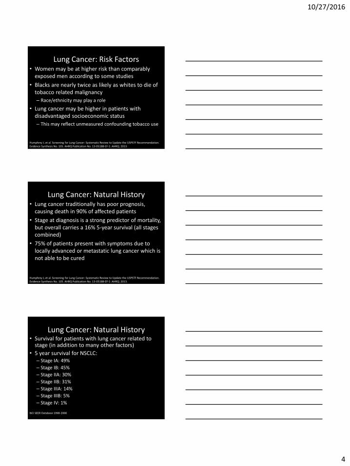
Lung Cancer • Lung cancer is abnormal proliferation arising within
the airways or tissue of the lung
• Traditionally divided into non-small cell and small cell carcinoma
• Small cell carcinoma accounts for approximately 16% of all cases of lung cancer
• Unusual tumor types such as carcinoid tumor account for approximately 5% of cases
• The remainder of cases are attributable to non-small cell carcinoma
Humphrey L et al. Screening for Lung Cancer: Systematic Review to Update the USPSTF Recommendation. Evidence Synthesis No. 105. AHRQ Publication No. 13-05188-EF-1: AHRQ; 2013.

10/27/2016
2
Lung Cancer: Epidemiology • Lung cancer is the second most common cancer in
the United States
• Lung cancer is the #1 cause of cancer-related mortality in the United States
• The American Cancer Society (ACS) estimates that in the United States in 2012: – 226, 160 new cases of lung cancer
– 160,340 lung cancer related deaths
– Lung cancer will account for 28% of all cancer-related deaths
Humphrey L et al. Screening for Lung Cancer: Systematic Review to Update the USPSTF Recommendation. Evidence Synthesis No. 105. AHRQ Publication No. 13-05188-EF-1: AHRQ; 2013.
Lung Cancer: Epidemiology • Lung cancer is the leading cause of years of life lost
to cancer in the United States
– Estimated 15 years of life lost on average per person dying of lung cancer
Humphrey L et al. Screening for Lung Cancer: Systematic Review to Update the USPSTF Recommendation. Evidence Synthesis No. 105. AHRQ Publication No. 13-05188-EF-1: AHRQ; 2013.
Lung Cancer: Epidemiology • It is estimated that 7% of everyone born today will
be diagnosed with lung cancer during their lifetime and nearly 6% will die from lung cancer
• Lung cancer rates have been been increasing steadily in recent years throughout the world, and differences between countries have largely mirrored differences in smoking rates
• Worldwide estimated 1.6 million new cases and 1.4 million deaths from lung cancer in 2008
Humphrey L et al. Screening for Lung Cancer: Systematic Review to Update the USPSTF Recommendation. Evidence Synthesis No. 105. AHRQ Publication No. 13-05188-EF-1: AHRQ; 2013.
10/27/2016
3
Lung Cancer: Risk Factors • Smoking is greatest risk factor for lung cancer
• Smoking is responsible for approximately 85% of lung cancer cases in the United States
• Worldwide, smoking accounts for 75-80% of cases of lung cancer in men and not less than 50% of cases in women
• Smoking most associated with small cell and squamous cell carcinoma and less associated with adenocarcinoma, including adenocarcinoma in site (AIS)
Humphrey L et al. Screening for Lung Cancer: Systematic Review to Update the USPSTF Recommendation. Evidence Synthesis No. 105. AHRQ Publication No. 13-05188-EF-1: AHRQ; 2013.
Lung Cancer: Risk Factors • How many smokers are there?
– NCI estimated that in 2006-2007, approximately 37% of the US population were current or former smokers
– In 2008, an estimated 7 million Americans between ages 55-75 had at least a 30 pack-year smoking history
– Estimated 19% of US population in 2010 were current smokers.
– Projected that 17% of the US population will be current smokers by the year 2020
Humphrey L et al. Screening for Lung Cancer: Systematic Review to Update the USPSTF Recommendation. Evidence Synthesis No. 105. AHRQ Publication No. 13-05188-EF-1: AHRQ; 2013.
Lung Cancer: Risk Factors • Other risk factors for lung cancer:
– Age: Incidence of lung cancer significantly increases with age
– Environmental radon exposure
– Family history
– COPD
– Pulmonary fibrosis
– Exposure to second hand smoke
– Indoor cooking fumes
– Occupational exposures including asbestos, arsenic, chromium, and coal tar
Humphrey L et al. Screening for Lung Cancer: Systematic Review to Update the USPSTF Recommendation. Evidence Synthesis No. 105. AHRQ Publication No. 13-05188-EF-1: AHRQ; 2013.
10/27/2016
4
Lung Cancer: Risk Factors • Women may be at higher risk than comparably
exposed men according to some studies
• Blacks are nearly twice as likely as whites to die of tobacco related malignancy
– Race/ethnicity may play a role
• Lung cancer may be higher in patients with disadvantaged socioeconomic status
– This may reflect unmeasured confounding tobacco use
Humphrey L et al. Screening for Lung Cancer: Systematic Review to Update the USPSTF Recommendation. Evidence Synthesis No. 105. AHRQ Publication No. 13-05188-EF-1: AHRQ; 2013.
Lung Cancer: Natural History • Lung cancer traditionally has poor prognosis,
causing death in 90% of affected patients
• Stage at diagnosis is a strong predictor of mortality, but overall carries a 16% 5-year survival (all stages combined)
• 75% of patients present with symptoms due to locally advanced or metastatic lung cancer which is not able to be cured
Humphrey L et al. Screening for Lung Cancer: Systematic Review to Update the USPSTF Recommendation. Evidence Synthesis No. 105. AHRQ Publication No. 13-05188-EF-1: AHRQ; 2013.
Lung Cancer: Natural History • Survival for patients with lung cancer related to
stage (in addition to many other factors)
• 5 year survival for NSCLC: – Stage IA: 49%
– Stage IB: 45%
– Stage IIA: 30%
– Stage IIB: 31%
– Stage IIIA: 14%
– Stage IIIB: 5%
– Stage IV: 1%
NCI SEER Database 1998-2000
10/27/2016
5
Lung Cancer: Natural History • 5 year survival for SCLC:
– Stage I: 31%
– Stage II: 19%
– Stage III: 8%
– Stage IV: 2%
NCI SEER Database 1988-2001
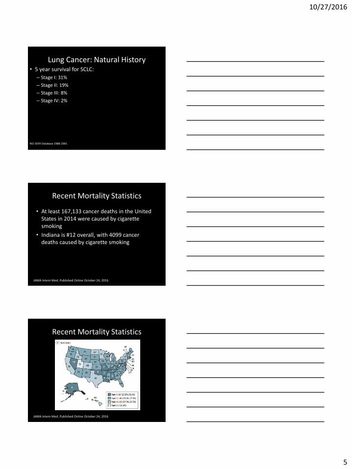
Recent Mortality Statistics
• At least 167,133 cancer deaths in the United States in 2014 were caused by cigarette smoking
• Indiana is #12 overall, with 4099 cancer deaths caused by cigarette smoking
JAMA Intern Med. Published Online October 24, 2016
Recent Mortality Statistics
JAMA Intern Med. Published Online October 24, 2016
10/27/2016
6
What Makes Lung Cancer a Good Target for Screening?
• High Morbidity and Mortality
• Relatively high prevalence in high risk populations
• Mortality and survival in lung cancer related to initial stage of diagnosis, therefore, early diagnosis may be beneficial
• Based on this, an effective screening program for early diagnosis and treatment may have a significant impact on the high mortality of lung cancer
Humphrey L et al. Screening for Lung Cancer: Systematic Review to Update the USPSTF Recommendation. Evidence Synthesis No. 105. AHRQ Publication No. 13-05188-EF-1: AHRQ; 2013.
What Makes a Good Screening Test? • The condition or disease should be an important health problem • There should be an identifiable latent or early symptomatic stage • The natural history of the condition or disease should be
adequately understood, including progression from latent to symptomatic disease
• There should be an accepted treatment for the condition and an agreed upon policy on whom to treat
• High sensitivity and specificity • Acceptable to patients and providers • Cost effective • Non-invasive or minimally invasive • Facilities for diagnosis and treatment should be widely available • Screening should be a continuous process rather than a “once and
done” proposition
Humphrey L et al. Screening for Lung Cancer: Systematic Review to Update the USPSTF Recommendation. Evidence Synthesis No. 105. AHRQ Publication No. 13-05188-EF-1: AHRQ; 2013.
Wilson JMG et al. Principles and Practice of Screening for Disease. Geneva, Switzerland; 1968. Report No.: Public Health Papers No. 34.
10/27/2016
7
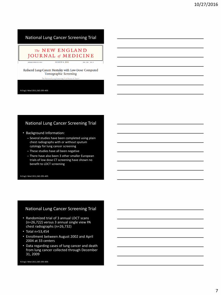
National Lung Cancer Screening Trial
N Engl J Med 2011;365:395-409.
• Background Information:
– Several studies have been completed using plain chest radiographs with or without sputum cytology for lung cancer screening
– These studies have all been negative
– There have also been 3 other smaller European trials of low dose CT screening have shown no benefit to LDCT screening
National Lung Cancer Screening Trial
N Engl J Med 2011;365:395-409.
• Randomized trial of 3 annual LDCT scans (n=26,722) versus 3 annual single view PA chest radiographs (n=26,732)
• Total n=53,454
• Enrollment between August 2002 and April 2004 at 33 centers
• Data regarding cases of lung cancer and death from lung cancer collected through December 31, 2009
National Lung Cancer Screening Trial
N Engl J Med 2011;365:395-409.
10/27/2016
8
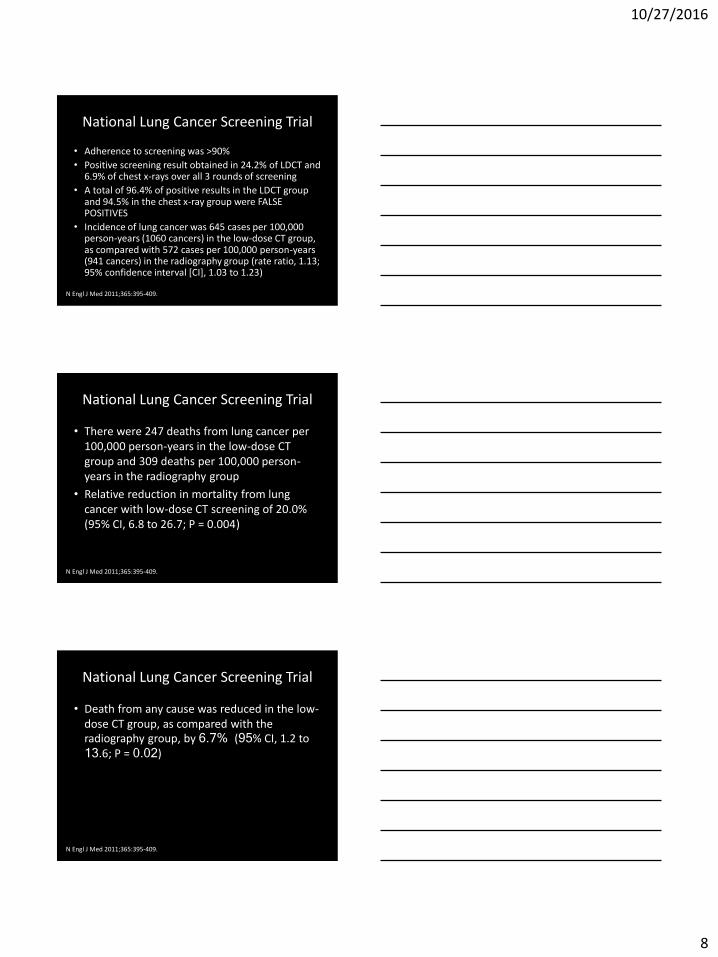
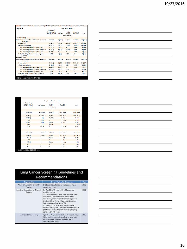
• Adherence to screening was >90%
• Positive screening result obtained in 24.2% of LDCT and 6.9% of chest x-rays over all 3 rounds of screening
• A total of 96.4% of positive results in the LDCT group and 94.5% in the chest x-ray group were FALSE POSITIVES
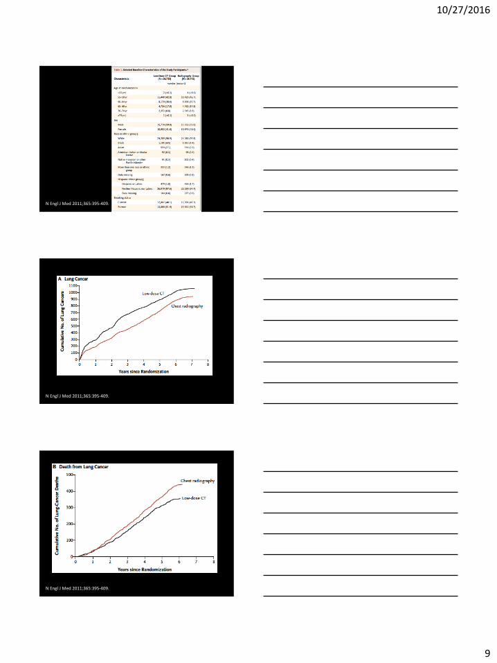
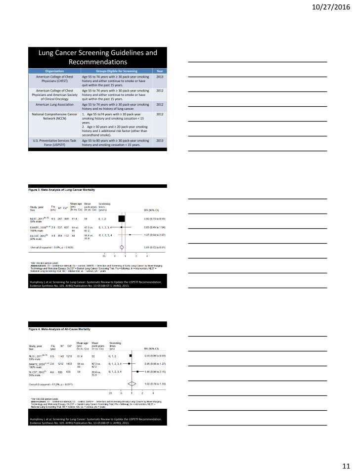
• Incidence of lung cancer was 645 cases per 100,000 person-years (1060 cancers) in the low-dose CT group, as compared with 572 cases per 100,000 person-years (941 cancers) in the radiography group (rate ratio, 1.13; 95% confidence interval [CI], 1.03 to 1.23)
National Lung Cancer Screening Trial
N Engl J Med 2011;365:395-409.
• There were 247 deaths from lung cancer per 100,000 person-years in the low-dose CT group and 309 deaths per 100,000 person-years in the radiography group
• Relative reduction in mortality from lung cancer with low-dose CT screening of 20.0% (95% CI, 6.8 to 26.7; P = 0.004)
National Lung Cancer Screening Trial
N Engl J Med 2011;365:395-409.
• Death from any cause was reduced in the low-dose CT group, as compared with the radiography group, by 6.7% (95% CI, 1.2 to 13.6; P = 0.02)
National Lung Cancer Screening Trial
N Engl J Med 2011;365:395-409.
10/27/2016
9
N Engl J Med 2011;365:395-409.
N Engl J Med 2011;365:395-409.
N Engl J Med 2011;365:395-409.
10/27/2016
10
N Engl J Med 2011;365:395-409.
N Engl J Med 2011;365:395-409.
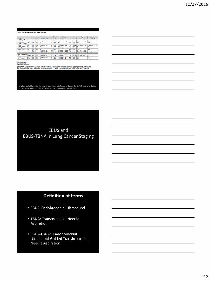
Lung Cancer Screening Guidelines and Recommendations
Organization Groups Eligible for Screening Year
American Academy of Family Practice
Evidence is insufficient to recommend for or
against screening.
2013
American Association for Thoracic Surgery
1. Age 55 to 79 years with ≥ 30 pack-year smoking history. 2. Long-term lung cancer survivors who have completed 4 years of surveillance without recurrence, and who can tolerate lung cancer treatment in order to detect second primary lung cancer until the age of 79. 3. Age 50 to 79 years with a 20 pack-year smoking history and additional comorbidity that produces a cumulative risk of developing lung cancer ≥ 5% in 5 years.
2012
American Cancer Society Age 55 to 74 years with ≥ 30 pack-year smoking history, either currently smoking or have quit within the past 15 years, and who are in relatively good health.
2013
10/27/2016
11
Lung Cancer Screening Guidelines and Recommendations
Organization Groups Eligible for Screening Year
American College of Chest Physicians (CHEST)
Age 55 to 74 years with ≥ 30 pack-year smoking history and either continue to smoke or have quit within the past 15 years.
2013
American College of Chest Physicians and American Society
of Clinical Oncology
Age 55 to 74 years with ≥ 30 pack-year smoking history and either continue to smoke or have quit within the past 15 years.
2012
American Lung Association Age 55 to 74 years with ≥ 30 pack-year smoking history and no history of lung cancer.
2012
National Comprehensive Cancer Network (NCCN)
1. Age 55 to74 years with ≥ 30 pack-year smoking history and smoking cessation < 15 years. 2. Age ≥ 50 years and ≥ 20 pack-year smoking history and 1 additional risk factor (other than secondhand smoke).
2012
U.S. Preventative Services Task Force (USPSTF)
Age 55 to 80 years with ≥ 30 pack-year smoking history and smoking cessation < 15 years.
2013
Humphrey L et al. Screening for Lung Cancer: Systematic Review to Update the USPSTF Recommendation. Evidence Synthesis No. 105. AHRQ Publication No. 13-05188-EF-1: AHRQ; 2013.
Humphrey L et al. Screening for Lung Cancer: Systematic Review to Update the USPSTF Recommendation. Evidence Synthesis No. 105. AHRQ Publication No. 13-05188-EF-1: AHRQ; 2013.
10/27/2016
12
Humphrey L et al. Screening for Lung Cancer: Systematic Review to Update the USPSTF Recommendation. Evidence Synthesis No. 105. AHRQ Publication No. 13-05188-EF-1: AHRQ; 2013.
EBUS and EBUS-TBNA in Lung Cancer Staging
Definition of terms
• EBUS: Endobronchial Ultrasound
• TBNA: Transbronchial Needle Aspiration
• EBUS-TBNA: Endobronchial Ultrasound Guided Transbronchial Needle Aspiration
10/27/2016
13
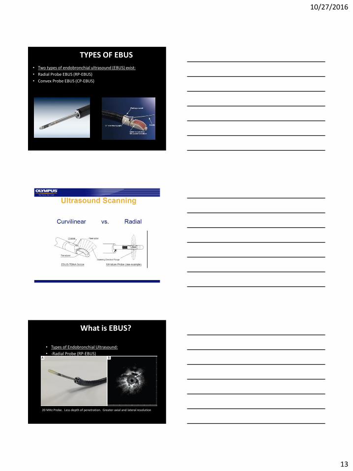
TYPES OF EBUS
• Two types of endobronchial ultrasound (EBUS) exist:
• Radial Probe EBUS (RP-EBUS)
• Convex Probe EBUS (CP-EBUS)
What is EBUS?
• Types of Endobronchial Ultrasound:
• -Radial Probe (RP-EBUS)
20 MHz Probe. Less depth of penetration. Greater axial and lateral resolution
10/27/2016
14
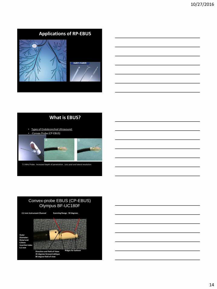
Applications of RP-EBUS
What is EBUS?
• Types of Endobronchial Ultrasound:
• -Convex Probe (CP-EBUS)
7.5 MHz Probe: Increased depth of penetration. Less axial and lateral resolution
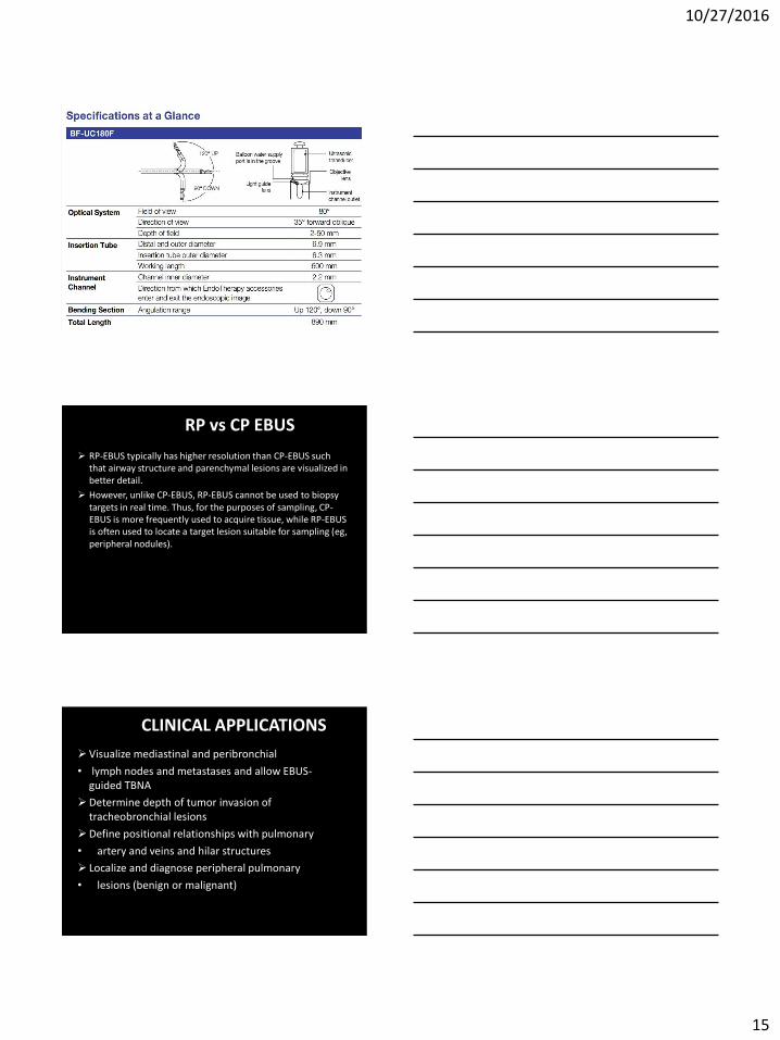
Convex-probe EBUS (CP-EBUS)
Olympus BF-UC180F
Scanning Range: 50 degrees 2.2 mm Instrument Channel
Direction and field of View: 35 degrees forward oblique 80 degree field of view
Outer Diameter: Distal end: 6.9mm Insertion tube: 6.3 mm
Ridges for balloon
10/27/2016
15
RP vs CP EBUS
RP-EBUS typically has higher resolution than CP-EBUS such that airway structure and parenchymal lesions are visualized in better detail.
However, unlike CP-EBUS, RP-EBUS cannot be used to biopsy targets in real time. Thus, for the purposes of sampling, CP-EBUS is more frequently used to acquire tissue, while RP-EBUS is often used to locate a target lesion suitable for sampling (eg, peripheral nodules).
CLINICAL APPLICATIONS
Visualize mediastinal and peribronchial
• lymph nodes and metastases and allow EBUS- guided TBNA
Determine depth of tumor invasion of tracheobronchial lesions
Define positional relationships with pulmonary
• artery and veins and hilar structures
Localize and diagnose peripheral pulmonary
• lesions (benign or malignant)
10/27/2016
16
Staging of non-small cell lung cancer (NSCLC)
Diagnosis and to evaluate pathologic conditions like pulmonary sarcoma and pulmonary embolism
Extrinsic compression of the airway by peri-bronchial process
Submucosal disease
Diagnosis of endobronchial lesions with necrotic tumor, hemorrhagic tumor, predicting line of surgical resection
Follow up of small cell tumors, lymphoma
CLINICAL APPLICATIONS
ADVANTAGES
EBUS is a minimally-invasive
Real-time imaging permits the sampling of lymph nodes that are smaller than 10 mm in short axis and/or near major blood vessels
High diagnostic yield
Safe procedure that can be performed on an outpatient basis using local anesthesia and conscious sedation or general anesthesia.
EBUS can access a wide range of mediastinal lymph nodes as well as hilar lymph nodes (2R, 2L, 3P, 4R, 4L, 7, 10R, 10L, 11R, 11L) , and sample centrally located pulmonary lesions with high sensitivity.
Complications are uncommon, while sampling is performed in real time.
DISADVANTAGES
EBUS cannot image or sample subaortic and para-aortic lymph
nodes (stations 5 and 6).
Its availability is institution-specific, and expertise is required to interpret images and obtain diagnostic samples.
Small sample size.
10/27/2016
17
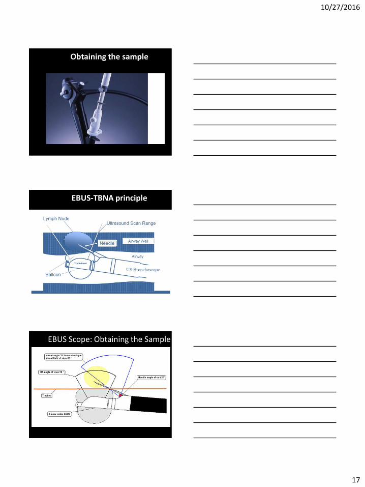
Obtaining the sample
EBUS-TBNA principle
EBUS Scope: Obtaining the Sample
10/27/2016
18
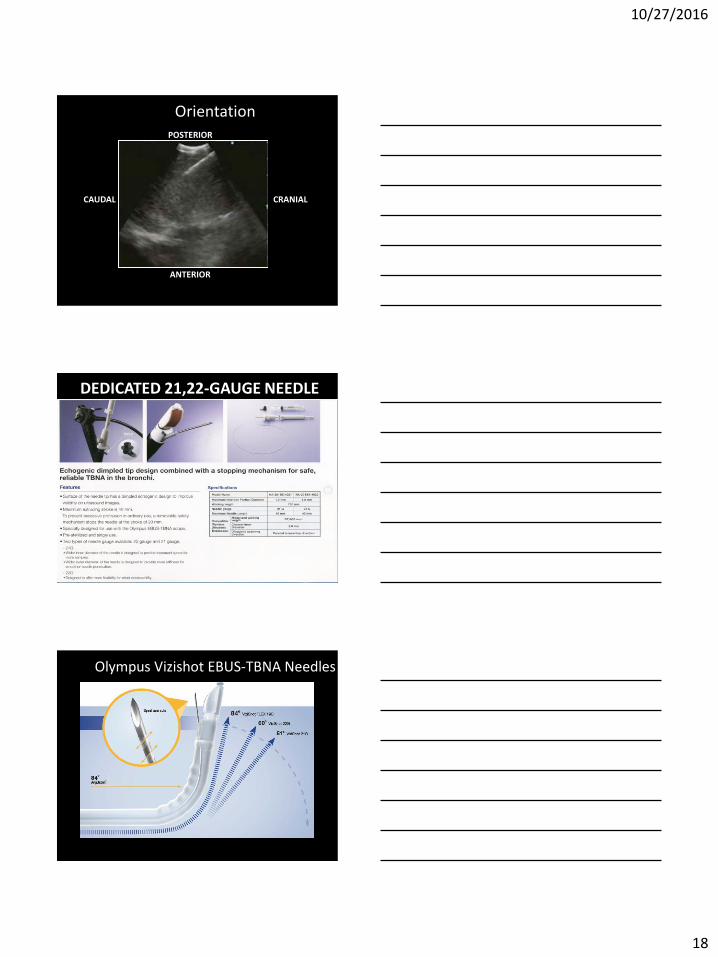
Orientation POSTERIOR
ANTERIOR
CRANIAL CAUDAL
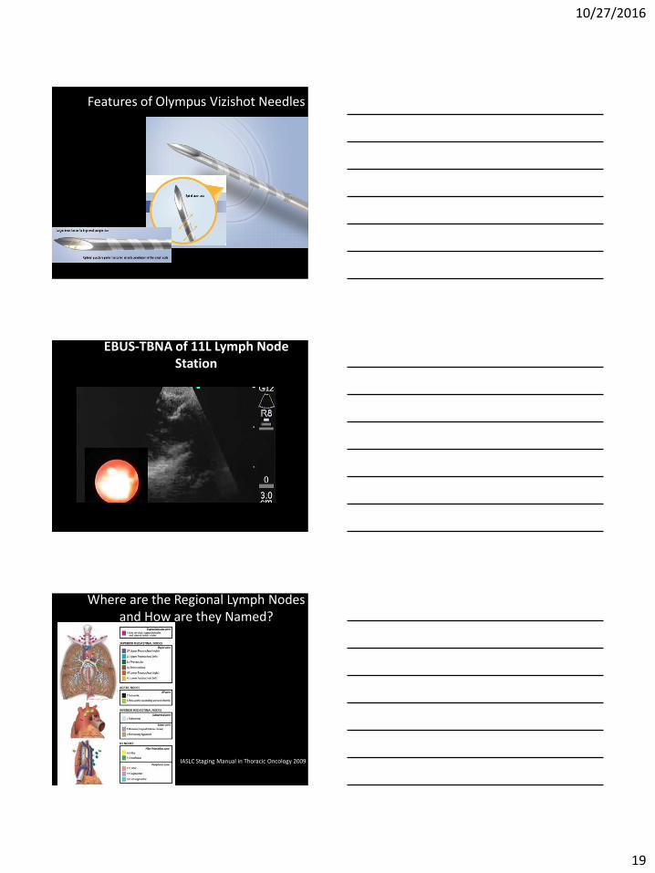
DEDICATED 21,22-GAUGE NEEDLE
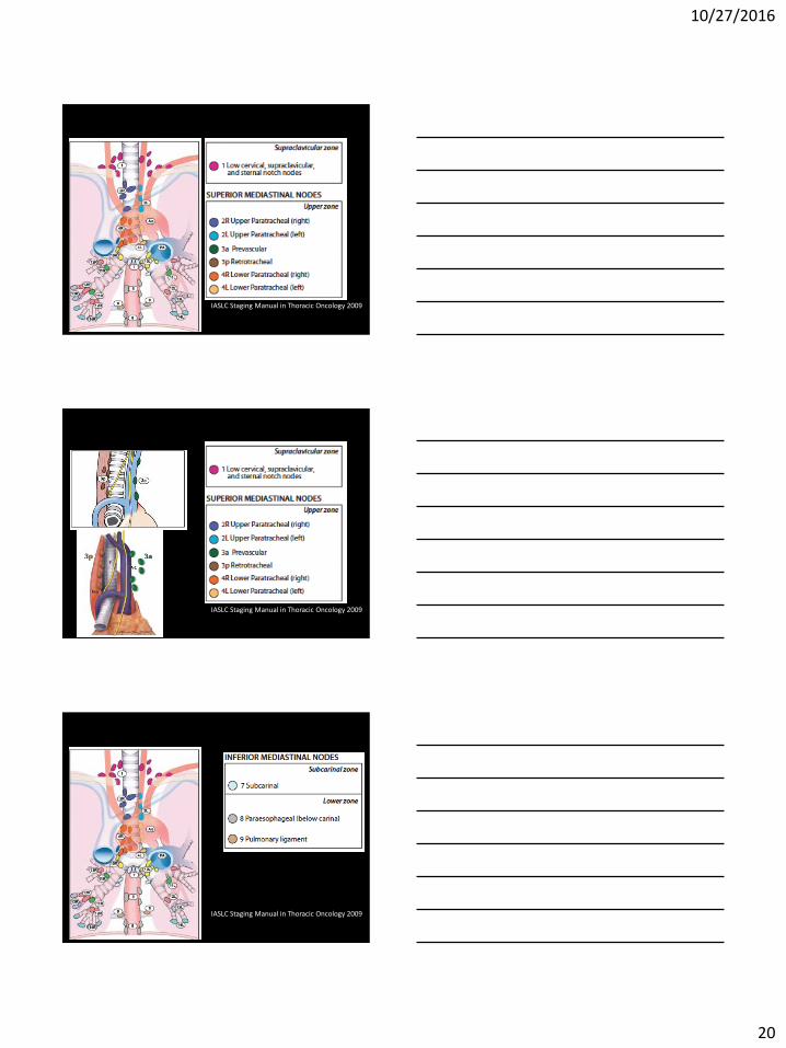
Olympus Vizishot EBUS-TBNA Needles
10/27/2016
19
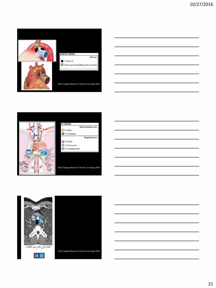
Features of Olympus Vizishot Needles
EBUS-TBNA of 11L Lymph Node Station
Where are the Regional Lymph Nodes and How are they Named?
IASLC Staging Manual in Thoracic Oncology 2009
10/27/2016
20
IASLC Staging Manual in Thoracic Oncology 2009
IASLC Staging Manual in Thoracic Oncology 2009
IASLC Staging Manual in Thoracic Oncology 2009
10/27/2016
21
IASLC Staging Manual in Thoracic Oncology 2009
IASLC Staging Manual in Thoracic Oncology 2009
IASLC Staging Manual in Thoracic Oncology 2009

10/27/2016
22
IASLC Staging Manual in Thoracic Oncology 2009
IASLC Staging Manual in Thoracic Oncology 2009
IASLC Staging Manual in Thoracic Oncology 2009
10/27/2016
23
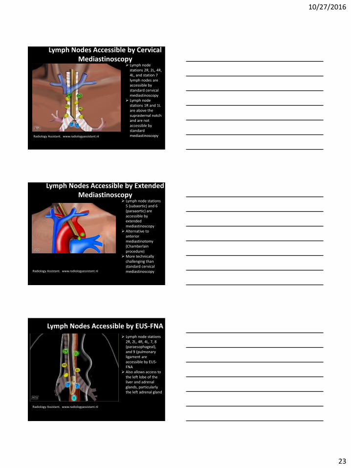
Lymph Nodes Accessible by Cervical Mediastinoscopy
Radiology Assistant. www.radiologyassistant.nl
Lymph node stations 2R, 2L, 4R, 4L, and station 7 lymph nodes are accessible by standard cervical mediastinoscopy
Lymph node stations 1R and 1L are above the suprasternal notch and are not accessible by standard mediastinoscopy
Lymph Nodes Accessible by Extended Mediastinoscopy
Radiology Assistant. www.radiologyassistant.nl
Lymph node stations 5 (subaortic) and 6 (paraaortic) are accessible by extended mediastinoscopy
Alternative to anterior mediastinotomy (Chamberlain procedure)
More technically challenging than standard cervical mediastinoscopy
Lymph Nodes Accessible by EUS-FNA
Radiology Assistant. www.radiologyassistant.nl
Lymph node stations 2R, 2L, 4R, 4L, 7, 8 (paraesophageal), and 9 (pulmonary ligament are accessible by EUS-FNA
Also allows access to the left lobe of the liver and adrenal glands, particularly the left adrenal gland
10/27/2016
24
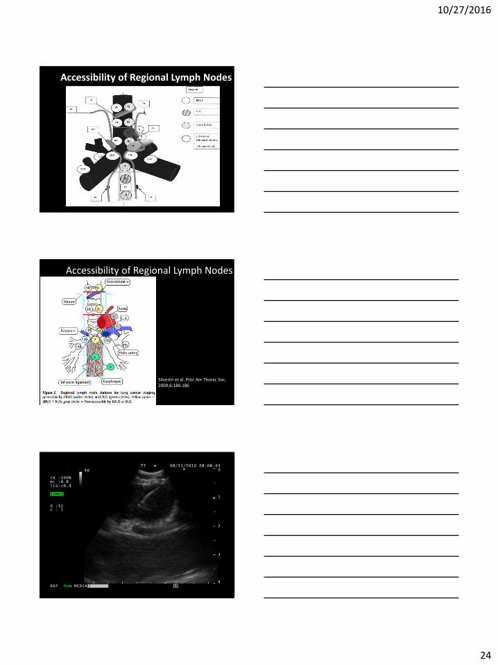
Accessibility of Regional Lymph Nodes
Accessibility of Regional Lymph Nodes
Silvestri et al. Proc Am Thorac Soc. 2009;6:180-186
10/27/2016
25
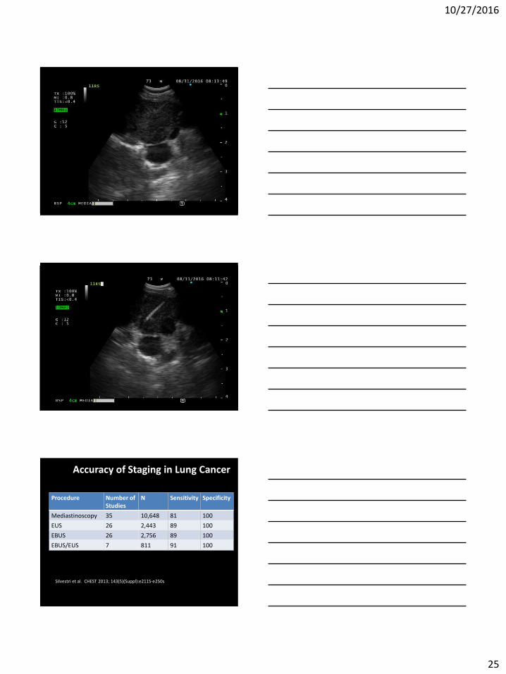
Accuracy of Staging in Lung Cancer
Procedure Number of Studies
N Sensitivity Specificity
Mediastinoscopy 35 10,648 81 100
EUS 26 2,443 89 100
EBUS 26 2,756 89 100
EBUS/EUS 7 811 91 100
Silvestri et al. CHEST 2013; 143(5)(Suppl):e211S-e250s
10/27/2016
26
ACCP Lung Cancer Guidelines 2013
• In patients with high suspicion of N2,3 involvement, either by discrete mediastinal lymph node enlargement or PET uptake (and no discrete metastases), a needle technique (EBUS-NA, EUS-NA, or combined EBUS/EUS-NA) is recommended over surgical staging as a best first test
Silvestri et al. CHEST 2013; 143(5)(Suppl):e211S-e250s
Safety and Complications
Overall, very safe with minimal reported complications
Complications of conscious sedation/general anesthesia
Rarely, infection has been reported following EBUS-TBNA including infectious pericarditis/pericardial effusion, tumor bed infection, mediastinal abscess, mediastinitis
Mechanism likely related to introduction of oral contaminants
Rare bleeding complications, even in a series which did not hold Plavix prior to EBUS-TBNA
Safety and complications
EBUS pneumothorax rate 0.2%
TBBx pneumothorax rate 1-6% (4.0%)
EBUS-TBNA bleeding rate 0.2%
TBBx bleeding rate 2-9% (2.1%)
10/27/2016
27
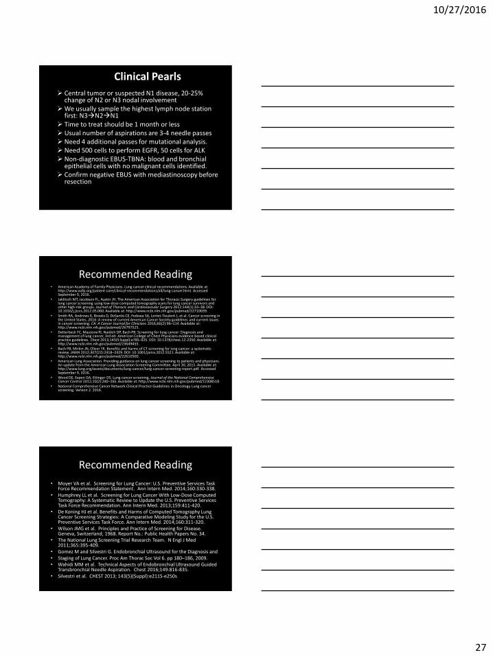
Clinical Pearls
Central tumor or suspected N1 disease, 20-25% change of N2 or N3 nodal involvement
We usually sample the highest lymph node station first: N3N2N1
Time to treat should be 1 month or less Usual number of aspirations are 3-4 needle passes Need 4 additional passes for mutational analysis. Need 500 cells to perform EGFR, 50 cells for ALK Non-diagnostic EBUS-TBNA: blood and bronchial
epithelial cells with no malignant cells identified. Confirm negative EBUS with mediastinoscopy before
resection
Recommended Reading
• American Academy of Family Physicians. Lung cancer clinical recommendations. Available at: http://www.aafp.org/patient-care/clinical-recommendations/all/lung-cancer.html. Accessed September 9, 2016.
• Jaklitsch MT, Jacobson FL, Austin JH. The American Association for Thoracic Surgery guidelines for lung cancer screening using low-dose computed tomography scans for lung cancer survivors and other high-risk groups. Journal of Thoracic and Cardiovascular Surgery 2012;144(1):33–38. DOI: 10.1016/j.jtcvs.2012.05.060. Available at: http://www.ncbi.nlm.nih.gov/pubmed/22710039.
• Smith RA, Andrews K, Brooks D, DeSantis CE, Fedewa SA, Lortet-Tieulent J, et al. Cancer screening in the United States, 2016: A review of current American Cancer Society guidelines and current issues in cancer screening. CA: A Cancer Journal for Clinicians 2016;66(2):96–114. Available at: http://www.ncbi.nlm.nih.gov/pubmed/26797525.
• Detterbeck FC, Mazzone PJ, Naidich DP, Bach PB. Screening for lung cancer: Diagnosis and management of lung cancer, 3rd ed: American College of Chest Physicians evidence-based clinical practice guidelines. Chest 2013;143(5 Suppl):e78S–92S. DOI: 10.1378/chest.12-2350. Available at: http://www.ncbi.nlm.nih.gov/pubmed/23649455.
• Bach PB, Mirkin JN, Oliver TK. Benefits and harms of CT screening for lung cancer: a systematic review. JAMA 2012;307(22):2418–2429. DOI: 10.1001/jama.2012.5521. Available at: http://www.ncbi.nlm.nih.gov/pubmed/22610500.
• American Lung Association. Providing guidance on lung cancer screening to patients and physicians. An update from the American Lung Association Screening Committee. April 30, 2015. Available at: http://www.lung.org/assets/documents/lung-cancer/lung-cancer-screening-report.pdf. Accessed September 9, 2016.
• Wood DE, Eapen GA, Ettinger DS. Lung cancer screening. Journal of the National Comprehensive Cancer Control 2012;10(2):240–265. Available at: http://www.ncbi.nlm.nih.gov/pubmed/22308518.
• National Comprehensive Cancer Network Clinical Practice Guidelines in Oncology. Lung cancer screening. Version 2. 2016.
Recommended Reading
• Moyer VA et al. Screening for Lung Cancer: U.S. Preventive Services Task Force Recommendation Statement. Ann Intern Med. 2014;160:330-338.
• Humphrey LL et al. Screening for Lung Cancer With Low-Dose Computed Tomography: A Systematic Review to Update the U.S. Preventive Services Task Force Recommendation. Ann Intern Med. 2013;159:411-420.
• De Koning HJ et al. Benefits and Harms of Computed Tomography Lung Cancer Screening Strategies: A Comparative Modeling Study for the U.S. Preventive Services Task Force. Ann Intern Med. 2014;160:311-320.
• Wilson JMG et al. Principles and Practice of Screening for Disease. Geneva, Switzerland; 1968. Report No.: Public Health Papers No. 34.
• The National Lung Screening Trial Research Team. N Engl J Med 2011;365:395-409.
• Gomez M and Silvestri G. Endobronchial Ultrasound for the Diagnosis and • Staging of Lung Cancer. Proc Am Thorac Soc Vol 6. pp 180–186, 2009. • Wahidi MM et al. Technical Aspects of Endobronchial Ultrasound Guided
Transbronchial Needle Aspiration. Chest 2016;149:816-835. • Silvestri et al. CHEST 2013; 143(5)(Suppl):e211S-e250s