luxations crown fracture crown/root fracture root fracture ... · •monitor for apical changes and...
TRANSCRIPT
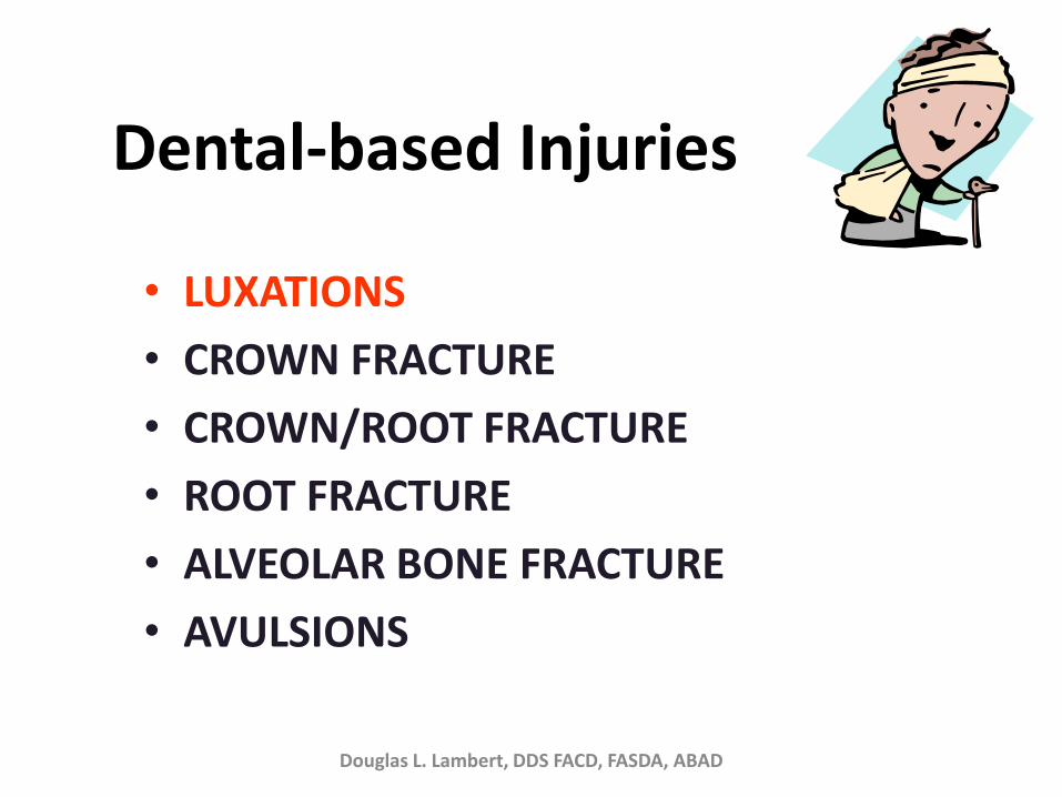
Dental-based Injuries
• LUXATIONS
• CROWN FRACTURE
• CROWN/ROOT FRACTURE
• ROOT FRACTURE
• ALVEOLAR BONE FRACTURE
• AVULSIONS
Douglas L. Lambert, DDS FACD, FASDA, ABAD

LUXATIONS The tooth is loose, now what?
1. Concussive-not loose or displaced, but tender to percussion
2. Subluxation-loose, but no displacement
3. Extrusive Luxation-partially out of socket
4. Lateral Luxation-displaced usually toward the palate
5. Intrusive Luxation-clinical crown appears shorter
Douglas L. Lambert, DDS FACD, FASDA, ABAD
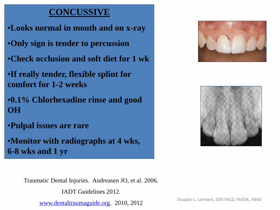
CONCUSSIVE
•Looks normal in mouth and on x-ray
•Only sign is tender to percussion
•Check occlusion and soft diet for 1 wk
•If really tender, flexible splint for
comfort for 1-2 weeks
•0.1% Chlorhexadine rinse and good
OH
•Pulpal issues are rare
•Monitor with radiographs at 4 wks,
6-8 wks and 1 yr
Traumatic Dental Injuries. Andreasen JO, et al. 2006.
IADT Guidelines 2012.
www.dentaltraumaguide.org. 2010, 2012 Douglas L. Lambert, DDS FACD, FASDA, ABAD

SUBLUXATION
•Looks normal in the socket on an x-
ray-similar to concussive
•Check occlusion and adjust
•Soft diet
•0.1%Chlorhexadine rinse and OH
•Flexible splint for 7-14 days for
patient comfort
•Good long term pulpal prognosis-
monitor at 4 wks, 6-8 wks and 1 yr
Traumatic Dental Injuries. Andreasen JO, et al. 2006.
IADT Guidelines 2012.
www.dentaltraumaguide.org. 2010, 2012 Douglas L. Lambert, DDS FACD, FASDA, ABAD

EXTRUSION LUXATION
•Apical portion of socket empty
•PDL is disrupted
•Reposition and check occlusion
•0.1% Chlorhexadine rinse and soft diet
•Flexible splint for 2 weeks (up to 3)
•Monitor for apical changes and
resorption with radiographs – 4 wks, 6-8
wks, 6 mo, and 1 yr. Pulpal necrosis
usually seen by 4 weeks
•Immature apex likely to revascularize,
mature apex minimal chance
Traumatic Dental Injuries. Andreasen JO, et al. 2006.
IADT Guidelines 2012.
www.dentaltraumaguide.org. 2010, 2012 Douglas L. Lambert, DDS FACD, FASDA, ABAD

LATERAL LUXATION
•Usually displaced palatally-root apex can be
palpated in vestibule on occasion
•Looks similar to extrusion on x-ray
•Labial plate may be fractured
•Firmly reposition with anesthesia (can be locked)
and check occlusion
•Flexible splint 3-4 weeks (due to bony fx), then
check for PDL changes. Monitor with x-rays
•May need 3-4 additional weeks (radiographs)
•0.1% Chlorhexadine rinse and OH
•Immature apex favorable; closed apex
unfavorable, 75% go necrotic
•Surface resorption frequent-esp. at apex
Traumatic Dental Injuries. Andreasen JO, et al. 2006.
IADT Guidelines 2012.
www.dentaltraumaguide.org. 2010, 2012 Douglas L. Lambert, DDS FACD, FASDA,
ABAD
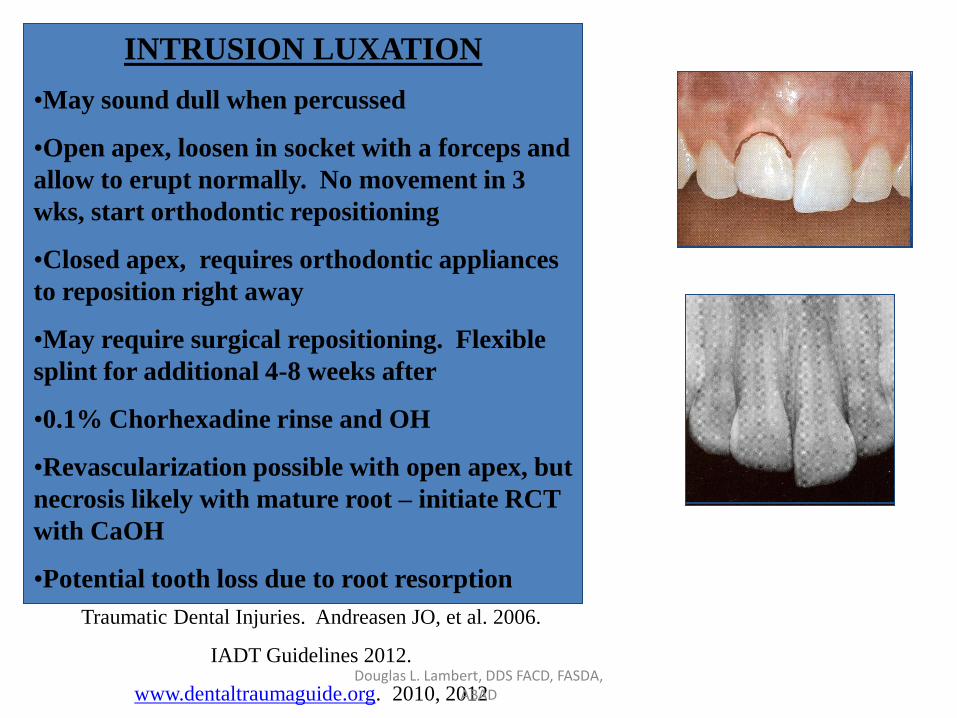
INTRUSION LUXATION
•May sound dull when percussed
•Open apex, loosen in socket with a forceps and
allow to erupt normally. No movement in 3
wks, start orthodontic repositioning
•Closed apex, requires orthodontic appliances
to reposition right away
•May require surgical repositioning. Flexible
splint for additional 4-8 weeks after
•0.1% Chorhexadine rinse and OH
•Revascularization possible with open apex, but
necrosis likely with mature root – initiate RCT
with CaOH
•Potential tooth loss due to root resorption
Traumatic Dental Injuries. Andreasen JO, et al. 2006.
IADT Guidelines 2012.
www.dentaltraumaguide.org. 2010, 2012 Douglas L. Lambert, DDS FACD, FASDA,
ABAD

ROOT FRACTURE • Complex injury to the PDL,
cementum, dentin, and pulp
• Tooth appears elongated clinically
• Radiolucent line(s) separate fragments-may be subtle
• Apical fragment usually undamaged
• Must reposition coronal fragment and splint, but no consensus on length of time
Douglas L. Lambert, DDS FACD, FASDA, ABAD

ROOT FRACTURE HARD TISSUE HEALING
• Dentin from
odontoblasts and cementum bridge the gap
• Normal tooth mobility
• Normal pulp test
• Slightly discernible fx line
Douglas L. Lambert, DDS FACD, FASDA, ABAD

ROOT FRACTURE CONNECTIVE TISSUE HEALING
• PDL cells invade the
entire fracture gap and enclose both segments
• Normal pulp test
• Increased mobility
• Obvious fx line
• Coronal pulp chamber obliterated
Douglas L. Lambert, DDS FACD, FASDA, ABAD

ROOT FRACTURE GRANULAR TISSUE HEALING
• Coronal pulp becomes necrotic
• Granulation tissue forms between the two fragments
• Necessitates removal of the coronal pulp tissue
• Coronal fragment treated with CaOH, then RCT or CaOH and MTA
Douglas L. Lambert, DDS FACD, FASDA, ABAD
ROOT FRACTURE HEALING OUTCOMES
• Key factor to healing is the stage of root development and degree of displacement of the coronal portion
• Immature apex heals by HT most likely
• Mature apex usually heals by CT and nonhealing by GT
• HT healing likely with fragments not displaced
• CT path to healing likely if fragment displaced or not repositioned properly
Douglas L. Lambert, DDS FACD, FASDA, ABAD

ALVEOLAR BONE FRACTURE
• Segment containing one or more teeth is displaced
• Occlusion is off
• Entire section is mobile
• Differentiate between root fx and alveolar fx by using radiographs-in a root fracture, fx position (line) will not move if beam angle changed
• Force needed to reposition segment
• Flexible splint for 3-4 weeks
• Monitor closely for necrosis especially with closed apex
Douglas L. Lambert, DDS FACD, FASDA, ABAD

At the office…tooth out of the mouth (dry time)
Extraoral Time
< 60 minutes
Open Apex
Extraoral Time
< 60 minutes
Closed Apex
Extraoral Time
> 60 Minutes
Open Apex
Extraoral Time
> 60 minutes
Closed Apex
OPTIONS

Out of the mouth < 60 minutes…
OPEN APEX • Revascularization possible
• Rinse off debris gently with saline
• Soak root surface for five minutes with topical abx (minocyline or doxycycline)*
• Replant-verify position w/ xray
• Do not initiate endodontic treatment at this point
• Flexible splint for 10-14 days
• ABX coverage, soft diet, Peridex, tetanus booster
CLOSED APEX • Revascularization unlikely, but
still good chance for periodontal healing
• Rinse off debris with saline and coagulum from socket
• Replant gently-xray to check
• Flexible splint for 10-14 days
• Initiate endo – CaOH paste to decrease chances of root resorption
• ABX coverage, soft diet, OH, etc..
IADT Guidelines 2012. *Experimentally successful Douglas L. Lambert, DDS FACD, FASDA, ABAD
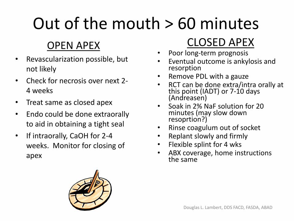
Out of the mouth > 60 minutes OPEN APEX
• Revascularization possible, but not likely
• Check for necrosis over next 2-4 weeks
• Treat same as closed apex
• Endo could be done extraorally to aid in obtaining a tight seal
• If intraorally, CaOH for 2-4 weeks. Monitor for closing of apex
CLOSED APEX • Poor long-term prognosis • Eventual outcome is ankylosis and
resorption • Remove PDL with a gauze • RCT can be done extra/intra orally at
this point (IADT) or 7-10 days (Andreasen)
• Soak in 2% NaF solution for 20 minutes (may slow down resoprtion?)
• Rinse coagulum out of socket • Replant slowly and firmly • Flexible splint for 4 wks • ABX coverage, home instructions
the same
Douglas L. Lambert, DDS FACD, FASDA, ABAD