lymphadenopathy - mans · 2016-12-26 · causes of lymphadenopathy l localized ... lymph node...
TRANSCRIPT

LYMPHADENOPATHY
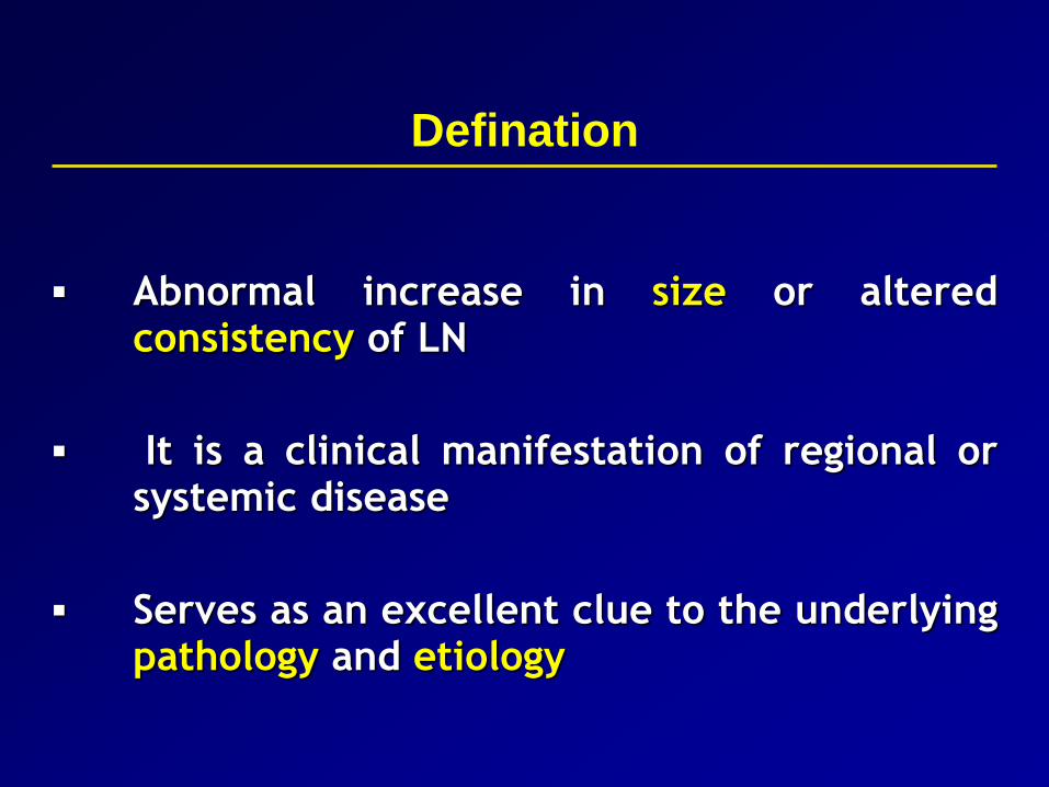
Defination
Abnormal increase in size or alteredconsistency of LN
It is a clinical manifestation of regional orsystemic disease
Serves as an excellent clue to the underlyingpathology and etiology

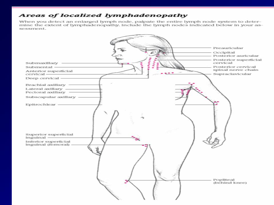

Lymph Node Groups
submental, submandibular, preauricular,
postauricular, occipital,
cervical: anterior, posterior
clavicular: supraclavicular, infraclavicular
axillary: anterior (pectoral), lateral, posterior
(subscapular), central
epitrochlear, inguinal

CAUSES OF LYMPHADENOPATHY
l
LOCALIZED
LN draining a septic focus
Cervical; axillary; inguinal; periauricular
Metastasis from carcinoma or other solid tumour
─ Hilar bronchus
─ Vichow stomach
─ Cervical thyroid, togue, parotid

CAUSES OF LYMPHADENOPATHY (localized)
Systemic infection
Viral: viral hepatitis (Rt. Supracalv. LN)
Bacterial: TB
Protozoal: Filarial infection
Generelized LN may start as localized LN. As in Hodgkin’s disease

CAUSES OF LYMPHADENOPATHY
GENERALISED
infections
- viral; infectious mononucleosis,CMV,HIV
- bacterial; tuberculosis,syphilis
- protozoal; toxoplasmosis
Leukemias
Especially chronic lymphocytic leukemia

CAUSES OF LYMPHADENOPATHY (Con.)
Lymohomas
- Hodgkin’s disease (HD)
- Non-Hodgkin’s lymphoma (NHL)
Collagen
- Rheumatoid artheritis
infitration; sarcoidosis
drugs; phenytoin

Characters of L.N enlargment
Infection: Acute, tender, warm
– Primary region drained also involved (e.g
neck nodes w/strep throat)
– Sometimes get diffuse enlargement in
response to generalized infection or
systemic inflammatory process (.e.g TB,
HIV, systemic mononucleosis)
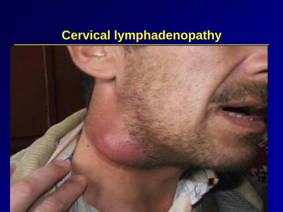
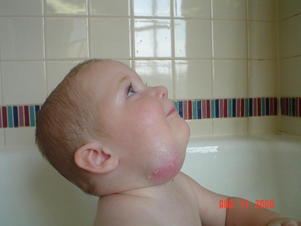
Cervical lymphadenopathy
Characters of L.N enlargment
Malignancy:
– Slowly progressive, firm, multiple nodes involved,stuck together & to underlying structures, not tender
–Primary site malignancy could be nodes(e.g.lymphoma)or adjacent region (e.g. intra-oral squamous cell ca)
- Associated with constitutional symptoms
- Pel Ebstein fever: in HD ( period of fever lasting forfew days or weeks alternating with aprexial period
Lymph Node Examination
Inspection:
- lymphedema
- surgical scars from cancer excision
- obvious masses
Palpation:
technique:
use the pads of the middle three fingers & move the
skin in circular motions over the underlying tissues in
each area
Lymph Node Examination
in abnormal nodes, describe in terms of
─ location
─ size
─ discrete or matted together
─ mobile or fixed
─ consistency (soft, hard, firm)
─ tenderness


Cervical Lymph Node Anatomy & Drainage
Ant Cerv: Throat, tonsils, post pharynx, thyroid
Post Cerv: Back of skull
Tonsillar: Tonsils, posterior pharynx
Sub-Mandibular: Floor of mouth
Sub-Mental: Teeth
Supra-Clavicular: Thorax
Pre-Auricular: Ear

Lymph Node Exam
Gently walk fingers
along general regions –
comparing R to L

Palpation of supraclavicular LN

Axillary L.N. Examination
Support the patient’s arm and
elbow with the non-examining
hand to maintain optimal
relaxation of musles
Axillary nodes are palpated at deep
pressure using a circular motion
with the pads of the three middle
fingers of the examining hand, in
all four aspects of the axilla.
4

Axillary LN examination
Proceeding down
the mid-axillary
chestwall, gently
move the pads of
the fingers
medially and
inside the border
of the pectoral
muscle
Continue by
palpating the
subscapular
nodes. Sweep
back up and
return to the
axilla with the
palm facing
laterally,
Check the
lateral nodes
with the palm of
the hand facing
the humeral
head
start palpating
the central nodes
deep in the apex
of the axilla. The
hand is straight
up, deep in the
underarm

Clinical Approach
Presentation:
-Swelling -Constitional symptoms
-Pressure symptoms -Mediastinal Syndrome
-Pressure on veins oedema
-Pressure on nerves pain
Age:
-TB: in children & young children
-HD: highest incidence () 20-40 years
- NHL: middle age & late life
-ALL: Highest in first 6 years

Clinical Approach
History:
of infectionss, drugs
Distribution:
Localized or generelized
Single or multiple groups affected
Other signs:
Fever: H.D, NHL. Leukemia
Jaundice:
Eye: infection, subconjunctival Hage, exophalmos
Clinical Approach
Mouth: Tonsils, infection, parotid , gum hyperplasia
Skin:
pruritis: H.D, NHL
skin nodules: CLL, NHL
Herpes zoster
Tenderness of sternum: in CML
Bone tenderness
Abdominal examination:
- Ascitis & masses - liver
- Spleen: huge in CML

