lymphatic dysregulation in patients with heart failure
TRANSCRIPT

THE PRESENT AND FUTURE
JACC REVIEW TOPIC OF THE WEEK
Lymphatic Dysregulation in PatientsWith Heart FailureJACC Review Topic of the Week
Marat Fudim, MD, MHS,a,b Husam M. Salah, MD,c Janarthanan Sathananthan, MBCHB, MPH,d Mathieu Bernier, MD,e
Waleska Pabon-Ramos, MD, MPH,f Robert S. Schwartz, MD,g Josep Rodés-Cabau, MD, PHD,e,h François Côté, MD,i
Abubaker Khalifa, MD,j Sean A. Virani, MD, MSC, MPH,d Manesh R. Patel, MDa,b
ABSTRACT
The lymphatic system is an integral part of the circulatory system and plays an important role in the volume homeostasis
of the human body. The complex anatomy and physiology paired with a lack of simple diagnostic tools to study the
lymphatic system have led to an underappreciation of the contribution of the lymphatic system to acute and chronic heart
failure (HF). Herein, we discuss the physiological role of the lymphatic system in volume management and the evidence
demonstrating the dysregulation of the lymphatic system in HF. Further, we discuss the opportunity to target the
lymphatic system in the management of HF and different potential approaches to accessing the lymphatic system.
(J Am Coll Cardiol 2021;78:66–76) © 2021 by the American College of Cardiology Foundation.
T he circulatory system consists of the cardio-vascular system and the lymphatic system.The cardiovascular system is a closed, high-
pressure circulatory system with the heart acting as acentral pump, whereas the lymphatic system is anopen, low-pressure circulatory system with no centralpump (1). The lymphatic vessels are present in almostall tissues except bone marrow, cartilage, and cornea(1). Several liters of fluids are filtered via the semiper-meablemembrane of the capillaries into the interstitialspace every day. The filtered fluid has important phys-iological functions, such as tissue nutrition and hydra-tion. An increase in the amount of the filtered fluid can
lead to interstitial edema with clinical manifestationssuch as extremity and tissue edema, including pulmo-nary edema. The amount of filtered fluids depends onthe Starling equation for fluid filtration (2):
Jv ¼ LpS½ðpc �piÞ� sðpc �piÞ�
where JV is the filtration volume per second, Lp is thehydraulic conductance of the membrane, S is surfacearea for filtration, pc is the capillary hydrostaticpressure, pi is the interstitial hydrostatic pressure, s isthe reflection coefficient, pc is the oncotic pressure ofthe plasma protein, and pi is the oncotic pressure ofthe interstitial protein (Figure 1).
ISSN 0735-1097/$36.00 https://doi.org/10.1016/j.jacc.2021.04.090
From the aDepartment of Medicine, Division of Cardiology, Duke University Medical Center, Durham, North Carolina, USA; bDuke
Clinical Research Institute, Durham, North Carolina, USA; cDepartment of Medicine, University of Arkansas for Medical Sciences,
Arkansas, USA; dCentre for Cardiovascular Innovation and Division of Cardiology, University of British Columbia, Vancouver,
British Columbia, Canada; eQuebec Heart and Lung Institute, Laval University, Quebec City, Quebec, Canada; fDepartment of
Radiology, Division of Interventional Radiology, Duke University Medical Center, Durham, North Carolina, USA; gMinneapolis
Heart Institute, Minneapolis, Minnesota, USA; hHospital Clinic of Barcelona, Barcelona, Spain; iInterventional Radiology
Department, CHU de Quebec, Laval University, Quebec City, Quebec, Canada; and the jDepartment of Medicine, Joseph Brant
Hospital, McMaster University, Hamilton, Ontario, Canada.
Anita Deswal, MD, served as Guest Associate Editor for this paper. Christie Ballantyne, MD, served as Guest Editor-in-Chief for this
paper.
The authors attest they are in compliance with human studies committees and animal welfare regulations of the authors’
institutions and Food and Drug Administration guidelines, including patient consent where appropriate. For more information,
visit the Author Center.
Manuscript received March 30, 2021; accepted April 13, 2021.
Listen to this manuscript’s
audio summary by
Editor-in-Chief
Dr. Valentin Fuster on
JACC.org.
J O U R N A L O F T H E A M E R I C A N C O L L E G E O F C A R D I O L O G Y VO L . 7 8 , N O . 1 , 2 0 2 1
ª 2 0 2 1 B Y T H E A M E R I C A N CO L L E G E O F C A R D I O L O G Y F O U N DA T I O N
P U B L I S H E D B Y E L S E V I E R

One of the main functions of the lymphaticvascular system is to collect filtered fluid that accu-mulates in the interstitial space (mainly water, salts,and plasma proteins) and return it to the centralvenous system. To prevent interstitial edema, thereturn of filtered lymph fluid occurs at a rate similarto the rate of fluid production/accumulation in theinterstitial space (2). After fluid enters the lymphaticvascular system, it becomes lymph and passesthrough lymph nodes, where foreign matter is filteredand neutralized by the immune system cells (eg,dendritic cells, macrophages, and lymphocytes) (2).
Although lymph composition was thought to besimilar to that of the plasma, proteomic mappinghas shown unique composition of the tissue-derived proteins in the lymph (3). Lymph containshigh concentrations of proteins that are involved incell catabolism and apoptosis, extracellular matrix
remodeling, and innate immunity (3). Be-sides its role in fluid hemostasis, lymph actsas a chemical buffer system, facilitates im-mune cell trafficking, and transports pro-teomes to draining lymph nodes (3).
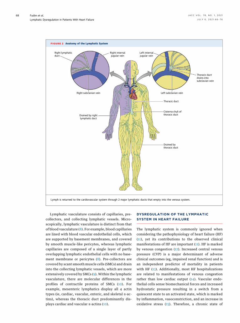
Lymph is returned to the cardiovascularsystem through 2 major lymphatic ducts thatempty into the venous system (ie, the rightlymphatic duct and the thoracic duct) (4). Theright lymphatic duct drains lymph from the right sideof thorax, right upper extremity, and right side ofhead and neck, and empties into the junction of theright internal jugular vein and right subclavian vein(Figure 2) (4). The thoracic duct runs superiorly fromthe superior aspect of the cisterna chyli to the lowercervical spine, drains lymph from all the body exceptparts that are drained by the right lymphatic duct,and empties into the junction of the left internal ju-gular vein and the left subclavian vein (5). Thethoracic duct terminates as a single duct in the ma-jority of cases (72%), or less frequently as double(8.5%), triple (1.8%), or quadruple (2.2%) ducts (6).The thoracic duct typically returns approximately1.38 mL/kg/h of lymph to the central venous circula-tion (5). An increase in the filtration volume (arterial/venous congestion) is counteracted with an increasein the amount of filtered lymph fluid and a decreasein interstitial protein, thus decreasing the oncoticpressure of the interstitium (7).
FIGURE 1 Basic Principles of Fluid Shifts Between Individual Compartments and Lymph Production
Alveolus
Palv
PH
POnc
POnc
PHCapillaryNet outward
fluid filtration
Net outward fluid filtration = K (PH capillary − PH interstitium) − Kσ (Ponc capillary − Ponc interstitium)
Lymphaticdrainage
LymphInterstitium
The amount of filtered fluids depends on the Starling equation. Fluid extravasation depends on the balance of hydrostatic pressure, and
oncotic pressure in the capillaries and the interstitium. K ¼ constant; Palv ¼ alveolar pressure; Ph ¼ hydrostatic pressure; Ponc ¼ oncotic
pressure.
HIGHLIGHTS
� The lymphatic system is integral to vol-ume hemostasis.
� The lymphatic system is involved in manyof the clinical manifestations of HF.
� In patients with HF, therapeutic targetingof the lymphatic system could reducecongestive symptoms.
AB BR E V I A T I O N S
AND ACRONYM S
CRS = cardiorenal syndrome
CVP = central venous pressure
HF = heart failure
PCWP = pulmonary capillary
wedge pressure
SMC = smooth muscle cells
J A C C V O L . 7 8 , N O . 1 , 2 0 2 1 Fudim et al.J U L Y 6 , 2 0 2 1 : 6 6 – 7 6 Lymphatic Dysregulation in Patients With Heart Failure
67
Lymphatic vasculature consists of capillaries, pre-collectors, and collecting lymphatic vessels. Micro-scopically, lymphatic vasculature is distinct from thatof blood vasculature (8). For example, blood capillariesare lined with blood vascular endothelial cells, whichare supported by basement membranes, and coveredby smooth muscle-like pericytes, whereas lymphaticcapillaries are composed of a single layer of partlyoverlapping lymphatic endothelial cells with no base-ment membrane or pericytes (8). Pre-collectors arecovered by scant smoothmuscle cells (SMCs) and draininto the collecting lymphatic vessels, which are moreextensively covered by SMCs (9). Within the lymphaticvasculature, there are molecular differences in theprofiles of contractile proteins of SMCs (10). Forexample, mesenteric lymphatics display all 4 actintypes (ie, cardiac, vascular, enteric, and skeletal a-ac-tins), whereas the thoracic duct predominantly dis-plays cardiac and vascular a-actins (10).
DYSREGULATION OF THE LYMPHATIC
SYSTEM IN HEART FAILURE
The lymphatic system is commonly ignored whenconsidering the pathophysiology of heart failure (HF)(11), yet its contributions to the observed clinicalmanifestations of HF are important (11). HF is markedby venous congestion (12). Increased central venouspressure (CVP) is a major determinant of adverseclinical outcomes (eg, impaired renal function) and isan independent predictor of mortality in patientswith HF (13). Additionally, most HF hospitalizationsare related to manifestations of venous congestionrather than low cardiac output (14). Vascular endo-thelial cells sense biomechanical forces and increasedhydrostatic pressure resulting in a switch from aquiescent state to an activated state, which is markedby inflammation, vasoconstriction, and an increase inoxidative stress (15). Therefore, a chronic state of
FIGURE 2 Anatomy of the Lymphatic System
Right lymphaticduct
Right internaljugular vein
Right subclavian vein
Drained by rightlymphatic duct
Drained bythoracic duct
Cisterna chyli ofthoracic duct
Thoracic ductdrains intosubclavian vein
Thoracic duct
Left subclavian vein
Left internaljugular vein
Lymph is returned to the cardiovascular system through 2 major lymphatic ducts that empty into the venous system.
Fudim et al. J A C C V O L . 7 8 , N O . 1 , 2 0 2 1
Lymphatic Dysregulation in Patients With Heart Failure J U L Y 6 , 2 0 2 1 : 6 6 – 7 6
68

venous congestion can lead to organ damage, such aspulmonary vascular remodeling, hepatic injury, andrenal injury.
MECHANISMS OF LYMPHATIC CONGESTION
IN HF
Similar to venous congestion, lymphatic congestion isa hallmark of HF and drives both symptom manifes-tation and adverse outcomes in this population. InHF, a number of parallel mechanisms contribute tothe accumulation of interstitial fluid, which manifestsitself clinically as lower and upper extremity edema,pulmonary edema, hepatic congestion with subse-quent ascites, renal failure, and increased gut
permeability and decreased absorption (16) (CentralIllustration).
1. Increased filtration. Higher capillary hydrostaticpressure (aka, venous congestion in the tissue)results in increased fluid filtration, and thus,greater extravasation of fluid in the interstitialspace. In the absence of an equivalent increase inlymph fluid clearance from the tissue, there is bothacute and chronic extravascular fluidaccumulation.
2. Decreased drainage. An increase in CVP preventsthe emptying of lymph via the thoracic duct intothe central venous circulation. Central venouslymph drainage is a passive process and depends
CENTRAL ILLUSTRATION Dysregulation of the Lymphatic System in Heart Failure
Increased lymphproduction
Impaired lymphvessel integrityand compliance
High centralvenous pressure
Impaired renalfunction due to
capsular restraint
Salt and waterretention
Impaired thoracicduct drainage
Increased lymphproduction
Excessextravasatedfluid
Fudim, M. et al. J Am Coll Cardiol. 2021;78(1):66–76.
A number of parallel mechanisms contribute to the accumulation of interstitial fluid, which manifests itself clinically as lower and upper
extremity edema, pulmonary edema, hepatic congestion with subsequent ascites, renal failure, and increased gut permeability and decreased
absorption.
J A C C V O L . 7 8 , N O . 1 , 2 0 2 1 Fudim et al.J U L Y 6 , 2 0 2 1 : 6 6 – 7 6 Lymphatic Dysregulation in Patients With Heart Failure
69

on a negative pressure gradient from tissue /
thoracic duct / central veins.3. Impaired lymph vessel integrity and compliance.
Increased vascular permeability (aka, vascularleakage) enhances the extravasation of plasma andprotein with resultant accumulation of interstitialfluid. HF is characterized by a systemic pro-inflammatory state (17). Systemic inflammation,irrespective of the underlying etiology (eg, sepsis,HF, cancer) increases vascular permeability causedby disintegration of the vascular barrier; thus,large molecules, such as proteins, can leak into theinterstitial space (18). This subsequently decreasesthe oncotic pressure of plasma and increases theinterstitial oncotic pressure, with a net increase infiltration volume. In response to the increase ininterstitial fluid accumulation and the need to in-crease lymph flow, lymphatics adapt via a changein contractile activity (19). Although this adapta-tion seems to be effective in acute inflammation,lymph transport decreases significantly in chronicinflammation (eg, in HF), which may signifyimpaired lymph vessel integrity and compliance inthese states (19). Further, infiltrating neutrophilsduring inflammation release neutrophil elastasethat degrades elastin microfibril interfacer 1, thusweakening the intercellular junctions of lymphaticendothelial cells with subsequent lymphatic vesselcollapse (20). Additionally, in certain conditions,such as radiation-induced heart disease, impairedlymph vessel integrity can contribute to some ofthe manifestations of HF (21); radiation candecrease lymphangiogenesis resulting in extra-vascular volume accumulation (eg, pericardialeffusion) (21).
4. Dysfunctional lymphatic and lymphovenousvalves. Lymphatic vessels contain lymphaticvalves, which regulate a unidirectional lymphflow. Dysfunction of the lymphatic valves can leadto lymph reflux and lymphedema (22). Addition-ally, lymphovenous valves regulate the return oflymph to the cardiovascular system (22), althoughthe role of lymphatic and lymphovenous valves inHF has not been studied. The chronic increase incentral venous pressure (as seen in HF) may lead todysfunctional lymphatic and lymphovenousvalves caused by a retrograde increase in thelymphatic pressure.
5. Dysregulated renal lymphodynamics. Elevatedinferior vena cava pressure in HF with subsequentelevation in renal vein pressure results in an in-crease in renal lymph flow and sodium content anda decrease in urinary sodium content (23). Further,increased renal lymph flow accelerates washing
out interstitial proteins with subsequent decreasein the renal interstitial colloid osmotic pressure,thus promoting passive sodium reabsorption (23).These changes result in increased sodium and fluidretention in HF.
6. Maladaptive lymphangiogenesis resulting inmyocardial remodeling. There is a growing body ofevidence suggesting an important role for thelymphatic system in counteracting myocardialedema and inflammation in various ischemic andnonischemic heart disease conditions. Insufficientlymphangiogenesis (eg, after myocardial infarc-tion) can lead to myocardial interstitial fibrosis,cardiac remodeling, and cardiac dysfunction (24).
Individually, these components are unlikely tolead to accumulation of interstitial fluid, given thatcompensatory mechanisms allow for regulation oflymph flow across a broad range of perturbations. Yet,in HF, the previously listed derangements likelyoccur in parallel, overwhelming the homeostasis oflymph production and drainage.
Lymphatic dysfunction and remodeling haverepeatedly been demonstrated in HF-related comor-bidities, such as type 2 diabetes mellitus (25), hyper-tension (26), and obesity (27), and with increased age(28). Diet-induced obesity animal models are associ-ated with reduced lymphatic capillary density andreduced dermal lymphatic collecting vessel pumpingrates. Further, increased immune cell accumulationsurrounding lymphatic vessels and impaired vesseldilatation (reduced local nitric oxide production)have been described. Decreased lymphangiogenesisand impaired vessel integrity have been described inanimal models of diabetes mellitus (29). Notably,impaired lymph drainage can also directly affect theheart, leading to chronic myocardial edema, inflam-mation, and fibrosis with resultant cardiac dysfunc-tion, as shown in animal models of myocardialinfarction (30).
Evidence for an impaired lymphatic system inhumans with HF is limited. Houston et al. (31) showedthat the level of the lymphangiogenic factor vascularendothelial growth factor-D is positively correlatedwith the left heart filling pressures and duration of HFdiagnosis. This finding might suggest a compensatorymechanism to augment lymphatic clearance of thecongested pulmonary tissue. Recent evidence sug-gests that in patients with HF and preserved ejectionfraction, the number of lymphatic vessels isdecreased, but diameters are increased (likely causedby elevated backward pressure from the centralvenous system) (32). Impaired lymphatic vesselcompliance likely contributes to a reduced filtration
Fudim et al. J A C C V O L . 7 8 , N O . 1 , 2 0 2 1
Lymphatic Dysregulation in Patients With Heart Failure J U L Y 6 , 2 0 2 1 : 6 6 – 7 6
70

coefficient and thus tissue clearance of extravasatedfluid (32).
As fluid builds in the interstitial space, more fluidwould be expected to be drained by the lymphaticsystem. In the setting of normal plasma oncoticpressure, a gradual increase in capillary hydrostaticpressure is typically compensated by an increase inlymph flow (7). After a certain capillary hydrostaticpressure threshold is reached, the lymphatic vascularsystem fails to compensate for any further increase inhydrostatic pressure or increase in filtration volumewithin the interstitial space.
In the lungs, for example, an acute increase incapillary hydrostatic pressure to a value >25 mm Hgresults in pulmonary edema and decreased lungcompliance (7). When hydrostatic pressure is chron-ically elevated, lymph flow may increase up to 30times the normal rate (7,33). This may, in part,explain the absence of pulmonary edema in patientswith HF who have chronically elevated pulmonarycapillary wedge pressure (PCWP) as opposed to pa-tients with acute elevation in PCWP (eg, in acute
mitral regurgitation) who tend to develop pulmonaryedema even with a small increase in PCWP (33).Although there is no direct evidence that over-whelming of the lymphatic system is the driver ofpulmonary edema formation in acute HF, severalstudies showed an important role of the lymphaticsystem in the management of pressure changesassociated with HF. One of the early studies thatexamined this concept was in a dog model (34),showing that an acute rise in left atrial pressure isassociated with an increase in right duct lymph flow.The same observation applies to right-sided HF; inan experiment (35) studying cor pulmonale in dogs,an increase in systemic venous pressure resulted ingreater formation of capillary filtrate, accumulationof fluids in the lymphatic reservoir, and increase inthoracic duct lymph flow. Szabo et al. (36) showedthat thoracic duct pressure increased in parallel tojugular vein pressure in dogs. This increase inthoracic duct pressure was associated with an in-crease in regional lymphatic pressure and abdominallymphatic pressure.
FIGURE 3 Magnetic Resonance Lymphangiography of the Central Lymphatic System
(A) Normal single thoracic duct (white arrow). (B) Bilateral thoracic ducts (black arrows) and abnormal pulmonary lymphatic flow from
thoracic ducts toward lung parenchyma (black arrowheads). Reproduced from Itkin et al. (61).
J A C C V O L . 7 8 , N O . 1 , 2 0 2 1 Fudim et al.J U L Y 6 , 2 0 2 1 : 6 6 – 7 6 Lymphatic Dysregulation in Patients With Heart Failure
71

The significance of passive lymph flow impairmentleading to lymphatic congestion has been demon-strated in animals and humans. In an experimentalsheep model (37), an increase in left atrial pressureand systemic venous pressure resulted in reducedlymph flow in the efferent duct of the caudal medi-astinal lymph nodes and increase in pulmonarycongestion. In an invasive hemodynamic study inpatients with HF and preserved ejection fraction (38),patients who developed lung congestion during ex-ercise had a similar increase in pulmonary blood flowas those who did not develop lung congestion. How-ever, patients who developed lung congestion hadhigher pulmonary capillary wedge pressures andhigher CVP than those who did not (38). This suggeststhat the development of lung congestion in thesepatients was largely driven by impaired lymphaticdrainage of the lungs caused by elevated CVP. Finally,evidence of interstitial myocardial edema in patientswith HF is directly linked to venous congestion, andresolution of interstitial myocardial edema followscardiac decongestion (39).
THE LYMPHATIC SYSTEM IN HF:
CARDIORENAL SYNDROME
Cardiorenal syndrome (CRS) is a clinical syndrome inwhich dysregulation of the heart and/or the kidneysleads to acute or chronic dysfunction of the otherorgan (40). The pathophysiology of CRS is poorlyunderstood. The most common accepted explanationfor the classical type 1 CRS is renal hypoperfusionwith subsequent renin-angiotensin-aldosterone sys-tem and sympathetic nervous system activation andincrease in arginine vasopressin secretion (40).However, this concept may only partially explain theCRS, as arterial hypotension is uncommon in thesetting of acute HF (41), which would likely suggest alow likelihood of renal hypoperfusion. Venouscongestion appears to be a far greater contributor tothe pathophysiology of CRS. In patients with cardiacdysfunction secondary to pulmonary hypertension,Damman et al. (42) showed that CVP and renal bloodflow were independent determinants of glomerularfiltration rate. In patients admitted with
FIGURE 4 Thoracic Duct Imaging and Cannulation
(A) Fluoroscopic image demonstrating opacification of the thoracic duct with a post-operative left cervical lymph leakage (white arrow) is
shown. (B) The microcatheter is advanced down to the thoracic duct. Embolization coils were deployed by this retrograde access to stop the
leakage.
Fudim et al. J A C C V O L . 7 8 , N O . 1 , 2 0 2 1
Lymphatic Dysregulation in Patients With Heart Failure J U L Y 6 , 2 0 2 1 : 6 6 – 7 6
72

decompensated HF, Mullens et al. (43) showed thatworsening renal function was associated with greaterCVP.
Similar to the discussion of the lymphatic systemfor tissue congestion in HF, lymph accumulation alsooccurs in the kidneys. Renal lymphatic inflow may beoverwhelmed in the setting of raised venous pressure(ie, venous congestion in HF) or augmented capillarypermeability (eg, systemic inflammation). Finally,renal lymphatic outflow into the central venous sys-tem may be impaired caused by the increase in theCVP, acting as a functional outflow barrier to thehighly congested thoracic duct (23). The result is renalinterstitial edema (12). What makes the kidney uniqueis its capsule that limits organ stretch, thus increasingintrarenal pressure and ultimately causing renaldysfunction. Renal venous congestion decreases uri-nary flow and urinary sodium concentration, which isnot merely a reflection of a reduced gradient acrossthe kidney, but rather is a consequence of increasedrenal pressure (44). Increased venous pressure raiseslymph production and clearance from the renalinterstitium up to 4-fold. However, lymph flow rea-ches a plateau (around w21 mm Hg) beyond whicheven decreasing outflow pressure does not changelymph flow (45).
CLINICAL EVALUATION OF THE
LYMPHATIC SYSTEM
LYMPHATIC IMAGING. Part of the reason why thelymphatic system is not at the forefront when wethink of HF, vascular congestion, and volume man-agement is the inherent complexity in visualizing it.The lymphatic system was not visualized until 1952,when Kinmonth described pedal lymphangiographyas a method of outlining the lymphatic system (46).Key features of the lymphatic system in HF are anincrease in size of the thoracic duct and lymph nodes(47) and an increase in the lymph flow rate (48,49).Several imaging modalities exist today to evaluatethe central lymphatic system. Although none of theseimaging modalities are clinically used in HF, they canpotentially aid in the assessment of HF severity andstratify patients who might benefit fromlymphatic intervention.
Pedal lymphangiography involves cannulization oflymphatic ducts through small incisions on the dorsumof the feet using 30-gauge needles (50). Ethiodized oil(radiopaque) is injected through these needles fol-lowed by normal saline using a special pump; thiseventually results in opacification of the cisterna chyliand thoracic duct (50). Intranodal lymphangiographyis another technique that involves access of the
bilateral inguinal lymph nodes using 25-gauge spinalneedles under ultrasound guidance followed by in-jection of oil-based contrast under fluoroscopic guid-ance (50). Dynamic contrast enhanced magneticresonance lymphangiography is an evolving imagingtechnique that involves ultrasound-guided injectionof gadolinium-based contrast into the inguinal lymphnodes followed by imaging of the chest and abdomenusing a 3-dimensional imaging protocol with highspatial resolution (51) (Figure 3).
THORACIC DUCT CANNULATION. Thoracic ductcannulation has important diagnostic and therapeuticvalues (49). Diagnostically, thoracic duct cannulationcan be used to calculate the lymph flow rate andpressure within the thoracic duct, characterize thecomposition of lymph in the thoracic duct, and aid inthe differential diagnosis of different lymphatic-related disorder (49). Thoracic duct cannulation alsohas several potential therapeutic uses, such as man-agement of ascites in hepatic cirrhosis, limitingedema in acute pancreatitis, and possibly with con-trol of fluid volume in HF (49).
Techniques for accessing the thoracic duct haveevolved over the years. Historically, access to thethoracic duct is achieved through surgical cannula-tion of the cervical portion of the duct under localanesthesia using a polyethylene or silastic catheter(48,49,52). The catheter can be left in the thoracicduct for many days as needed. It can then be removedat bedside with the application of pressure dressing(48,49,52). This technique is currently not in clinicaluse.
Recently, interventional radiologists have accessedthe cervical thoracic duct using a direct percutaneousaccess (53,54). Following lymphangiography, fluoro-scopic images of the cervical portion of the thoracicduct (left neck area) typically show opacification ofthe thoracic duct. Using a combination of ultrasound
TABLE Future Directions and Key Unanswered Questions
Can interventional drainage of lymph from the thoracic duct be used in the therapeuticmanagement of heart failure (shunting or externalizing)?
What are the implications of molecular and structural differences within the lymphaticvasculature on congestion in heart failure?
Are there different degrees of these differences among patients? Would that explain differentmanifestations of heart failure in different patients with the same degree of volume overload(eg, more ascites than pedal edema in some patients, whereas others have no ascites)?
What is the role of inflammation in lymphatic disturbance in heart failure and the potential roleof anti-inflammatory agents?
What is the role of pro-lymphangiogenic factors (eg, vascular endothelial growth factor C and D)in counteracting cardiac remodeling in various conditions, such as ischemia, hypertension,and aging?
Are patients with congenital lymphatic disorders at increased risk of heart failure?
What is the role of genetics as a driver of heart failure through lymphatic disruption?
J A C C V O L . 7 8 , N O . 1 , 2 0 2 1 Fudim et al.J U L Y 6 , 2 0 2 1 : 6 6 – 7 6 Lymphatic Dysregulation in Patients With Heart Failure
73
and fluoroscopic guidance, a needle can be advancedinto the cervical portion of the thoracic duct followedby a guidewire exchanged for a microcatheter, whichcan be left in place for several days to drain lymph asneeded (53–55). Interventional radiologists areaccustomed to use this technique for the manage-ment of thoracic duct leakage (eg, thoracic ductembolization) with the point of entry most frequentlybeing the cisterna chylie (56) (Figure 4). Thoracic ductcannulation is an overall safe procedure with acomplication rate of 3% (eg, leg edema) (56).
TARGETING THE LYMPHATIC SYSTEM IN THE
MANAGEMENT OF HF
Tissue and organ congestion in HF is not merelyattributable to the central venous congestion, butintegrally involves the lymphatic system as well.Management of acute decompensated HF focusesinitially on venous decongestion via diuretic therapyand venodilation/vasodilation. Many therapies tar-geting vasodilation, augmented diuresis, or ultrafil-tration, in the acute phase of the disease, have failedto demonstrate improved outcomes compared withthe current standard of care. Diuretic resistance is acommon barrier to achieving euvolemia and increasestime to both symptom resolution and hemodynamicstability (57). Notably, despite best efforts, clinicaloutcomes in hospitalized HF patients remain poor,and readmission rates for HF consistently top anyother diagnosis in the United States. Thus, we pro-pose that interventions directly targeting deconges-tion of the lymphatic system could provide a novelpathway to relieve tissue congestion and improvetarget organ function.
To date, a number of studies have investigated thefeasibility and effectiveness of lymphatic drainage inHF. Cole et al. (58) constructed a lymphovenousanastomosis via thoracic duct-to-pulmonary veinshunt in dogs with right-sided HF. The shunt resultedin reduced systemic venous pressure, increased uri-nary sodium excretion within a few hours, and sig-nificant reduction in ascites in 77% of the dogs. In asheep model (59), pulmonary edema was induced bymaintaining a left atrial pressure of 35 mm Hg. Sheepwith thoracic duct drainage had significantly lesspulmonary edema and smaller pleural effusioncompared with sheep without thoracic duct drainage.
Human studies using a therapeutic approach tolymph drainage are summarized in the following text:
1. In a study in 1963, in patients with intractable HFwho failed to improve with medical therapy,
cervical thoracic duct cannulation resulted in asignificant drop in venous pressures and improve-ment in symptoms and signs of HF (ie, distendedneck veins, peripheral edema, ascites, dyspnea,and orthopnea) (48). Thoracic duct cannulation inthese patients also provided important diagnosticvalues about thoracic duct changes in patients withHF; the diameter of the duct in these patients was2-4 times the normal diameter of about 2 mm, andthe lymph flow rate was 4-12 times the normal rate.After resolution of signs of HF in these patients,the investigators reduced the flow rate to thenormal rate (1 mL/min) for several hours; thisresulted in reappearance of signs of HF in thesepatients (48).
2. In 1969, a second study (52) tested cervical thoracicduct cannulation in patients with advanced HF.Thoracic duct drainage reduced symptom burdenand signs of volume overload, decreased the CVPfrom a mean of 32 to 14 cm H2O, and increased theurinary output (52). The diameter of the thoracicduct in all of these patients was enlarged up to 6times the normal diameter.
3. Although lymphovenous anastomosis has not beenstudied in humans as a method to manage HF, astudy from 1975 used a thoracic duct-to-internaljugular vein shunt to treat patients with cirrhosisand intractable ascites. In all of the patients, thethoracic duct was dilated an average of 3 times thenormal diameter and demonstrated an increasedintraductal pressure. Almost one-half of the pa-tients had improvement in their ascites followingthe procedure, and 25% of the patients had signif-icant reduction in the frequency of therapeuticparacentesis (60).
CONCLUSIONS
The lymphatic system plays a central role in volumemanagement, and dysregulation of the lymphaticsystem underlies most of the classical signs andsymptoms in HF, such as lower extremity and pul-monary edema and cardiorenal syndrome. Therefore,we propose that targeting the lymphatic system in HFcan potentially provide a novel pathway to decongesttissue and improve target organ function. We providepreclinical and clinical data to support the feasibilityof targeting the lymphatic system in HF through anumber of potential approaches. Novel device-basedinterventions are under active investigation toenhance thoracic duct drainage in acute decom-pensated HF (Table).
Fudim et al. J A C C V O L . 7 8 , N O . 1 , 2 0 2 1
Lymphatic Dysregulation in Patients With Heart Failure J U L Y 6 , 2 0 2 1 : 6 6 – 7 6
74

FUNDING SUPPORT AND AUTHOR DISCLOSURES
Dr Fudim was supported by K23HL151744 from the National Heart,
Lung, and Blood Institute (NHLBI), the American Heart Association
grant no. 20IPA35310955, Mario Family Award, Duke Chair’s Award,
Translating Duke Health Award, Bayer, and BTG Specialty Pharma-
ceuticals; and has received consulting fees from AstraZeneca, Axon-
Therapies, CVRx, Daxor, Edwards LifeSciences, Galvani, and NXT
Biomedical. Dr Sathananthan has served as a consultant for NXT
Biomedical. Dr Pabon-Ramos has served as a consultant for NXT
Biomedical, Guerbet, and Medtronic. Dr Schwartz has served as a
consultant for NXT Biomedical. Dr Khalifa is an employee of NXT
Biomedical. Dr Patel has received research grants from AstraZeneca,
Bayer, Janssen, Procyrion, and Heartflow; and is on the advisory
board for Bayer, Janssen, Mytonomy, and Procyrion. NXT Biomedical
has applied for a patent related to therapy targeting the lymphatic
system. All other authors have reported that they have no relation-
ships relevant to the contents of this paper to disclose.
ADDRESS FOR CORRESPONDENCE: Dr Marat Fudim,2301 Erwin Road, Durham, North Carolina 27713, USA.E-mail: [email protected]. Twitter: @FudimMarat.
RE F E RENCE S
1. Breslin JW, Yang Y, Scallan JP, Sweat RS,Adderley SP, Murfee WL. Lymphatic vesselnetwork structure and physiology. Compr Physiol2018;9(1):207–99.
2. Moore JEJ, Bertram CD. Lymphatic systemflows. Annu Rev Fluid Mech 2018;50:459–82.
3. Santambrogio L. Lymph formation and compo-sition. In: Lee B-B, Rockson SG, Bergan J, editors.Lymphedema: A Concise Compendium of Theoryand Practice. Springer International Publishing,2018:139–52.
4. Manda N, Agarwal M. Anatomy, LymphaticSystem StatPearls; 2021. https://www.ncbi.nlm.nih.gov/books/NBK513247/. Accessed May 22,2021.
5. Ilahi M, St Lucia K, Ilahi TB. Anatomy, Thorax,Thoracic Duct. 2021. StatPearls 2021. Available at:https://www.ncbi.nlm.nih.gov/books/NBK513227/.Accessed May 22, 2021.
6. Ratnayake CBB, Escott ABJ, Phillips ARJ,Windsor JA. The anatomy and physiology of theterminal thoracic duct and ostial valve in healthand disease: potential implications for interven-tion. J Anat 2018;233(1):1–14.
7. Verbrugge FH, Guazzi M, Testani JM,Borlaug BA. Altered hemodynamics and end-organdamage in heart failure: impact on the lung andkidney. Circulation 2020 Sep 8;142(10):998–1012.
8. Choi I, Lee S, Hong Y-K. The new era of thelymphatic system: no longer secondary to theblood vascular system. Cold Spring Harb PerspectMed 2012;2(4):a006445.
9. Wang Y, Jin Y, Mäe MA, et al. Smooth musclecell recruitment to lymphatic vessels requiresPDGFB and impacts vessel size but not identity.Development 2017;144(19):3590–601.
10. Muthuchamy M, Gashev A, Boswell N,Dawson N, Zawieja D. Molecular and functionalanalyses of the contractile apparatus in lymphaticmuscle. FASEB J 2003;17(8):1–25.
11. Houck P, Dandapantula H, Hardegree E,Massey J. Why we fail at heart failure: lymphaticinsufficiency is disregarded. Cureus 2020;12(6):e8930.
12. Dupont M, Mullens W, Tang WHW. Impact ofsystemic venous congestion in heart failure. CurrHeart Fail Rep 2011;8(4):233.
13. Damman K, van Deursen VM, Navis G,Voors AA, van Veldhuisen DJ, Hillege HL.Increased central venous pressure is associatedwith impaired renal function and mortality in abroad spectrum of patients with cardiovasculardisease. J Am Coll Cardiol 2009;53(7):582–8.
14. Fonarow GC, Heywood JT, Heidenreich PA,Lopatin M, Yancy CW. Temporal trends in clinicalcharacteristics, treatments, and outcomes forheart failure hospitalizations, 2002 to 2004:findings from Acute Decompensated Heart FailureNational Registry (ADHERE). Am Heart J 2007;153(6):1021–8.
15. Ganda A, Onat D, Demmer RT, et al. Venouscongestion and endothelial cell activation in acutedecompensated heart failure. Curr Heart Fail Rep2010;7(2):66–74.
16. Varun S, Fang JC. Gastrointestinal and liverissues in heart failure. Circulation 2016;133(17):1696–703.
17. Murphy SP, Kakkar R, McCarthy CP, Januzzi JL.Inflammation in heart failure. J Am Coll Cardiol2020;75(11):1324–40.
18. Joris I, Cuénoud HF, Doern GV, Underwood JM,Majno G. Capillary leakage in inflammation. Astudy by vascular labeling. Am J Pathol 1990;137(6):1353–63.
19. Al-Kofahi M, Yun JW, Minagar A, Alexander JS.Anatomy and roles of lymphatics in inflammatorydiseases. Clin Exp Neuroimmunol 2017;8(3):199–214.
20. Pivetta E, Wassermann B, Belluz LDB, et al.Local inhibition of elastase reduces EMILIN1cleavage reactivating lymphatic vessel function ina mouse lymphoedema model. Clin Sci 2016;130(14):1221–36.
21. Allam O, Park KE, Chandler L, et al. The impactof radiation on lymphedema: a review of theliterature. Gland Surg 2020;9(2):596–602.
22. Geng X, Cha B, Mahamud MR, Srinivasan RS.Intraluminal valves: development, function anddisease. Dis Model Mech 2017;10(11):1273–87.
23. Russell PS, Hong J, Windsor JA, Itkin M,Phillips ARJ. Renal lymphatics: anatomy, physi-ology, and clinical implications. Front Physiol2019;10:251.
24. Brakenhielm E, González A, Díez J. Role ofcardiac lymphatics in myocardial edema andfibrosis: JACC review topic of the week. J Am CollCardiol 2020;76(6):735–44.
25. Liao S, Cheng G, Conner DA, et al. Impairedlymphatic contraction associated with immuno-suppression. Proc Natl Acad Sci U S A 2011;108(46):18784–9.
26. Zawieja SD, Gasheva O, Zawieja DC,Muthuchamy M. Blunted flow-mediated responsesand diminished nitric oxide synthase expression inlymphatic thoracic ducts of a rat model of meta-bolic syndrome. Am J Physiol Heart Circ 2016;310(3):H385–93.
27. Nitti MD, Hespe GE, Kataru RP, et al. Obesity-induced lymphatic dysfunction is reversible withweight loss. J Physiol 2016;594(23):7073–87.
28. Cuijpers I, Simmonds SJ, van Bilsen M, et al.Microvascular and lymphatic dysfunction in HFpEFand its associated comorbidities. Basic Res Cardiol2020;115(4):39.
29. Scallan JP, Hill MA, Davis MJ. Lymphaticvascular integrity is disrupted in type 2 diabetesdue to impaired nitric oxide signaling. CardiovascRes 2015;107(1):89–97.
30. Henri O, Pouehe C, Houssari M, et al. Selectivestimulation of cardiac lymphangiogenesis reducesmyocardial edema and fibrosis leading toimproved cardiac function following myocardialinfarction. Circulation 2016;133(15):1484–97; dis-cussion 1497.
31. Houston BA, Tedford RJ, Baxley RL, et al.Relation of lymphangiogenic factor vascularendothelial growth factor-D to elevated pulmo-nary artery wedge pressure. Am J Cardiol 2019;124(5):756–62.
32. Rossitto G, Mary S, McAllister C, et al. Reducedlymphatic reserve in heart failure with preservedejection fraction. J Am Coll Cardiol 2020;76(24):2817–29.
33. Uhley HN, Leeds SE, Sampson JJ, Friedman M.Role of pulmonary lymphatics in chronic pulmo-nary edema. Circ Res 1962;11:966–70.
34. Uhley HN, Leeds SE, Sampson JJ, Friedman M.Right duct lymph flow in experimental heart fail-ure following acute elevation of left atrial pres-sure. Circ Res 1967;20(3):306–10.
J A C C V O L . 7 8 , N O . 1 , 2 0 2 1 Fudim et al.J U L Y 6 , 2 0 2 1 : 6 6 – 7 6 Lymphatic Dysregulation in Patients With Heart Failure
75
35. Wegria R, Entrup RW, Jue J, Hughes M. A newfactor in pathogenesis of edema of cardiac origin.Am J Physiol Content 1967;213(1):94–101.
36. Szabo G, Magyar Z. Effect of increased sys-temic venous pressure on lymph pressure andflow. Am J Physiol Content 1967;212(6):1469–74.
37. Laine GA, Allen SJ, Katz J, Gabel JC, Drake RE.Effect of systemic venous pressure elevation onlymph flow and lung edema formation. J ApplPhysiol 1986;61(5):1634–8.
38. Reddy YN v, Obokata M, Wiley B, et al. Thehaemodynamic basis of lung congestion duringexercise in heart failure with preserved ejectionfraction. Eur Heart J 2019;40(45):3721–30.
39. Verbrugge FH, Bertrand PB, Willems E, et al.Global myocardial oedema in advanced decom-pensated heart failure. Eur Heart J CardiovascImaging 2017;18(7):787–94.
40. Rangaswami J, Bhalla V, Blair JEA, et al.Cardiorenal syndrome: classification, patho-physiology, diagnosis, and treatment strate-gies: a scientific statement from the AmericanHeart Association. Circulation 2019;139(16):e840–78.
41. Adams KFJ, Fonarow GC, Emerman CL, et al.Characteristics and outcomes of patients hospi-talized for heart failure in the United States:rationale, design, and preliminary observationsfrom the first 100,000 cases in the AcuteDecompensated Heart Failure National Registry(ADHERE). Am Heart J 2005;149(2):209–16.
42. Damman K, Navis G, Smilde TDJ, et al.Decreased cardiac output, venous congestion andthe association with renal impairment in patientswith cardiac dysfunction. Eur J Heart Fail 2007;9(9):872–8.
43. Mullens W, Abrahams Z, Francis GS, et al.Importance of venous congestion for worseningof renal function in advanced decompensatedheart failure. J Am Coll Cardiol 2009;53(7):589–96.
44. Katz YJ, Cockett AT. Elevation of inferior venacava pressure and thoracic lymph and urine flow.Circ Res 1959 Jan;7(1):118–22.
45. Rohn DA, Stewart RH, Elk JR, Laine GA,Drake RE. Renal lymphatic function followingvenous pressure elevation. Lymphology 1996;29(2):67–75.
46. Kinmonth JB. Lymphangiography in man; amethod of outlining lymphatic trunks at operation.Clin Sci 1952;11(1):13–20.
47. Ngom A, Dumont P, Diot P, Lemarié E. Benignmediastinal lymphadenopathy in congestive heartfailure. Chest 2001;119(2):653–6.
48. Dumont AE, Clauss RH, Reed GE, Tice DA.Lymph drainage in patients with congestive heartfailure. N Engl J Med 1963;269(18):949–52.
49. Dumont AE, Witte MH. Clinical usefulness ofthoracic duct cannulation. Adv Intern Med 1969;15:51–71.
50. Nadolski GJ, Itkin M. Lymphatic interventions.In: IR Playbook: A Comprehensive Introduction toInterventional Radiology. Cham: Springer, 2018:259–67.
51. Pimpalwar S, Chinnadurai P, Chau A, et al.Dynamic contrast enhanced magnetic resonancelymphangiography: categorization of imagingfindings and correlation with patient management.Eur J Radiol 2018;101:129–35.
52. Witte MH, Dumont AE, Clauss RH, Rader B,Levine N, Breed ES. Lymph circulation in conges-tive heart failure. Circulation 1969;39(6):723–33.
53. Reis SP, MacFarlane J, Anene A, Pillai AK.Direct percutaneous access to the cervical portionof the thoracic duct, an alternative to traditionalaccess through the cisterna chyli. J Vasc IntervRadiol 2015;26(12):1902–4.
54. Arslan B, Masrani A, Tasse JC, Stenson K,Turba ÜC. Superselective retrograde lymphaticduct embolization for management of post-operative lymphatic leak. Diagn Interv Radiol2017;23(5):379–80.
55. Guevara CJ, Rialon KL, Ramaswamy RS,Kim SK, Darcy MD. Guided, direct puncture retro-grade thoracic duct access, lymphangiography,and embolization: feasibility and efficacy. J VascInterv Radiol 2016;27(12):1890–6.
56. Itkin M, Kucharczuk JC, Kwak A, Trerotola SO,Kaiser LR. Nonoperative thoracic duct emboliza-tion for traumatic thoracic duct leak: experience in109 patients. J Thorac Cardiovasc Surg 2010;139(3):584–90.
57. Reed BN, Devabhakthuni S. Diuretic resistancein acute decompensated heart failure: a chal-lenging clinical conundrum. Crit Care Nurs Q 2017;40(4):363–73.
58. Cole WR, Witte MH, Kash SL, Rodger M,Bleisch WR, Muelheims GH. Thoracic duct-to-pulmonary vein shunt in the treatment of experi-mental right heart failure. Circulation 1967 Oct;36(4):539–43.
59. Allen SJ, Drake RE, Laine GA, Gabel JC. Effectof thoracic duct drainage on hydrostatic pulmo-nary edema and pleural effusion in sheep. J ApplPhysiol 1991;71(1):314–6.
60. Udwadia TE. Lymphovenous anastomosis forintractable ascites in cirrhosis of the liver. Int Surg1975;60(10):537–9.
61. Itkin M, Nadolski GJ. Modern techniques oflymphangiography and interventions: currentstatus and future development. CardioVasc IntervRadiol 2018;41(3):366–76.
KEY WORDS congestion, heart failure,lymphatic system
Go to http://www.acc.org/jacc-journals-cme to takethe CME/MOC/ECME quizfor this article.
Fudim et al. J A C C V O L . 7 8 , N O . 1 , 2 0 2 1
Lymphatic Dysregulation in Patients With Heart Failure J U L Y 6 , 2 0 2 1 : 6 6 – 7 6
76