lytechinus pictus corina dumitrescu
TRANSCRIPT

CHLORATE DISRUPTS GASTRULATION AND AXIAL
PATTERNING IN EMBRYOS OF THE SEA URCHIN
LYTECHINUS PICTUS
Corina Dumitrescu
B.Sc., University of British Columbia, 2000
THESIS SUBMITTED IN PARTIAL FULFILLMENT OF
THE REQUIREMENTS FOR THE DEGREE OF
MASTER OF SCIENCE
in the Department
0 f
Molecular Biology and Biochemistry
O Corina Dumitrescu
August 2003
All rights reserved. This work may not be reproduced in whole or in part, by photocopy
or other means, without permission of the author.

Approval
Name:
Degree:
Title of thesis:
Corina Dumitrescu
Master of Science
Chlorate disrupts gastrulation and axial patterning in embryos of the sea urchin Lytechinus pictus
Examining Committee:
Chair: Dr. Andrew J . Bennet
- Dr. Bruce P. Brandhorst Senior Supervisor
Dr. Michael J.@mith Professor Department of Molecular Biology and Biochemistry
-
Dr. Nicholas Harden Assistant Professor Department of Molecular Biology and Biochemistry
~~.~n~aWklns miner
Date approved:

I hereby grant to Simon Fraser University the right to lend my thesis,
project or extended essay (the title of which is shown below) to users of
the Simon Fraser University Library, and to make partial or single copies
only for such users or in response to a request from the library of any
other university, or other educational institution, on its own behalf or for
one of its users. I further agree that permission for multiple copying of
this work for scholarly purposes may be granted by me or the Dean of
Graduate Studies. It is understood that copying or publication of this
work for financial gain shall not be allowed without my written
permission.
Title of ThesislProjectlExtended Essay: . .
and a- In embrvos of
fie seaurchin I fie-
Author: (signature)
Corina Dumitrescu (name)

Abstract
Heparan sulfate proteoglycans (HSPGs) are abundant cell-surface
molecules that have been shown to bind several growth factors,
mediating their presentation to the appropriate receptors. These
extracellular matrix molecules have been implicated in signaling and
developmental patterning. To investigate the role of HSPGs in sea urchin
development, an inhibitor of sulfation of proteoglycans, sodium chlorate,
was used to treat embryos of the sea urchin Lytechinus pictus. Instead of
two bilaterally symmetric skeletal spicules, treated embryos form
multiple spicules in a ring, indicating radialization of the ectoderm. This
was confirmed by the reduced and radialized expression of the LpSl
gene, a marker for aboral (dorsal) ectoderm. Gastrulation was also
inhibited: treated embryos form a short gut, lacking obvious
differentiation. Embryos are sensitive to the effects of chlorate treatment
from the time of hatching to late gastrula stage, implying that properly
sulfated HSPGs are necessary during this period. These effects can be
rescued by co-treating with platelet derived growth factor-BB (PDGF-BB),
suggesting that growth factor signaling via HSPGs is involved in axial
patterning and gastrulation.
Subtractive hybridizations of cDNAs were done to detect genes
other than LpSl whose expression is altered in response to chlorate
treatment. A gene that is considerably upregulated in response to
. . . 111

chlorate treatment was discovered, and is referred to here as CS 1-1.13.
A sequence of approximately 1.4 kb of the CS 1-1.13 cDNA was
determined and appears to be part of the 3'-untranslated region. The CS
1-1.13 gene is expressed throughout development, reaching a peak at
the mesenchyme blastula stage.

Acknowledgments
I am extremely grateful to Dr. Bruce Brandhorst for his support
and supervision, and for allowing me the opportunity to pursue this
project in his laboratory. I would also like to thank the other members of
my supervisory committee: Dr. Michael Smith and Dr. Nick Harden for
their guidance throughout my project, as well as Dr. Nancy Hawkins for
taking on the role of my Public Examiner.
I wish to thank my laboratory mate, Cory Bishop for helpful
discussions. A heartfelt thank you to my other laboratory mate, Sharon
Hourihane for all of her support and kindness, and especially for all of
her suggestions over tea. I cannot express how much your help has
meant to me.
Bari Zahedi, Darrell Bessette, and Daniela Ginta all provided me
with much appreciated advice and assistance. Thank you for all of your
technical expertise, and for even letting me use your reagents for specific
protocols. And a big thank you to Dr. Nick Harden for allowing me to
use his laboratory space for the radioactive work that was done for this
project.
My parents deserve a special acknowledgement for supporting me
in all of my endeavors for so many years.
I would also like to thank my partner, Scott Gryba, for encouraging
me and giving me the strength to pursue my dreams.
Thank you to all of the friends I made over the years a t SFU, who
made my time here more enjoyable.

Lastly, I would like to thank all of my non-scientific friends who
had the patience to listen to me when I talked about my project, even
though they did not always understand what I was talking about.

Table of Contents
. . Approval ............................................................................................... 11
... Abstract .............................................................................................. ill
Acknowledgements ................................................................................ v
Table of Contents ................................................................................. vii
List of Figures ........................................................................................ x
List of Abbreviations ............................................................................. xi
Chapter 1: Introduction .................................................................. 1
Embryogenesis of the sea urchin embryo ..................... 1
The animal-vegetal axis ................................................ 4
The oral-aboral axis ..................................................... 9
Aboral ectoderm-specific genes ................................... 16
The extracellular matrix ............................................. 20
................................... Heparan sulfate proteoglycans 22
Chlorate as an inhibitor of sulfation ........................... 29
The specific objectives of this thesis ........................... 33
Chapter 2: Perturbation of gastrulation. spiculogenesis. and axial patterning in chlorate-treated L . pictus embryos and
................................................... rescue with PDGF-BB 34
2.1 Introduction ............................................................... 34
2.2 Materials and Methods ............................................... 38
............................................... Embryo culture 38
...................... Removal of fertilization envelope 39
........................ Chlorate treatment of embryos 39
........ Cytoplasmic RNA isolation from embryos 40
............................. Agarose gel electrophoresis 41
............................ Overnight bacterial cultures 42
Plasmid DNA isolation and restriction enzyme
......................................................... digestion 42
DNA agarose gel electrophoresis ...................... 43
Description of hybridization riboprobes ........... 43
vii

2.2.10 Preparation of 32P-labeled LpS l hybridization probe .............................................................. 44
2.2.1 1 Preparation of DIG-labeled LpSl hybridization
riboprobe ........................................................ 45
2.2.12 32P Northern blotting ....................................... 47
............................................... 2.2.13 cDNA synthesis 48
2.2.14 RT-PCR analysis of cDNA expression ............... 49
2.2.15 Whole mount in situ hybridization ................... 51
...................................................................... 2.3 Results 54
2.3.1 Chlorate interferes with development: concentration dependence ............................... 54
............................... 2.3.2 Chlorate sensitivity period 57
2.3.3 LpSl expression in 30 mM chlorate-treated
embryos .......................................................... 62
2.3.4 In situ hybridizations of 30 mM chlorate-
treated embryos .............................................. 65
2.3.5 PDGF rescues of 30 mM chlorate-treated
embryos .......................................................... 65 ................................................................. 2.4 Discussion 71
2.4.1 The effects of chlorate on L . pictus embryos ..... 71
2.4.2 The sensitive period to chlorate ....................... 73
2.4.3 The role of growth factors in development ....... 75
Chapter 3: The use of subtractive hybridization and 5'-RACE to characterize a gene that is upregulated in chlorate- treated L . pictus embryos ........................................ 79
3.1 Introduction ............................................................... 79
............................................... 3.2 Materials and Methods 81
...................................... 3.2.1 Poly-A+ RNA isolation 81
3.2.2 Subtractive hybridization ................................ 82
3.2.3 Preparing cDNA probes ................................... 86
3.2.4 Plasmid DNA blotting ...................................... 86
3.2.5 Preparation of polyubiquitin and CS 1-1.13
DIG-labeled riboprobes ................................... 87
3.2.6 DIG Northern blotting ..................................... 87
... V l l l

3.2.7 Removing the riboprobe from Northern blots .......................................... for rehybridization 89
3.2.8 Depurination and denaturation of DNA gel
before blotting ................................................. 89 .......................................................... 3.2.9 5'-RACE 90
3.2.10 Cloning of subtractive hybridization and 5'-
............................................... RACE products 92 ...................................................................... 3.3 Results 94
3.3.1 Subtractive hybridizations of control and
chlorate-treated embryo cDNA ........................ 94
3.3.2 Verification of subtractive hybridization .......... 97
........... 3.3.3 Northern blot with CS 1-1.13 riboprobe 97
.................. 3.3.4 Sequence obtained from 5'-RACE 102
3.3.5 Northern blot of various developmental stages
............................. using CS 1-1.13 riboprobe 102 ............................................................... 3.4 Discussion 107
3.4.1 Subtractive hybridization using control and
................................... chlorate-treated cDNA 107
......................................... 3.4.2 5'-RACE sequence 107
3.4.3 The temporal expression of CS 1-1.13 ............ 108
Chapter 4: Conclusions and Future Work .................................... 109
............................................................. 4.1 Conclusions 109 ............................................................ 4.2 Future Work 111
References ............................................................................ 114

List of Figures
Figure 1:
Figure 2:
Figure 3:
Figure 4:
Figure 5:
Figure 6:
Figure 7:
Figure 8:
Figure 9:
The effects of various concentrations of sodium chlorate on
L. pictus embryos ................................................................ 56
The effects of the addition of 30 mM chlorate a t different
stages during development ... . . . . . . . . . . . . . . . . . . . . . . . . . . . . . . . . . . . . . . . . . . . . . .. 59
The effects of the removal of 30 mM chlorate a t different
stages during development . . . . . . . . . . . . . . . . . . . . . . . . . . . . . . . . . . . . . . . . . . . . . . . . . . 6 1
Expression of the LpSl gene in embryos treated with
NaC103 or NiC12 ................................................................... 64 In situ hybridizations using an LpSl riboprobe .................... 67
Rescues done with growth factors on chlorate-treated embryos . . . . . . . . . . . . . . . . . . . . . . . . . . . . . . . . . . . . . . . . . . . . . . . . . . . . . . . . . . . . . . . . . . . . . . . . . . . . . . 70
Flowchart illustrating the steps taken to carry out a
subtractive hybridization ..................................................... 85 Dot blot analysis of reverse subtracted cDNA library ............ 96
Southern blot using pooled, forward and reverse subtracted cDNA digested with RsaI and hybridized with an LpSl
riboprobe . . . . . . . . . . . . . . . . . . . . . . . . . . . . . . . . . . . . . . . . . . . . . . . . . . . . . . . . . . . . . . . . . . . . . . . . . . . - 9 9 Figure 10: Expression of CS 1-1.13 in chlorate-treated embryos . . . . . . . . . 10 1
Figure 1 1 : The 1.4 kb sequence of CS 1-1.13 compiled from fragments
obtained through 5'-RACE, with highlighted GSPs ............. 104
Figure 12: Expression of CS 1-1.13 a t various developmental stages.. . 106

List of Abbreviations
AMV-RT - Avian Myeloblastosis Virus reverse transcriptase
A/V - animallvegetal
BAPN - p-aminoproprionitrile
BCIP - 5-bromo-4-chloro-3-indolyl phosphate
CSPG - chondroitin sulfate proteoglycan
DEPC - diethylpyrocarbonate
DIG - Digoxygenin
DNA - deoxyribonucleic acid
DNase - deoxyribonuclease
DMF - dimethylformamide
DTT - dithiothrietol
ECM - extracellular matrix
ECM RE - extracellular matrix response element
EDTA - ethylenediarninetetraacetic acid
EGF - epidermal growth factor
GSP - gene specific primer
HSPG - heparan sulfate proteoglycan
IPTG - isopropylthio-P-D-galactoside
kb - kilobases
LB - Luria-Bertani broth
MMLV-RT - Moloney Murine Leukemia Virus reverse transcriptase
NBT - nitro blue tetrazolium
NUP - nested universal primer
O/Ab - oral/ aboral
ORF - open reading frame

PABA - para-aminobenzoic acid
PBS - phosphate buffered saline
PBST - PBS plus 0.1% Tween-20
PCR - polymerase chain reaction
PDGF - platelet derived growth factor
PMC - primary mesenchyme cell
RACE - rapid amplification of cDNA ends
RNA - ribonucleic acid
RNase - ribonuclease
RT-PCR - reverse transcriptase PCR
SDS - sodium dodecyl sulfate
SMC - secondary mesenchyme cell
TB - terrific broth
TGF - transforming growth factor
UPM - universal primer mix
UTR - untranslated region
UV - ultraviolet
X-gal - X-galactose
xii
Chapter 1
Introduction
1.1 Embryogenesis of the sea urchin embryo
Sea urchin embryos have long provided a good model system for
studying normal embryonic development. Embryos develop externally
from spawned gametes and are nearly transparent, making observation
extremely simple. The cleavage patterns and various stages of sea urchin
embryo development are well known to developmental biologists such that
any deviations are readily identified.
The spawned gametes from adult sea urchins may be combined in
sea water to initiate fertilization. With fertilization, blocks to polyspermy
are triggered, the visible one being the elevation of the fertilization
envelope. The fertilized egg then begins its well-characterized cleavages.
When Lytechinus pictus embryos are cultured at 16OC, first cleavage takes
place after approximately 90 minutes.
In the case of sea urchin fertilization, sperm can fuse with the egg at
any point on the surface of the egg. There is no correlation between the
position of the first cleavage plane of the fertilized egg and the site of
fertilization (reviewed by Wilt, 1987). However, the planes of division relate

to the location of the egg jelly canal, an area of discontinuity in the jelly
layer surrounding sea urchin eggs formed by the extrusion of polar bodies
during meiosis. The jelly canal marks the animal pole of the egg, and the
first cleavage plane is along the animal-vegetal axis of radial symmetry
(Schroeder, 1980).
Thus, the first cleavage is meridional, as is the cleavage occurring
after it, and these first two cleavages are equal and perpendicular to each
other. The third cleavage is equatorial, separating the animal and vegetal
halves. Up to this point, cleavage has produced daughter cells of equal
volume. At the fourth cleavage, however, the cells of the vegetal pole divide
unequally and obliquely to produce four large cells, the macromeres, and
four smaller cells, the micromeres; the cells of the animal pole divide
equally to produce the mesomeres. After the sixth cleavage, the embryo is
composed of 60 cells that are arranged in five cellular tiers: an l , an2,
vegl, veg2, and the micromeres. The division of the micromeres is
delayed, but they eventually cleave to form the small micromeres most
vegetally, and the large micromeres. Subsequent cleavages produce a
ciliated blastula.
At approximately 18 hours post fertilization, the blastula is able to
dissolve its fertilization envelope due to the secretion of hatching enzyme,
emerging as a free-swimming hatched blastula (Lepage et al., 1992b).
During the hatched blastula stage the vegetal pole flattens and forms the

vegetal plate. The primary mesenchyme cells (PMCs) begin to ingress from
the vegetal plate during the mesenchyme blastula stage. The PMCs are
descendents of the large micromeres, and they later fuse to form syncytial
cables that lay down the calcium carbonate larval skeleton. After
ingression, the PMCs begin to migrate along the epithelial wall of the
blastocoel to the two ventrolateral regions where they will form the initial
triradiate skeletal spicules.
Gastrulation normally begins about 30 hours after fertilization with
the buckling of the thickened vegetal plate; this is the first stage of
gastrulation. The second stage of gastrulation involves the elongation of
the archenteron, which is thought to occur through the rearrangement
and changes in shape of cells (Ettensohn, 1985). Secondary mesenchyme
cells (SMCs) form at the tip of the archenteron and extend filopodia to
make contact with the blastocoel wall. This process may be involved in the
localization of the stomadeum, or future mouth (Wilt, 1987; Hardin and
McClay, 1990). A s the tip of the archenteron approaches the blastocoelar
wall, SMCs ingress into the blastocoel. They later form coelomic pouches,
pigment cells, blastocoelar cells, and muscle (reviewed by Ettensohn and
Sweet, 2000). The tip of the archenteron fuses with the stomadeal
ectoderm, forming the mouth. Gastrulation thus produces an embryo
having three primary germ layers, including a digestive tube having a
mouth and an anus, typical to deuterostomes.

During the prism stage the archenteron differentiates into a tri-
partite gut consisting of the esophagus, stomach, and intestine. The
skeletal rods also extend; these will provide structural support for the
arms of the pluteus larva that are involved in feeding. The skeletal
structures formed include two oral rods, two anal rods, two transverse
rods, and two body rods. With the use of these arms and ciliary bands,
the larva begins to feed and consequently grow. Eventually, the ectoderm
on the left side of the larva invaginates to form a vestibule, which together
with the hydrocoel develops into an adult rudiment. The rudiment grows
inside the larva until metamorphosis occurs, when it is released as a
juvenile sea urchin. Upon metamorphosis, many larval cells are destroyed
through resorption.
1.2 The animal-vegetal axis
The sea urchin embryo has three axes of polarity: animal-vegetal,
oral-aboral, and left-right. The primary axis, which is specified in the
unfertilized egg, is known as the animal-vegetal (A/V) axis. The formation
of this axis is due to the polarization of maternal factors within the sea
urchin egg. This polarity is seen in the jelly canal, in the extrusion of the
polar bodies at the animal pole, and in species of sea urchins whose
unfertilized eggs contain polarized distributions of pigment granules

(Boveri, 1901; Schroeder, 1980). The polarity along this axis was
demonstrated in early egg bisection experiments; the fertilized animal half
of the egg gave rise to a ciliated Dauerblastula lacking endoderrn and
mesoderm, while the fertilized vegetal half of the egg had the ability to form
a fairly normal pluteus larva (Horstadius, 1939).
The classical model of patterning along the A/V axis was supported
by the experiments of Horstadius (1973). This model proposed two
diffusible morphogens, each concentrated a t either the animal or the
vegetal pole. The animalizing morphogen would be concentrated at and
diffuse from the animal pole while the vegetal morphogen would be
concentrated in micromeres at the vegetal pole and diffuse toward the
animal pole. The ratio of concentrations of these morphogens would
determine the fate of the tiers of cells along the A/V axis. This model was
later challenged by Ransick and Davidson (1993), who confirmed that
micromeres from a 16-cell embryo that were transplanted to the animal
pole of another embryo induced a second archenteron to form. They
argued that there is no animalizing agent, and that micromeres can
organize a vegetal plate in animal ectoderm by secreting inducing
substances.
Vegetal morphology and expression of markers that are normally
only expressed in vegetal blastomeres can be evoked in animal
blastomeres by treatment with LiCl (Herbst, 1892; Livingston and Wilt,

1989). The pathway involved in vegetal signaling was later discovered to
involve the nuclear localization of p-catenin in a vegetally graded fashion
(Wikramanayake et al., 1998; Logan et al., 1999) that acts as a
transcriptional co-activator with TCF (Vonica et al., 2000). Lithium is
thought to inhibit the activity of GSK-3, resulting in localization of P-
catenin to nuclei and activation of expression of vegetal genes (Emily-
Fenouil et al., 1998). Entry of p-catenin into nuclei occurs by a cell
autonomous mechanism that is yet uncharacterized, but is most likely
maternally inherited (reviewed by Brandhorst and Klein, 2002). Recently,
several proteins that are concentrated at the animal pole in early sea
urchin embryos were identified, suggesting that the opposing gradient
model may be correct. This discovery began with the identification of
several genes that are activated as early as the 8-cell stage. These were
found to be the genes encoding hatching enzyme (SpHE and HE) in
Strongylocentrotus pulpuratus (Reynolds et al., 1 992) and Paracentrotus
lividus (Lepage et al., 1992b) and a protease related to Tolloid and BMPl in
S. pulpuratus (SPAN) (Reynolds et al., 1992) and P. lividus (BP 10) (Lepage
et al., 1992a). Having identified the regulatory regions of these two genes,
it was possible to identify transcription factors involved in their expression.
SpEts4 was found to bind the SpHE promoter region and positively
regulate expression of SpHE in animal cells (Wei et aL, 1999), while
SpSoxB1, an HMG-domain protein, is an essential positive regulator of

SPAN expression although it does not act as a transcription factor (Kenny
et al., 1999). The proteins encoded by these genes came to be referred to
as animalizing transcription factors (ATFs). As would be expected of
maternal factors, both SpEts4 and SpSoxBl mRNAs are present in the
unfertilized egg; however, what is strange is that they are evenly
distributed in the egg and the embryo u p until the fourth cleavage
(reviewed by Angerer and Angerer, 2000).
With the discovery of the involvement of 13-catenin and TCF in
vegetal signaling, researchers began to look into possible downstream
effectors. One such effector is the S. purpuratus Krox-like gene, SpKroxl.
SpKroxl mRNA is first seen in macromeres of 16-cell stage embryos but is
later restricted to cells of the developing vegetal plate. Its expression is
lost with the invagination of vegetal plate cells, which suggests that its role
may be in the initial establishment of the vegetal plate rather than in
endoderm differentiation (Wang et al., 1996). The S. purpuratus Kriippel-
like gene, SpKrl, and the paired-class homeodomain protein, pmarl, are
also activated by the 13-catenin/TCF complex (Howard et al., 2001; Oliveri
et al., 2002). However, both of these protein products appear to be acting
as repressors a t the vegetal pole. It has been suggested that SpKrl is
involved in the clearing of ATFs such as SpSoxBl from the mesendoderm
region (reviewed by Angerer and Angerer, 2003), while Pmar 1 is thought to

act in the micromeres to repress the action of a broadly distributed
repressor of micromere fate (Oliveri et aL, 2002).
The mesendoderm region of the early sea urchin embryo must
eventually differentiate into both secondary mesenchyme and endoderm.
Thus, multiple studies have focused on the various signaling events
involved in these specifications. At present, the best characterized
pathway in this region involves the L. vanegatus Notch pathway. At the
early blastula stage, the Notch receptor is present on the surface of cells in
the animal half of the embryo, but then shifts its localization in the
mesenchyme blastula embryo to a ring surrounding the central vegetal
plate. Notch is internalized into a ring of cells that will become SMCs,
while it remains apical on a ring of cells that will become endoderm.
Therefore, it is possible to define the boundary between presumptive SMC
precursors and presumptive endoderm (Sherwood and McClay, 1997).
LvDelta, the gene encoding the ligand for the Notch receptor, is expressed
in the micromere descendents during the blastula stage, and in the
macromere descendents during the mesenchyme blastula and early
gastrula stages. It has been demonstrated that expression of LvDelta by
micromere descendants is both necessary and sufficient for the
development of mesodermal pigment cells and blastocoelar cells, while
macromere-derived LvDelta is necessary for blastocoelar cell and muscle
cell development. (Sweet et aL, 1999; 2002). Sherwood and McClay (1999)

demonstrated that overexpression of activated LvNotch increases SMC
specification, whereas a loss of LvNotch signaling eliminates SMC
specification. The expansion of SMCs seen with activation of LvNotch
signaling was at the expense of presumptive endoderm cells, while loss of
SMC specification resulted in the endoderm expanding into territory where
SMCs usually arise. This suggests that signaling through LvNotch is
necessary for the differentiation of SMCs in the mesendoderm region.
1.3 The orataboral axis
The secondary axis of the sea urchin embryo, the oral-aboral (O/Ab)
(aka, the dorsal-ventral axis), does not seem to be established until after
fertilization, and becomes evident during late cleavage with the localization
of differential gene expression in the territories of the presumptive oral and
aboral ectoderm (reviewed by Davidson et al., 1998). The oral ectoderm
eventually gives rise to the larval nervous system, mouth, and facial
epithelium, and the aboral ectoderm forms squamous epithelium (Coffman
and Davidson, 2001). The region that marks the border between the oral
and aboral ectoderm forms the ciliary band of the pluteus larva
(Wikramanayake and Klein, 1997).
The mechanism by which the O/Ab axis becomes specified remains
somewhat of a mystery. There does appear to be a correlation between the

first cleavage plane and the future position of the O/Ab axis, at least in
several sea urchin species. In L. pictus, the first cleavage plane normally
corresponds to the plane of bilateral symmetry of the larva. Contrastingly,
in Strongylocentrotus purpuratus embryos, the O/Ab axis usually forms
450 clockwise with respect to the A/V axis when viewed from the animal
pole. However, there are some species that have no relationship between
cleavage planes and the future O/Ab axis (Henry et al., 1992).
The fact that the future O/Ab axis has a correlation with the first
cleavage plane in some species of sea urchins suggests that in these
particular species the O/Ab is specified before cell division begins.
However, classical experiments demonstrate that this axis is certainly not
determined, as it can be respecified under certain conditions. For
example, when two or four cell embryos are dissociated, every blastomere
has the ability to produce a pluteus larva with a normal O/Ab axis. Also,
when meridional halves of eggs or embryos are fused, the pluteus larvae
generated have a single O/Ab axis (Horstadius, 1973).
There are differences between the oral and aboral side of the embryo
quite early in development. Czihak (1963) demonstrated that at the 16-
cell stage the blastomeres of the future oral pole exhibit a higher rate of
respiration, as illustrated by a gradient of cytochrome oxidase. Later it
was shown that this respiratory gradient can be used to respecify the
O/Ab axis. Embryos can be immobilized in clusters of four such that a

redox gradient is then established. The side of the embryos facing the
outside of the cluster tends to become the oral side, while the side facing
inside tends to become aboral (Coffman and Davidson, 2001). However,
the embryos used in this experiment were clustered a t the 4-cell stage,
which has already been shown to contain a plastic O/Ab axis. In fact, it
has been suggested that the O/Ab axis does not become irreversibly fured
(determined) until gastrulation. Hardin et al. (1992) treated L. variegatus
embryos with NiC12, an agent that appears to alter commitment of
ectodermal cells along the O/Ab axis. When embryos were treated at
various times, so as to determine the period during which NiCl2 has an
effect on patterning, it was found that embryos were sensitive from the
hatched blastula stage to early gastrula stage. This result was quite
surprising, as it implies that the O/Ab axis can be perturbed up until the
early gastrula stage, which is relatively late in development.
Until recently, genes specifically involved in driving the
differentiation of the ectoderm were unknown. With the discovery of a
positive regulatory region in the enhancer region of the aboral ectoderm-
specific gene Spec2a, the S. purpuratus orthodenticle-like gene, SpOtx,
came to light. Spec2a was shown to contain three SpOtx consensus
binding sites, and Spec2a expression was lost when these sites were
mutated (Mao et al., 1994). Two SpOtx proteins have been identified, a
and p, and these are generated by alternative RNA splicing from the single

SpOtx gene. The spatial and temporal patterns of SpOtx mRNA were
studied, and showed a surprising pattern. SpOtxa transcripts are initially
present in all cells of the embryo during cleavage, but they gradually
become restricted to oral ectoderm; whereas, the three Spot@ transcripts
accumulate primarily in ectoderm a t the mesenchyme blastula stage, with
restriction to oral ectoderm and vegetal plate territories later (Gan et al.,
1995; Li et al., 1997). The SpOtx proteins are initially present in the
cytoplasm of early cleavage stage embryos but are then translocated into
nuclei during the early blastula stage, which corresponds to the time of
Spec gene activation (Mao et al., 1996). The role of SpOtx as an activator
of Spec2a transcription was also shown by the injection of SpOtx mRNA
into eggs. Once fertilized, these embryos developed into epithelial balls of
aboral ectoderm suggesting that SpOtx redirected all cells to an aboral
ectoderm fate (Mao et al., 1996).
SpGsc, a homologue of goosecoid, has been identified by Angerer et
al. (2001), and has been shown to also play a role in the differentiation of
ectoderm in the sea urchin embryo. Morpholino-mediated inhibition of
SpGsc translation produced embryos that failed to gastrulate or establish
O/Ab polarity. These embryos were shown to be expressing Spec1 in all
ectodermal cells, which suggests that the role of SpGsc is as a repressor of
aboral ectoderm differentiation in the oral region. A s SpGsc transcripts
are first detectible a t the hatched blastula stage, quite a bit later than the

transcriptional activation of SpOtx, the authors suggest that SpOtx may
first specify all ectoderm as aboral, but that SpGsc may then act to
respecify the oral region.
A s the establishment of the O/Ab axis appears to be related to the
position of the A/V axis, there have been several investigations into how
disrupting the A/V axis affects the O/Ab axis. Wikramanayake et
aL(1995) cultured animal caps from both S. purpuratus and L. pictus, and
compared the expression of markers for the differentiation of oral and
aboral ectoderm in the Dauerblastulae that formed Intriguingly, even
though a marker for oral ectoderm was detected throughout the S.
purpuratus animal cap embryoids, and no stomodeum or ciliary band was
formed, they did express an aboral specific marker a t about the same time
as control embryos. However, L. pictus Dauerblastulae did not express the
aboral ectoderm specific marker, suggesting that the animal cap in this
species is not autonomous in its ability to form the O/Ab axis and to form
aboral ectoderm. When L. pictus embryos were treated with lithium,
vegetal structures were formed, and the ectoderm began to differentiate.
To test whether endoderm formation is necessary for the differentiation of
aboral ectoderm when animal caps are treated with lithium,
Wikramanayake and Klein (1997) optimized the lithium treatment to
ensure that no endoderm formed, and demonstrated that the treatment
resulted in the polarized expression of the aboral ectoderm specific gene.

In contrast, Yoshikawa (1997) showed that when normal sea urchin
embryos are vegetalized by treatment with lithium, the differentiation of
ectoderm into oral and aboral is also disrupted.
Because SpOtx and SpGsc are thought to act upstream of the oral
and aboral tissue specific markers, the effects of disrupting nuclear
localization of p-catenin on the expression of SpGsc was also studied. It
was discovered that the accumulation of SpGsc in oral ectoderm depends
on proper A/V patterning involving the nuclear localization of p-catenin.
This was determined through the dissociation of embryos and the injection
into embryos of cadherin, an inhibitor of nuclear localization of p-catenin;
in both of these cases, SpGsc transcripts were undetectable (Angerer et al.,
2001). The transcript BMP2/4, which is normally enriched in oral
ectoderm, also links the patterning of the two axes. BMP2/4 mRNA
injection into both L. pictus and S. purpuratus eggs not only suppresses the
commitment of cells to a vegetal fate, but it also causes differentation of
ectoderm into the aboral type. On the other hand, injection of NOGGIN
mRNA, which encodes an inhibitor of BMP4, causes the opposite effects.
That is, the ectoderm/endoderm border is shifted toward the animal pole
and causes ectodermal differentiation into ciliated band and oral
ectoderm. Thus, all of these reports imply that the proper establishment
of the A/V axis is necessary for the appropriate differentiation of ectoderm
into oral and aboral types.

Recently, three T-box genes involved in O/Ab patterning were
characterized in two species of sea urchins: LvBrac (bracyury) and
LvTbx2/3 in L. variegatus (Gross and McClay, 200 1; Gross et aL, 2003),
and coquillette, a member of the Tbx2 subfamily, in P. lividus (Croce et al.,
2003). These genes encode transcription factors that are part of the T-
domain family. LvTbx2/3 was localized using a polyclonal antiserum and
nuclear localization was seen in the aboral territories of all three germ
layers starting at the mesenchyme blastula stage (Gross et al., 2003).
Coquillette had a dynamic localization pattern: at the blastula stage it was
expressed in prospective aboral ectoderm and endoderm, wheras starting
from the gastrula stage, it was only expressed in aboral primary
mesencyme cells (Croce et al., 2003). Both of these genes appear to be
downstream of the O/Ab axis-specifying proteins. LvTbx2/3 expression is
lost when BMP2/4 is overexpressed (Gross et al., 2003), and
overexpression of goosecoid blocks coquillette expression (Croce et al.,
2003). Brachyu y, on the other hand, is expressed in endoderm vegetally,
and oral ectoderm animally. Vegetal expression of Lvbrac requires proper
p-catenin signaling in the micromeres (Gross and McClay 2001); thus
LvBrac appears to be another gene involved in the correct positioning of
the O/Ab axis with respect to the A/V axis.

1.4 Aboral ectoderm-specific genes
With the discovery of tissue specific genes in sea urchin embryos,
researchers were able to rely on these markers of differentiation rather
than on only observations of morphological differences in perturbed
embryos. Since the first discoveries of tissue specific genes by Lynn et aL
(1983), Carpenter et aL (1984) and Angerer and Davidson (1984), many
tissue specific genes have been characterized. One of the first tissue
specific genes to be classified is Specl, an aboral ectoderm specific marker
(Carpenter et aL, 1984). Therefore, when focusing on the specification of
the O/Ab axis, the most common genes studied are the Spec family and
their homolgue in L. pictus, LpSl.
The S. purpuratus Spec gene family is made up of Specl and six or
seven related Spec2 genes (Hardin et aL, 1985; Hardin et aL, 1988). It is
suspected that the multiple copies of genes in the family arose from fairly
recent gene duplications within the species. The products encoded by the
Spec family belong to the troponin C/ calmodulin/ myosin light chain group
of calcium binding proteins and vary in size from 14 kD to 17 kD. The
Spec proteins each contain four EF-hand (or helix-loop-helix) domains,
which are involved in binding calcium ions. Although the function of the
Spec genes remains unknown, it has been suggested that they may be

involved in the transport of calcium ions across the ectoderm to be used in
the construction of the calcium carbonate spicules (Hardin et al., 1987).
Specl mRNA is present at low levels in the early embryo, but begins
to accumulate in the prospective aboral ectoderm at the early blastula
stage (Bruskin et al., 1981; Bruskin et al., 1982; Hardin et al., 1985;
Hardin et al., 1988). This is before the differentiation of aboral ectoderm,
as detected by its squamous nature, is even visible. This makes Specl a
good molecular marker in the study of aboral ectoderm territory
specification during the course of development. Hardin et al. (1988) made
measurements of the Spec transcripts during S. purpuratus embryogenesis
and showed that Specl is the most abundant mRNA of the family. Specl
expression reaches a peak at the midgastrula stage, but then declines two-
to three-fold by the pluteus larva stage. The transcripts of most of the
various other members of the family, the Spec2 mRNAs, begin to
accumulate detectably several hours later at the late blastula-early
gastrula stage; however, collectively they only reach about 40 to 60% the
levels of Specl. The Spec2d gene has a different expression timeline - the
transcripts accumulate mostly during the gastrula and pluteus stages
with levels reaching only 2% those of Specl (Hardin et aL, 1988).
The L. pictus Spec homologue, LpSI, was identified and cloned by
Xiang et al. (1988). It was later discovered that there are two LpSl genes
of almost identical sequence, LpSl a and LpSIP, that generate transcripts
of about 2.1 kb (Xiang et al., 199 lb). Two similar LpSl proteins were
detected by Brandhorst et aL (1991) using two-dimensional
electrophoresis. The LpSl protein is able to cross-react with Specl
antibodies, and LpSl and Specl mRNAs display a similar spatial and
temporal pattern of expression. However, the LpSl protein was found to
be 34 kDa rather than 17 kDa in size. LpSl was also shown to contain
eight EF-hand domains which share structural homology with the Specl
or Spec2 EF-hand domains; the rest of the sequence is highly divergent,
suggesting that the overall structural features of the Spec family of
proteins are more conserved than the amino acid sequences (Xiang et al.,
1991b). Therefore, it is believed that the LpSl genes arose from a
duplication of an ancestral Spec gene (Xiang e t al., 1988; 1991b).
Proper temporal and spatial expression of the LpSlP gene requires
only 762 bp of the 5' flanking DNA sequence and 17 bp of 5'-untranslated
leader sequence, based on expression promoter-reporter transgenes (as
both LpSl genes have indistinguishable upstream sequences, it is
assumed that LpSla is regulated in an identical manner). Deletions to
either -51 1 or -368 bp resulted in a 3-4 fold decrease in chlorarnphenicol
acetyltransferase (CAT) reporter gene activity and perturbed the exclusive
expression of a lac2 reporter gene in aboral ectoderm (Xiang et aL, 1991b).
Two guanine-rich DNA elements (G-strings) appear to be involved in
transcriptional regulation of the LpSl genes: a proximal positive cis-

regulatory element at 70 bp to -75 bp and a similar, more distal element at
-72 1 bp to -726 bp (Xiang et al., 199 la). The proximal G-string element is
able to bind the mammalian transcription factor IF1, and a similar
transcription factor may activate LpSl expression in aboral ectoderm
(Xiang et aL, 1991a). Seid et aL (1996) reported that mutations in the
distal G-string cause over a two-fold increase in CAT gene activity and the
expression of a green fluorescent protein reporter in non-aboral ectoderm
cells in L. pictus embryos. These results suggest that a repressor normally
binds to the distal G-string in non-aboral ectoderm cells to inhibit LpSl
expression. Wang and Klein (1996) later reported that there is a G/C-rich
positive regulatory sequence beside the proximal G-string, and that both
G-strings and the G/C-rich region are required for the positive control of
LpSlP transcription. In accordance with Seid et al. (1996), they stated
that the distal G-string also appears to act as a negative regulator as it
contains a mesenchyme cell repressor element. This indicates that the
LpS 1 genes are positively regulated in aboral ectoderm cells and negatively
regulated in mesenchyme cells.
George et al. (1996) determined that Lytechinus embryos synthesize
two distinct upstream stimulatory factors (USFs) that bind to other DNA
regulatory sites - USF sites - within the LpSl promoter region. Reporter
gene constructs containing a mutated USF binding site were expressed in
all cell types in gastrula and pluteus stage embryos, while wild-type

constructs were expressed primarily in the aboral ectoderm (Seid et al.,
1996). The USF proteins are expressed in all cells of the early embryo and
in all tissues except the aboral ectoderm in later embryos. Therefore, the
USF proteins appear to be acting as a repressor of LpSl expression in
Lytechinus embryos.
1.5 The extracellular matrix
The extracellular matrix (ECM) is composed of an interlocking
network of macromolecules. The composition and organization of the ECM
has been implicated in influencing cell migration, adhesion, and
differentiation in many biological processes including development
(reviewed by Hay, 1989). The sea urchin blastula is made up of a
spherical epithelium with two faces - the apical and basal epithelia. Both
of these surfaces are coated with complex multilayered ECMs made up of
macromolecules such as collagens, heparan sulfate proteoglycans,
laminins, and fibronectin-like proteins (reviewed by Har-el and Tanzer,
1993; Spiegel et al., 1983; Spiegel et al., 1989).
The lathrytic agent P-aminopropionitrile (BAPN) has been shown to
reduce the cross-linking of collagen molecules by inhibition of lysyl oxidase
activity, leading to the proteolytic digestion and removal of collagen from
the matrix (Kleinman et al., 1981). When sea urchin embryos were treated
with BAPN, no invagination of the archenteron occurred, but when BAPN
was removed gastrulation was initiated with the acculmulation of collagen.
The failure of treated embryos to gastrulate was interpreted as a lack of
endoderm differentiation, and was confirmed with staining for expression
of a cell surface glycoprotein associated with differentiating endoderm,
End01 (Wessel and McClay, 1987). When the expression of LpSl was
examined in BAPN treated L. pictus embryos, it was found that disruption
of the ECM inhibits transcription of these genes. When the agent was
removed, however, transcripts began to accumulate once more.
Interestingly, transcription of the Spec genes in S. purpuratus was
unaffected by BAPN treatment (Wessel et al., 1989). These results
suggested that the LpSl genes may be regulated by a growth factor
signaling pathway mediated by the ECM in L. pictus embryos; however,
this does not appear to be the case in S. purpuratus. Seid et al. (1997)
reported that the LpSlP gene contains an ECM response element (ECM
RE) in the proximal G-string regulatory sequence that appears to bind an
ECM-regulated repressor. They suggest that the intact ECM normally
transmits signals to inhibit repressor activity a t the proximal G-string
regulatory sequence in aboral ectoderm cells. This repressor was found to
normally be active in oral ectoderm, as embryos injected with mutated
proximal G-string-LpS 1-CAT constructs showed staining for CAT in both
the aboral as well as the oral ectoderm. Inconsistencies with respect to

whether the proximal G-string is a positive or negative regulator have yet
to be worked out.
1.6 Heparan sulfate proteoglycans
Heparan sulfate proteoglycans (HSPGs) have been shown to have
profound roles in both vertebrates and invertebrates (Esko and Lindahl,
200 1). They have been implicated in developmental processes such as cell
proliferation, differentiation, motility, and morphogenesis (reviewed by
Bernfield et al., 1999). Although HSPGs were previously thought to lack
specificity for binding particular ligands at the cell surface, that idea is
now changing as their functions become better characterized (Perrimon
and Bernfield, 2000). Due to the potential for numerous variations in the
structure of these molecules, HSPGs have been shown to have extremely
specific functions.
An HSPG is a complex structure, composed of a core protein
coupled to several glycosaminoglycan (GAG) chains. There are five main
classes of HSPGs, based on their core proteins: syndecans, glypicans,
perlecans, agrins, and type XVIII collagen (Iozzo, 200 1). These proteins
have heparan sulfate (HS) chains of various lengths attached to them at
particular serine-glycine residues found within defined amino-acid
sequences (Lindahl et al., 1998). HS is composed of alternating

glucuronic/iduronic acid and glucosamine disaccharide units connected
to the core protein by a linkage region (galactose-galactose-xylose) . The
individual saccharide units of the HS chain can undergo numerous
modifications such as N-sulfation, 0-sulfation, epimerization, and
deacetylation (Perrimon and Bernfield, 2000). It is apparent that due to
the diversity of core protein type and HS chain composition, length, and
modification, individual HSPGs are extremely distinct. The expression of
the core proteins is tissue-specific and the molecules bound by the HS
side chains are extremely exact; therefore, a precise signaling pathway can
be initiated. HS chains, and therefore HSPGs, have been shown to be
implicated in several signaling pathways due to their ability to bind growth
factors such as of the fibroblast growth factors (FGFs), platlet derived
growth factor (PDGF), heparin-binding epidermal growth factor (HB-EGF),
transforming growth factor P (TGF-P), bone morphogenetic protein 2 (BMP
2), as well as members of the Wnt and Hedgehog families (Guimon et al..,
1993; Kelly et al., Joseph et al., 1996; Paria et al., 1999; Jackson et al.,
1997; Ruppert et al., 1996; Dhoot et al., 200 1 ; Lin and Perrimon, 1999).
HSPGs are generally found associated with cell surfaces, basement
membranes or extracellular matrices (ECMs), where they present their
extracellular ligands to the appropriate receptors. Syndecans have
transmembrane domains, glypicans contain a carboxy-terminal glycosyl
phosphatidylinositol (GPI) linkage to plasma membrane lipids, and

perlecans and agrins are secreted into basement membranes (Iozzo, 1998).
HSPG protein cores range from about 32 to almost 500 kDa, while HS
chains vary from approximately 5 to 70 kDa. The HS chains also have the
ability to move laterally fairly freely, allowing bound growth factors to
reach receptors that may be located several hundred nanometers away
(Iozzo, 2001). It is interesting to note that syndecan core proteins also
have the capability of being proteolytically cleaved close to their
transmembrane domain, releasing the ectodomain of the protein, which
contains the HS chains. This process could be involved in mediating
movement of HSPG-bound growth factors within extracellular spaces
(Subramanian et al., 1 997).
The roles of HSPGs in developmental processes have recently gained
more attention. Studies have been conducted on the role of HSPGs in the
normal development of Drosophila, Xenopus, quail, and mice. It has been
shown that defects in the genes or disruptions in the accumulation of
mRNA from the genes encoding either the core proteins to which HS
chains are attached, or the enzymes involved in HS chain modification
have profound effects on development (Nakato et al., 1995; Kramer and
Yost, 2002; Haerry et al., 1997; Binari et al., 1997; Bullock et al., 1998;
Dhoot et al., 200 1; Sen et al., 1998).
Several genetic studies on Drosophila have implicated HSPGs in
various signaling pathways due to the effects of any alterations in enzymes

involved in biosynthesis of HS chains. Embryos having mutations in a
gene encoding a UDP-glucose dehydrogenase were discovered in several
different labs. This is a protein that produces UDP-glucuronic acid from
UDP-glucose, which is essential for heparan side chain elongation. When
the structures of GAG chains were examined in Drosophila embryos
carrying mutations in the gene that encodes this protein - sugarlesss -
there were shown to be great reductions in heparan sulfate levels (Toyoda
et al., 2000). Previously, two papers had reported the effects of mutation
of sugarless. Cuticles prepared from mutant Drosophila embryos showed a
loss of naked cuticle and a mirror-image duplication of denticle belts. The
expression of both wg and engrailed (en) was examined because they
mutually support each other's expression through a feedback loop that
involves hedgehog (hh) (review in Martinez Arias, 1993). Haerry et al.
(1997) found that the phenotype caused by a mutation in a gene they
referred to as suppenkasper (ska) showed a marked similarity to the
phenotype due to a mutant wg gene. Binari et a1.(1997) also analyzed the
role of this gene, which they called kiwi, and showed that its activity is
critical for Wg signaling. They also found that the phenotypes observed
due to the different mutant alleles of kiwi showed similarity to wg mutant
phenotypes. Sugarless, sku, and kiwi are the same gene.
Furthermore, the biochemical effects of two other Drosophila
mutations that are know to have an impact on signaling involving Wnt,

TGF-P, Hh, and FGF families have been examined. Mutations in the gene
suvateless, which encodes a protein with similarity to the vertebrate N-
deacetylase/N-sulfotransferase, have a strong impact on the normal
production of HSPGs: embryos were found to have an absence of N-, 6-0-
and 2-0-sulfated disaccharides (Toyoda et al., 2000a). Sulfateless is
essential for Wg signaling, and mutations in this gene show a similar
segment-polarity cuticle phenotype (Lin and Perrimon, 1999). The
Drosophila homologue of the vertebrate gene EXTI, tout-velu, has been
shown to be selectively involved in Hh signaling (The et al., 1999). EXT1
encodes a heparan sulfate co-polymerase that has been shown to be a
transmembrane glycoprotein that functionally alters the synthesis and
display of HS chains (McCormick et al., 1998). Analysis of HSPGs from ttv
mutant larvae has revealed a dramatic reduction in the levels of HS chains
(Toyoda et al., 2000b).
While the enzymes required for the synthesis of HS chains have
been shown to be necessary for the proper function of HSPGs during
development, mutations in the core proteins can also cause developmental
problems. The Drosophila gene division abnormally delayed (dally) has
homology to a family of glypican-related integral proteins, and it has been
shown to be required for proper cell division patterning in the development
of the nervous system. Dally mutants have a delayed first division in
lamina precursor cells and failure of the second divion to enter S phase

(Nakato et al., 19%). In a later study, Lin and Perrimon (1999)
demonstrated that Dally is the protein core of an HSPG that is involved in
Wg signaling, and that Dally cooperates with the Wg receptor, Frizzled, to
mediate this pathway.
HSPGs have also been shown to play a role in axis patterning in
Xenopus embryos (Krarner and Yost, 2002; Kramer et al., 2002). Injection
of truncated, dominant-negative syndecan-2 mRNA results in randomized
left-right patterning, as does injection of syndecan mRNA containing
mutated serine HS attachment sites. This suggests that the syndecan-2
core protein and the attached HS chains are necessary for correct left-right
patterning. Through co-immunoprecipitation, sydencan-2 was also shown
to bind Vgl, a TGFP family member known to be involved in left-right
patterning (Kramer and Yost, 2002). Kramer et al. (2002) then
demonstrated that PKCy phosphorylation of syndecan-2 on the right side
of the Xenopus embryo is necessary for correct left-right patterning.
Through immunostaining using antibodies against phospospecific
syndecan-2 and a pan-syndecan antibody, they found that the
phosphorylation state of the cytoplasmic domain of syndecan-2 differs on
the left and right sides of the ectoderm in the embryo. The differential
phosphorylation is essential for establishing left-right asymmetry.
The first putative connection between the ECM and HSPGs in sea
urchin embryos was revealed by Ramachandran et aL (1993). They found

that human recombinant platelet derived growth factor-BB (PDGF-BB) and
transforming growth factor-a (TGF-a) synergistically rescue BAPN treated
L. pictus and L. variegatus embryos so that development and accumulation
of LpSl proceed. Later, Govindarajan et aL (1995) used anti-human
PDGF-BB and TGF-a antibodies to treat L. pictus and L. vanegatus
embryos, and discovered that addition of these antibodies from the four-
cell to the hatching blastula stage inhibited gastrulation and
spiculogenesis in these embryos. When embryos were treated with a
synthetic peptide containing the HSPG binding sequence on human
PDGF-BB, gastrulation was inhibited and multiple radialized spicules were
formed, suggesting that Lytechinus embryos normally use PDGF-BB
bound to HSPGs in the ECM for signaling involved in gastrulation and
patterning of the O/Ab axis. Rarnachandran et al. (1995) used
immunostaining to localize PDGF-like receptors in Lytechinus embryos
during the gastrula stage, and found that they are located on the primary
mesenchyme cells, the gut, and most prominently on the secondary
mesenchyme cells and the stomadeum. EGF-like receptors were also
discovered in the ECM of embryos; however, these receptors stained less
intensely and were present on the gut and the primary and secondary
mesenchyme cells.
A gene encoding a sea urchin syndecan core protein was cloned
from the Japanese species Anthocidaris crassispina (Tomita et aL, 2000).

Immunostaining indicated that this proteoglycan is present a t a constant
level from the unfertilized egg stage to the pluteus larva stage, and it is
localized on the apical and basal surfaces of the epithelia in embryos at
the blastula and gastrula stages. The effects of culturing A. crassispina
embryos in the antibody against the syndecan core protein were later, and
it was concluded that syndecan is necessary for proper elongation of the
postoral arms during the late pluteus stage (Tomita et al., 2002).
1.7 ChZorate as an inhibitor of suZfation
The sulfation of various molecules in vivo involves a complex series
of steps which are driven by various enzymes. Chlorate has been shown
to be an inhibitor of ATP-sulfurylase, the first enzyme in the biosynthesis
of the high energy sulfate donor 3'-phosphoadenosine 5'-phosphosulfate
(PAPS) (Farley et al., 1978). The in vivo sulfation pathway involves the
transport of sulfate to the cytosol, where it reacts with adenosine
triphosphate (ATP) to form adenosine 5'phosphatesulfate, and then
phosphorylation of this molecule to form PAPS (Hirchsberg et al., 1998).
Sulfate is transferred from PAPS onto various substrates by the action of
the enzyme tyrosylprotein sulfotransferase (Lee and Huttner, 1983). In
this pathway, chlorate acts as a competitive inhibitor due to its structural

similarity to sulfate, thereby interfering with formation of the sulfate donor
(Farley et al., 1978; Hortin et al., 1988).
Due to the importance of sulfation in biochemical events, chlorate
has been used to investigate the effects of the inhibition of this process in
cultured cells (Baeuerle and Huttner, 1986; Hortin et aL, 1988; Greve et
al., 1988; Delehedde et al., 1996; Fjeldstad et al., 2002). Baeuerle and
Huttner (1986) showed that chlorate is an extremely potent inhibitor in the
sulfation of proteins by studying the incorporation of [35S] when cultured
cells were treated with an assortment of sulfation inhibitors. Of
significance is the fact that chlorate was shown to have no toxic effects,
unlike other commonly used sulfation inhibitors. Chlorate had no effect
on protein synthesis, and cell morphology and cell number did not vary
from controls. A dose dependent response in inhibition of sulfation was
apparent, as well as a synergistic inhibitory effect when cells were cultured
in the presence of chlorate and a medium low in sulfate. Hortin et al.
(1988) also looked at the effects of various sulfate inhibitors on cultured
cells. They investigated the incorporation of [35S] into proteins, but
studied various other sulfated macromolecules including glycoproteins,
and proteoglycans. Again they found that chlorate is an effective inhibitor
of the sulfation of proteins, as well as of glycoproteins and proteoglycans.
Therefore, several studies to date have used chlorate to investigate the
various roles of sulfated glycoproteins and proteoglycans.

Dhoot et al. (2001) used chlorate to determine the relationship
between Qsulfl, the avian orthologue of a family related to heparan-spcific
N-acetyl glucosamine sulfatases, and Wnt signaling using muscle
progenitor cells and a quantitative TCF luciferase reporter gene. They
found that Wnt signaling was activated 17-fold when C2C12 cells were
cocultured with Wntl-expressing cells, as compared to controls. However,
Wntl induction was inhibited by 1mM chlorate in all circumstances.
The role of HSPGs in various aspects of mouse development has also
been studied using chlorate to inhibit N- and 0-linked sulfation of HS
chains. Gritli-Linde et al. (2001) used chlorate to study the diffusion of
SHH in mouse tooth cultures due to the known association between Hh
proteins and HSPGs. Molars cultured in chlorate showed a dramatic
decrease or even a total ablation of SHH immunostaining in dental papilla
and in the inner enamel epithelium/preameloblasts as compared to
control molars; however, the expression of shh was unaffected. An
unexpected finding was an upregulation in patched I (Ptc I) expression in
chlorate treated molars. Yip et al. (2002) looked at the effects of chlorate
on spinal neurulation in the mouse embryo. It was found that treated
embryos experience accelerated neuropore closure, which can be rescued
with the addition of HS to the medium in which the embryos were
cultured. Interestingly, they also found that Ptc I expression is strongly

upregulated in treated embryos. There was strong Ptc 1 staining in the
notochord, with less intense staining in the midline neuroepithelium.
Although chlorate has been shown to inhibit the formation of PAPS, the
universal sulfate donor in cellular sulfation reactions, there have been
several experiments on cultured cells with results that suggest that
chlorate may be having a selective effect on sulfation. Safaiyan et al.
(1999) analyzed the effect of various concentrations of chlorate on the
structure of HS. They isolated HS chains from cultured cells, which were
then subjected to nitrous acid cleavage. It was discovered that when cells
were treated with 50 mM chlorate, overall 0-sulfation was inhibited by
about 70% while N-sulfation was not significantly affected. At lower
concentrations of chlorate (5 and 20 mM), 6-0-sulfation was selectively
inhibited while 2-0-sulfation was unaffected. On the other hand,
Fjeldstad et al. (2002) found that varying concentrations of chlorate have
an effect on what molecules are not being sulfated. At low concentrations
of chlorate (2-5 mM), they showed that HSPG sulfation was not affected
whereas sulfation of proteins and chondroitin sulfate proteoglycans
(CSPGs) was almost completely eliminated. At 15-30 mM chlorate, HSPG
sulfation was greatly decreased. It was suggested that HSPG sulfation
operates at a lower PAPS concentration than protein and CSPG sulfation.
Chondroitin sulfate is composed of alternating glucuronic acid and

glucosamine disaccharide units connected to the core protein by
glucosamine (Pedersen et al., 2000)
1 .8 The specific objectives of this thesis
In this project chlorate was used as an inhibitor of sulfation of
HSPGs during the normal developmental time course of L. pictus embryos.
In particular, I set out to answer several questions:
1) If chlorate has an effect on L. pictus embryos, what is this effect with
regard to morphology?
2) When is the period of sensitivity of L. pictus embryos to chlorate?
3) Can the effects of chlorate on L. pictus embryos be rescued by
treatment with HS or growth factors?
4) Are there any genes that experience either a downregulation or
upregulation due to chlorate treatment?
I report on the results of my experiments and investigations in the next
chapters of this thesis.

Chapter 2
Perturbation of gastrulation, spiculogenesis, and
axial patterning in chlorate-treated L. pictus embryos and
rescue with PDGF-BB
2.1 Introduction
The use of chlorate has increased our understanding of the effects
of a lack of sulfation of HSPGs during embryo development. The ECM of
animals contains various macromolecules, including HSPGs, which are
made u p of a protein core and heparan sulfate chains. The HSPGs have
been implicated in the binding and presentation of several growth factors
to their receptors (reviewed by Bernfield et al., 1999 and Perrimon and
Bernfield, 2000). The benefits of chlorate, in particular, lie in the fact
that it is a powerful inhibitor of sulfation, yet it does not have the toxic
effects of previously used sulfation inhibitors (Baeuerle and Huttner,
1986; Hortin et al., 1988).
While chlorate has recently been used to treat both mouse and
quail embryos (Yip et al., 2002; Dhoot et aL, 2001), its effects on sea
urchin embryos had never been investigated. The experiments done by
Ramachandran et al. (1993, l995), and Govindarajan et al. (1995)
revealed a possible connection between the ECM and growth factors such

as PDGF-BB, TGF-a, and EGF. Because the presence of intact HSPGs
has been shown to be necessary for the proper binding of various growth
factors to their receptors, and therefore in the function of signaling
pathways (reviewed by Bernfield et al., 1999 and Perrimon and Bernfield,
2000), a putative relationship between unaffected HSPGs and normal
development in the sea urchin embryo was proposed. Especially of
interest were the results that Govindarajan et al. (1995) obtained when
they treated L. pictus and L. variegatus embryos with a synthetic peptide
containing the HSPG binding sequence for human PDGF-BB. These
results implicated HSPG-mediated PDGF-BB signaling in multiple
developmental processes.
The previous data suggest an important role for HSPGs in sea
urchin development. We hypothesized that treating L. pictus embryos
with chlorate would alter normal development if it inhibits sulfation of
HSPGs. Thus, the first goal of this part of my research project was to
establish whether chlorate has an effect on the development of L. pictus
embryos, and if it does, to determine the time frame during which it
exerts this effect. Embryos were first cultured with several
concentrations of chlorate from fertilization to larval stage so that the
consequences of these treatments on the morphology of the embryos
could be examined. Then, embryos were treated with a set concentration

of chlorate for selected periods during their development. Chlorate
treatment disrupted gastrulation and radialized embryos.
It had been previously reported that the disruption of collagen
cross-linking in the ECM by the treatment of L. pictus embryos with
BAPN inhibits gastrulation and the expression of LpSl (Wessel et al.,
1989), and that the LpSlP gene actually contains an ECM RE (Seid et al.,
1997). With this in mind, the second goal of this part of my project was
to assess the effects of disrupting the ECM with chlorate on LpSl gene
expression. This was determined by RNA electrophoretic gel blot
hybridization (Northern blot hybridization) and by reverse transcription-
polymerase chain reaction (RT-PCR) (a more sensitive assay). LpSl
expression was also assessed in chlorate treated and control embryos
through in situ hybridization to see if the positioning and differentiation
of aboral ectoderm, and therefore the patterning of the O/Ab axis are
altered by chlorate treatment. LpSl expression was reduced and
radialized in chlorate-treated embryos.
The final goal of this part of my project was to determine whether
the morphology of chlorate treated embryos could be rescued with PDGF-
BB and/or TGF-a. Govindarajan et al. (1995) found that treatment of L.
pictus and L. variegatus embryos with antibodies to these two growth
factors had a strong effect on the morphology of treated embryos.
Therefore, these growth factors were used individually and concurrently

to attempt to rescue the morphology of chlorate treated L. pictus
embryos. PDGF-BB almost completely rescued chlorate-treated embryos.

2.2 M a t e r i a l s and M e t h o d s
2.2.1 Embryo Culture
Adult Lytechinus pictus were obtained from Marinus in Long
Beach, California. These animals were kept in aquarium tanks with
recirculating sea water at 12- 17•‹C. Gametes from adult animals were
collected through intracoelomic injection of approximately 200 pl of 0.5
M KC1. Eggs were collected in sea water and were then filtered into 4 1 of
sea water through a 153 pm mesh Nitex filter to remove debris such as
fallen spines. They were washed by settling and pouring off excess water
3 times in approximately 4 1 of sea water. Sperm was collected "dry" and
was stored on ice in a 1.5 ml microcentrifuge tube. Several drops of
sperm were activated by dilution in sea water; the activity of the sperm
was verified using microscopy. Eggs were fertilized by adding several
drops of diluted sperm to the suspension of eggs. A sample of the
suspension was taken to view fertilization, which was indicated by the
elevation of the fertilization envelope. Eggs with low fertilization rates
were discarded. Once fertilized, the suspension was washed once more
by filtering sea water through a 44 pm Nitex filter to remove any excess
sperm that may be present. The culture was diluted to a density of
approximately 1x10² embryos/ml, and divided up into beakers of

appropriate volumes. Embryos were cultured in an incubator a t 15-16•‹C
with stirring a t a speed of 60 rpm. Generally, under these conditions
embryos hatch 20 hours post fertilization. The mesenchyme blastula
stage is reached approximately 24 hours after fertilization, and
gastrulation begins a t around 32 hours. The pluteus larvae stage is
reached after approximately 72 hours; however, embryos were generally
cultured for a t least 96 hours to verify that development was not retarded
by any treatments that were used.
2.2.2 Removal of the fertilization envelope
Before fertilization, eggs were suspended in sea water containing
10 mM para-aminobenzoic acid (PABA), an inhibitor of the peroxidase
involved in hardening of the fertilization envelope. The PABA sea water
was aspirated through a 44 pm Nitex filter so as to prevent aspiration of
the eggs. The eggs were resuspended in PABA-sea water and fertilized.
To remove the soft fertilization envelope, fertilized eggs were passed
through a 54 pm Nitex filter.
2.2.3 Chlorate treatment of embryos
The appropriate amount of NaC103 was weighed out using a fine
scale. The chemical was dissolved in 1 ml of sea water before adding the

appropriate volume to each beaker of embryos. To define the sensitivity
period for chlorate, the chemical was added to and removed from
cultures a t various stages during development. The removal of chlorate
from an embryo culture required the aspiration of the sea water through
a 44 pm Nitex filter to concentrate the embryos and the addition of fresh
sea water to the culture. This was done a t least 3 times so as to
decrease the concentration of chlorate to a negligible amount. For
chlorate treatments, volumes of 150 ml were generally used.
2.2.4 Cytoplasmic RNA isolation from embryos
Total cytoplasmic RNA was extracted using Trizol (Invitrogen,
catalogue # 15596-026) and chloroform. Suspensions of pluteus stage
embryos were transferred to 50 ml conical tubes and centrifuged a t
around 300 g to loosely pellet embryos. Much of the sea water was
aspirated off; about 2 ml of water were left in each tube to resuspend
embryos. About 500 pl of embryos were transferred to each 1.5 ml
microcentrifuge tube and centrifuged again to pellet embryos; all excess
water was removed. The embryos were resuspended in 1 ml of Trizol,
and this mixture was repeatedly pipetted using a P- 1000 Pipetman until
it appeared homogenous. The mixture was incubated a t room
temperature for 5 minutes. Then 200 p1 of chloroform was added and
the tubes were vigorously shaken for approximately 15 seconds to mix
40

the Trizol and chloroform. The tubes were again left to incubate at room
temperature for 3 minutes. To separate the two phases, the tubes were
centrifuged at 12,000 g for 15 minutes at 24•‹C. The colourless upper
phase was transferred to a clean tube and 0.5 ml of isopropanol was
added. The solution was left to incubate at room temperature for 10-20
minutes so that the RNA would precipitate. The tubes were centrifuged
at 12,000 g for 10 minutes at 2-4•‹C such that the precipitated RNA could
be pelleted. The pellets were washed twice with 500p1 of 75% ethanol,
centrifuging at 7,500 g for 5 minutes at 2-4•‹C each time. Pellets were
then air-dried for about 10 minutes, redissolved in DEPC
(diethy1pyrocarbonate)-treated water, and aliquotted. Tubes of RNA were
frozen at -80•‹C to inhibit RNase activity.
2.2.5 Agarose gel electrophoresis of RNA
RNA samples were quantified using an Ultrospec I11 (Pharmacia)
spectrophotometer. Samples containing 10 pg of RNA were dried in a
vacuum spin drier until most of the DEPC-water had evaporated. The
RNA was resuspended in 50% (v/v) deionized formamide (BRL,
ultrapure), 6% (v/v) deionized formaldehyde (Sigma), 1 x MOPS (3-[N-
morpholino]propanesulfonic acid, Sigma) buffer (20 mM MOPS at pH 7, 5
mM sodium acetate, 1mM EDTA), and heat-denatured at 65•‹C for 15
minutes. RNA samples were separated by electrophoresis on 1.2%
4 1

agarose (Bio-Rad, molecular analytical grade) gel containing 1 x MOPS
buffer and 3.3% formaldehyde and 1 pg/ml ethidium bromide in 1 x
MOPS buffer a t 3-4 V/cm. After electrophoresis, the gel was washed
twice, for ten minutes each, in distilled water to remove formaldehyde.
Gels were visualized using a 300 nm UV transilluminator and
photographed before blotting. Photographs were taken using the Scion
Image system.
2.2.6 Overnight bacterial cultures
To produce LpSl or ubiquitin plasmid, 5 ml of either Luria-Bertani
broth (LB) or terrific broth (TB) containing 100 pg/ml ampicillin was
inoculated with frozen transformed DH5a cells. The cells were cultured
overnight in 15 ml Falcon round-bottom tubes a t 37OC in a 225 rpm
shaking incubator.
2.2.7 Plasmid DNA isolation and restriction enzyme digestion
Plasmid DNA was extracted from bacterial cultures using The
Magic Minipreps DNA Purification Kit (Promega), as described in the
manufacturer's instructions. DNA was eluted with 50 p1 of distilled
water. To check the extracted plasmid DNA, restriction digests were
carried out with several enzymes (Invitrogen) known to cut the plasmid.

Typical digestions were carried out a t 37OC for approximately 90 minutes
a t a n enzyme concentration of 1 unit per pg recombinant plasmid DNA.
The sizes of the DNA fragments produced from the digests were analyzed
by electrophoresis on agarose gels.
2.2.8 DNA agarose gel electrophoresis
A 1% (w/v) agarose gel in l x TAE buffer (40 mM Tris-acetate, ImM
EDTA) containing 1 pg/ml ethidium bromide was used to separate DNA
fragments according to size. Loading buffer was added to the samples at
a ratio of 1:6 before gel loading. Gels were run a t 1-5 V/cm and were
visualized using a 300 nm transilluminator. Photographs were taken by
using the Scion Image system.
2.2.9 Description of hybridization riboprobes
In this project, both Digoxigenin- and 32P-labeled riboprobes were
used for Northern blot hybridization and in situ hybridization assays. For
this chapter, only 32P-labeled riboprobes were used for Northern blots;
therefore, a description of Northern blotting using DIG-labeled probes
can be found in the next chapter. A plasmid containing an LpSl DNA
insert was used to produce both types of riboprobes. This plasmid
includes a 0.44 kb LpSl DNA fragment corresponding to part of the

coding sequence of the LpSl gene from the (h)gtlO clone. The sequence
was subcloned into the EcoRI site of Bluescript (BRL) by Xiang et al.
(1988).
2.2.10 Preparation of 32P-labeled LpS 1 hybridization probe
To prepare 32P-labeled probes, the Rediprime I1 DNA Labeling
System (Amersham, catalogue #27-9250-01) was used according to the
manufacturer's instructions with some modifications. To begin,
approximately 1 pg of LpSl plasmid was cut using the restriction enzyme
EcoRI to excise the insert from the vector. The DNA was separated by
electrophoresis on an agarose gel, and the fragments were visualized
using a 360 nm gel box, to minimize UV damage to the DNA. The band
corresponding to the 0.44 kb coding sequence was excised from the gel
using a clean razor blade, and the DNA was purified using the QIAquick
Gel Extraction Kit (Qiagen, catalogue #28704) according to the
manufacturer's instructions. The DNA was eluted into a microcentrifuge
tube using only 5 pl of warmed distilled water so that it would be more
concentrated.
The purified coding sequence DNA was denatured by heating to
95•‹C for 5 minutes, and placed on ice for 2 minutes to prevent
reannealing. The tube was briefly centrifuged and 10 pl of reagent mix, 5
p1 (50 pCi) of [a-32Pl-dCTP (Perkin-Elmer NEN), 29 p1 of distilled water
44

and 1 p1 of 5- 10 units/pl FPLCpure Klenow fragment were added. The
tube was placed in a 37•‹C water bath and the reaction was left to
proceed for 60-90 minutes. Unincorporated nucleotides from the
labeling reaction were removed using Micro Bio-Spin 30 Chromatography
Columns (Bio-Rad, catalogue #732-6202). The probe was heated to 95OC
for 5 minutes, placed on ice for 2 minutes, and added directly to
hybridization buffer.
2.2.11 Preparation of DIG-labeled LpS 1 hybridization riboprobe
DIG-labeled riboprobes were used for the whole mount in situ
hybridization assays in this section of the project. Riboprobes were
synthesized by using labeling components from Roche Molecular
Biochemicals and T7 or T3 RNA polymerase (Ambion, catalogue #2082
and #2063, respectively).
To begin, the LpSl plasmid was linearized with either the
restriction enzyme BamHI or Hind111 to produce antisense or sense
riboprobes, respectively. 10-20 pg of plasmid DNA were digested,
producing 5' overhangs a t the appropriate restriction site. Each
restriction site is in the multiple cloning site on either side of the LpSl
coding sequence. The antisense riboprobe was transcribed from the T7
phage promoter, while the sense riboprobe was transcribed from the T3
phage promoter.
45

After restriction enzyme digestion, the linearized template was
purified using chloroform. An equal volume of chloroform was added to
each tube and vortexed. The tubes were briefly centrifuged to separate
the aqueous and organic phases. The top phase, containing the purified
DNA was removed to a clean tube. The linearized plasmid could be used
as a template for the in vitro transcription reaction to make DIG-labeled
riboprobe.
Briefly, DEPC-treated water was added to 1-2 pg of linearized
template DNA to bring the volume up to 13 p1. Then 2 p1 of DIG-labeling
mix, 2 p1 of 10 x transcription buffer, 1 p1 of RNaseOUT RNase inhibitor
(Invitrogen, catalogue # 107770 19), and 2 p1 RNA polymerase were added
so that the final volume was 20 p1. The solution was quickly centrifuged,
and the reaction was carried out at 37•‹C for 2 hours. After this time, the
DNA template was digested by adding 1 p1 of DNase I (Invitrogen,
catalogue # 180470 19) and incubating at 37•‹C for 15 minutes.
Unincorporated nucleotides were removed from labeled RNA by passing
the reaction mixture through Micro Bio-Spin 30 Chromatography
Columns (Bio-Rad). The riboprobe was aliquotted and stored at -80•‹C
for future use.
The DIG-labeled riboprobe was characterized before use to ensure
that the probe is full length and to verify the yield of the in vitro
transcription. To confirm the size of the riboprobe, electrophoresis was

carried out. To quantify the yield of the in vitro transcription, serial
dilutions (1/ 1, 1/ 10, 1/ 100, 1/ 1,000 and 1/ 10,000) of both the probe
and the DIG-labeled control RNA were spotted onto a positively charged
nylon membrane and fixed onto the membrane with UV light using a
Stratalinker 2400 (Stratagene) on the Auto Cross Link setting (120
mJoules). After cross-linking, the procedure used to treat the membrane
was identical to that used in DIG Northern blotting after hybridization
(see section 3.2.6). After the film was developed, the dilutions of
riboprobe were compared to the dilutions of DIG-labeled control RNA to
estimate the yield of riboprobe.
DIG-labeled probes may be used several times once they are
diluted in hybridization buffer. After 2 or 3 uses, more probe may be
added to the hybridization buffer to replenish the concentration. The
riboprobe in hybridization buffer may be stored a t -80•‹C until the next
time it is used.
2.2.12 32P Northern blotting
The RNA gel was blotted overnight onto a positively charged nylon
membrane (Hybond) in 10X SSC (1.5 M NaCl and 0.15 M sodium citrate,
pH 7.0) using the unidirectional capillary method. After it had been
transferred, RNA was UV cross-linked to fur it onto the membrane.

The membrane was prehybridized at 65•‹C in hybridization buffer
consisting of 50 mM PIPES (piperazine-N-N'-bis [a-ethane sulfonic acid]),
100 mM NaC1, 50 mM sodium phosphate, and 1 mM EDTA. The
prehybridization buffer was discarded, and the membrane was
hybridized overnight at 65•‹C in fresh hybridization buffer containing 106
cpm/ml of probe. All hybridizations were carried out in glass tubes
using a rotating hybridization incubator.
Following hybridization, the membrane was washed for 10 minutes
in 50 ml of wash buffer (5% SDS in 1 X SSC) at room temperature. After
this, the membrane was washed twice at 65•‹C for 30 minutes each time
in wash buffer. The membrane was wrapped in Saran Wrap and exposed
to Kodak BioMax MS film (Amersham) overnight at -80•‹C.
2.2.13 cDNA synthesis
cDNA was prepared from extracted total RNA to be used for RT-
PCR. Briefly, 2 pg of RNA was combined with 1 p1 of oligo dT primer
(Invitrogen), 1 p1 of RNaseOUT RNase inhibitor (Invitrogen) diluted 1:4 in
DEPC-treated water, and enough DEPC-treated water to bring the
reaction volume up to 10 p1. This solution was heated to 70•‹C for 10
minutes to denature the RNA and then chilled on ice. To the same tube
was added 2 p1 of 10 x first-strand buffer (500 mM Tris-C1, pH 8.3, 750
mM KC1, and 30 mM MgCh), 1 pl of lOmM dNTPs (purchased from
48

Amersham as 100 mM each, and diluted to the final concentration), 1 p1
diluted RNase inhibitor, 2p1 of 100 mM dithiothreitol (DTT), 2 p1 DEPC-
treated water, and 2 p1 AMV-RT (Invitrogen, catalogue # 18020-0 16). The
tube was briefly centrifuged and incubated a t 37•‹C for approximately 2
hours to allow the reverse transcription to proceed. After this time, the
RNA templates were digested by adding RNase A and incubating a t 37•‹C
for 15 minutes. The tube was centrifuged, and the solution was
aliquoted and frozen a t -20•‹C to be used for PCR.
2.2.14 RT-PCR analysis of cDNA expression
RT-PCR was carried out to quantify LpSl gene expression, with
polyubiquitin expression acting as a loading control; the prevalence of
polyubiquitin mRNA is constant during embryonic development and is not
altered by many experimental treatments. The LpSl primers used were
taken from Xiang et al. (1988), and are shown below:
Forward 5'-GACAACTATGACACAAACAAAGACG-3'
Reverse 5'-ACCATCGCCATCCTTGTCTGCTTCT-3'
It should be noted that these primers are outside of the coding sequence
contained in the LpSl plasmid; therefore, a positive control was not
possible for LpSl PCR.
The polyubiquitin primers were designed using the program Oligo
4.1 to verify that primers are not capable of self-annealing. The primers
49

made were within the repeat sequences of polyubiquitin. The sequences
are shown below:
Forward 5'-CTCACAGGCAAGACCATCACTC-3'
Reverse 5'-CACGGAGCCTGAGAACAAGATG-3'
Both sets of primers were ordered from Invitrogen Custom Primers.
PCR reaction solutions were made up as a master mixture, and 23
p1 of this mixture was added to 2 p1 of each template cDNA. For each
PCR reaction, the following was added to the master mixture: 2.5 p1 of
10X Idaho PCR buffer, 2.5 p1 of 2 mM dNTPs, 1 p1 of 10 pM forward
primer, 1 p1 of 10 pM reverse primer, 15.8 p1 of PCR-grade water, and 0.2
p1 of Taq DNA Polymerase (Invitrogen, catalogue # 18038-0 18).
All PCR reactions were carried out using capillary tubes in a Idaho
Technology thermal cycler. The conditions used for the LpSl PCR were
94•‹C for 1 minute for the denaturation, 60•‹C for 1 minute for the
annealing, and 72•‹C for 45 seconds for the elongation; these conditions
were taken from Wikramanayake and Klein (1997). The PCR conditions
used for polyubiquitin were determined empirically. They were 94•‹C for
30 seconds for the denaturation, 55•‹C for 30 seconds for the annealing,
and 72•‹C for 90 seconds for the elongation. The reactions were carried
out separately due to the differences in PCR conditions.

2.2.15 Whole mount in situ hybridization
L. pictus embryos were fixed at desired stages in 10 volumes of ice-
cold fix containing 2.5% gluteraldehyde in phosphate buffered saline
(PBS), pH 7.4 for 2 hours. Embryos were washed 3 times with 10
volumes of PBS at room temperature. They were then dehydrated with
washes of 20%, 35%, 50%, and 70%. At this point, fixed embryos were
stored in 70% ethanol at -20•‹C.
Aliquots of approximately 300 p1 of fixed embryos were further
dehydrated by 1 ml washes of 80%, 90%, and 2X 100% ethanol. To
delipidate embryos and therefore increase probe accessibility, they were
resuspended in 100% toluene for 10 minutes. After the delipidation,
embryos were washed in 100% ethanol, and then rehydrated with 1 ml
washes of 90%, 80%, 7094, 50%, 35%, and 10% ethanol. Embryos were
washed twice in PBST (PBS, 0.1% Tween-20) to remove all traces of
ethanol, and were resuspended in 500 p1 of proteinase K solution (40
pg/ml proteinase K in PBST) for 10 minutes at room temperature. The
digestion was stopped by removing the proteinase K solution and adding
1 ml of stop solution (2 mg/ml glycine in PBST). Embryos were washed
twice in PBST and were post-fixed for 30 minutes on ice in freshly
prepared 4% paraformaldehyde in PBST. To remove paraformaldehyde,
embryos were thoroughly washed in PBST 4-5 times.

After washing, hybridization buffer consisting of 50% deionized
formamide (Sigma), 10% polyethylene glycol (Sigma), 0.6 M NaC1, 5 mM
EDTA, 20 mM Tris-C1, 500 pg/ml yeast tRNA (Boehringer Mannheim), 2X
Denhardt's solution (Sigma), and 0.1 % Tween-20 was added sequentially
to the embryos a t 0.25X, 0.5X, 0.75X, and 1X concentrations. Embryos
were prehybridized for 1-4 hours a t 50•‹C. To hybridize, an equal volume
of hybridization buffer containing 100 ng/ml of riboprobe was added to
the embryos. Thus, the final concentration of riboprobe was 50 ng/ml.
Embryos were hybridized for 4-5 days a t 50•‹C with gentle rotation. After
hybridization, the buffer was diluted by adding PBST drop wise.
Embryos were washed twice in PBST for 20 minutes a t 50•‹C, three times
in 0.5X SSC for 30 minutes a t the same temperature, and once more in
PBST a t room temperature.
To block non-specific antibody binding, the embryos were
resuspended in blocking solution (PBST containing 2% sheep serum) for
30 minutes a t room temperature. They were then incubated in a 1:500
dilution of anti-Digoxigenin Fab fragments (Roche) in blocking solution
overnight. To remove unbound antibody, embryos were washed twice in
PBST and twice in alkaline phosphatase buffer (100 mM NaC1, 100 mM
Tris pH 9.5, 50 mM MgC12, 1mM levamisole, 0.1% Tween-20). To stain,
embryos were incubated in the dark in 1 ml of alkaline phosphatase
buffer containing 3.375 pl NBT (100 mg/ml nitro blue tetrazolium in 70%

dimethylformamide, DMF) and 3.5 pl BCIP (50 mg/ml 5-bromo-4-chloro-
3-indolyl phosphate in 100% DMF). Samples were checked often for
colour development, which usual took several hours. If staining was
proceeding slowly, embryos would sometimes be left overnight. The
reaction was stopped with 1 mM EDTA in PBST.

Resu Zts
2.3.1 Chlorate interferes with development: concentration dependence
L. pictus embryos were cultured in various concentrations of
sodium chlorate to assess its effects on the morphology of embryos. The
concentrations of chlorate used were 1 mM, 2.5 mM, 5 mM, 10 mM, 20
mM, 30 mM, 40 mM, and 50 mM. Embryos were cultured for a t least 1
day after the control embryos had reached pluteus larva stage to verify
that chlorate is not simply slowing development. Morphology was
recorded using differential interference contrast and polarized light
imaging on a n Olympus Vanox microscope.
A s can be seen in Fig. 1, as the concentration of chlorate was
increased, the ventrolateral pair of birefingent spicules in treated
embryos began to form extra branches and then increased in number.
This implied a radialization of the embryos with a loss of bilateral
symmetry and the O/Ab axis a t higher concentrations of chlorate. With
respect to gastrulation, the gut became shorter, broader, and apparently
failed to differentiate into pharynx, stomach, and intestine as the
chlorate concentration was increased. The concentration of 50 mM
chlorate appeared to be lethal to L. pictus embryos.

Figure 1: The effects of various concentrations of sodium chlorate on L.
pictus embryos. Embryos were treated from fertilization until 24 hours
after controls had reached the pluteus larval stage. Each embryo was
photographed using differential interference contrast (X) and polarized
light imaging (X3. (A, A') 1 mM chlorate, (B, B3 2.5 mM chlorate, (C, C3 5
mM chlorate, (D, D3 10 mM chlorate, (E, E3 20 mM chlorate, (F, F3 30
mM chlorate, (G, G3 40 mM chlorate. (H, H3 are control pluteus larvae.
While E, F, and G are shown from a lateral view, E', F7, and G7 which are
shown from a vegetal view to better illustrate spicule arrangement.

.)- ;+J:;.%: e l .
2.3.2 Chlorate sensitivity period
Chlorate was either added to (Fig. 2 ) , or removed from (Fig. 3),
cultures of L. pictus embryos at set times during embryonic development.
The stages used were hatched blastula, early mesenchyme blastula
(when primary mesenchyme cells have just begun to ingress), late
mesenchyme blastula (when primary mesenchyme cells have begun to
migrate), early gastrula, mid gastrula, and late gastrula.
It was discovered that chlorate could be removed and have no
effect up until the hatched blastula stage. To verify that this was not due
to an inability of chlorate to pass through the fertilization membrane
prior to hatching, the membrane was removed prior to treatment; this
made no difference. Thus, the hatched blastula stage signified the
beginning of the chlorate sensitive period. Chlorate could be added to
cultures and produce an effect up until the late gastrula stage; therefore,
this signified the end of the sensitive period. This implies that there is a
window of sensitivity to chlorate in L. pictus embryos that extends from
hatched blastula to late gastrula.

Figure 2: The effects of the addition of 30 mM chlorate at different stages
(hatched blastula, early mesenchyme blastula, late mesenchyme
blastula, early gastrula, mid-gastrula, and late gastrula) during
development. Percentages of the various morphologies observed from
each treatment are shown to the right.
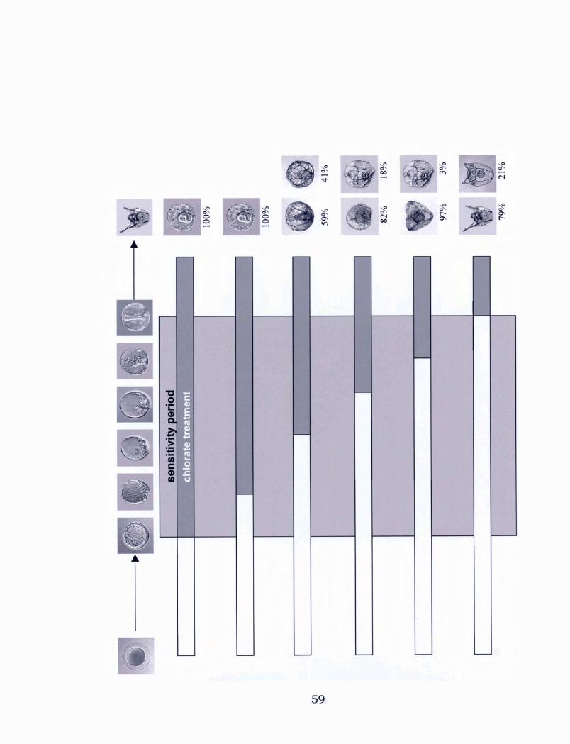

Figure 3: The effects of the removal of 30 mM chlorate a t different stages
during development. Percentages of the various morphologies observed
from each treatment are shown to the right.


2.3.3 LpSl expression in 30 mM chlorate-treated embryos
The concentration of 30 mM chlorate was used to treat embryos
and examine changes in the expression of LpSl as compared to control
embryos. Chlorate-treated embryos appeared radialized, and Hardin et
al. (1992) had previously reported a decrease in LpSl expression after
treatment of L. uariegatus embryos with the radializing agent, NiC12.
Northern blotting and RT-PCR detected a decrease in LpSl RNA per
embryo, implying a reduction in aboral ectoderm, as is illustrated by Fig.
4.
By Northern blots there appeared to be a complete loss of LpSl in
chlorate-treated embryos, but the more sensitive RT-PCR method
detected LpSl mRNA in chlorate-treated embryos, although at a much
lower level. Using RT-PCR, LpSl expression in chlorate-treated embryos
was also compared to its expression in nickel-treated embryos, as nickel
is known to cause a severe reduction in LpSl transcripts. Chlorate
appeared to reduce LpSl expression even more strongly than nickel
treatment.
The loading control for the Northern blot was the ethidium bromide
stained rRNA that was visualized prior to blotting, while for RT-PCR
polyubiquitin primers were used to confirm that equivalent amounts of
RNA were compared.

Figure 4: Expression of the LpSl gene in embryos treated with NaC103 or
NiC12. (A) Northern blot using an LpSl 32P-labeled probe. (B) rRNA
visualized with ethidium bromide to serve as a loading control for the
Northern blot. (C) RT-PCR using LpSl primers. (D) RT-PCR using
polyubiquitin primers to serve as a control for the LpSl RT-PCR. Multiple
bands are seen due to the amplification of different numbers of ubiquitin
repeat units.

control NaClO3

2.3.4 In situ hybridizations of 30 mM chlorate-treated embryos
In situ hybridizations using the LpSl antisense riboprobe were
preformed on 30 mM chlorate-treated embryos and control embryos to
determine the localization of LpSl expression. Embryos hybridized with
sense riboprobe served as controls. Normally, spicules form a t the
boundary between oral and aboral ectoderm, which is why only two
spicules form in the control embryos. Because 30 mM chlorate-treated
embryos form a ring of spicules, it was anticipated that aboral ectoderm,
which expresses LpS 1, would also be found in a ring around these
embryos. A s was expected, LpS 1 expression did encircle the embryos,
but its position along the A/V axis varied (see Fig. 5). This indicates that
the chlorate-treated embryos are radialized: that is, they lack bilateral
symmetry and an O/A axis, and a reduced amount of aboral ectoderm
appears to form.
2.3.5 PDGF rescues of 30 rnM chlorate-treated embryos
Based on the literature regarding HSPGs and their involvement in
signaling, we hypothesized that growth factors might rescue the
effects seen from chlorate treatment. Embryos were cultured for at least
96 hours in 30 mM of chlorate and 25 ng of PDGF-BB or TGF-a, or a
combination of the two growth factors. Although TGF-a did not have a

Figure 5: In situ hybridizations using an LpSl probe. (A) Control embryo
showing the normal, polarized pattern of LpSl expression in aboral
ectoderm. (B) and (C) LpSl expression patterns in chlorate-treated
embryos illustrating how the position of the ring of its expression varies
along the A/V axis. (D) Control embryo hybridized with LpSl sense
riboprobe. ( E ) Chlorate-treated embryo hybridized with LpSl sense
riboprobe.


significant effect, PDGF-BB (or PDGF-BB and TGF-a) had an effect on
chlorate-treated embryos. In these cultures, gastrulation was partially
rescued as was seen by a more elongated gut (see Fig. 6), when compared
to embryos treated only with 30 mM chlorate. The gut appeared to
attach randomly in these cultures, and apparently did not go on to
differentiate. Skeletal and ectodermal patterning did not appear to be
rescued, as multiple spicules were still seen.
Because PDGF-BB seemed to have a more dramatic rescuing effect
on chlorate-treated embryos than TGF-a, it was used in later
experiments. Chlorate and PDGF-BB were added to a culture after
fertilization and were removed at the end of the chlorate sensitivity period
(late gastrula). When this was done, the morphology of the embryos
formed was similar to control embryos. Fig. 6 illustrates that gut
differentiation and proper attachment were rescued and bilateral
spiculogenesis was rescued as well. Some abnormal branching of the
spicules was still visible when larvae were viewed under polarized light,
as can be seen in Fig. 6.

Figure 6: Rescues done with growth factors on chlorate-treated embryos.
(A) L. pictus embryo cultured in PDGF-BB and chlorate, which were
added after fertilization and left for a t least 96 hours. (B) Embryo
cultured in PDGF-BB, TGF-a, and chlorate, which were added after
fertilization and left for a t least 96 hours. (C) Control late gastrula
embryo included to illustrate normal gastrulation. (D) Embryo cultured
in PDGF-BB and chlorate, which were added after fertilization but
removed a t late gastrula stage. (E) Same embryo as in (D), but visualized
with polarized light to show spicule morphology. (E) Control pluteus
larva.


2.4 Discussion
2.4.1 The effects of chlorate on L. pictus embryos
Sea urchin embryos have been used extensively to observe the
effects of various drugs on development. With the discovery of chlorate's
effectiveness as an in vivo inhibitor of sulfation (Baeuerle and Huttner,
1986; Hortin et al., 1998), researchers began to use this drug to
investigate the role of sulfation. In this project, chlorate was used to
observe the consequences of putative inhibition of sulfation of HSPGs.
Chlorate appears to disrupt two major processes during L. pictus
development - gastrulation and patterning of the O/Ab axis, and as a
result, spiculogenesis. There are few known treatments that perturb the
O/Ab axis in sea urchin embryos; NiC12 has been shown to do this
(Hardin et al., 1992). The treatment of L. pictus embryos with 30 mM
chlorate produces a morphology which resembles that of L. variegatus
embryos treated with 0.5 mM NiC12. The most striking similarity is the
excessive number of spicules formed, their radial distribution, and their
reduced size. In both cases there are always more than two triradiate
spicules present, and these form in a ring inside the blastocoel. A s it is
clear that the O/Ab axis is disrupted, it is not surprising that LpSl/ LvSl
expression is reduced and radialized. This finding suggests that with

both chlorate and nickel treatment, excess oral ectoderm is formed a t the
expense of aboral ectoderm.
While some of the similarities between the morphology of chlorate-
treated embryos and the morphology of nickel-treated embryos are quite
evident, it is unknown whether the underlying biochemical effects of the
two treatments are connected, as the underlying action of nickel is
unknown. A s chlorate has been shown to act as an inhibitor of sulfation,
treated embryos might be expected to resemble those cultured in sulfate-
free sea water. An obvious similarity is that embryos in sulfate-free sea
water do not gastrulate properly, but often undergo a limited form of
invagination (reviewed by Wilt, 1987), as is the case in chlorate-treated
embryos. However, gastrulation occurs normally in nickel-treated
embryos, forming a fully extended archenteron, although targetting of the
tip of the stomadeum may be altered (Hardin et al., 1992).
When embryos are cultured in sulfate-free sea water, primary
mesenchyme cells ingress, but their capacity to migrate is greatly
reduced (Katow and Soursh, 198 1). Solursh et al. (1986) found a similar
effect when embryos were treated with D-P-xyloside, which inhibits the
processing of CSPGs: PMC migration was again disrupted. A s chlorate is
a more potent inhibitor of HS than CS sulfation, it is possible that CSPGs
are necessary for PMC migration while HSPGs are involved in the
specification of the location to which PMCs should migrate.
A s HSPGs are modified through both sulfation and glycosylation, it
is interesting to consider the morphology of sea urchin embryos in which
glycosylation has been disrupted. Schneider et al. (1977) treated Arbacia
punctulata sea urchin embryos with tunicamycin, an inhibitor of N-linked
glycosylation. Tunicamycin has been shown to affect both glycoprotein
and proteoglycan synthesis (Yanagishita, 1986). Treatment of A.
punctulata embryos with tunicamycin early in development (unhatched
blastula to early gastrula) seemed to have no detrimental effects on
morphology until embryos reached the early gastrula stage. At this
point, it became evident that the PMCs in the blastocoel were unable to
migrate, and that extension of the archenteron (which had invaginated
slightly) was arrested. When embryos were treated after the completion
of gastrulation, there was an arrest of spicule formation and arm
outgrowth. A s with chlorate, disruption of HSPGs by tunicamycin may
have a significant inhibitory effect on gastrulation and spiculogenesis in
sea urchin development. Although chlorate-treated embryos are capable
of forming multiple spicules, they are reduced in size. This is similar to
the effects of post-gastrula tunicamycin treatment on spicule formation.
2.4.2 The sensitive period to chlorate
Hardin et al. (1992) discovered that L. variegatus embryos are
sensitive to 0.5 mM nickel treatment from the hatched blastula stage

until early gastrula. While chlorate treatment also appears to exert its
earliest effects at hatched blastula, the end of the sensitive period of L.
pictus to chlorate extends to late gastrula. However, different aspects of
development are affected by the presence of chlorate at various stages.
A s previously mentioned, gastrulation is always inhibited by the presence
of chlorate until late gastrula stage. If chlorate is acting to disrupt the
sulfation of HSPGs, then this result suggests that HSPGs are crucial for
the elongation of the archenteron.
In the case of spiculogenesis, multiple spicules were generally
formed if chlorate was present prior to primary cell migration. Therefore,
when chlorate is added at the late mesenchyme blastula stage (when
primary mesenchyme cells have ingressed and migrated but gastrulation
has not yet begun) the majority of treated embryos form only two
spicules. HSPGs of the extracellular matrix are probably involved in
patterning migrating PMCs, so adding chlorate after PMCs have already
migrated to the sites where the first two triradiate spicules form produces
a majority of embryos with only two spicules. Transplantation
experiments using embryos treated with NiC12 performed by Armstrong et
al. (1993) reinforce this idea. They recombined normal or NiCl2-treated
PMCs with either normal or NiC12-treated PMC-less host embryos to
establish whether spicule patterning is regulated by the PMCs
themselves or the ectoderm of the blastula wall. They observed that it is

the ectoderm that is responsible for specifying the number and
arrangement of spicules made by the PMCs. They also determined that
spicule size is controlled by the ectoderm, as cultured PMCs produce
larger spicules than in vivo. Spicule size also responds to experimental
perturbation of embryo size.
Interestingly, even when chlorate is present later in development
(after primary mesenchyme cells have migrated), spicules still become
branched and twisted. This effect is seen up until the late gastrula
stage. Ettensohn and McClay (1986) injected primary mesenchyme cells
into hosts of the same sea urchin species that were at different stages in
development. They were able to demonstrate that the migration of PMCs
and the correct deposition of the carbonate skeleton were dependent
upon the stage of the host, and that the interactions of PMCs with the
blastocoel environment (especially the inner wall of the ectoderm) are
important until the late gastrula stage. Thus, chlorate may be affecting
the patterning of HSPGs in the ECM later in development such that
spicule elongation and patterning is abnormal.
2.4.3 The role of growth factors in development
There are several growth factors that have been shown to be
involved in the proper development of sea urchin embryos. Through
previous work, TGF-a, EGF, and PDGF-BB have all been implicated in
75

gastrulation and the formation of spicules (Ramachandran et al., 1993;
1995; 1997; Govindarajan et al., 1995). The addition of anti-human
PDGF-BB and TGF-a antibodies to Lytechinus embryos inhibits
gastrulation, spiculogenesis, and differentiation along the O/Ab axis
(Govindarajan et al., 1995), as does the expression of a dominant-
negative human PDGF receptor-P lacking the cytoplasmic domain
(Ramachandran et al., 1997). The most striking connection was seen
between chlorate and PDGF-BB. The morphology of embryos treated
with a synthetic peptide representing the HSPG binding sequence on
human PDGF-BB (Govindarajan et al., 1995) showed a remarkable
similarity to the morphology of embryos treated with chlorate. This
peptide presumably competed for binding sites with the endogenous
PDGF-like growth factor, thereby inhibiting PDGF signaling. Treatment
of Lytechinus embryos with this peptide inhibited gastrulation and
caused radially arranged multiple spicules to form. The rescue of
chlorate-treated embryos by PDGF-BB treatment further illustrates the
importance of PDGF signaling, which probably occurs through HSPGs,
during sea urchin development.
Katow and Aim (2002) demonstrated a connection between growth
factors and their effects on downstream proteins. They determined that
suramin, an inhibitor of growth factor receptors, inhibits the migration of
PMCs in the blastocoel and the second phase of gastrulation in H.

pulchenimus sea urchin embryos. Cotreatment with PDGF-AB
significantly restored the length of the archenteron in these suramin-
treated embryos. These rescued embryos had a remarkable similarity to
embryos that were cultured in both chlorate and PDGF-BB: the
archenteron was partially extended, but it attached randomly along the
blastocoel wall rather than extending straight toward the animal pole.
Suramin treatment was shown to decrease the incorporation of BrdU
(indicating decreased cell division), but PDGF-AB did not rescue this
effect. Thus, the partial restoration of archenteron length was not
associated with restoration of cell proliferation, but probably rather had
to do with the rearrangement of cells that occurs during convergent
extension of the archenteron. It was also suggested that PDGF signaling
might have a role in the activation of extacellular signal-regulated kinase
1 (ERK-I), as PDGF-AB treatment brought about a re-activation of the
kinase.
It is interesting that adding chlorate and PDGF to embryo cultures
produces only a partial rescue, while their removal at the end of the
chlorate sensitive period results in a nearly complete rescue. One of the
problems with introducing growth factors into the sea water culture
medium is that this is not a site-specific process; the exogenous growth
factor is presented throughout the embryo which is not representative of
the precise binding of growth factors by HSPGs that presumably

normally occurs. It is possible that an overall distribution of a low
concentration of PDGF is sufficient to partially rescue gastrulation
(archenteron extension), but that a higher concentration of PDGF a t
certain sites is necessary for full rescue, including O/Ab polarization, to
occur. Another possible explanation lies in the role of HSPGs as co-
receptors in signaling (reviewed by Perrimon and Bernfield, 2000). For
proper signaling to occur, PDGF may require binding to both its
appropriate receptor and HSPGs. Once chlorate is removed, HSPGs may
be assembled and then locally bind the exogenous PDGF that is present
in the blastocoel leading to a more complete rescue.
Ramachandran et al. (1995) used antibodies against mammalian
receptors for immunostaining to visualize Lytechinus PDGF-like
receptors. They revealed that these receptors are concentrated on the
PMCs, the gut, and most evidently on the SMCs and a t the stomadeum
(where the mouth forms after fusion with the tip of the gut). The putative
localization of PDGF-like receptors on the gut and the stomadeum
suggest a possible role of growth factor signaling in locating the correct
position for archenteron attachment. If PDGF signaling is, in fact,
involved in this process, this might explain why the archenteron attached
a t random sites on the blastocoel wall when L. pictus cultures were
treated with chlorate and PDGF for the duration of their development.

Chapter 3
The use of subtractive hybridization and 5'-RACE to
characterize a gene that is upregulated in chlorate-treated
L. pictus embryos
Introduction
Subtractive hybridization is a powerful technique for discovering
genes that are differentially expressed in two populations. Originally,
Sargent and Dawid (1983) used a primitive version of subtractive
hybridization to determine transcripts that are present in a
developmental stage or tissue, but are not present or are much less
abundant in another stage or tissue in Xenopus laeuis embryos. The
subtractive hybridization techniques currently available are extremely
sensitive to even slight changes in gene expression, making it possible to
characterize even rare messages.
Although several marker genes have been characterized in
Lytechinus embryos, in the strictest sense there are presently no RNA
markers for oral ectoderm. Because I discovered that chlorate treatment
appears to cause a decrease in the expression of LpSl, a marker gene for
aboral ectoderm (Xiang et al., 1988) and potentially causes the formation
of oral ectoderm at the expense of aboral ectoderm, it is possible that

expression of oral ectoderm-specific genes might increase in chlorate-
treated embryos. A subtractive hybridization screen might uncover
markers of oral ectoderm. Moreover, it might detect genes involved in
extension of the archenteron and specification of the O/Ab axis,
processes that are poorly understood.

Materials and Methods
3.2.1 Poly-A+ RNA isolation
Poly-A+ RNA was extracted from L. pictus embryos using a
Quickprep Micro mRNA Purification Kit (Amersham, catalogue #27-
92 55-0 1) following the manufacturer's instructions. Approximately 500
p1 of embryos were homogenized in 400 p1 of Extraction Buffer, and then
diluted with 800 p1 of Elution Buffer. This solution was centrifuged for 1
minute at 13,000 rpm to separate the two phases. The oligo(dT)-cellulose
pellet that resulted from centrifuging a tube provided by the
manufacturer and removing its supernatant was overlain with the clear
supernatant from the homogenate. The tube was inverted to resuspend
the cellulose. The mixture was incubated for 5- 10 minutes and then
centrifuged at 13,000 rpm briefly. At this point, the polyadenylated RNA
(mRNA) should be bound to the cellulose, so the supernatant was
removed and the pellet was washed three times with 1 ml of High-Salt
Buffer and three times with 1 ml of Low-Salt Buffer. The tube was
centrifuged for several seconds and the supernatant removed between
each washing. Finally, the pellet was resuspended in 300 p1 of Low-Salt
Buffer and transferred to a Microspin column. The column was briefly
centrifuged at 13,000 rpm, and it was placed into a sterile 1.5 ml

microfuge tube. To elute the poly-A+ RNA, 200 pl of pre-warmed Elution
Buffer was added to the resin bed and the column was centrifuged for 5
seconds. In order to increase the yield of mRNA, the elution was
repeated. A volume of 16 p1 was removed to be used for quantifying the
mRNA using a Ultrospec I11 (Pharmacia) spectrophotometer, and then 10
pl of glycogen, 38.4 pl of potassium acetate and 1 ml of 95% ethanol was
added to the tube. This solution was placed a t -20•‹C for 30 minutes to
precipitate the mRNA. The poly-A+ RNA was pelleted a t 4OC a t 14,000
rpm for 5 minutes. The supernatant was removed, and the pellets were
allowed to air dry before being redissolved in DEPC-treated water.
3.2.2 Subtractive hybridization
Clontech's PCR-Select cDNA Subtraction Kit (catalogue #K1804- 1)
was used as recommended by the manufacturer. This protocol allows
the comparison of two populations of mRNA and the cloning of portions
of genes that are expressed a t different levels in the populations. As the
protocol is complex, a flowchart is shown to illustrate the steps taken
during this procedure. Note that the reference cDNA is referred to as the
"driver" cDNA, and the sample with upregulated transcripts is known as
the "tester" cDNA.
The significance of the Clontech system is that only target
molecules are exponentially amplified. After digestion of both tester and
82

driver cDNA with the blunt cutter Rsal, the tester is subdivided into two
populations which are each ligated with a different cDNA adaptor
(Adaptor 1 and Adaptor 2R). Two hybridizations are performed following
ligation. In the first, an excess of driver is added to each sample tester;
the samples are then heat denatured and allowed to anneal. Already, the
pool is enriched for differentially expressed sequences a s some tester
cDNA cannot reanneal with driver cDNA and remains single stranded. In
the second hybridization, the two primary hybridization samples are
mixed together but are not denatured. At this point, the single stranded
tester cDNA from both primary subtractions is able to reassociate, and
double stranded hybrids with different ends (due to the two adaptors) are
formed. After filling in the ends of the double stranded cDNAs, the
differentially expressed tester sequences will be the only ones that
contain annealing sites for the two nested primers. cDNAs with the same
adaptor on both ends undergo suppression PCR. In this case, during
each primer annealing, the formation of a secondary structure
resembling a pan handle (due to annealing of the adaptors on the same
strand) is kinetically favored to primer annealing.
In this project, cDNA made from chlorate treated embryos was
used as both tester and driver cDNA. In this way, it was possible isolate
genes that are both upregulated and downregulated in chlorate treated
embryos.

Figure 7: Flowchart illustrating the steps taken to carry out a subtractive
hybridization.

cDNA synthesis
mRNA from two populations is used to make tester and driver cDNA
+ Rsal digestion
Tester and driver cDNA are digested to produce shorter, blunt-ended ds cDNA which is optimal for subtraction
Adaptor ligation
Two different adaptors (Adaptor 1 and Adaptor 2R) are ligated to only tester cDNA
+ First hybridization
An excess of driver cDNA is added to tester cDNA, to allow hybridization and enrichment of differentially expressed sequences
* Second hybridization
I Driver cDNA is added to further enrich for differentially expressed sequences I
First PCR amplification
Differentially expressed sequences are amplified exponentially
+ Second PCR amplification
Differentially expressed sequences are further enriched, and background is reduced

3.2.3 Preparing cDNA probes
DIG-labeled cDNA probes were prepared through the reverse
transcription of cytoplasmic RNA using DIG-labeled dUTP (Boehringer
Mannheim). Before beginning the reaction, 1.5 pg of RNA were
denatured a t 65•‹C for 15 minutes and placed on ice for 5 minutes. For
the reverse transcription, the following were added to a sterile microfuge
tube: 3 p1 of 10 x first strand buffer, 1.5 p1 of RNase inhibitor, 3 p1 of 5
mM dNTP mixture (dATP, dCTP, and dGTP only), 6 p1 of 0.65 mM dTTP, 6
p1 of 0.35 mM DIG-dUTP, 3 p1 of oligo-dT, and the heat-denatured RNA.
The volume was made u p to 28 p1 with DEPC water, and then 2 p1 of
AMV-RT was added for a final volume of 30 p1. The tube was briefly
centrifuged, and the reaction was left to proceed for 2 hours at 37•‹C.
After this time, RNase A was added to the tube to degrade the template
RNA at 37•‹C for 15 minutes.
3.2.4 Plasmid DNA blotting
The plasmid samples were first heated to 95OC for 10 minutes and
snap-cooled on ice to denature the DNA. Each sample was spotted onto
a positively-charged membrane and cross-linked with UV light. The
membrane was pre-hybridized a t 65OC for 30-60 minutes, and
hybridridized using cDNA probes made from control embryos. The
procedure followed for detection is the same as for RNA blots. The same
membrane was stripped and reprobed using cDNA probes from chlorate
treated embryos to enable a comparison of expression levels.

3.2.5 Preparation of polyubiquitin and CS 1 - 1.13 DIG-labeled riboprobes
DIG-labeled riboprobes were made using the ubiquitin and pCR1-
1.13 plasmids. The ubiquitin plasmid was digested with EcoRl to
linearize the DNA, while either Hind111 or BamHI was used with the
pCR1.1-13 plasmid. Both riboprobes were transcribed using T7 RNA
polymerase. The transcription was carried out as described in section
2.2.1 1 of the previous chapter.
3.2.6 DIG Northern blotting
RNA was resolved by electrophoresis as was described in section
2.2.5. The RNA gel was blotted overnight onto a positively charged nylon
membrane (Boehringer Mannheim) in 20X SSC (3.0 M NaCl and 0.3 M
Sodium citrate, pH 7.0) using the unidirectional capillary method. After
it had been transferred, RNA was UV cross-linked to flur it onto the
membrane.
The membrane was then pre-hybridized in pre-warmed
hybridization buffer [which consists of 50% deionized formamide, 5 x
SSC, 0.1% (w/v) N-lauroylsarcosine, 0.02% (v/v) SDS, 2% blocking
reagent (Boehringer Mannheim)], for 30-60 minutes at 55•‹C. After the
pre-hybridization, the membrane was hybridized with denatured DIG-
labeled riboprobe at a concentration of 100 ng/ml in pre-warmed

hybridization buffer overnight at 55OC. The next day, the membrane was
washed twice with low stringency wash buffer (2 x SSC plus 0.1% SDS)
at room temperature for 10 minutes each time, twice with pre-warmed
high stringency wash buffer (0.1 x SSC plus 0.1% SDS) at 55OC for 30
minutes each time.
To detect the labeled riboprobe, the membrane was washed briefly
in maleic acid buffer [O. 1 M maleic acid (Sigma) and 0.15 M NaC1, pH
7.51 plus 0.3% (v/v) Tween-20 (Sigma)] for several minutes, then
incubated in 1X blocking solution [1% (w/v) blocking reagent in maleic
acid buffer] for 30-60 minutes. After blocking, the membrane was
incubated in anti-DIG-alkaline phosphatase conjugate diluted 1: 10,000
in blocking solution for 30-60 minutes. To remove unbound antibody,
the membrane was washed twice in maleic acid buffer plus 0.3% (v/v)
Tween-20 for 30 minutes each time. The membrane was equilibrated in
detection buffer (0.1 M Tris-C1 and 0.1 M NaC1, pH 9.5), and it was
incubated for 15 minutes in CSPD, a chemiluminescent alkaline
substrate (Boehringer Mannheim) diluted 1: 100 in detection buffer. After
the membrane was removed from the CSPD solution, it was wrapped up
in Saran Wrap to keep it from drying out. The luminescent signal was
enhanced by incubating the membrane at 37•‹C for 15 minutes. The
membrane was placed onto Hyperfilm ECL (Amersham) for 5 seconds to
30 minutes, and the film was developed.

3.2.7 Removing the nboprobe from Northern blots for rehybridization
To rehybridize a membrane with a different probe, it was necessary
to remove the previously used DIG-labeled riboprobe. Therefore, 100 ml
of 0.1% SDS was heated until it was almost boiling, and was poured onto
the membrane. The membrane was incubated in this solution for 10
minutes on a rocker without further heating. After the membrane had
been stripped, it was washed for 5 minutes in washing buffer at room
temperature. At this point, the membrane was ready to be prehybridized
for the next hybridization.
3.2.8 Depurination and denaturation of DNA gel before blotting
DNA was resolved by electrophoresis as was described in section
2.2.8. Before blotting, DNA was depurinated and denatured to make
transfer and hybridization easier. Depurination is achieved with a
controlled acid treatment, and is optional for DNA fragments smaller
than 10 kb in size. I chose to use this treatment to increase transfer
efficiency.
The gel was submerged in 250 mM HC1 with shaking for 10
minutes, and was rinsed with distilled water. To denature the DNA, the
gel was submerged in denaturation solution (0.5 N NaOH and 1.5 M
NaC1) twice, for 15 minutes each time. The gel was again rinsed with
water before its pH was neutralized by being submerged in neutralization

buffer (0.5 M Tris-HC1, pH 7.5 and 3 M NaC1) twice, for 15 minutes each
time.
DNA was blotted in the same was as RNA, and DNA blots using
DIG-labeled probe were treated in the same way as RNA blots using DIG-
labeled probe (see section 3.2.6) except that any water used with DNA
was not DEPC-treated.
Clontech's SMART RACE cDNA Amplification Kit (catalogue
#K18 1 1- 1) was used as recommended by the manufacturer, with some
deviations. Briefly, 1 pg of poly A+ RNA prepared from L. pictus a t
pluteus larva stage was combined with 1 pl of 10 pM 5'-cDNA Synthesis
Primer, 1 pl of 10 pM SMART I1 oligo (5'-AAG CAG TGG TAA CAA CGC
AGA GTA CGC GGG-37, and sterile water to bring the volume u p to 5 p1.
The reaction mixture was heated to 70•‹C and cooled on ice. The first
strand synthesis was carried out by adding 2 pl of 5 x first strand buffer,
1 pl of 20 mM DTT, 1 pl of 10 mM dNTP mix, and 1 pl of Superscript I1
MMLV reverse transcriptase (Invitrogen, catalogue # 18064-022) to the
reaction mixture and incubating the tube for 1.5 hours a t 42OC in an air
incubator to reduce evaporation. The Superscript I1 MMLV-RT possesses
a terminal transferase activity that adds 3-5 dC residues to the 3' end of
the first strand cDNA. This allows the 3' dGs of the SMART I1 oligo to
anneal a t the end of the template mRNA and provide template for further
extension. A s a positive control for the cDNA synthesis, the kit contained
control human placental total RNA to be used to make RACE-Ready
cDNA. After the first strand synthesis, the first strand reaction product
90

was diluted with Tricine-EDTA buffer (10 mM Tricine-KOH, pH 8.5 and 1
mM EDTA) to 250 p1 and heated to 72•‹C for 7 minutes to destroy MMLV-
RT activity.
5' RACE was carried out using several gene specific primers (GSP)
to extend the 5' end of the cloned cDNA. The various GSPs also served
as nested primers as the RACE PCRs appeared to generate clearer bands
when a secondary PCR was done. For the primary PCR, a master mix
was made containing 17.25 p1 PCR-grade water, 2.5 p1 10 x Advantage 2
PCR Buffer, 0.5 p1 of 10 mM dNTP mix, and 0.5 pl50 x Advantage 2
Polymerase Mix per PCR being done. The 5' RACE sample comprised of
20.75 p1 of this master mix combined with 1.25 p1 of 5'-RACE-Ready
cDNA, 2.5 p1 of 10 x universal primer mix (UPM), and 0.5 pl of GSP. A
positive control was done on cDNA made from the control human
plactental RNA using a 10 pM 5'-RACE TFR Primer. The negative
controls used only the GSP or only the UPM.
After the primary PCR, the product was purified using the
QIAquick PCR Purification Kit (Qiagen) according to the manufacturer's
instructions. During the purification, DNA was eluted using 50 pl of
water which was diluted into 200 p1 of Tricine-EDTA buffer. 2.5 p1 of
diluted primary PCR product was used for a nested PCR using 1 pl of the
nested universal primer (NUP) and a nested GSP.
The PCR reactions were carried out following a hotstart PCR
protocol consisting of 30 cycles of 5 seconds at 94•‹C for the
denaturation, 10 seconds at 66•‹C for the annealing, and 3 minutes at
70•‹C for the elongation. All PCR reactions were done using a Perkin-
Elmer GeneAmp Systems 2400 thermal cycler. The various GSPs used

for the 5'-RACE reactions were designed using Oligo 4.1 and were
purchased from Sigma Genosys. They are shown below:
GSP 1 5'-CCT ACM CCA CTC TTG CAA ATT CAT TC-3' (M = A or C)
GSP 2 5'-CTG GCA GTA AAA CCT ATC GCT ATG T-3'
GSP 3 5'-TAA AGA ATG CTG TTG ACC TAC CA-3'
GSP 4 5'-CCT TTA CCA GCT TCT TCG TTC GC-3'
3.2.10 Cloning of subtractive hybridization and 5'-RACE products
The various products from the subtractive hybridization and 5'-
RACE reactions were cloned into the pCR 2.1 vector using the Original
TA Cloning Kit (Invitrogen, catalogue #45-0046) according to the
manufacturer's directions. The TA cloning system takes advantage of the
activity of Taq polymerase that adds a single dA to the 3' ends of PCR
products; therefore the vector contains 3'-overhangs of dT residues.
PCR products were first ligated into the pCR 2.1 vector using
components from the kit. The reaction mixture contained 1 p1 of 10 X
Ligation Buffer, 2 p1 of pCR vector (25 ng/pl), 1 p1 of fresh PCR product
(approximately 10 ng), 5 p1 of sterile water, and 1 pl of T4 DNA Ligase.
The reaction was incubated a t 14•‹C overnight.
The next day, a vial of One Shot cells was thawed on ice, and 4 p1
of the ligation mixture was pipetted into the vial and stirred gently. The
vial was incubated on ice for 30 minutes and then heat shocked for 30
seconds in a 42•‹C water bath. 250 p1 of SOC medium was added to the
vial before putting it into a 37OC shaking incubator a t 225 rpm for
exactly 1 hour. The vial was placed on ice to discourage more bacterial
growth. TB plates containing 100 pg/ml ampicillin were prepared by
92

spreading 40 p1 of 40 mg/ml X-Gal and 40 p1 of 100 mM IPTG onto their
surfaces. Varying volumes of cells (generally 50 p1- 100 p1) were then
plated onto them. Plates were incubated a t 37•‹C for a t least 18 hours;
they were then incubated a t 4OC to aid with blue colour development in
negative colonies. Plasmid DNA extractions were performed, and the
DNA was digested with EcoRI to verify that an insert of the expected size
was present. All sequencing was done at the University of Calgary.

3.3 Results
3.3.1 Subtractive hybridizations of control and chlorate-treated ernb y o
Subtractive hybridizations were performed using cDNA made from
control and chlorate-treated L. pictus embryos to determine whether any
other genes besides LpSl experience a change in expression. Both
forward and reverse subtractive hybridizations were done such that
decreases and increases in gene expression of chlorate-treated embryos
would be detected.
Once the cDNAs were subtracted, the products were amplified by
PCR. These subtraction products were then cloned, and approximately
20 colonies were chosen a t random from both the forward and reverse
reaction for dot blotting using cDNA probes made from chlorate-treated
and control embryos.
Fig. 8 shows the results of the dot blots done to compare the
various expression levels. In the dot blots for the forward subtraction
(not shown), which represents genes that are potentially downregulated
in chlorate-treated embryos, there were no substantial differences in
gene expression observed. However, in the dot blots for the reverse
subtraction, which represents genes that are upregulated in chlorate-
treated embryos, there were two cDNAs that had noticeable differences in
expression. I chose to focus on the cDNA with the largest apparent
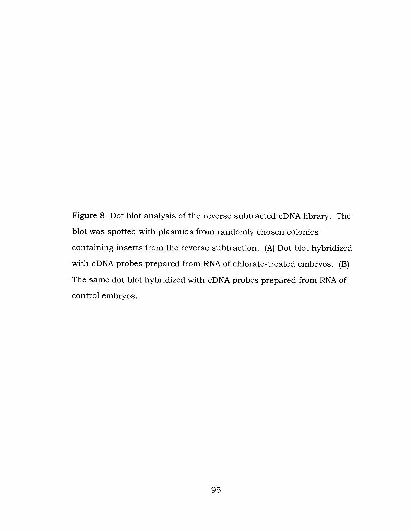
Figure 8: Dot blot analysis of the reverse subtracted cDNA library. The
blot was spotted with plasmids from randomly chosen colonies
containing inserts from the reverse subtraction. (A) Dot blot hybridized
with cDNA probes prepared from RNA of chlorate-treated embryos. (B)
The same dot blot hybridized with cDNA probes prepared from RNA of
control embryos.


increase in expression, which is denoted by an arrow. From this point
on, this cDNA will be referred to as pCR1-I. 13. Most cDNAs in the
subtracted library have not been characterized.
3.3.2 Verification of su btractive hybridization
A s a test of the effectiveness of the subtractive hybridization, the
pool of amplified cDNA products from the forward and reverse
subtractions were run on a gel, blotted and probed with LpSl riboprobe.
LpSl riboprobe was used because its expression was known to decrease
as a result of chlorate treatment. It was predicated that the LpSl probe
would recognize a band in the forward subtraction pool of approximately
1 kb in size due to the RsaI digestion used in the subtractive
hybridization protocol. Fig. 9 confirms that the subtractive hybridization
effectively detected the downregulation of LpSI.
3.3.3 Northern blot with C S 1 - 1.13 riboprobe
To verify the difference in gene expression of pCR1-I. 1 3 seen in the
cDNA dot blots, a Northern blot hybridization was performed using
mRNA from control and chlorate-treated embryos and pCR1-1.13
riboprobe. The blot was stripped and reprobed with polyubiquitin
riboprobe, which served as a loading control. Again, a significant
upregulation of pCRl-1.13 was evident in chlorate-treated embryos as
compared to control embryos (see Fig. 10).

Figure 9: Southern blot using pooled forward and reverse subtracted
cDNA digested with RsaI, and hybridized with an LpSl riboprobe.

forward
LpSl cDNA fragment -1 kb 0
reverse

Figure 10: Expression of CS 1-1.13 in chlorate-treated embryos. (A)
Northern blot hybridized with CS 1-1.13 riboprobe. (B) Northern blot
hybridized with polyubiquitin, used as a loading control.

control C103

3.3.4 Sequence obtained from 5'-RACE
Approximately 1.4 kb of pCR1- 1.13 sequence was obtained by
using 5'-RACE, as is shown in Fig. 11. Because the cloned sequences all
overlapped somewhat, this 1.4 kb sequence was verified several times.
The original clone from the subtractive hybridization contained a poly-A+
tail, indicating that this clone contained the 3' end of the mRNA. There
is no long open reading frame (ORF); thus, the sequence obtained so far
appears to be part of the 3' untranslated region (UTR) of the mRNA.
3.3.5 Northern blot of various developmental stages using CS 1-1.13
riboprobe
In order to see the temporal expression levels of CS 1-1.13 during
various stages of development, an ontongeny Northern blot was done,
with rRNA being used as a loading control (see Fig. 12). It appears that
CS 1-1.13 is expressed throughout embryonic development, reaching a
peak in expression at the mesenchyme blastula stage, and declining
during the later stages of development.

Figure 1 1 : The 1.4 kb sequence of CS 1-1.13 compiled from fragments
that were obtained through 5'-RACE, with highlighted GSPs. The
sequence reads 5' to 3' starting in the top left corner.

IGene Specific Primer 11
Gene Specific Primer 2

Figure 12: Expression of CS 1-1.13 at various developmental stages. (A)
Northern blot showing CS 1-1.13 expression. E : egg, HB : hatched
blastula, MB : mesenchyme blastula, MG : mid gastrula, PL: pluteus
larva. (B) Ethidium bromide stained rRNA, used as a loading control.


3.4 Discussion
3.4.1 Subtractive hybridization using control and chlorate-treated cDNA
The forward and reverse subtractive hybridizations gave some
significant results. The 1 kb LpSl cDNA fragment that would be
recognized by the 0.44 kb sequence used to make LpSl riboprobe was
shown to be present in the forward subtraction pool, but absent in the
reverse subtraction pool, which was a confirmation of the validity of the
procedure. The forward subtraction clones that were chosen did not
show any significant differences in gene expression when they were
probed with control and chlorate-treated cDNA probes; however, there
were two cloned plamids in the reverse subtraction that illustrated an
obvious upregulation of gene expression when they were hybridized with
chlorate-treated cDNA probes as compared to control cDNA probes.
I chose to focus my work on the plasmid that demonstrated the
largest difference in expression, which was named CS 1-1.13. The clone
obtained from the subtractive hybridization contained the poly-A+ tail
and no open reading frame, suggesting that this was the 3'-UTR of the
mRNA.
3.4.2 5'-RACE sequence
With 5'-RACE, it was possible to obtain a total of approximately 1.4
kb of sequence. This sequence was used in BLAST searches on both the
NCBI database and the sea urchin genome database (S. purpuratus), but
107

no significant matches were found. It appears that the sequence I have
gathered up until this point does not contain an open reading frame
(ORF). This makes it extremely difficult to compare the CS 1-1.13
sequence with anything in the databases as UTRs vary significantly
between species.
This project will be continued in order to acquire sequence from
the ORF that can be used to determine which gene is upregulated as a
result of chlorate treatment.
3.4.3 The temporal expression of CS 1 - 1.13
The expression level of CS 1-1.13 appears to change during various
stages of L. pictus embryo development, making the gene potentially
interesting for studying development. It seems that the CS 1-1.13 mRNA
is present at all stages, although its levels are extremely low in eggs and
early embryos. Expression appears to peak at the mesenchyme blastula
stage and then decline until the pluteus stage. It is interesting that the
period of peak expression corresponds to the period of sensitivity of the
embryos to chlorate. The gene may have a role in oral ectoderm
specification or differentiation, or gastrulation.

Chapter 4
Conclusions and Future Work
Through this project I set out to address the following questions:
1) If chlorate has an effect on L. pictus embryos, what is this effect with
regard to morphology?
2) When is the sensitive period of L. pictus embryos to chlorate?
3) Can the effects of chlorate on L. pictus embryos be rescued by
treatment with H S or growth factors?
4) Are there any genes that experience either a downregulation or
upregulation due to chlorate treatment?
Using the results that have been presented in this thesis, several
conclusions can be drawn.
Chlorate has several clear effects on the development of L. pictus
embryos, and the severity of these effects increase with higher
concentrations of chlorate. At 30 mM chlorate, embryos are unable to
complete gastrulation and normal O/Ab axis patterning is disrupted,
resulting in multiple radially distributed spicules.

L. pictus embryos appear to be sensitive to chlorate for an extensive
period of time. Embryos first become sensitive at the hatched blastula
stage, which was confirmed by treating embryos that had had their
fertilization envelopes removed. They remain sensitive until the late
gastrula stage, after which the addition of chlorate has no effect on
development. The extent to which gastrulation and spiculogenesis are
affected depends on when chlorate is present during development.
It was possible to rescue the effects of chlorate treatment by also
adding PDGF-BB to embryo cultures. The presence of chlorate and
PDGF throughout development produced only a partial rescue, as the
archenteron was more elongated in this case. However, the removal of
chlorate and PDGF-BB at the end of the chlorate sensitive period brought
about a nearly full rescue. Addition of HS to sea waterdid not seem to
rescue the effects of chlorate, although it is unclear whether this was due
to the inability of HS to penetrate into the blastocoel or the inability of
HS to actually rescue chlorate-treated embryos.
Through the use of subtractive hybridization, gene expression
variations in chlorate-treated and control embryos were studied, and a
gene whose expression is upregulated in chlorate-treated embryos was
discovered. Although it is unknown what protein encoded by this
particular gene and how this protein is involved in development, there

appears to be a peak in expression of CS 1-1.13 during the mesenchyme
blastula stage.
4.2 Future Work
There are several further investigations that can be carried out to
further our understanding of the effects of chlorate.
Although we assume that the effects of chlorate are seen due to an
inhibition of HSPG sulfation, this has not been verified. It is probable
that chlorate is acting in this manner based on previous research
reviewed in Chapter 1. This could be verified by doing experiments using
35s to label HSPGs in control and chlorate-treated embryos. Similarly, it
would be beneficial to know whether HS has the capacity to rescue the
effects of chlorate. Although HS added to chlorate-treated sea urchin
embryo cultures did not provide a rescue, it is possible that this is due to
the inability of HS to cross the blastocoel wall. Thus, injection of HS into
the blastocoel might bring about a rescue.
It is quite interesting that chlorate-treated embryos form multiple
spicules, and that embryos that are exposed to chlorate after PMC
migration form branched and twisted spicules. There appears to be
some patterning signal present on the blastocoel wall, and HSPGs may
have a role in providing this signal. It would be beneficial to be able to
visualize the pattern of HSPGs binding PDGF, perhaps by
immunostaining for PDGF or core proteins in the blastocoel. This may
provide some insight into how proper growth factor binding to HSPGs
mediates normal development.

With respect to the O/Ab axis, due to the expression pattern of
LpSl and spicule formation in chlorate-treated embryos, it is clear that
the normal patterning of the O/Ab axis is perturbed. However, it would
be useful to verify that LpC2 actin, another aboral ectoderm specific gene,
is also affected in a similar manner by chlorate treatment.
The DNA sequence of the cDNA or gene corresponding to CS I -
1.13, is not finished. It would be interesting to determine the identity of
this gene, and how it is involved in normal development. This could be
accomplished through a continuation of the 5'-RACE that was originally
carried out to determine its sequence or through hybridization screening
of a cDNA library for full length inserts. Once CS 1-1.13 is characterized,
the potential for further experiments would increase.
Presumably, several genes that experience a difference in
expression due to chlorate treatment were discovered through
subtractive hybridization. While I chose to focus on a gene that
experienced a large upregulation in chlorate-treated embryos, there is at
least one other gene that is certainly upregulated and several others that
appear to be upregulated. In addition, there are probably genes that
become downregulated with chlorate treatment, as is the case with LpSl,
although they were not among the few subtracted cDNA clones randomly
chosen for characterization. A hybridization screen using a cDNA library
for L. pictus would be a high-throughput method for identifying other
genes that are up- or down-regulated in response to chlorate treatment.
Such genes have potentially interesting roles in axis specification,
ectoderm differentiation, gastrulation, and ECM function.
Joseph Yost (University of Utah) has offered our lab polyclonal
antibodies against the HSPG syndecan-2 core protein and two of its
112

phosphorylated forms. His lab has found a connection between left-right
patterning in Xenopus embryos and the phosphorylation state of
syndecan-2 (Kramer et al., 2002). Although chlorate has only been
shown to inhibit sulfation of HSPGs (and not phosphorylation), it would
be interesting to immunostain normal and chlorate-treated embryos to
assess the distribution of syndecan-2 and its state of phosphorylation.
The putative inhibition of sulfation that may be occurring in L. pictus
embryos due to the addition of chlorate may also affect localization and
modification of HSPGs. I t will be interesting to know the distribution of
other HSPG and CSPG core proteins in urchin embryos. Creation of
appropriate reagents will be facilitated by the completion of the sea
urchin genome project.

References
Angerer, L.M. and Angerer, R.C. (2000). Animal-vegetal axis patterning mechanisms in the early sea urchin embryo. Dev. Biol. 218, 1- 12.
Angerer, L.M. and Angerer, R.C. (2003). Patterning the sea urchin embryo: Gene regulatory networks, signaling pathways, and cellular interactions. Cum. Top. Dev. Biol. 53: 159- 198.
Angerer, R.C. and Davidson, E.H. (1984). Molecular indices of cell lineage specification in the sea urchin embryo. Science 226, 1 153- 1 160.
Angerer, L.M., Oleksyn, D.W., Levine, A.M., Li, X., Klein, W.H., and Angerer, R.C. (200 1). Sea urchin goosecoid function links fate specification along the animal-vegetal and oral-aboral embryonic axes. Development 128, 4393-4404.
Armstrong, N., Hardin, J . , and McClay, D.R. (1993). Cell-cell interactions regulate skeleton formation in the sea urchin embryo. Development 119, 833-840.
Baeuerle, P.A. and Huttner, W.B. (1986). Chlorate - a potent inhibitor of protein sulfation in intact cells. Biochem. Biophys. Res. Commun. 141, 870-877.
Bernfield, M., Gotte, M., Park, P.W., Reizes, O., Fitzgerald, M.L., Lincecum, J., and Zako, M. (1999). Functions of cell surface heparan sulfate proteoglycans. Annu. Rev. Biochem. 68, 729-777
Brandhorst, B.P., Filion, M., Nisson, P.E., and Crain, W.R. (199 1). Restricted expression of the Lytechinus pictus Spec 1 gene homologue in reciprocal hybrid embryos with Strongylocentrotus purpuratus. Dev. Biol. 144, 405-4 11.
Brandhorst BP, Klein WH. (2002). Molecular patterning along the sea urchin animal-vegetal axis. Int. Rev. Cytol. 2 13, 183-232.
Bullock, S.l., Fletcher, J.M. Beddington, R.S.P., and Wilson, V.A. (1998). Renal agenesis in mice homozygous for a gene trap mutation in the gene encoding heparin sulfate 2-sulfotransferase. Genes Dev. 12, 1894- 1906.

Cameron, R.A., Hough-Evans, B.R., Britten, R.J., and Davidson, E.H. (1987). Lineage and fate of each blastomeres of the eight-cell sea urchin embryo. Genes Dev 1, 75-85.
Carpenter, C.D., Bruskin, A.M., Hardin, P.E., Keast, M.H., Anstrom, J., Tyner, A.L., Brandhorst, B.P., and Klein, W.H. (1984). Novel protein belonging to the troponin C superfamily are encoded by a set of mRNA in the sea urchin embryo. Cell 36, 663-67 1.
Coffman, J.A. and Davidson, E.H. (200 1). Oral-aboral axis specification in the sea urchin embryo. I. Axis entrainment by respiratory asymmetry. Dev. Biol. 230, 18-28.
Croce, J . , Lhomond, G., and Gache, C. (2003). Coquillette, a sea urchin T-box gene of the Tbx2 subfamily, is expressed asymmetrically along the oral-aboral axis of the embryo and is involved in skeletogenesis. Mech. Dev. 120, 561-572.
Davidson, E.H., Cameron, R.A., and Ransick, A. (1998). Specification of cell fate in the sea urchin embryo: summary and some proposed mechanisms. Development 125, 3269-3290.
Delehedde, M., Deudon, E., Boilly, B., and Hondermarck, H. (1996). Heparan sulfate proteoglycans play a dual role in regulating fibroblast growth factor-2 mitogenic activity in human breast cancer cells. Exp. Cell Res. 229, 398-406.
Dhoot, G.K., Gustafsson, M.K., Ai, X., Sun, W., Standiford, D.M., and Emerson, C.P. J r . (2001). Regulation of Wnt signaling and embryo patterning by an extracellular sulfatase. Science 293, 1663- 1666.
Emily-Fenouil, F., Ghiglione, C., Lhomond, G., Lepage, T., and Gache, C. (1998). GSK3betalshaggy mediates patterning along the animal- vegetal axis of the sea urchin embryo. Development 125, 2489-2498.
Esko, J . and Lindahl, U. (2001). Molecular diversity of heparan sulfate. J. Clin. Invest. 108, 169- 173.
Ettensohn, C.A. (1985). Gastrulation in the sea urchin embryo is accompanied by the rearrangement of invaginating epithelial cells. Dev Biol. 112, 383-390.
Ettensohn, C.A. and McClay, D.R. (1986). The regulation of primary mesenchyme cell migration in the sea urchin embryo: transplantations of cells and latex beads. Dev. Biol. 1 17, 380-39 1.

Ettensohn, C.A. and Sweet, H.C. (2000). Patterning the early sea urchin embryo. C u r . Top. Dev. Biol. 50, 1-44.
Farley, J.R., Nakayama, G., Cryns, D., and Segal, I.H. (1978). Adenosine triphosphate sulfurylase from Penicillium chrysogenum equilibrium binding, substrate hydrolysis, and isotope exchange studies. Arch Biochem Biophys. 185, 376-390.
Fjeldstad, K., Pedersen, M.E., Vuong, T.T., Kolset, S.O., Nordstrand, L.M., and Prydz, K. (2002). Sulfation in the Golgi lumen of Madin- Darby canine kidney cells is inhibited by brefeldin A and depends on a factor present in the cytoplasm and on Golgi membranes. J. Biol. Chem. 277, 36272-36279.
Gan, L., Mao, C.A., Wikramanayake, A., Angerer, L.M., Angerer, R.C., and Klein, W .H. (1995). An orthodenticle-related protein from Strongylocentrotus purpuratus. Dev. Biol. 167, 5 17-528.
George, J.M., Seid, C.A., Lee, H., Tomlinson, C.R. (1996). Two distinct forms of USF in the Lytechinus sea urchin embryo do not play a role in LpSl gene inactivation upon disruption of the extracellular matrix. Mol. Reprod. Dev. 45, 1-9.
Govindarajan, V., Ramachandran, R.K., George, J.M., Shakes, D.C., and Tomlinson, C.R. (1995). An ECM-bound, PDGF-like growth factor and a TGF-a-like growth factor are required for gastrulation and spiculogenesis in the Lytechinus embryo. Dev. Biol. 172, 54 1-55 1.
Greve, H., Cully, Z., Blumberg, P., and Kresse, H. (1988). Influence of chlorate on proteoglycan biosynthesis by cultured human fibroblasts. J. Biol. Chem. 263, 12886-12892.
Gritli-Linde, A., Lewis, P., McMahon, A.P., and Linde, A. (200 1). The whereabouts of a morphogen: Direct evidence for short- and graded long-range activity of hedgehog signaling peptides. Dev. Biol. 236, 364-386.
Gross, J.M., and McClay, D.R. (2001). The role of Brachyury (T) during gastrulation movements in the sea urchin Lytechinus variegatus. Dev. Biol. 239, 132-147.
Gross, J . M . , Peterson, R.E., Wu, S.Y., and McClay, D.R. (2003). LvTbx213: a T-box family transcription factor involved in formation

of the oral/aboral axis of the sea urchin embryo. Development 130, 1989- 1999.
Guimon, S., Maccarana, M., Olwin, B.B., Lindahl, U., and Rapraeger, A.C. (1993). Activating and inhibitory heparin sequences for FGF-2 (basic FGF). Distinct requirements for FGF- 1, FGF-2, and FGF-4. J. Biol. Chem. 268, 23906-239 14
Hardin, J . , Coffman, J.A., Black, S.D., and McClay, D.R. (1992). Commitment along the dorsoventral axis of the sea urchin embryo is altered in response to NiC12. Development 1 16, 67 1-685.
Hardin, J . and McClay, D.R. (1990). Target recognition by the archenteron during sea urchin gastrulation. Dev. Biol. 142, 87- 105.
Hardin, P.E., Angerer, L.M., Hardin, S.H., Angerer, R.C., and Klein, W.H. (1988). Spec2 genes in Strongylocentrotus purpuratus: Structure and differential expression in embryonic aboral ectoderm. J. Mol. Biol. 202, 4 17-43 1.
Hardin, S.H., Carpenter, C.S., Hardin, P.E., Bruskin, A.M., Klein, W.H. (1985). Structure of the Spec 1 gene encoding a major calcium- binding protein in the embryonic ectoderm of the sea urchin Strongylocentrotus purpuratus. J. Mol. Biol. 186, 243-255.
Hardin, S.H., Keast, M. J. , Hardin, P.E., Klein, W.H. (1987). Use of consensus oligonucleotides for detecting and isolating nucleic acids encoding calcium binding domains of the troponin C superfamily. Biochemistry 26, 35 18-3523.
Har-el, R. and Tanzer, M.L. (1993). Extracellular matrix 3: Evolution of the extracellular matrix in invertebrates. FASEB J. 7 , 1 1 15- 1 123.
Hay, E.D. (1989). Extracellular matrix, cell skeletons, and embryonic development. Am. J. Med. Genet. 34, 14-29.
Henry, J . J . , Klueg, K.M., and Raff, R.A. (1 992). Evolutionary dissociation between cleavage, cell lineage and embryonic axes in sea urchin embryos. Development 114, 93 1-938.
Herbst, C. (1892). Experimentalle untersuchungen iiber den einfluss der veranderten chemischen zusammensetzung des ungebenden mediurns auf die entwicklung der thiere I teil. Versuche an seeigeleiern. 2. Wiss. 2001. 55, 446-518.

Horstadius, S. (1939) The mechanics of sea urchin development, studied by operative methods. Bio. Rev. Cambridge Phil. Soc. 14, 132- 179.
Horstadius, S. (1973) Experimental Embryology of Echinoderms. Oxford: Clarendon Press.
Horstadius, S. and Gustafson, T. (1954). The effect of three antimetabolites on sea urchin development. J. Embryol. Exp. Morph. 2, 2 16-226.
Hortin, G.L., Schilling, M., and Graham, J.P. (1988). Inhibitors of the sulfation of proteins, glycoproteins, and proteoglycans. Biochem. Biophys. Res. Commun. 150, 342-348.
Howard, E.W., Newman, L.A., Oleksyn, D.W., Angerer, R.C., Angerer, L.M. (2001). SpKrl: a direct target of p-catenin regulation required for endoderm differentiation in sea urchin embryos. Development 128, 365-375.
Iozzo, R.V. (200 1). Heparan sulfate proteog1ycans:intricate molecules with intriguing functions. J. Clin. Invest. 108, 165-167.
Iozzo, R.V. (1998). Matrix proteog1ycans:from molecular design to cellular function. Annu. Rev. Biochem. 67, 609-652.
Jackson, S.M., Nakato, H., Sugiura, M., Jannuzi, A., Oakes, R., Kaluza, V., Golden, C., and Selleck, S.B. (1997) Dally, a Drosophila glypican, controls cellular responses to the TGF-beta-related morphogen, Dpp. Development 124, 4 1 13-4 120.
Joseph, S.J., Ford, M.D., Barth, C., Portbury, S., Bartlett, P.F., Nurcombe, V., and Greferath, U. (1996). A proteoglycan that activates fibroblast growth factors during early neuronal development is a perlecan variant. Development 122, 3443-3452.
Katow, H. and Aizu, G. (2002). Essential role of growth factor receptor - mediated signal transduction through the mitogen-activated protein kinase pathway in early embryogenesis of the echinoderm. Develop. Grwoth Differ. 44, 437-455.
Katow, H. and Solursh, M. (1981). Ultrastructural and time-lapse studies of primary mesenchyme cell behavior in normal and sulfate- deprived sea urchin embryos. Exp. Cell Res. 136, 233-245.

Kleinman, H.K., Klebe, R.J., and Martin, G.R. (1981). Role of collagenous matrices in the adhesion and growth of cells. J. Cell Biol. 88, 473- 485.
Kelly, J.L., Sanchez, A., Brown, G.S., Chesterman, C.N., and Sleigh, M . J . (1993). Accumulation of PDGF B and cell-binding forms of PDGF A in the extracellular matrix. J. Cell Biol. 121, 1 153- 1 163.
Kenny, A.P., Kozlowski, D., Oleksyn, D.W., Angerer, L.M., Angerer, R.C. (1999). SpSoxB 1, a maternally encoded transcription factor asymmetrically distributed among early sea urchin blastomeres. Development 126, 5473-5483.
Kramer, K.L., Barnette, J.E., and Yost, H. J. (2002). PKCy regulates syndecan-2 inside-out signaling during Xenopus left-right development. Cell 11 1, 981 -990.
Kramer, K.L. and Yost, H.J . (2002). Ectodermal syndecan-2 mediates left-right axis formation in migrating mesoderm as a cell- nonautonomous Vg 1 cofactor. Dev. Cell 2, 1 15- 124.
Lee, R.W.H and Huttner, W.B. (1983). Tyrosine-0-sulfated proteins of PC12 pheochromocytoma cells and their sulfation by a tyrosylprotein sulfotransferase. J. Biol. Chem. 258, 1 1326- 1 1334.
Lepage, T., Ghiglione, C., Gache, C. (1992a). Spatial and temporal expression pattern during sea urchin embryogenesis of a gene coding for a protease homologous to the human protein BMP- 1 and to the product of the Drosophila dorsal-ventral patterning gene tolloid. Development 114, 147- 163.
Lepage, T., Sardet, C., and Gache, C. (1992b). Spatial expression of the hatching enzyme gene in the sea urchin embryo. Dev. Biol. 150, 23- 32.
Li, X., Chuang, C.K., Mao, C.A., Angerer, L.M., Klein, W.H. (1997). Two Otx proteins generated from multiple transcripts of a single gene in Strongylocentrotus purpuratus. Dev. Biol. 187, 253-266.
Lin, X. and Perrimon, N. (1999). Dally cooperates with Drosophila Frizzled 2 to transduce Wingless signaling. Nature 400, 28 1-284.
Lindahl, U., Kusche-Gullberg, M., and Kjellen, L. (1 998). Regulated diversity of heparan sulfate. J. Biol. Chem. 273, 24979-24982.

Livingston, B.T. and Wilt, F.H. (1989). Lithium evokes expression of vegetal-specific molecules in the animal blastomeres of sea urchin embryos. Roc Natl Acad Sci U S A 86, 3669-3673.
Logan, C.Y., Miller, J.R., Ferkowicz, M.J., and McClay, D.R. (1999). Nuclear p-catenin is required to specify vegetal cell fates in the sea urchin embryo. Development 126, 345-357.
Lynn, D.A., Angerer, L.M., Bruskin, A.M., Klein, W.H., and Angerer, R.C. (1983). Localization of a family of mRNAs in a single cell type and its precursors in sea urchin embryos. Roc. Natl. Acad. Sci. U S A 80, 2656-2660.
Mao, C.A., Gan, L., and Klein, W.H. (1994). Multiple Otx binding sites required for expression of the Strongylocentrotus purpuratus Spec2a gene. Dev. Biol. 165, 229-242.
Mao, C.A., Wikramanayake, A.H., Gan, L., Chuang, C.K., Summers, R.G., and Klein, W.H. (1996). Altering cell fates in sea urchin embryos by overexpressing SpOtx, an orthodenticle-related protein. Development 122, 1489- 1498.
Nakato, H., F'utch, T.A., and Selleck, S.B. (1995). The division abnormally delayed (dally) gene: a putative integral membrane proteoglycan required for cell division patterning during postembryonic development of the nervous system in Drosophila. Development 12 1, 3687-3702.
Oliveri, P., Carrick, D.M., and Davidson, E.H. (2002). A regulatory gene network that directs micromere specification in the sea urchin embryo. Dev. Biol. 246, 209-228.
Paria, B.C., Elenius, K., Klagsbrun, M., and Dey, S.K. (1999). Heparin- binding EGF-like growth factor interacts with mouse blastocysts independently of ErbB 1: a possible role for heparan sulfate proteoglycans and ErbB4 in blastocyst implantation. Development 126, 1997-2005.
Pedersen, L.C., Tsuchida, K., Kitagawa, H., Sugahara, K., Darden, T.A., and Negishi, M. (2000). Heparan/ chondroitin sulfate biosynthesis. Structure and mechanism of human glucuronyltransferase I J. Biol. Chem. 275, 34580-34585.
Perrimon, N. and Bernfield, M. (2000). Specificities of heparan sulfate proteoglycans in developmental processes. Nature 404, 725-728.

Ramachandran, R.K., Govindarajan, V., Seid, C.A., Patil, S., and Tomlinson, C.R. (1995). Role for platelet-derived growth factor-like and epidermal growth factor-like signaling pathways in gastrulation and spiculogenesis in the Lytechinus sea urchin embryo. Dev. Dyn. 204, 77-88.
Ramachandran, R.K., Seid, C.A., Lee, H., and Tomlinson, C.R. (1993). PDGF-BB and TGF-alpha rescue gastrulation, spiculogenesis, and LpS 1 expression in collagen-disrupted embryos of the sea urchin genus Lytechinus. Mech. Dev. 44,33-40.
Ramachandran, R.K., Wikramanayake, A.H., Uzman, J.A., Govindarajan, V., and Tomlinson, C.R. (1997). Disruption of gastrulation and oral- aboral ectoderm differentiation in the Lytechinus pictus embryo by a dominant/negative PDGF receptor. Development 124, 2355-2364.
Ransick, A. and Davidson, E.H. (1993). A complete second gut induced by transplanted micromeres in the sea urchin embryo. Science 259, 1134-1 138.
Reynolds, S.D., Angerer, L.M., Palis, J., Nasir, A., and Angerer, R.C. (1992). Early mRNAs, spatially restricted along the animal-vegetal axis of sea urchin embryos, include one encoding a protein related to tolloid and BMP- 1. Development 114, 769-786.
Romancino, D.P., Montana, G., Dalmazio, S., and Di Carlo, M. (2001). Bep4 protein is involved in patterning along the animal-vegetal axis in the Paracentrotus lividus embryo. Dev. Biol. 234, 107- 1 19.
Romancino DP, Montana G, Di Carlo M. (1998). Involvement of the cytoskeleton in localization of Paracentrotus lividus maternal BEP mRNAs and proteins. Exp Cell Res 238, 10 1 - 109.
Ruppert, R., Hoffmann, E., and Sebald, W. (1996). Human bone morphogenetic protein 2 contains a heparin-binding site which modifies its biological activity. Eur. J. Biochem. 237, 295-302.
Safaiyan, F., Kolset, S.O., Prydz, K., Gottfridsson, E., Lindahl, U., and Salmivirta, M. (1999). Selective effects of sodium chlorate treatment on the sulfation of heparan sulfate. J. Biol. Chem. 274, 36267- 36273.

Schneider, E.G., Nguyen, H.T., and Lennarz, W.J. (1977). The effect of tunicamycin, an inhibitor of protein glycosylation, on embryonic development in the sea urchin. J. Biol. Chem. 253, 2348-2355.
Schroeder, T.E. (1980). The jelly canal marker of polarity for sea urchin oocytes, eggs, and embryos. Exp Cell Res. 128, 490-494.
Seid, C.A., George, J.M., Sater, A.K., Kozlowski, M.T., Lee, H., Govindarajan, V., Ramachandran, R.K., and Tomlinson, C.R. (1996) USF in the Lytechinus sea urchin embryo may act as a transcriptional repressor in non-aboral ectoderm cells for the cell lineage-specific expression of the LpSl genes. J. Mol. Biol. 264, 7- 19.
Seid, C.A., Ramachandran, R.K., George, J.M., Govindarajan, V., Gonzalez-Rimbau, M .F., Flytzanis, C. N., and Tomlinson, C. R. (1997). An extracellular matrix response element in the promoter of the LpS 1 genes of the sea urchin Lytechinus pictus. Nucleic Acids Res. 25, 3175-3182.
Shenvood, D .R. and McClay, D.R. (1 997). Identification and localization of a sea urchin Notch homologue: insights into vegetal plate regionalization and Notch receptor regulation. Development 124, 3363-3374.
Shenvood, D.R. and McClay, D.R. (1999). LvNotch signaling mediates secondary mesenchyme specification in the sea urchin embryo. Development 126,1703- 1713.
Solursh, M., Mitchell, S.L., Katow, H. (1986). Inhibition of cell migration in sea urchin embryos by P-D-xyloside. Dev. Biol. 118, 325-332.
Spiegel, E., Burger, M.M., and Spiegel, M. (1983). Fibronectin and laminin in the extracellular matrix and basement membrane of sea urchin embryos. Exp. Cell Res. 144, 47-55.
Spiegel, E., Howard, L., and Spiegel, M. (1989). Extracellular matrix of the sea urchin and other marine invertebrate embryos. J. Morphol. 199, 71-92.
Subramanian, S.V., Fitzgerald, M.L., and Bernfield, M. (1997). Regulated shedding of syndecan-1 and -4 ectodomains by thrombin and growth factor activation. J. Biol. Chem. 273, 1 1563- 1 1569.

Sweet, H.C., Gehring, M. and Ettensohn, C.A. (2002). LvDelta is a mesoderm-inducing signal in the sea urchin embryo and can endow blastomeres with organizer-like properties. Development 129, 1945- 1955.
Sweet HC, Hodor PG, and Ettensohn CA. (1999). The role of micromere signaling in Notch activation and mesoderm specification during sea urchin embryogenesis. Development 126, 5255-5265.
The, I., Bellaiche, Y., and Perrimon, N. (1999). Hedgehog movement is regulated through tout velu-dependent synthesis of a heparan sulfate proteoglycan. Mol. Cell 4, 633-639.
Tomita, K., Yamasu, K., and Suyemitsu, T. (2000). Cloning and characterization of cDNA for syndecan core protein in sea urchin embryos. Dev. Growth Dffer. 42, 449-458.
Tomita, K., Yamasu, K., and Suyemitsu, T. (2002). Role of syndecan in the elongation of postoral arms in sea urchin larvae. Dev. Growth Dffer. 44, 45-53.
Toyoda, H., Kinoshita-Toyoda, A., Fox, B., and Selleck, S.B. (2000a). Structural analysis of glycosaminoglycans in animals bearing mutations in sugarless, sulfateless, and tout-velu. Drosophila homologues of vertebrate genes encoding glycosaminoglycan biosynthetic enzymes. J. Biol. Chem. 275, 2 1856-2 186 1.
Toyoda, H., Kinoshita-Toyoda, A., and Selleck, S.B. (2000b). Structural analysis of glycosaminoglycans in Drosophila and Caenorhabditis elegans and demonstration that tout-velu, a Drosophila gene related to EXT tumor suppressors, affects heparan sulfate in vivo. J. Biol. Chem. 275, 2269-2275.
Vonica, A., Weng, W., Gumbiner, B.M., and Venuti, J.M. (2000). TCF is the nuclear effector of the beta-catenin signal that patterns the sea urchin animal-vegetal axis. Dev. Biol. 2 17, 230-243.
Wang, W. and Klein, W.H. (1996). A G/C-rich DNA-regulatory element controls positive expression of the sea urchin Lytechinus pictus aboral ectoderm-specific LpSl gene. DNA Cell Biol. 15, 133- 145.
Wang, W., Wikrarnanayake, A.H., Gonzalez-Rimbau, M., Vlahou, A., Flytzanis, C.N., and Klein, W.H. (1996). Very early and transient vegetal-plate expression of SpKrox 1, a Kruppel/ Krox gene from Stronglyocentrotus purpuratus. Mech. Dev. 60, 185- 195.

Wei, Z, Angerer RC, and Angerer LM. (1999). Identification of a new sea urchin ets protein, SpEts4, by yeast one-hybrid screening with the hatching enzyme promoter. Mol. Cell Biol. 19, 127 1- 1278.
Wessel, G.M. and McClay, D.R. (1987). Gastrulation in the sea urchin embryo requires the deposition of crosslinked collagen within the extracellular matrix. Dev. Biol 12 1, 149- 165.
Wessel, G.M., Zhang, W., Tomlinson, C.R., Lennarz, W.J., and Klein, W.H. (1989). Transcription of the Spec 1-like gene of Lytechinus is selectively inhibited in response to disruption of the extracellular matrix. Development 106, 355-365.
Wessel, G.M., Zaydfudim, V., Hsu, Y.J., Laidlaw, M., Brooks, J.M. (2000). Direct molecular interaction of a conserved yolk granule protein in sea urchins. Dev. Growth Differ. 42, 507-517.
Wikramanayake, A.H., Huang, L., and Klein, W.H. (1998). p-Catenin is essential for patterning the maternally specified animal-vegetal axis in the sea urchin embryo. Proc. Natl. Acad. Sci. USA 95, 9343-9348.
Wikramanayake, A.H., and Klein, W .H. (1 997). Multiple signaling events specify ectoderm and pattern the oral-aboral axis in the sea urchin embryo. Development 124, 13-20.
Wilt, F.H. (1987). Determination and morphogenesis in the sea urchin embryo. Development 100, 559-575.
Xiang, M., Lu, S.Y., Musso, M., Karsenty, G., and Klein, W.H. (1991a). A G-string positive cis-regulatory element in the LpSl promoter binds two distinct nuclear factors distributed non-uniformly in Lytechinus pictus embryos. Development 113, 1345- 1355.
Xiang, M.Q., Ge, T., Tomlinson, C.R., and Klein, W.H. (199 1 b). Structure and promoter activity of the LpSl genes of Lytechinuspictus. Duplicated exons account for LpS1 proteins with eight calcium binding domains. J. BioL Chem. 266, 10524- 10533.
Xiang, M.Q., Bedard, P.A., Wessel, G., Filion, M., Brandhorst, B.P., and Klein, W.H. (1 988). Tandem duplication and divergence of a sea urchin protein belonging to the troponin C superfamily. J Biol Chem 263, 17173-17180.

Yanagishita, M, ( 1986). Tunicamycin inhibits proteoglycan synthesis in rat ovarian granulosa cells in culture. Arch. Biochem. Biophys. 251, 287-298.
Yip, G.W., Ferretti, P., and Copp, A.J . (2002). Heparan sulphate proteoglycans and spinal neurulation in the mouse embryo. Development 129, 2 109-2 1 19.
Yoshikawa, S. (1997). Oral/aboral ectoderm differentiation of the sea urchin embryo depends on a planar or secretory signal from the vegetal hemisphere. Dev. Growth Dvfer. 39, 3 19-327.