made - jb.asm.org · justina h. hill and edwin c. white undergoing plasmolysis, included the...
TRANSCRIPT

SODIUM CHLORIDE MEDIA FOR THE SEPARATION OFCERTAIN GRAM-POSITIVE COCCI FROM GRAM-
NEGATIVE BACILLI
JUSTINA H. HILL AND EDWIN C. WHITE
From the James Buchanan Brady Urological Institute, Johns Hopkins Hospital,Baltimore, Maryland
Received for publication, February 10, 1929
In culturing mixed clinical specimens, such as urines, in whichthere are both Gram-positive cocci and Gram-negative bacilli,the difficulty of obtaining pure cultures of the cocci by the usualmethods of plating is at times insurmountable. This is es-pecially so when the cocci are present in relatively small numbers,or when the bacilli belong in such genera as Proteus or Pseudo-monas. The recent work of one of the authors (White, 1929)showed that colon group organisms could be inhibited or killedby concentrations of urea or of sodium chloride which failed todestroy Staphylococcus aureus. This finding suggested the pos-sibility of using such substances in media for the culturing ofspecimens from cases of mixed infection.Study of the effect of sodium chloride upon bacteria has been
undertaken by many workers. Martens (1888) found thatstaphylococci were viable on transfer from 30 per cent solution.Petterson (1900), Guillermard (1908, 1909), and others have ob-served the selective action of salt. So far as we can determine,however, the application of such data for the isolation of saltresistant organisms does not seem to have been considered before.Fischer (1903) classified bacteria in two groups, according to thepermeability of their membranes. Among those with permeablemembranes, that is, which could not be plasmolyzed, he placed,among others, the Gram-positive spore-formers studied, Proteus,Esherichia acidilactici, the sarcinae and the staphylococci. Theorganisms with impermeable membranes, that is, capable of
43
on Decem
ber 26, 2019 by guesthttp://jb.asm
.org/D
ownloaded from

JUSTINA H. HILL AND EDWIN C. WHITE
undergoing plasmolysis, included the spirilla, Eberthela typhi,Esherichia coli-communis, Pseudomonas pyocyanea and others.Lewandowsky (1904) believed that the action of high percentagesof salt upon bacteria was due to the molecular concentrations ofthe solutions. Holzinger (1908) has shown the inhibitory actionof osmosis upon bacteria. The bactericidal action of physio-logical salt solutions has been demonstrated especially by Duthoit(1923a, 1923b), who found that Staphylococcus aureus was themost resistant to physiological salt solution of the organismsstudied. In a third paperDuthoit (1923c) showed the retarda-tion of this bactericidal action of sodium chloride by the additionof calcium chloride. Schmidt (1924) transferring to liquid in-stead of solid media, was unable to confirm Duthoit's results, adifference which may possibly be explained by this variation inmethod. Neither Duthoit nor Schmidt has considered the reac-tion of the test solutions.
This report consists of the study of 50 cultures on media con-taining sodium chloride in concentrations from 1 through 25 percent. Tests have been made with pure cultures, with culturesmixed in different proportions in the laboratory, and with clinicalepecimens which showed microscopically the presence of two ormore types of organisms.The following media have been used:1. Sodium chloride agars. These were beef infusion agars,
containing 500 grams of ground beef per liter of water, 1.5 percent agar, 1 per cent peptone and from 1 through 20 per centsodium chloride. This medium was adjusted to pH 6.0, tubed,autoclaved and slanted.
2. Sodium chloride broths. These were beef infusion broths,containing 500 grams of ground beef per liter of water, 1 per centpeptone and from 1 through 25 per cent sodium chloride. Thpsebroths were adjusted to pH 6.0, placed in flasks in 200 cc.amounts and autoclaved. For the tests, 1 cc. amounts weretransferred to small sterile tubes, sterility controls being madeat 37.50C.Tubes which stood more than twenty-four hours before use
were plugged with sterile rubber stoppers, in order to prevent
44
on Decem
ber 26, 2019 by guesthttp://jb.asm
.org/D
ownloaded from
MEDIA FOR SEPARATION OF COCCI FROM BACILLI
evaporation, while flasks were kept with their cotton plugswrapped in tightly fastened rubber.The hydrogen ion concentration was invariably 6.0 because
this had been found by White (1929) to be about the averagehydrogen concentration of dog urines in which differential bac-teriostasis was first obtained. It is possible that further workwill result in the use of a different hydrogen ion concentration, asother combinations of the variables are studied.The following cultures have been studied:
Numbaistraism
Staphylococcw aureus......................... 5Staphylococcus albus .......................... 4Micrococci............................. 5
Eberthella typhi.............................. 1Salmonella paratyphi.......................... 1Salmonella schottmulleri........................ 1Eberthella paradysenteriae Flexner............. 1Eberthella dysenteriae Shiga................... 1Pseudomonas aeruginosa (B. pyocyaneus) ...... 5Proteus vulgaris Hauser....................... 4Klebsiella pneumoniae (B. pneumoniae)........ 1Colon group bacilli........................... 18genus Escherichia........................... 10
genus Aerobacter........................... 8
Bacillus anthracis............................. 1Corynebacterium pseudodiphtheriticum ......... 2
rof
(nos. 1, 2, 3, 209, 12)(nos. 5, 6, 8, 11)(nos. 4, 7, 9, 10, 13)
(nos. 28, 32, 127, 153,1, 9, 38, 75, 92, 138)
(nos. 25, 89, 170, 73,90, 96; 190, 8)
TESTS WITH PURE CULTURES
1. Salt agarsSeries of slants, prepared as described, and containing from 1
through 20 per cent sodium chloride, were inoculated with 1standard loopful (5 mm. oise) of an eighteen-hour pH 7.6 brothculture of the test organism. The effect of the acidity of themedium was controlled by comparison with the growth on asimilarly inoculated slant of 1 per cent salt, pH 7.6 agar. Theeffect of the salt concentration was controlled by the 1 per centsalt pH 6.0 slant. Readings were made after twenty-four andforty-eight hours of incubation at 37.5°C. At the end of this
45
on Decem
ber 26, 2019 by guesthttp://jb.asm
.org/D
ownloaded from
JUSTINA H. HILL AND EDWIN C. WHITE
time, transfers were made to 10 cc. of pH 7.6 broth from slantswhich showed no visible growth, the surface of the slants beingcarefully scraped. These transfer tubes were incubated forforty-eight hours. We were therefore able to determine three orfour zones of action; first, the salt concentrations which gave novisible inhibition of growth; second, the zone of inhibited butdefinite growth; third, the zone in which there was no visiblegrowth, but in which there was inhibition without completekilling, as evidenced by positive transfers; and fourth, the zoneof salt concentrations in which there was actual killing of theorganisms, the transfers being sterile. The results of these testsare recorded in bar diagram I. In this, the solid black barsrepresent the zones of uninhibited growth, the stripped bars thezones of visible but inhibited growth, and the outlined bars thezones of no visible growth from which positive transfers were ob-tained. In some cases the bars are not closed, indicating thattransfers from the highest salt concentration studied, 20 per cent,were positive. A vertical line closing the outlined bar indicatesthat transfers from greater concentrations were sterile.
It will be seen from this diagram that there is a sharp differen-tiation between the cocci and both the Gram-negative and theGram-positive bacilli. All of the 14 cultures of cocci studiedgrew heavily through 8 per cent salt, while none of the Gram-negative bacilli grew heavily on higher than 6 per cent salt, someshowing inhibition in as low as 4 per cent. Moreover, 3 strainsof the cocci grew heavily on 10 per cent salt; 7, or 50 per cent ofthe strains showed no inhibition on 11 per cent; and 1 strain wasuninhibited on 13 per cent. That is, the break between unin-hibited and inhibited growth of the cocci lay at about 11 percent salt, while with the Gram-negative bacilli it came at 5 or 6per cent. Similarly, the zone of visible but inhibited growth ofthe cocci lay between 9 and 19 per cent salt as compared with4 through 8 per cent for the Gram-negative bacilli. Thethird zone, that of complete inhibition of visible growth, withoutkilling, as shown by positive transfers, began with the cocci at12 per cent salt. Its upper range was not determined for theseorganisms, as all of the cocci grew on transfer from 20 per cent
46
on Decem
ber 26, 2019 by guesthttp://jb.asm
.org/D
ownloaded from

- I I I 7.I I - I- T T -
-cII- - - _ _
I - -
Ii00.
V)z
I I,ge _e_sc. _ _t_- __--
47
I.ak tDA%1qAUI
@61
or
@Z. *t
68
06 9
gIg * *
Sc- 'U64 ''v
SZ91 *
£LZ * "
SL" .
T£ *
S A
Isen
Z "U
.cis N 9
Jv^
9 ",w
0Z *
LAo 9
01 @4 4
£1 h
65 9
8 #
frf
w
C4
0
[4E4
04
4
0
[44P4
[4
400
O.004O
04:9
4030
m
1---
.1
LII
Ii
I I
I I
on Decem
ber 26, 2019 by guesthttp://jb.asm
.org/D
ownloaded from

JUSTINA H. HILL AND EDWIN C. WHIT
salt. With the Grain-negative bacilli, however, this third zonebegan at 6 per cent and in no case extended beyond 14, alltransfers from greater concentrations being sterile. This seemsto demonstrate clearly the differential bacteriostasis of sodiumchloride, to which the cocci are more resistant than the bacilli.
In regard to the Gram-positive bacilli, it is of interest to notethat their growth is inhibited more easily than any of the otherorganisms studied, the two diphtheroids growing heavily onlythrough 3 per cent salt, the vegetative and sporulating anthraxculture showing inhibition above 1 per cent salt. However, thezone of visible but inhibited growth with all of the cultures ofGram-positive bacilli equals or slightly exceeds the similar zonefor the Gram-negative bacilli. The third zone, with the Gram-positive bacilli, is more comparable to that of the cocci, althoughthe vegetative anthrax culture and 1 of the diphtheroids werekilled by 19 per cent salt. These results seem to indicate anintermediate position for the Gram-positive bacilli, but it is im-possible to draw further conclusions in regard to these organismsuntil a large number of strains have been studied. Koch, 1881,was the first to show the resistance of anthrax spores to salt,while both de Freytag, 1890, and Stadler, 1899, confirming Koch'sstatement in regard to spores, found that the vegetative forms ofB. anthracis were not resistant to salt. By de Freytag's, 1890,method diphtheria bacilli were not killed in three weeks bysalt, Stadtler's, 1899, transfers of this organism being positiveafter four and one-half weeks of salting. Schmidt, 1924, how-ever, found that this organism was soon killed by different saltconcentrations in comparison with his results with other organ-isms. None of these authors, however, so far as can be deter-mined, noted the reaction of their media.
2. Salt brothsOne cubic centimeter of broth, prepared as described, was
placed in a small tube and inoculated with 1 standard loopful ofan eighteen-hour pH 7.6 broth culture of the test organism. Saltconcentrations from 1 through 25 per cent were used. Theeffect of the acidity of the medium was controlled by comparison
48
on Decem
ber 26, 2019 by guesthttp://jb.asm
.org/D
ownloaded from

MEDIA FOR SEPARATION OF COCCI FROM BACILLI
0619006£4
@1.I619
If
6 "
Sl
£. a
Zla h
nUJtAd 4,.-
* a
I, *Ad v
iso$.4jALjg
LI: "
1 to+ 'I01 O.0IW9 .9 ,S 4,
IF '#
z
-j
z
&
CLPwU. Gz
h <a:
qU)
I.-iv, - L-
9=lx.x
ml
(aw w
Ln!2>
-V ".1.4 --do
_I
-f =
_ _ =
C#gCg#CCvUNC*4'- ---
49
I-b
I I I I
. II I - * - I I
4Gom
4
m44
0Ez
0
4
4m
I I
,1 _
_ff
L-
_
I
r
-
Im
I
p It
a,
it
A,
v
4p
Al
It
N I
.I
II I I
ZI.1 n
I I
on Decem
ber 26, 2019 by guesthttp://jb.asm
.org/D
ownloaded from
JUSTINA H. HILL AND EDWIN C. WHITE
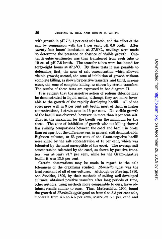
with growth in pH 7.6, 1 per cent salt broth, and the effect of thesalt by comparison with the 1 per cent, pH 6.0 broth. Aftertwenty-four hours' incubation at 37.50C., readings were madeto determine the presence or absence of visible growth. One-tenth cubic centimeter was then transferred from each tube to10 cc. of pH 7.6 broth. The transfer tubes were incubated forforty-eight hours at 37.5°0. By these tests it was possible todetermine; first, the zone of salt concentration which allowedvisible growth; second, the zone of inhibition of growth withoutcomplete killing, as shown by positive transfers; and third, in somecases, the zone of complete killing, as shown by sterile transfers.The results of these tests are expressed in bar diagram II.
It is evident that the selective action of sodium chloride maybe demonstrated in liquid media, although they are more favor-able to the growth of the rapidly developing bacilli. All of thecocci grew well in 9 per cent salt broth, most of them in higherconcentrations, 1 strain even in 16 per cent. No visible growthof the bacilli was observed, however, in more than 9 per cent salt.That is, the maximum for the bacilli was the minimum for thecocci. The zone of inhibition of growth without killing showedless striking comparisons between the cocci and bacilli in broththan on agar, but the difference was, in general, still demonstrable.Eighteen cultures, or 53 per cent of the Gram-negative bacilliwere killed by the salt concentration of 15 per cent, which wastolerated by the most susceptible of the cocci. The average saltconcentration tolerated by the cocci, as shown by positive trans-fers, was at least 21.7 per cent, while for the Gram-negativebacilli it was 13.6 per cent.
Certain observations may be made in regard to the salttolerances of the organisms studied. Eberthella typhi was theleast resistant of all of our cultures. Although de Freytag, 1890,and Stadtler, 1899, by their methods of salting well-developedcultures, obtained positive transfers after long periods of time,other authors, using methods more compaiable to ours, have ob-tained results similar to ours. Thus, Matzuschita, 1900, foundthe growth of Eberthella typhi good on from 0 to 3.5 per cent salt,moderate from 4.5 to 5.5 per cent, scarce on 6.5 per cent and
50
on Decem
ber 26, 2019 by guesthttp://jb.asm
.org/D
ownloaded from

MEDIA FOR SEPARATION OF COCCI FROM BACILLI
slight or none above this. K. von Karaffa-Korbutt, 1912, foundthat this organism grew in 7 per cent salt in peptone broth, butnot in 8 per cent. Although Schmidt (1924) found Eberthellatyphi viable in 1.5 per cent salt after ten days, Duthoit (1923a),reported that in 0.9 per cent salt two thousand bacilli were re-duced to six within six and one-half hours. It is possible thata study of a number of strains of this organism and of the relatedforms will reveal some specific differences.The effect of salt upon organisms of the genera Pseudomonas
and Proteus was striking. Of the 5 cultures of Pseudomonasaeruginosa (Bacillus pyocyaneus) studied, 3 grew heavily on agaronly through 3 per cent salt, the other 2 cultures being in-hibited above 5 per cent. These 5 cultures were killed on saltagar by a concentration of not more than 11 per cent salt, ascompared with growth on transfer of all the cocci from 20 percent. The four Proteus strains behaved alike on salt agar, thatis, they all grew heavily on 6 per cent salt, with inhibitionthrough 8 per cent, no transfer being positive above 14 per cent.The findings with both of these genera on salt broth showed ageneral parallelism, with a somewhat greater tolerance. Therepression of chromogenesis in the Pseudomonas cultures was in-variable. Matzuchita, 1900, also found Proteus more resistantto salt than Pseudomonas, growth of the former being poorabove 8.5 per cent, the latter, scarce at 6.5 per cent.
In analyzing the findings with organisms of the colon group andits related form, Klebsiella pneumoniae, our lack of adequateclassification makes it impossible to draw accurate comparisons.There are no marked differences between cultures of the genusEscherichia and those of the genus Aerobacter. These organisms,regardless of their genera, have been studied by India ink ex-aminations for the presence of capsules, in fact some of them wereselected from a collection of 200 cultures on account of their en-capsulation. Of the 19 organisms, including the Friedlanderbacillus 9, or 47.3 per cent were heavily encapsulated and viscidin growth, 10, or 52.6 per cent showed no capsules, or very slightones and were not viscid in growth. Of the 14 cultures whichwere killed by 9 per cent agar, 9, or 64.2 per cent were the 9
51
on Decem
ber 26, 2019 by guesthttp://jb.asm
.org/D
ownloaded from

JUSTINA H. HILL AND EDWIN C. WHITE
thickly encapsulated cultures. That is, all of the thickly en-capsulated cultures belonged in the group most easily killedby salt.
TESTS WITH MIXED CULTURES
The preceding experiments indicated the possibility of usingsalt agars to inhibit bacilli present in mixed cultures, or even, insome instances, to isolate Gram-positive cocci from such mix-tures of organisms. Eighteen-hour pH 7.6 broth cultures of thetest organisms were mixed in two proportions. Series A con-sisted of 1 cc. of the culture of a bacillus and 1 cc. of the cultureof a coccus, while Series B consisted of 1 cc. of the bacillus cultureand 1 standard loopful of the coccus culture. One loopful ofsuch mixtures was placed on each of the following agar slants; 1per cent salt, pH 6.0 agar, the control and on 6, 8, 10 and 15 percent salt, pH 6.0 agars. After forty-eight hours of incubation,smears of the slants showing visible growth were examined.Transfers were made to 10 cc. of pH 7.6 broth from the 10 and15 per cent salt agars, whether or not there was any visiblegrowth. The use of a liquid medium was favorable to thedevelopment of any bacilli which might be viable.Enrichment of the cocci was obtained on all of the salt agars
tested. The controls on 1 per cent .alt, pH 7.6 agar, on bothseries, had heavy growth, either of both organisms, or of an over-whelming number of bacilli. In the A series of salt agar tests,in which the inoculum contained equal parts of the bacillus andof the coccus cultures, examinations of smears of the culturesshowed only cocci in 75 per cent of the tests on 6 per cent salt;71.4 per cent on 8 per cent salt; 89.2 per cent on 10 per cent saltand 92.8 per cent on 15 per cent salt. Transfers in this series topH 7.6 broth gave pure cultures of the cocci in 17.8 per centfrom 10 per cent salt and in 39.2 per cent from 15 per cent salt.In the B series of experiments, in which the inoculum was 1loopful of a mixture of 1 cc. of the bacillus culture and 1 loopfulof the coccus culture, the results on 8, 10 and 15 per cent saltwere fully as good, in some cases better, than in series A. On 6per cent salt, in series B, 67.6 per cent of the smears showed only
52
on Decem
ber 26, 2019 by guesthttp://jb.asm
.org/D
ownloaded from

MEDIA FOR SEPARATION OF COCCI FROM BACILLI
cocci; on 8 per cent salt, the number of smears showing onlycocci increased to 91.1 per cent. On 10 per cent salt, 94.1 percent of the series B tests showed only cocci, but only 11.6 percent of the transfers gave pure cultures of cocci. On 15 per centsalt all of the tests in series B which had any growth, showedonly cocci, while of the 27 viable transfers from this concentra-tion, 16 or 59.2 per cent were pure cultures of cocci.
It is of interest to note that the amount of growth on saltagar seems to be determined by the number of cocci in theinoculum. In the B series, in which there were fewer cocci, thegrowth was generally lighter, although the number of bacilliwas relatively larger than in the A series. This finding correlateswith the results of examinations of smears of the cultures.
TESTS WITH MIXED INFECTIONS
In the application of the previous findings for the enrichmentof cocci in mixed infections, the salt concentrations employedmust be of sufficiently wide range to cover the unknown varia-tions in the number of organisms present. In general resultswith lower concentrations of salt may be obtained from clinicalmaterial than from pure or artificially mixed cultures. This isprobably on account of the smaller number of organisms present.
In table 1 are suimmarized the findings from 20 cases of mixedinfection.The organisms found in these infections were staphylococci,
streptococci of the Gamma type, colon group bacilli and Pseudo-monos pyocyanea, from 2 to 4 types of organisms being present.In 16 of these 20 cases, pure cultures of the cocci were obtainedfrom 6 through 15 per cent salt agar. Of the 4 cases in whichthere was enrichment of the cocci, but in which pure cultureswere not obtained, 3 were not placed on high salt concentrations.In the fourth case, no. 20, a streptococcus was present in verysmall numbers with Ps.- pyocyanea and a colon group bacillus.It seems evident from these 20 cases that the use of salt agars isof definite advantage in culturing many specimens from mixedinfections.
53
on Decem
ber 26, 2019 by guesthttp://jb.asm
.org/D
ownloaded from

JUSTINA H. HILL AND EDWIN C. WHITE
0
100b oot.oo-
10p.4
0'
4).,4
Ca
e 0 0Zo oo
004~~~~~~ r~~~~t0.t 0
00'iIII0o
* .oo U c c
0o .. 0X as 0 o ^
04
0 0~~~
1 ~~~~~~~k b- 1 m tXo
0 0 ~ 0 0 0
0sD4s 001 00
0
E* 0 0 0 00 0
4 0 0O
P -
co 0
-4 to32 m an to3D m
2¢~~ ~~C cis as as as0O V 48OVVmmm mV2 AV EmMO, a
0O ~ O
.5~~~~~~~
3m *- 00000 0 0)0)0 00 0
-) - -eco k4 to_- _ - _- _- _- _- -
54
0
*.:*4)0
'II0%OD0Go
0
z 0b.
;s0
00
00I1b
aP4
_. Nq C* lw Lo co
on Decem
ber 26, 2019 by guesthttp://jb.asm
.org/D
ownloaded from

MEDIA FOR SEPARATION OF COCCI FROM BACILLI
Ias t0 m- 4)
0 a30 an 7bs:e co
a s.0
tz"c' .
ID0
0
z
0
D- t0 0 0-cc z c
ft00
b- t- tz-
cc co Ck
a
34 2_4 1
.^
o0 Z
AD o
v2
0-4
00
O-I.
.I 3C4
0
oi a
0
.0.4D
0
*^ 4
=I
0 *a
to
00
°3 i3 40 0. .0 040r 0
tz 00p.4 p4
55
on Decem
ber 26, 2019 by guesthttp://jb.asm
.org/D
ownloaded from

JUSTINA H. HILL AND EDWIN C. WHITE
DISCUSSION
These experiments demonstrate some of the possibilities ofthe use of salt agars. Should the method prove of value, it maybe applied to other organisms than those included here. A studyof the use of different salt concentrations in special media forstreptococci is indicated from the few cases we have observed.While there is little evidence that salt offers any aid to generic orspecific identification, although this was once claimed by Dubois,1910, for Escherichia coli, it is possible that further studies willreveal such differences. We hope that others will test thismethod or some modification of it on similar or on differenttypes of specimens.
SUMMARY AND CONCLUSIONS
1. It has been found that pH 6.0 sodium chloride agars, withsalt concentrations from 2 through 20 per cent, exert markedinhibitory action on the growth of bacilli of the typhoid, para-typhoid, dysentery, and colon groups, on species of Proteus,Pseudomonas, on diphtheroids and on Bacillus anthra8is. TheGram-positive cocci studied tolerate high salt concentrations, allbeing positive on transfer from 20 per cent sodium chloride agar.
2. In pH 6.0 broths, with salt concentrations from 2 through25 per cent, the same differential bacteriostasis may be observed,although to a lesser degree than on agar.
3. It has been found that when mixtures of cocci and bacilliin different proportions are cultured on appropriate salt agars,the cocci invariably outgrow the bacilli and may sometimes berecovered in pure culture.
4. The use of 6, 8, 10 and 15 per cent salt agars greatly facili-tates the isolation of Gram-positive cocci from specimens frommixed infections.
5. The use of such salt agars is therefore suggested for theinhibition of Gram-negative bacilli and for the isolation of Gram-positive cocCi.
56
on Decem
ber 26, 2019 by guesthttp://jb.asm
.org/D
ownloaded from

MEDIA FOR SEPARATION OF COCCI FROM BACILLI 57
REFERENCESDE FREYTAG: Arch. Hyg., 1890, 11, 60.DUBOIS, R.: Compt. rend. Soc. biol., 1910, 68, 26.DUTHOIT, A.: Compt. rend. Soc. biol., 1923a, 89, 548.DUTHOIT, A.: Compt. rend. Soc. biol., 1923b, 89, 550.DIJTHOIT, A.: Compt. rend. Soc. biol., 1923c, 89, 553.FisCHER, A.: Vorlesungen uber Bakterien, Zweite Vermehrte, Chapt. III, Die
Bakterienzelle als osmotisches System, Gustav Fischer, Jena, 1903, 20.GUILLEMARD, A.: Compt. rend. Acad. Sc.,. 1908, 117, 1177.GUILLEMARD, A.: Compt. rend. Soc. biol., 1909, 67, 538.HOLZINGER, F.: Centralb. Bakt., II Abt., 1908, 21, 449.KARAFFA-KORBUTT, K. VON: Zeitschr. Hyg., 1912, 71, 161.KOCH, R.: Mittheil. a. d. kais. Gesundheitsamte, 1881, i, 273, cited by de Freytag,
1890LEWANDOWSKY, F.: Arch. Hyg., 1904, 60, 47.MARTENS, G.: Virch. Arch., 1888, 112, 341.MATZUSCHITA, I.: Zeitschr. Hyg., 1900, 35, 495.PETTERSON, A.: Arch. Hyg., 1900, 37, 171.SCHMIDT, H.: Centralb. Bakt., I Abt. Orig., 1924, 91, 510.STADLER, E.: Arch. Hyg., 1899, 35, 40.WHTE, E. C.: Jour. Urol., 1929, in press.
on Decem
ber 26, 2019 by guesthttp://jb.asm
.org/D
ownloaded from