magdalena kucharska, developing a model of …fibtex.lodz.pl/2012/6b/115.pdfdeveloping a model of...
TRANSCRIPT

Kucharska M, Niekraszewicz A, Kardas I, Marcol W, Właszczuk A , Larysz-Brysz M, Lewin-Kowalik J. Developing a Model of Peripheral Nerve Graft Based on Natural Polymers.FIBRES & TEXTILES in Eastern Europe 2012; 20, 6B (96): 115-120.
115
Developing a Model of Peripheral Nerve Graft Based on Natural Polymers
Magdalena Kucharska, Antoni Niekraszewicz,
Iwona Kardas, *Wiesław Marcol,
*Adam Właszczuk,*Magdalena Larysz-Brysz,
*Joanna Lewin-Kowalik
Institute of Biopolymers and Chemical Fibres ul. M. Skłodowskiej-Curie 19/27, 90-570 Łódź, Poland
E-mail: [email protected]
*Medical University of Silesia, Department of Physiology,
ul. Medyków 18, 40-752 Katowice Ligota, Poland
AbstractPresented are the results of investigations into the preparation of a peripheral nerve pros-thesis. The prosthesis is built up of a multichannel core having a diameter of 2 to 5 mm. The core prepared by freeze drying is housed in a polymeric sleeve. The prosthesis core is made of microcrystalline chitosan (MCCh) while the sleeve is prepared from poly(DL-lactide-co- glycolide) copolymer. The usefulness of the prepared biomaterial was assessed by in vivo testing on animals
Key words: biopolymers, peripheral nerve, prosthesis, chitosan.
millimetres) of the spinal cord were re-moved and the resulting gap was instant-ly bridged with an alginate sponge [14].
Nowadays, only one commercial artificial nerve is used in human therapy under the trade name of NeuraGenR. The implant is made up of type I collagen in the form of a hollow tube and is only suitable for bridging short (3 - 5 mm) nerve defects [1, 5]. This explains why many research centres are working on the preparation of more efficient solutions to the problem.
The aim of the research presented was to elaborate a model of nerve prosthesis that showed neutral immunity, was bio-degradable and resorbable, and with an internal structure allowing facilitated re-growth of the parallel nerve fibres. The prosthesis is expected to prevent the pa-tient from further suffering caused by the uptake of a nerve fragment from his body for the reconstruction. The authors hope to identify the preparation of a prototype of a multichannel prosthesis based on natural polymers; chitosan from the po-ly-amino-saccharide family in particular. Such construction of the implant and the used material were adopted on the basis of a literature search and an introductory investigation by the authors, which had highlighted the beneficial impact of the microcrystalline form of chitosan upon the regeneration of nerve fibres [18].
n MaterialsMicrocrystalline chitosan (MCCh)Microcrystalline chitosan was prepared according to a method elaborated at IBWCh [19]. MCCh of high purity, with varying molecular mass and pH, was used in the investigation.
tunnels have been mostly used in nerve repair with varying success, most notably:n connective tissue/collagen, hollow or
filled with fibrin, n synthetic: silicone or polyethylene
with varied permeability of the walls [1, 5, 6,10, 11].
Attempts were made to use tunnels with parallel arranged carbon fibres or fibres made up of resorbable poly(glycolic acid) to improve the orientation of the regenerating nerve fibres [1, 7]. These tunnels were also filled with neurotropic substances and/or populations of vari-ous cells (Schwann cells, mother cells) that are subjected to various modifica-tions. In the last two years, tubes and nerve prostheses without an external coat (substitute of perneurium) prepared from sole chitosan or alginate have also been investigated [2 - 4, 8, 9, 10, 12, 13]. A hydroxyapatite-chitosan complex was used and the cross-section of the pros-thesis changed to a triangle to improve the tenacity of the tubes [14, 15]. Tests conducted on animals have shown that the chitosan tubes induce an effective regeneration of the peripheral nerves and undergo a stepwise resorption without in-flammatory reactions [10]. In the therapy of spinal cord damage in animals, inter-esting investigations on the preparation of an implant of poly–β-hydroxy butyrate with a content of alginate hydroxy-gel enriched with fibronectin are presented [16, 17]. A lyophilised alginate gel cov-ered with a mesh made of poly(glycolic acid) was used in the regeneration of ex-tensively damaged nerves in cats. That solution allowed the perfect regeneration of the damaged nerves; however, strong inflammatory reactions could be ob-served during the process of implant bio-degradation [2]. In other, more inspiring experiments on animals, fragments (few
n IntroductionPeripheral nerves are often seriously impaired or even disrupted in the ever increasing number of injuries, occurring mainly in traffic accidents. It may sig-nificantly affect the motoric system and cause atrophy of the denervated mus-cles. Unprotected fibres in the proximal segment of the disrupted nerve have the ability to regenerate. The process is, however, spontaneous and chaotic, lead-ing in most cases to neuroma or micro-neuroma [1, 2]. Disordered sensory fibres build improper joints causing mechanical and chemical excitations resulting in pain sensation [6, 7]. The phenomenon is known as neuropathic pain resistant to pharmacological therapy and poses a se-rious therapeutic problem [7].
The problem of repairing extensively im-paired peripheral nerves is being studied in many research centres throughout the world. The most difficult clinical prob-lems are the so-called “gap injuries” in which the gap in the nerve continuity hin-ders its immediate sewing [1]. The gaps are often bridged with a nerve fragment taken up primarily from the patient’s skin with the negative effect of additional mutilation. Construction of nerve pros-theses, or more precisely, of nerve graft substitutes, would be the optimal solu-tion to the problem. An ideal prosthesis is expected to satisfy following demands: 1. display neutral immunity, 2. allow easy joining with the nerves stumps, 3. be elastic, 4. have an internal multichannel structure enabling the re-growth of the parallel nerve fibres, 5. be tight enough to prevent the in-growth of connective tissue, 6. enable the incorporation of neu-ralgic and neurotropic substances to the prosthesis structure, and 7. indicate slow biodegradation and resorption of the ma-terial [1 - 6]. Up to now, so-called natural

FIBRES & TEXTILES in Eastern Europe 2012, Vol. 20, No. 6B (96)116
The physicochemical parameters of the prepared MCCh are as follows:n MCCh/278: Mv = 423 kD,
deacetylation degree (DD) = 82%, polymer content = 2.5%,
water retention value (WRV) = 4600%, ash content = 0.09%, pH = 7.0,nMCCh/171: Mv=287 kD,
DD = 80%, polymer content = 2.5%, WRV = 3990%, ash content = 0.04%, pH = 7.5.
Resomer RG 755S - poly(DL-lactide-co-glycolide) copolymerCopolymers of D,L-lactide and glycol-ide type Resomer RG 755S supplied by Boehringer Ingelheim Co. were used in the research. They were characterised as having the following composition: 75 mol% of DL-lactide and 25 mol% of glycolide residues. Inherent viscosity = 0.68 dl/g.
Polypropylene fibres The PP fibres were melt-spun on an ex-truder spinning bank, which is in the possession of IBWCh. The prepared monofilament PP fibres exhibited a good smoothness and their set diameter was between 0.16 and 0.22 mm. The fi-bres served as temporary component of the matrix and were removed after the channel in the prosthesis core had been formed. Therefore, the mechanical pa-rameters of the fibres were not measured.
Silicon mouldCommercial silicone sleeves with a di-ameter between 2 and 5 mm were used in the construction of the matrix of the prosthesis core.
GlycerolGlycerol (Fluka Co.), analytically pure according to Ph. Eur., was used as chi-tosan plasticiser.
n MethodsEstimation of the average molecular mass of chitosan (Mv) - Viscometric method The viscometric average molecular mass was calculated on the basis of intrinsic viscosity number [η] values. Viscos-ity was measured on a dilution viscom-eter with capillary No. 1, K ≈ 0.01 at 25 ± 0.1 °C. The average molecular mass
- Department of Physiology of the Sile-sian Medical University, Katowice.
The functional assessment of rats’ pe-ripheral nerve regeneration with prosthe-ses was conducted in the region of ex-tensive defects in the sciatic nerve. The experiments were carried out with Wistar C male rats.
In all anaesthetised animals (Avertine) the right sciatic nerve was exposed and its fragment, 7 mm in length, was re-moved. The defect was then replaced with a prosthesis fragment of a similar length, which was affixed to the perineu-rium of the proximal and distal stump (Figure 1). The prosthesis was filled up with saline, and then the postoperative wound was sutured.
was calculated using the Mark-Houwink [20] equation.
Estimation of the deacetylation degree of the chitosan (DD) - method of the first derivative of UV spectrum analysisThe deacetylation degree was estimated through a spectrophotometric method in which the maximum of the first derivative was found of the UV spectrum, which led to the calculation of DD according to a procedure prepared at IBWCh.
Estimation of ash contentAsh content in chitosan was estimated gravimetrically after annealing the poly-mer at 800 °C according to the IBWCh procedure.
Estimation of water retention value (WRV)WRV was estimated according to stand-ard method [21].
Estimation of polymer content in the microcrystalline chitosan (MCCh)The polymer content in the MCCh was estimated gravimetrically according to the IBWCh procedure.
Estimation of heavy metal contentHeavy metal content was estimated with the use of Atomic Absorption Spectros-copy (AAS).
Assessment of mechanical propertiesTests of mechanical properties were con-ducted in the accredited Laboratory of Metrology of IBWCh – PCA certificate No AB 388 Metrological Testing.
nnerve prostheses made by freeze dry-ing were tested according to ISO standard 7198:1998
nprosthesis sleeves made up of poly(DL-lactido-co-glycolide) co-polymer film were tested according to standards PN-ISO 4593:1999 (film thickness), PN-EN ISO 527-3:1998 (strength parameters).
Inspection of the structureThe surface and cross-section of the nerve prostheses were analysed by means of Scanning Electron Microscope SEM Quanta 200 (FEI Co., USA).
In vivo testing on animals Biological in vivo examinations were performed in the specialised medical unit
Figure 1. View of the in situ grafted mul-tichannel chitosan prosthesis.
The animals were euthanised after seven weeks and the grafted prosthesis was col-lected, together with the adjoining frag-ments of the proximal and distal sciatic nerve.
Preparing the specimens for histological assessmentNerve fragments were fixed together with the prostheses in a formalin solu-tion, dewatered in a saccharose solution and immersed in a medium for the histo-logical experiments, before being frozen. The prepared specimens were cut trans-versally or longitudinally with a freezing microtome into tiny scraps, 10 μm thick, and placed onto microscope glass plates.
Histological assessment of prosthesis – grafted nervesThe histological assessment of prosthe-sis-grafted nerves was performed using light and fluorescence microscopy.

117FIBRES & TEXTILES in Eastern Europe 2012, Vol. 20, No. 6B (96)
Assessment of the extent of neuropathic pain Assessment of the extent of neuropathic pain was done on the basis of the autot-omy phenomenon occurring in animals (gnawing the denervated paws’ toes due to the neuropathic pain).
n Results of investigationPreparation of a peripheral prosthesis modelThe prosthesis core was made of micro-crystalline chitosan by means of a freeze drying technique [22]. An assumed num-ber of smooth tensioned polypropylene fibres was placed in the interior of a sili-con mould, 2 or 5 mm in diameter. The mould was then filled up with a suspen-sion of microcrystalline chitosan and frozen at –25 °C for 15 minutes. Next, the silicon mould was removed and the obtained cylindrical core with the fibres was subjected to freeze drying. PP fibres were removed after drying leaving a prosthesis, 5 - 10 mm long with a diam-eter of 2 or 5 mm, containing channels in an amount depending upon the number of the earlier inserted (and removed) fi-bres. After grafting, the parallel channels serve to indicate the growth direction of the disrupted nerve fibres (Figure 2). To ease the connection of the prosthesis with the stumps of the defected nerve, the formed multichannel core was inserted in a sleeve made up of a resorbable poly-mer. The sleeve reached beyond the core for 4 mm on each side (Figure 3).
The prosthesis sleeves were made of a film of either microcrystalline chitosan (MCCh) or Resomer RG 755S poly(DL-lactide-co-polyglycolide) copolymer.
MCCh containing 1.7% of the polymer and with pH of 6.1 - 6.2 with the addi-tion of glycerol was used to prepare the sleeves for the nerve prostheses. The gel was cast onto Teflon plates and dried for 24 hours at ambient temperature. The obtained film was neutralized with Na2CO3 solution containing glycerol, and rinsed with a mixture of water and glycerol, before being freeze dried for 24 hours at -25 °C and under pressure of 0.1 - 0.57 hPa.
For the preparation of a film of poly(DL-lactide-co-polyglycolide), solutions of the copolymer in 1,4-dioxan with the ad-dition of polyethylene glycol 600 were used. The solutions were cast on Teflon plates, dried for 24 hours at ambient tem-perature and then for 4 days at 50 °C, under a pressure of 0.07 MPa, until the complete removal of the solvent.
It was found during the investigation that the proper selection of the diameter of the PP fibres that shape the channels of the prosthesis is of profound importance. Fibres with a diameter of ca. 0.16 mm formed channels with too small a diame-ter, which were prone to deformation and closure after the freeze drying. The use of 0.22 mm diameter PP fibres produced better results allowing the formation of
prostheses with evenly arranged channels with uniform diameters (Figure 2).
Silicone moulds with 2 and 5 mm diam-eters were used in the preparation of the prostheses. With the 5 mm moulds, cores could be made with as many as 37 chan-nels. However, as a result of consulta-tions with neurosurgeons, it was decided to fix the prosthesis with a diameter of 2 mm. With such a small diameter, proto-types of the implant were prepared con-taining from 7 to 13 channels.
It appeared that, during the formation of the prosthesis, a proper content of the polymer in the microcrystalline chitosan is important as well. The use of MCCh containing 2% polymer yielded pros-thesis with a developed surface, while a 2.5% concentration of the polymer re-sulted in a more compact structure. Such a structure is expected to largely prevent the overgrowth of connective tissue through the prosthesis surface.
Estimation of the mechanical properties of peripheral nerve prosthesesMechanical properties of the core of the peripheral nerve prosthesis made of MCCh by freeze drying were examined (Table 1). The biomaterial of MCCh/278
Figure 2. SEM microphotographs of the core cross-section of the multichannel prosthesis of peripheral nerve made of MCCh by means of freeze drying. Diameter of the prosthesis – 5 mm; diameter of the channels – 0.25 mm.
A B
Figure 3. Prosthesis of a peripheral nerve prepared by freeze drying; A-multichannel core, B- outer sleeve.
Table 1. Mechanical properties of the core of chitosan prostheses with 7 and 13 channels.
Tested parameterTest results
7 channels 13 channelsOuter diameter, mm 1.72 1.76Thickness variation coefficient, % 6.05 6.48Max. drawing force, N 4.42 4.02Drawing force variation coefficient, % 28.5 31.4Tenacity, MPa 6.21 5.65Elongation at max. stress, % 0.622 0.708Elongation variation coefficient, % 49.6 55.5

FIBRES & TEXTILES in Eastern Europe 2012, Vol. 20, No. 6B (96)118
was in the form of a cylinder 7 mm long with ca. 2 mm diameter, with 7 or 13 channels. Tests revealed that the core of the prepared prosthesis shows a tenac-ity above 5 MPa (it was somewhat better in the 7-channel version). Such tenac-ity ensures safe bridging of the defected nerve. When dry, the prostheses revealed a rather low elasticity, which is expected to improve after being implanted, filled with saline and contacted with systemic fluids. It should, under such conditions, not impair the limb’s mobility.
The sleeves of the nerve prostheses in the form of a film made up of microcrystal-line chitosan and Resomer RG 755S were also tested. The goal of the testing was to fix variation of mechanical resistance parameters of the film in dependence on the environment. It was especially im-portant to learn whether the mechanical resistance of the prostheses materials will remain on the level acceptable for a cor-rect joining with the perineurium after the grafting to a living organism (moist
environment). Results of the testing are shown in Table 2.
Results presented in Table 2 show a sub-stantial decrease of tenacity of both films in moist conditions. The drop amounts to 85% and 80% for the MCCh film and Re-somer RG 755S films, respectively. On the other hand, elasticity was improved in wet conditions by 71% and 45% for the MCCh and Resomer films, respec-tively. It can also be seen that the films of Resomer are about 49% stronger than those of chitosan in wet conditions. This was the reason that the films of Resomer RG 755S were selected for the construc-tion of the outer sleeve in the further ex-periments.
Biological assessment of the multichannel prostheses of peripheral nerves made of microcrystalline chitosanThe biological examination of periph-eral nerve prostheses was carried out on
Wistar C rats, of which three experimen-tal groups were formed:n Group 7A - 11 animals with implant-
ed chitosan prosthesis 7-channels. Implant core - MCCh (Mv = 423 kD, pH = 7.0), Implant sleeve - Resomer RG 755 S.
n Group 7B - 8 animals with implanted chitosan prosthesis 7-channels. Im-plant core - MCCh (Mv = 287 kD, pH = 7.5), Implant sleeve - Resomer RG 755 S.
n Group 13A – 9 animals with implant-ed chitosan prosthesis 13-channels. Implant core-MCCh (Mv = 423 kD, pH = 7.0), Implant skin - Resomer RG 755 S.
In the identification symbol, the number identifies the amount of channels and the letter describes the kind of MCCh used.
Examination of the medical suitability of the peripheral nerve prostheses con-firmed that the prototypes chosen for the experiments did not pose any difficulty in the course of grafting. The sleeve of the prosthesis provided the chance of easy and proper joining of the implant with the perineurium. The multichannel chitosan core revealed perfect air permeability.
Autotomy assessment of the animals after the implantation was made, which serves as an indication of the neuropathic pain. The extent of autotomy was as-sessed using the 13-point Wall scale. The examination did not reveal any essential difference in the extent of autotomy be-tween individual groups of the animals (Figure 4). The average extent of autot-omy in the three groups of animals after the implantations of selected prosthesis prototypes was in the range of 1.89 to 3.00 in the 13-point Wall scale. Hence, it may be deduced that the sensory fibres
Table 2. Mechanical properties of film made of MCCh and poly(DL-lactide-co-glycolide) copolymer.
Condition Parameter tested MCCh poly(DL-lactide-co-glycolide) copolymer
Dry
Film thickness, mm 0.83 0.052Thickness variation coefficient, % 13.3 14.0Max. drawing force, N 58.9 59.8Drawing force variation coefficient, % 21.7 19.9Tenacity, MPa 47.2 69.0Elongation at max. stress, % 43.0 49.3Elongation variation coefficient, % 27.9 21.9
Wet (immersed in demi-water for 60 s)
Film thickness, mm 0.84 0.058Variation coefficient of thickness, % 2.38 14.7Max. drawing force, N 8.90 10.6Drawing force variation coefficient, % 26.1 32.3Tenacity, MPa 7.04 13.8Elongation at max. stress, % 114 94.6Elongation variation coefficient, % 17.9 12.8
3 2,82
1,89
0 0,5
1 1,5
2 2,5
3 3,5
Group 7B Group 7A Group 13A
Figure 4. Intensification of neuropatic pain of animals.
89
54
25
0 10 20 30 40 50 60 70 80 90
100
Group 7B Group 7A Group 13A
Figure 5. The percentage of animals in which nerve growth into the prosthesis was observed.
Group 7B Group 7A Group 13A
3.5
3.0
2.5
2.0
1.5
1.0
0.5
0.0
3.02.82
1.89
Group 7B Group 7A Group 13A
100
80
60
40
20
0
25
54
89
119FIBRES & TEXTILES in Eastern Europe 2012, Vol. 20, No. 6B (96)
of the prosthesis-generated nerves do not cause strong mechanical and chemical excitations which evoke pain. It must be stressed that subsequent morphologi-cal assessment concerning regeneration of the peripheral nerve showed that the intensification of autotomy in the test groups of animals corresponded with the regeneration degree of the nerve. The better the organisation of the regenerat-ing nerve fragment the lower the autoto-my, meaning a milder neuropathic pain.
Histological examinationAssessment of the growth of nerve fibres into the prosthesis channelsThe percentage of animals in which the nerve grew from its edge on a length of at least 2 mm into the depth of the prosthe-sis counting from the end of the proximal nerve was estimated. It was found that the highest growth of the nerve into the prosthesis reached the highest percent-age of about 89% of animals in group 13A including 9 rats. These animals were grafted with 13-channel prostheses made of microcrystalline chitosan marked MCCh/278. The lowest growth of the nerve fibres occurred in group 7B. In that group, the percentage of rats in which the growth of the nerve reached 2mm into the prosthesis was as low as 25%. The ani-mals in the 7B group were grafted with a 7-channel prosthesis made of microc-rystalline chitosan marked MCCh/171. In group 7A, in which the animals were grafted also with the 7-channel prostheses but made of the microcrystalline chitosan MCCh/278, the percentage amounted to 54%. Since the chemical purity of both chitosans MCCH/278 and MCCh/171 is equal, it may be assumed that, with the same number of channels, the rate of nerve growth depended upon the pH of the material. This was 7.5 and 7.0 for
MCCh/171 and MCCh/278, respectively. Supposedly, the parameter plays an im-portant role in the process of nerve regen-eration. However, the presumption calls for proof in further research.
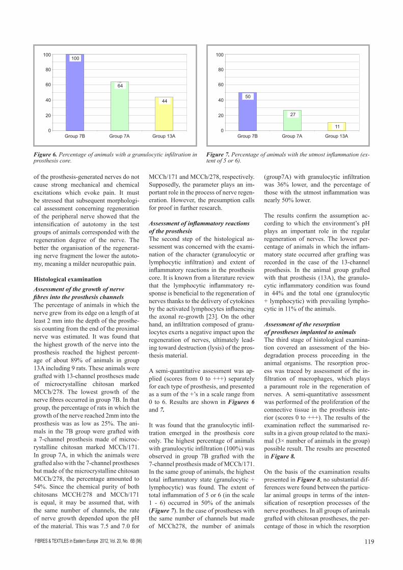
Assessment of inflammatory reactions of the prosthesisThe second step of the histological as-sessment was concerned with the exami-nation of the character (granulocytic or lymphocytic infiltration) and extent of inflammatory reactions in the prosthesis core. It is known from a literature review that the lymphocytic inflammatory re-sponse is beneficial to the regeneration of nerves thanks to the delivery of cytokines by the activated lymphocytes influencing the axonal re-growth [23]. On the other hand, an infiltration composed of granu-locytes exerts a negative impact upon the regeneration of nerves, ultimately lead-ing toward destruction (lysis) of the pros-thesis material.
A semi-quantitative assessment was ap-plied (scores from 0 to +++) separately for each type of prosthesis, and presented as a sum of the +’s in a scale range from 0 to 6. Results are shown in Figures 6 and 7.
It was found that the granulocytic infil-tration emerged in the prosthesis core only. The highest percentage of animals with granulocytic infiltration (100%) was observed in group 7B grafted with the 7-channel prosthesis made of MCCh/171. In the same group of animals, the highest total inflammatory state (granulocytic + lymphocytic) was found. The extent of total inflammation of 5 or 6 (in the scale 1 - 6) occurred in 50% of the animals (Figure 7). In the case of prostheses with the same number of channels but made of MCCh278, the number of animals
(group7A) with granulocytic infiltration was 36% lower, and the percentage of those with the utmost inflammation was nearly 50% lower.
The results confirm the assumption ac-cording to which the environment’s pH plays an important role in the regular regeneration of nerves. The lowest per-centage of animals in which the inflam-matory state occurred after grafting was recorded in the case of the 13-channel prosthesis. In the animal group grafted with that prosthesis (13A), the granulo-cytic inflammatory condition was found in 44% and the total one (granulocytic + lymphocytic) with prevailing lympho-cytic in 11% of the animals.
Assessment of the resorption of prostheses implanted to animalsThe third stage of histological examina-tion covered an assessment of the bio-degradation process proceeding in the animal organisms. The resorption proc-ess was traced by assessment of the in-filtration of macrophages, which plays a paramount role in the regeneration of nerves. A semi-quantitative assessment was performed of the proliferation of the connective tissue in the prosthesis inte-rior (scores 0 to +++). The results of the examination reflect the summarised re-sults in a given group related to the maxi-mal (3× number of animals in the group) possible result. The results are presented in Figure 8.
On the basis of the examination results presented in Figure 8, no substantial dif-ferences were found between the particu-lar animal groups in terms of the inten-sification of resorption processes of the nerve prostheses. In all groups of animals grafted with chitosan prostheses, the per-centage of those in which the resorption
Figure 6. Percentage of animals with a granulocytic infiltration in prosthesis core.
44
64
100
0 10 20 30 40 50 60 70 80 90
100
Group 7B Group 7A Group 13A
11
27
50
0 10 20 30 40 50 60 70 80 90
100
Group 7B Group 7A Group 13A
Figure 7. Percentage of animals with the utmost inflammation (ex-tent of 5 or 6).
Group 7B Group 7A Group 13A
100
80
60
40
20
0
100
64
44
Group 7B Group 7A Group 13A
100
80
60
40
20
0
50
27
11

FIBRES & TEXTILES in Eastern Europe 2012, Vol. 20, No. 6B (96)120
Received 27.10.2011 Reviewed 10.05.2012
had been enhanced, was contained in the range from ca. 42% to 50%.
n Conclusions1. The positive results of the investiga-
tion confirm that the prepared prosthe-sis of the peripheral nerve is a promis-ing candidate for use in neurosurgery.
2. It is advisable to continue the research with the goal of a further improvement of the implant, optimisation of the ma-terial parameters and estimation of the medical usefulness of the prosthesis throughout the entire regeneration cycle.
AcknowledgmentThe investigation were carried out within the research project No N N507 4612 33 supported by the Ministry of Science and High Education.
References 1. Battiston B, Geuna S, Ferrero M, Tos P.
Nerve repair by means of tubulisation: literature review and personal clinical experience comparing biological and synthetic conduits for sensory nerve re-pair. Microsurgery. 2005; 25(4): 258-67.
2. Suzuki Y, Tanihara M, Ohnishi K, Suzuki K, Endo K, Nishimura Y. Cat peripheral nerve regeneration across 50 mm gap repaired with a novel nerve guide com-posed of freeze-dried alginate gel. Neu-rosci Lett 1999, 259(2): 75-8.
48 42
50
0 10 20 30 40 50 60 70 80 90
100
Group 7B Group 7A Group 13A
Figure 8. Intensi-fication of biode-gradable implants.
3. Rosales-Cortes M, Peregrina-Sandoval J, Banuelos-Pineda J, Sarabia-Estrada R, Gomez Rodiles CC, Albarran-Rod-riguez E, Zaitseva GP, Pita-Lopez ML. Immunological study of a chitosan pros-thesis in the sciatic nerve regeneration of the axotomised dog. J Biomater Appl. 2003, 18(1): 15-23.
4. Wang X, Hu W, Cao Y, Yao J, Wu J, Gu X.: Dog sciatic nerve regeneration across a 30-mm defect bridged by a chitosan/PGA artificial nerve graft. Brain 2005, 128(Pt 8): 1897-910.
5. Williams LR, Danielsen N, Müller H et al..: Influence of the acellular fibrin matrix on nerve regeneration success within the silicon chamber model. In: The current status of peripheral nerve regeneration. Alan R Liss, Inc, 1988, pp. 111-122.
6. Windebank AJ, Poduslo JF: Neuronal growth factors produced by adult periph-eral nerve after injury. Brain Res 1986, 385: 197-200.
7. Zimmermann M.: Pathobiology of neu-ropathic pain. Eur. J. Pharmacol 2001, 429: 23-37.
8. Yan Q, Elliott Jl, Mathenson C et al..: Influences of neurotrophins on mamma-lian motor neurons in vivo. J Neurobiol 1993, 24: 1555-1577.
9. Stoll G, Müller H: Nerve injury, axonal de-generation and neural regeneration: ba-sic insight. Brain Pathol 1999, 9: 313-25.
10. Yamaguchi I, Itoh S, Suzuki M, Osaka A, Tanaka J, The chitosan prepared from crab tendons: II. The chitosan/apa-tite composites and their application to nerve regeneration. Biomaterials 2003; 24: 3285-3292.
11. Santos FX, Bilbao G, Rodrigo J et al.: Experimental model for local administra-tion of nerve growth factor in microsurgi-cal nerve reconnections. Microsurgery 1995, 16: 71-76.
12. Hashimoto T, Suzuki Y, Kitada M, Kataoka K, Wu S, Suzuki K, Endo K, Nishimura Y, Ide C. Peripheral nerve re-generation through alginate gel: analy-sis of early outgrowth and late increase in diameter of regenerating axons. Exp Brain Res 2002, 146(3): 356-68.
13. Qiang AO, Wang A, Cao W, Zhao Ch, GongY, Zhao N, Zhang X. Fabrication and characterisation of chitosan nerve conduits with microtubular architectures. Tsinghua Science and Technology ISSN 1007-0214 06/20 pp. 435-438 Vol 10, No 4, August 2005.
14. Itoh S, Yamaguchi I, Suzuki M, Ichinose S, Takakuda K, Kobayashi H, Shinomiya K, Tanaka J. Hydroxyapatite-coated ten-don chitosan tubes with adsorbed laminin peptides facilitate nerve regeneration in vivo. Brain Res 2003; 993: 111-123.
15. Itoh S, Yamaguchi I, Shinomiya K, Tana-ka J. Development of the chitozan tube prepared from crab tendon for nerve regeneration, Sci Technol Adv Mater 2003; 4: 261-268.
16. Novikov LN, Novikowa LN, Mosahebi A, Wiberg M, Terenghi G, Kellerth JO. A novel biodegradable implant for neu-ronal rescue and regeneration after spi-nal cord injury, Biomaterials 2002; 23: 3369-3376
17. Hert AM, Wiberg M, Terenghi G. Exog-enous leukaemia inhibitory factor en-hances nerve regeneration after late secondary repair using a bioartificial nerve conduit. Brit Assoc Plastic Surg 2003; 56: 444-450.
18. Marcol W, Larysz-Brysz M, Kucharska M, Niekraszewicz A, Ślusarczyk W, Kot-ulska K, Właszczuk P, Właszczuk A, Je-drzejowska-Szypulka H, Lewin-Kowalik J: Reduction of post-traumatic neuroma and epineural scar formation in rat sci-atic nerve by application of microcrystal-lic chitosan. Microsurgery 2011; 31(80): 642-649.
19. Polish Patent PL 281975 (1989)20. Domszy JG, Roberts G: Macromol. Che-
mie,1985, 186, 1671. 21. Struszczyk H, J. Appl Polymer Sci.
1987, 33, 171. 22. Polish Patent Appl. P-391898 (2010) 23. Lu X, Richardson PM: J Neurosci 1991;
11: 972.
Group 7B Group 7A Group 13A
100
80
60
40
20
0
5042
48