magnetic resonance imaging
TRANSCRIPT

MAGNETIC RESONANCE IMAGING (MRI) IS A MEDICAL IMAGING TECHNIQUE USED IN RADIOLOGY TO FORM PICTURES OF THE ANATOMY AND THE PHYSIOLOGICAL PROCESSES OF THE BODY IN BOTH HEALTH AND DISEASE. MRI SCANNERS USE STRONG MAGNETIC FIELDS, RADIO WAVES, AND FIELD GRADIENTS TO GENERATE IMAGES OF THE INSIDE OF THE BODY.
MAGNETIC RESONANCE IMAGING

MRI has a wide range of applications in medical diagnosis and over 25,000 scanners are estimated to be in use worldwide.[1] MRI affects diagnosis and treatment in many specialties although the effect on improved health outcomes is uncertain.
MEDICAL USES

MRI is in general a safe technique but the number of incidents causing patient harm has risen.[citation needed]
Contraindications to MRI include most cochlear implants and cardiac pacemakers, shrapnel and metallic foreign bodies in the eyes.

MRI is the investigative tool of choice for neurological cancers, as it has better resolution than CT and offers better visualization of the posterior fosse.
NEUROIMAGING

Cardiac MRI is complementary to other imaging techniques, such as echocardiography, cardiac CT and nuclear medicine. Its applications include assessment of myocardial ischemia and viability, cardiomyopathies, myocarditis, iron overload, vascular diseases and congenital heart disease.
CARDIOVASCULAR

Applications in the musculoskeletal system includes spinal imaging, assessment of joint disease and soft tissue tumors.
MUSCULOSKELETAL

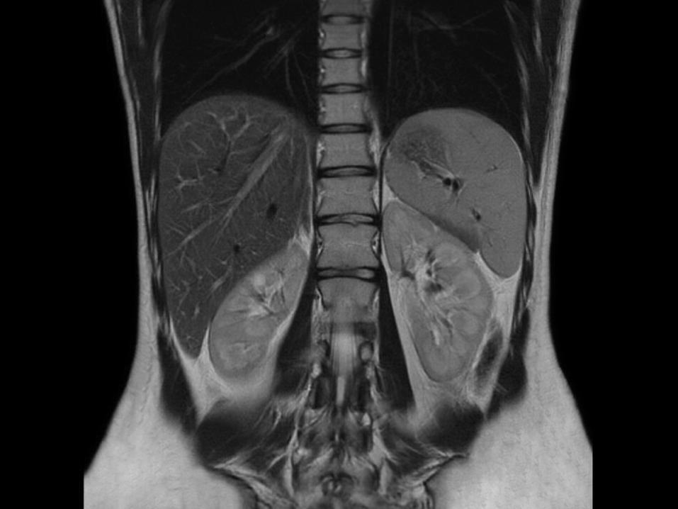
Hepatobiliary MR is used to detect and characterize lesions of the liver, pancreas and bile ducts. Focal or diffuse disorders of the liver may be evaluated using diffusion-weighted, opposed-phase imaging and dynamic contrast enhancement sequences.
LIVER AND GASTROINTESTINAL IMAGING MRI

Functional MRI (fMRI) is used to understand how different parts of the brain respond to external stimuli or passive activity in a resting state. Blood oxygenation level dependent (BOLD) fMRI measures the hemodynamic response to transient neural activity resulting from a change in the ratio of oxyhemoglobinand deoxyhemoglobin.
FUNCTIONAL MRI

MRI is the investigation of choice in the preoperative staging of rectal and prostate cancer, and has a role in the diagnosis, staging, and follow-up of other tumors.
ONCOLOGY

In an effort to standardize the roles and responsibilities of MRI professionals, an international consensus document, written and endorsed by major MRI and medical physics professional societies from around the globe, has been formally published. The document outlines specific responsibilities for the following positions:
STRUCTURE AND CERTIFICATION

Phase Contrast MRI (PC-MRI) is used to measure flow velocities in the body. It is mainly used to measure blood flow in the heart and throughout the body. PC-MRI can be considered a method of Magnetic Resonance Velocimetry. Since modern PC-MRI is typically time-resolved, it can also be referred to as 4D imaging (three spatial dimensions plus time).
PHASE CONTRAST MRI
1.MR Medical Director / Research Director (MRMD) - This individual is the supervising physician who has oversight responsibility for the safe utilization of MRI services.2. MR Safety Officer (MRSO) - Roughly analogous to a radiation safety officer, the MRSO acts on behalf of, and on the instruction of, the MRMD to execute safety procedures and practices at the point of care.3. MR Safety Expert (MRSE) - This individual serves in a consulting role to both the MRMD and MRSO, assisting in the investigation of safety questions that may include the need for extrapolation, interpolation, or quantification to approximate the risk of a specific study.

All patients are reviewed for contraindications prior to MRI scanning. Medical devices and implants are categorized as MR Safe, MR Conditional or MR Unsafe.
IMPLANTS

Ferromagnetic foreign bodies such as shell fragments, or metallic implants such as surgical prostheses and ferromagnetic aneurysm clips are also potential risks. Interaction of the magnetic and radio frequency fields with such objects can lead to heating or torque of the object during an MRI.
Titanium and its alloys are safe from attraction and torque forces produced by the magnetic field, though there may be some risks associated with Lenz effect forces acting on titanium implants in sensitive areas within the subject, such as stapes implants in the inner ear.

The very high strength of the magnetic field can cause projectile effect (or "missile-effect") accidents, where ferromagnetic objects are attracted to the center of the magnet. Pennsylvania reported 27 cases of objects becoming projectiles in the MRI environment between 2004 and 2008. There have been incidents of injury and death.
OROJECTILE RISK
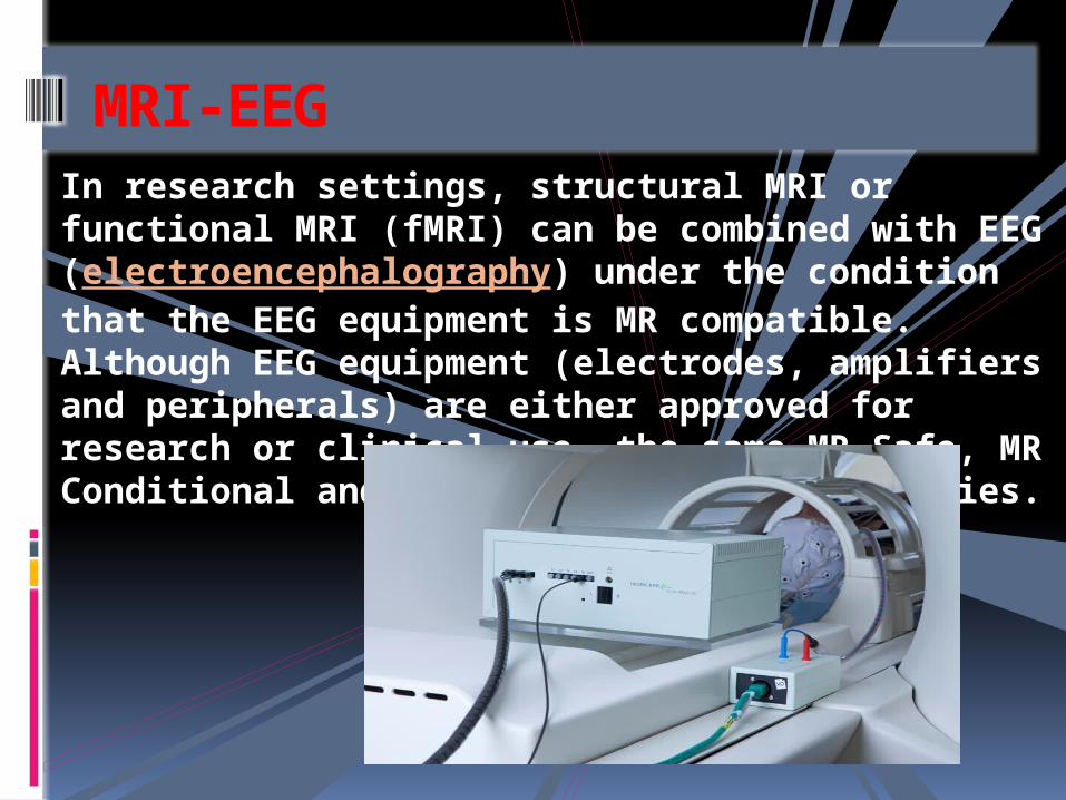
In research settings, structural MRI or functional MRI (fMRI) can be combined with EEG (electroencephalography) under the condition that the EEG equipment is MR compatible. Although EEG equipment (electrodes, amplifiers and peripherals) are either approved for research or clinical use, the same MR Safe, MR Conditional and MR Unsafe terminology applies.
MRI-EEG

There is no proven risk of biological harm from any aspect of a MRI scan, including very powerful static magnetic fields, gradient magnetic fields, or radio frequency waves.[46]
[47] Some studies have suggested possible geotaxis (i.e., potentially carcinogenic) effects of MRI scanning through micronuclei induction and DNA double strand breaks in vivo and in vitro.
GENOTOXIC EFFECTS

The rapid switching on and off of the magnetic field gradients is capable of causing nerve stimulation. Volunteers report a twitching sensation when exposed to rapidly switched fields, particularly in their extremities.
PERIPHERAL NERVE STIMULATION

Every MRI scanner has a powerful radio transmitter that generates the electromagnetic field that excites the spins. If the body absorbs the energy, heating occurs. For this reason, the transmitter rate at which energy is absorbed by the body must be limited (see Specific absorption rate).
HEATING CAUSED BY ABSORPTION OF RATIO WAVES

Switching of field gradients causes a change in the Lorentz force experienced by the gradient coils, producing minute expansions and contractions of the coil itself. As the switching is typically in the audible frequency range, the resulting vibration produces loud noises (clicking, banging, or beeping).
ACOUSTIC NOISE

As described in Physics of Magnetic Resonance Imaging, many MRI scanners rely on cryogenic liquids to enable the superconducting capabilities of the electromagnetic coils within. Though the cryogenic liquids used are non-toxic, their physical properties present specific hazards.
CRYOGENS
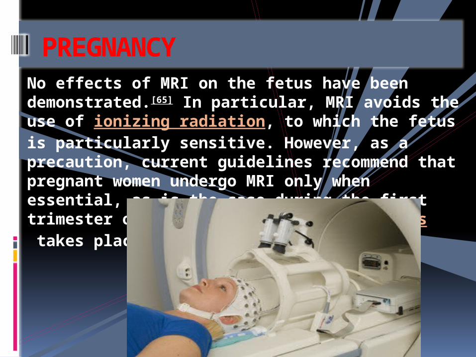
No effects of MRI on the fetus have been demonstrated.[65] In particular, MRI avoids the use of ionizing radiation, to which the fetus is particularly sensitive. However, as a precaution, current guidelines recommend that pregnant women undergo MRI only when essential, as is the case during the first trimester of pregnancy, when organogenesis takes place during this period.
PREGNANCY

Although painless, MRI scans can be unpleasant for those who are claustrophobic or otherwise uncomfortable with the imaging device surrounding them. Older closed bore MRI systems have a fairly long tube or tunnel. The part of the body being imaged must lie at the center of the magnet, which is at the absolute center of the tunnel. Because scan times on these older scanners may be long (occasionally up to 40 minutes for the entire procedure), people with even mild claustrophobia are sometimes unable to tolerate an MRI scan without management.
CLAUSTROPHOBIA AND DISCOMFORT

MRI and computed tomography (CT) are complementary imaging technologies and each has advantages and limitations for particular applications. CT is more widely used than MRI in OECD countries with a mean of 132 vs.
MRI VERSUS CT


Safety issues, including the potential for biostimulation device interference, movement of ferromagnetic bodies, and incidental localized heating, have been addressed in the American College of Radiology's White Paper on MR Safety, which was originally published in 2002 and expanded in 2004.
GUIDANCE

To perform a study, the person is positioned within an MRI scanner that forms a strong magnetic field around the area to be imaged.
PROCEDURE

Image contrast may be weighted to demonstrate different anatomical structures or pathologies.
CONTRAST

MRI for imaging anatomical structures or blood flow do not require contrast agents as the varying properties of the tissues or blood provide natural contrasts. However, for more specific types of imaging the most commonly used intravenous contrast agents are based on cheats of gadolinium.
CONTRAST AGENTS

Magnetic resonance imaging was invented by Paul C. Lautenberg in September 1971; he published the theory behind it in March 1973. The factors leading to image contrast (differences in tissue relaxation time values) had been described nearly 20 years earlier by Erik Odeblad (physician and scientist) and Gunnar Windstorm.
HISTORY


THANK
YOU