magnetic resonance imaging findings of cellular
TRANSCRIPT
Ntorkou et al. Journal of Medical Case Reports (2016) 10:71 DOI 10.1186/s13256-016-0861-3
CASE REPORT Open Access
Magnetic resonance imaging findings ofcellular angiofibroma of the tunica vaginalisof the testis: a case report
Alexandra A. Ntorkou1*, Athina C. Tsili1, Dimitrios Giannakis2, Anna Batistatou3, Sotirios Stavrou2,Nikolaos Sofikitis2 and Maria I. Argyropoulou1Abstract
Background: Cellular angiofibroma represents a rare mesenchymal tumor typically involving the inguinoscrotalarea in middle-aged men. Although the origin of this benign tumor is unknown, it is histologically classified as anangiomyxoid tumor. Cellular angiofibroma is characterized by a diversity of pathological and imaging features. Anaccurate preoperative diagnosis is challenging. Magnetic resonance imaging examination of the scrotum has beenreported as a valuable adjunct modality in the investigation of scrotal pathology. The technique by providing bothstructural and functional information is useful in the differentiation between extratesticular and intratesticulardiseases and in the preoperative characterization of the histologic nature of various scrotal lesions. There are fewreports in the English literature addressing the magnetic resonance imaging findings of cellular angiofibroma ofthe scrotum and no reports on functional magnetic resonance imaging data. Here we present the firstcase of a cellular angiofibroma arising from the tunica vaginalis of the testis and we discuss the value of amultiparametric magnetic resonance protocol, including diffusion-weighted imaging, magnetization transfer imagingand dynamic contrast-enhanced magnetic resonance imaging in the preoperative diagnosis of this rare neoplasm.
Case presentation: A 47-year Greek man presented with a painless left scrotal swelling, which had gradually enlargedduring the last 6 months. Magnetic resonance imaging of his scrotum displayed a left paratesticular mass, in closeproximity to the tunica vaginalis, with heterogeneous high signal intensity on T2-weighted images and no areas ofrestricted diffusion. The tumor was hypointense on magnetization transfer images, suggestive for the presence ofmacromolecules. On dynamic contrast-enhanced magnetic resonance imaging the mass showed intenseheterogeneous enhancement with a type II curve. Magnetic resonance imaging findings were strongly suggestive of abenign paratesticular tumor, which was confirmed on pathology following lesion excision.
Conclusions: Magnetic resonance imaging of the scrotum by combining conventional and functional magneticresonance data provides useful diagnostic information in the preoperative characterization of scrotal masses. A possiblediagnosis of a benign paratesticular tumor based on magnetic resonance imaging features may improve patient careand decrease the number of unnecessary radical surgical explorations.
Keywords: Cellular angiofibroma, Diffusion-weighted MRI, Magnetic resonance, Magnetization transfer contrastimaging, Tunica vaginalis
* Correspondence: [email protected] of Clinical Radiology, Medical School, University of Ioannina,45110 Ioannina, GreeceFull list of author information is available at the end of the article
© 2016 Ntorkou et al. Open Access This article is distributed under the terms of the Creative Commons Attribution 4.0International License (http://creativecommons.org/licenses/by/4.0/), which permits unrestricted use, distribution, andreproduction in any medium, provided you give appropriate credit to the original author(s) and the source, provide a link tothe Creative Commons license, and indicate if changes were made. The Creative Commons Public Domain Dedication waiver(http://creativecommons.org/publicdomain/zero/1.0/) applies to the data made available in this article, unless otherwise stated.

Fig. 1 Transverse T1-weighted image shows a well-demarcated leftparatesticular mass, lying adjacent to the scrotal wall. The lesion(arrowhead) demonstrated mainly similar signal intensity, whencompared to the ipsilateral displaced testis (arrow). Significant lefthydrocele (long arrow) was also observed
Ntorkou et al. Journal of Medical Case Reports (2016) 10:71 Page 2 of 6
BackgroundCellular angiofibroma (CA) or angiomyofibroblastoma(AMF)-like tumor is a rare benign mesenchymal tumor,typically involving the genital area of both genders. CAwas first described by Nucci et al. in 1997 and later byLaskin et al. in 1998 as a rare tumor arising in the para-testicular space in men and in the vulva in women [1, 2].Although the origin of this tumor is not clear, it hasbeen proposed that it derives from perivascular stemcells with potential to differentiate into fatty and myofi-broblastic tissue [1–8]. Only a few cases of CA of thescrotum have been reported to date [3–8].Magnetic resonance imaging (MRI) of the scrotum has
been proposed as a valuable alternative imaging techniquefor the evaluation of scrotal pathology [9–15]. Recently,functional MRI, including diffusion-weighted imaging(DWI), magnetization transfer imaging (MTI) and dy-namic contrast-enhanced (DCE) MRI have added import-ant additional diagnostic information in the interpretationof scrotal diseases [9–14]. Here we present the first caseof a CA of the tunica vaginalis of the testis, evaluatedwith a multimodal magnetic resonance (MR) protocol,including DWI, MTI and DCE MRI.
Case presentationA 47-year-old Greek man presented to our hospital witha painless left scrotal swelling, which he had for 2 yearsand which had gradually enlarged during the last 6months. No prior history of trauma or genitourinarytract infection was reported. A physical examinationrevealed a painless, firm scrotal mass, associated withhydrocele. No abnormal skin changes were observed.Laboratory tests, including serum tumor markers wereunremarkable.Sonographic examination of his scrotum showed the
presence of a large left extratesticular mass, of heteroge-neous echotexture. Significant hydrocele was alsodetected ipsilaterally. Color Doppler assessment revealedrich lesion vascularity.An MRI examination was performed on a 1.5-T magnet,
with the use of a circular surface coil. The MR protocol in-cluded axial spin-echo T1-weighted sequences and fastspin-echo T2-weighted sequences in three orthogonalplanes. Transverse diffusion-weighted (DW) images wereobtained using a single shot, multi-slice, spin-echo planarsequence with b-values of 0 and 900 second/mm−2.MTI was performed in the axial plane using a three-dimensional gradient-echo sequence, both with andwithout an on-resonance binomial prepulse to saturatethe broad resonance of immobile macromolecularprotons. The magnetization transfer ratio (MTR) wascalculated as follows: SIo–SIm/SIo × 100, where SIm andSIo refer to signal intensities with and without the satur-ation pulse, respectively. Coronal DCE subtraction MR
images were also obtained after an intravenous injectionof gadopentetate dimeglumine, with the use of athree-dimensional fast field-echo sequence. The patternsof contrast enhancement of both his normal testis and theextratesticular lesion were evaluated and time–signalintensity (TSI) curves were created.His spermatic cords, epididymis, and right testis were
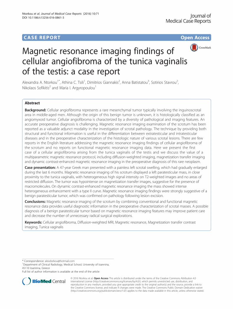
normal. A well-demarcated left paratesticular mass(Figs. 1, 2, 3, 4, and 5) in close proximity to his tunicavaginalis, displacing his ipsilateral testis was detected,measuring 5.5×4.8×4.3 cm. The tumor was inhomogen-eous, mainly with signal intensity similar and slightlyhigher than that of his normal testis on T1 (Fig. 1) andT2-weighted (Fig. 2) images, respectively. No areas ofrestricted diffusion were noted on DW images (Fig. 3).Magnetization transfer (MT) images showed low signalintensity for both the paratesticular tumor and thenormal testis, suggestive for the presence of macromole-cules (Fig. 4). On DCE sequences, the mass showedstrong heterogeneous enhancement (Fig. 5a) with a latepeak, followed by a plateau in the late contrast-enhanced period (type II curve, Fig. 5b). His left testisenhanced moderately and homogeneously with a linearincrease of signal intensity during the entire dynamicperiod (type I curve, Fig. 5c). MRI findings were stronglysuggestive of a benign paratesticular tumor.At left inguinal exploration a hard mass was seen in
his paratesticular region, separated from his testis andenucleation of the lesion was followed. Histopathologyrevealed a neoplasm confined to the parietal lamina of
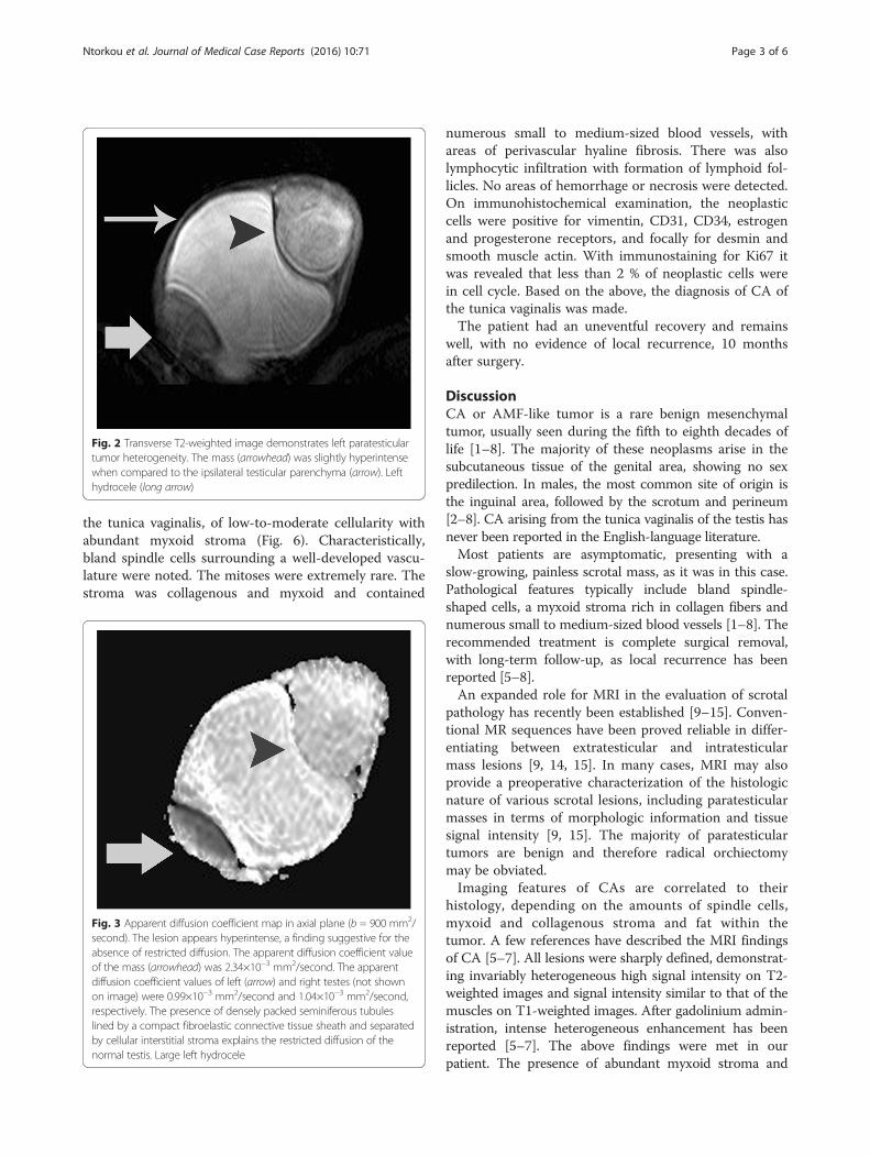
Fig. 2 Transverse T2-weighted image demonstrates left paratesticulartumor heterogeneity. The mass (arrowhead) was slightly hyperintensewhen compared to the ipsilateral testicular parenchyma (arrow). Lefthydrocele (long arrow)
Ntorkou et al. Journal of Medical Case Reports (2016) 10:71 Page 3 of 6
the tunica vaginalis, of low-to-moderate cellularity withabundant myxoid stroma (Fig. 6). Characteristically,bland spindle cells surrounding a well-developed vascu-lature were noted. The mitoses were extremely rare. Thestroma was collagenous and myxoid and contained
Fig. 3 Apparent diffusion coefficient map in axial plane (b = 900 mm2/second). The lesion appears hyperintense, a finding suggestive for theabsence of restricted diffusion. The apparent diffusion coefficient valueof the mass (arrowhead) was 2.34×10⁻³ mm2/second. The apparentdiffusion coefficient values of left (arrow) and right testes (not shownon image) were 0.99×10⁻³ mm2/second and 1.04×10⁻³ mm2/second,respectively. The presence of densely packed seminiferous tubuleslined by a compact fibroelastic connective tissue sheath and separatedby cellular interstitial stroma explains the restricted diffusion of thenormal testis. Large left hydrocele
numerous small to medium-sized blood vessels, withareas of perivascular hyaline fibrosis. There was alsolymphocytic infiltration with formation of lymphoid fol-licles. No areas of hemorrhage or necrosis were detected.On immunohistochemical examination, the neoplasticcells were positive for vimentin, CD31, CD34, estrogenand progesterone receptors, and focally for desmin andsmooth muscle actin. With immunostaining for Ki67 itwas revealed that less than 2 % of neoplastic cells werein cell cycle. Based on the above, the diagnosis of CA ofthe tunica vaginalis was made.The patient had an uneventful recovery and remains
well, with no evidence of local recurrence, 10 monthsafter surgery.
DiscussionCA or AMF-like tumor is a rare benign mesenchymaltumor, usually seen during the fifth to eighth decades oflife [1–8]. The majority of these neoplasms arise in thesubcutaneous tissue of the genital area, showing no sexpredilection. In males, the most common site of origin isthe inguinal area, followed by the scrotum and perineum[2–8]. CA arising from the tunica vaginalis of the testis hasnever been reported in the English-language literature.Most patients are asymptomatic, presenting with a
slow-growing, painless scrotal mass, as it was in this case.Pathological features typically include bland spindle-shaped cells, a myxoid stroma rich in collagen fibers andnumerous small to medium-sized blood vessels [1–8]. Therecommended treatment is complete surgical removal,with long-term follow-up, as local recurrence has beenreported [5–8].An expanded role for MRI in the evaluation of scrotal
pathology has recently been established [9–15]. Conven-tional MR sequences have been proved reliable in differ-entiating between extratesticular and intratesticularmass lesions [9, 14, 15]. In many cases, MRI may alsoprovide a preoperative characterization of the histologicnature of various scrotal lesions, including paratesticularmasses in terms of morphologic information and tissuesignal intensity [9, 15]. The majority of paratesticulartumors are benign and therefore radical orchiectomymay be obviated.Imaging features of CAs are correlated to their
histology, depending on the amounts of spindle cells,myxoid and collagenous stroma and fat within thetumor. A few references have described the MRI findingsof CA [5–7]. All lesions were sharply defined, demonstrat-ing invariably heterogeneous high signal intensity on T2-weighted images and signal intensity similar to that of themuscles on T1-weighted images. After gadolinium admin-istration, intense heterogeneous enhancement has beenreported [5–7]. The above findings were met in ourpatient. The presence of abundant myxoid stroma and

Fig. 4 Axial three-dimensional gradient-echo magnetic resonance image before (a) and after (b) the application of the magnetization transfer prepulse.The magnetization transfer ratio (in percent) of the left paratesticular mass (arrowhead) was 44.6, similar to that of the contralateral normal testis (46 %,not shown on images). The left testis (arrow) was displaced and compressed and the measurement of the magnetization transfer ratio was impossibledue to artifacts. Left hydrocele (long arrow)
Fig. 5 a Coronal dynamic contrast-enhanced subtracted image at early phase (180 seconds) and the corresponding time–signal intensity curvesof the b left paratesticular mass and the c ipsilateral testis. The tumor (arrowhead, a) enhanced strongly and heterogeneously, showing an initialupstroke followed by a plateau in the late contrast-enhanced phase (type II curve, b). Dynamic contrast-enhanced magnetic resonance findingswere strongly suggestive of benignity. Left testis (not shown on image) showed a linear increase of contrast enhancement throughoutthe examination (type I curve, c). Normal contralateral testis (arrow, a). AU arbitrary units
Ntorkou et al. Journal of Medical Case Reports (2016) 10:71 Page 4 of 6

Fig. 6 a Histopathologic evaluation revealed a highly vascularizedtumor, composed of bland spindle-shaped cells (hematoxylin andeosin ×100). b The stroma was mostly myxoid (periodic acid–Schiff –Alcian blue ×100)
Ntorkou et al. Journal of Medical Case Reports (2016) 10:71 Page 5 of 6
numerous tumoral vessels probably account for lesionhyperintensity on T2-weighted images and the patterns ofcontrast enhancement, respectively. However, conven-tional MRI findings are usually non-specific. Differentialdiagnosis should include aggressive angiomyxoma,AMF, solitary fibrous tumor, spindle-cell lipoma, well-differentiated liposarcoma and myxoid liposarcoma [1–8].The presence of intratumoral fat has been reported in
24 to 56 % of CAs [2–7]. Miyajima et al. concluded thatin the presence of a well-circumscribed hypervascularmass containing fat in the inguinal region of a male thediagnosis of CA should be considered [5]. No areas ofmacroscopic fat were observed within the neoplasm inour patient.In our case, a multiparametric MR protocol was used
to evaluate the paratesticular mass, including DWI, MTIand DCE MRI. DWI is an evolving technique that canbe used to improve tissue characterization when inter-preted in combination with conventional MRI findings[9–11]. Lesion detection and characterization on DWI isprimarily dependent on the extent of tissue cellularity,
and increased cellularity, mostly seen in malignancies, isassociated with restricted diffusion and reduced appar-ent diffusion coefficient (ADC) [9–11]. By combininghigh b-value DWI with conventional MRI findings ahigh accuracy has been reported in differentiating be-tween normal, benign and malignant scrotal contents[9–11]. Maruyama et al. reported a case of a 72-yearold man with AMF-like tumor of the scrotum, with-out areas of restricted diffusion on high b-value DWimages [6]. In our patient, his paratesticular tumorhad high ADC, a finding highly suggestive of thebenign nature of the lesion.MTI provides a different image contrast compared
to the conventional MR sequences, which dependsmainly on the concentration of macromolecules [12].Protons in tissues are divided into free water protonsand restricted protons, which are bound to proteinsand macromolecules. The MT phenomenon is deter-mined by the restricted macromolecular protons andis quantified by the MTR [13]. There is very limitedexperience in using MTI in the evaluation of scrotalpathology [12]. In the normal testis high MTR valuesrelated to macromolecular structures implicated withthe secretory activity have been previously reported[12]. In the present case, CA appeared hypointenseon MT images, with an MTR similar to that of nor-mal testicular parenchyma, and this could probably beexplained by the high collagen content seen on hist-ology. Collagen has been reported as an importantdeterminant of relaxation times in MTI, with largemolecular size and extensive intramolecular and inter-molecular cross-linking being the characteristics ofcollagen responsible for the MTI effect [12].DCE MRI provides information regarding the
characteristics of microvasculature of testicular car-cinomas, assessing tumor angiogenesis [13, 14]. DCEsubtracted MRI has been proved useful in differenti-ating testicular neoplasms and benign intratesticularlesions [9, 13, 14]. In a previous study, we classifiedthe progression of enhancement of testicular lesionsaccording to the shape of the TSI curves into threetypes: Type I curve, with a gradual increase ofcontrast enhancement during the entire dynamicperiod, found to represent patterns of enhancementof normal testis; type II curve, with an initial up-stroke, after which the signal intensity either plateausor gradually increases in the late contrast-enhancedperiod, suggestive of a benign diagnosis; and type IIIcurve, with an initial upstroke, followed by gradualwashout of the contrast medium, indicating the diag-nosis of malignancy [13]. In our patient, CA showedintense heterogeneous enhancement, followed by aplateau at the delayed phase (type II curve), closelycorrelating to the diagnosis of benignity.

Ntorkou et al. Journal of Medical Case Reports (2016) 10:71 Page 6 of 6
ConclusionsMRI by combining conventional and functional datamay provide valuable information in the pre-surgicalwork-up, helping in the characterization of the benignnature of paratesticular tumors. A possible diagnosisof benignity based on MRI features may obviateunnecessary radical orchiectomies. A multiparametricMR protocol, including DWI, MTI and DCE MRImay provide important additional diagnostic informa-tion in the interpretation of scrotal pathology.
ConsentWritten informed consent was obtained from the patientfor publication of this case report and accompanyingimages. A copy of the written consent is available forreview by the Editor-in-Chief of this journal.
AbbreviationsADC: apparent diffusion coefficient; AMF: angiomyofibroblastoma; CA: cellularangiofibroma; DCE: dynamic contrast-enhanced; DW: diffusion-weighted;DWI: diffusion-weighted imaging; MR: magnetic resonance; MRI: magneticresonance imaging; MT: magnetization transfer; MTI: magnetization transferimaging; MTR: magnetization transfer ratio; SIm: signal intensity after theapplication of the saturation pulse; SIo: signal intensity without the saturationpulse; TSI: time–signal intensity.
Competing interestsThe authors declare that they have no competing interests.
Authors’ contributionsAN, ACT, AB and MIA were major contributors in writing the manuscript. DG,SS and NS contributed to conception and data acquisition, and also inwriting this manuscript. All authors read and approved the final manuscript.
AcknowledgementsThis research received no specific grant from any funding agency in thepublic, commercial, or not-for-profit sectors.
Author details1Department of Clinical Radiology, Medical School, University of Ioannina,45110 Ioannina, Greece. 2Department of Urology, Medical School, Universityof Ioannina, 45110 Ioannina, Greece. 3Department of Pathology, MedicalSchool, University of Ioannina, 45110 Ioannina, Greece.
Received: 21 September 2015 Accepted: 2 March 2016
References1. Nucci MR, Granter SR, Fletcher CD. Cellular angiofibroma: a benign neoplasm
distinct from angiomyofibroblastoma and spindle cell lipoma. Am J SurgPathol. 1997;21:636–44.
2. Laskin WB, Fetsch JF, Mostofi FK. Angiomyofibroblastoma-like tumor of themale genital tract: analysis of 11 cases with comparison to femaleangiomyofibroblastoma and spindle cell lipoma. Am J Surg Pathol.1998;22:6–16.
3. Hara N, Kawaquchi M, Koike H, Nishiyama T, Takahashi K. Angiomyxoidtumor with an intermediate feature between cellular angiofibromaand angiomyofibroblastoma in the male inguinal region. Int J Urol.2005;12:768–72.
4. Shintaku M, Naitou M, Nakashima Y. Angiomyofibroblastoma-like tumor(lipomatous variant) of the inguinal region of a male patient. Pathol Int.2002;52:619–22.
5. Miyajima K, Hasegawa S, Oda Y, Toyoshima S, Tsuneyoshi M, Motooka M,et al. Angiomyofibroblastoma-like tumor (cellular angiofibroma) in the maleinguinal region. Radiat Med. 2007;25:173–7.
6. Maruyama M, Yoshizako T, Kitagaki H, Araki A, Igawa M. Magnetic resonanceimaging features of angiomyofibroblastoma-like tumor of the scrotum withpathologic correlates. Clin Imaging. 2012;36:632–5.
7. Aytaç B, Yalçinkaya U, Vuruşkan H. Angiomyofibroblastoma-like tumor ofthe scrotum: a case report and review of literature. Turk Patoloji Derg.2012;28:168–71.
8. Dikaiakos P, Zizi-Sermpetzoglou A, Rizos S, Marinis A. Angiofibroma of thespermatic cord: a case report and a review of the literature. J Med CaseRep. 2011;5:423.
9. Tsili AC, Giannakis D, Sylakos A, Ntorkou A, Sofikitis N, Argyropoulou MI. MRimaging of scrotum. Magn Reson Imaging Clin N Am. 2014;22:217–38.
10. Tsili AC, Argyropoulou MI, Giannakis D, Tsampalas S, Sofikitis N, Tsampoulas K.Diffusion-weighted MR imaging of normal and abnormal scrotum: preliminaryresults. Asian J Androl. 2012;14:649–54.
11. Tsili AC, Argyropoulou MI, Giannakis D, Sofikitis N, Tsampoulas K.Conventional and diffusion-weighted magnetic resonance imaging findingsof benign fibromatous paratesticular tumor: a case report. J Med Case Rep.2011;5:169.
12. Tsili AC, Ntorkou A, Baltogiannis D, Sylakos A, Stavrou S, Astrakas L, et al.Magnetization transfer imaging of normal and abnormal testis: preliminaryresults. Eur Radiol. 2015. doi:10.1007/s00330-015-3867-0.
13. Tsili AC, Argyropoulou MI, Astrakas LG, Ntoulia EA, Giannakis D, Sofikitis N,et al. Dynamic contrast-enhanced subtraction MRI for characterizingintratesticular mass lesions. AJR Am J Roentgenol. 2013;200:578–85.
14. Watanabe Y, Dohke M, Ohkubo K, Ishimori T, Amoh Y, Okumura A, et al.Scrotal disorders: evaluation of testicular enhancement patterns at dynamiccontrast-enhanced subtraction MR imaging. Radiology. 2000;217:219–27.
15. Akbar SA, Sayyed TA, Jafri SZ, Hasteh F, Neill JS. Multimodality imagingof paratesticular neoplasms and their rare mimics. Radiographics.2003;23:1461–76.
• We accept pre-submission inquiries
• Our selector tool helps you to find the most relevant journal
• We provide round the clock customer support
• Convenient online submission
• Thorough peer review
• Inclusion in PubMed and all major indexing services
• Maximum visibility for your research
Submit your manuscript atwww.biomedcentral.com/submit
Submit your next manuscript to BioMed Central and we will help you at every step: