malondialdehyde adduct to hemoglobin: a new marker of oxidative
TRANSCRIPT
Hindawi Publishing CorporationOxidative Medicine and Cellular LongevityVolume 2013, Article ID 694014, 6 pageshttp://dx.doi.org/10.1155/2013/694014
Clinical StudyMalondialdehyde Adduct to Hemoglobin: A New Marker ofOxidative Stress Suitable for Full-Term and Preterm Neonates
Cécile Cipierre,1,2 Stéphane Haÿs,2,3 Delphine Maucort-Boulch,4,5,6 Jean-Paul Steghens,3,7
and Jean-Charles Picaud2,3,5
1 Departement de Neonatologie, CHU Angers, 4 rue Larrey, 49100 Angers, France2Departement de Neonatologie, Hopital de la Croix Rousse, Hospices Civils de Lyon, 69004 Lyon, France3 Centre de Recherche en Nutrition Humaine Rhone-Alpes, Centre Hospitalier Lyon-Sud, 69310 Pierre Benite, France4Departement de Biostatistique, Hospices Civils de Lyon, 69003 Lyon, France5 Universite Claude Bernard Lyon I, 69100 Villeurbanne, France6CNRS UMR5558, Laboratoire de Biometrie et Biologie Evolutive, Equipe Biostatistique Sante, 69310 Piere-Benite, France7 Centre de Biologie Sud, UF Nutrition et Metabolisme, Centre Hospitalier Lyon-Sud, Hospices Civils de Lyon,69310 Pierre Benite, France
Correspondence should be addressed to Cecile Cipierre; [email protected]
Received 8 February 2013; Revised 13 April 2013; Accepted 16 April 2013
Academic Editor: Kota V. Ramana
Copyright © 2013 Cecile Cipierre et al.This is an open access article distributed under the Creative Commons Attribution License,which permits unrestricted use, distribution, and reproduction in any medium, provided the original work is properly cited.
Oxidative stress may play a central role in the onset of many diseases during the neonatal period. Malondialdehyde (MDA) isa marker of lipid peroxidation. The aim of this study was to evaluate a new marker, the malondialdehyde adduct to hemoglobin(MDA-Hb), which ismeasured in red blood cells (RBCs) and thus does not require that an additional blood sample be drawn. In thisprospective study, we first adapted themeasurementmethod previously described toHb solutions obtained fromwashed RBCs andthen evaluated the suitability of themethod for use in neonates.MDA-Hb concentrationsweremeasured by liquid chromatography-mass spectrometry. We compared the concentrations of MDA-Hb between preterm and term neonates. Erythrocyte samples werecollected at birth from 60 healthy neonates (29 full-term and 31 preterm), as well as from 50 preterm neonates with uncomplicatedpostnatal evolution during the first months of life. We found a significantly higher MDA-Hb concentration at birth in pretermneonates (𝑃 = 0.002). During the first months of life, MDA-Hb concentrations were 9.4 nanomol/g Hb in hospitalized pretermneonates. MDA-Hb could be used to assess oxidative stress in preterm neonates. Together with clinical variables, it could be auseful marker for oxidative stress exposition in these higher risk patients.
1. Introduction
Growing evidence indicates that an imbalance betweenoxidative stress (OS) and antioxidant defense mechanismsplays an important role in the onset of many diseases duringthe neonatal period [1, 2]. Birth is associated with a strongOS related to the rapid change from a relatively hypoxicintrauterine environment to the extrauterine environment(5-fold increase in alveolar PO
2) and several physiologic
processes involved in the delivery [3, 4]. These changes andprocesses greatly increase the production of free radicals,which must be controlled by the antioxidant defense systemthat has been maturing over the course of gestation [5, 6].
Free radicals are too short-lived to be detected directly inclinical systems, but oxygen free radicals react with lipids toproduce lipid peroxidation products, which when measuredserve as indirect biomarkers of in vivo oxidative stress statusand related diseases. Among these products, malondialde-hyde (MDA) is one of the principal and most studied low-molecular-weight end products. It is highly cytotoxic becauseof its ability to bind proteins or nucleic acids very quickly [7].
The thiobarbituric acid reactive substance method(TBAR test) has been frequently used to assess MDAconcentrations, but it lacks specificity as aldehydes otherthan MDA (formaldehyde, acetaldehyde, etc.) and nonlipid substances react with thiobarbituric acid [8, 9]. Other
2 Oxidative Medicine and Cellular Longevity
analytical techniques such as specific derivatization beforeliquid chromatography with UV or mass spectrometrydetection have been proposed to measure MDA moreprecisely [10]. It should be noted that all these techniquesreflect measures only at a specific moment, althoughperoxidation reactions fluctuate over time.
Determining adducts to proteins, particularly Hb, hasproved to be a useful approach for monitoring in vivoexposure to genotoxic compounds [11]. An adduct is formedwhen a low molecular compound binds with a biologicalmolecule [11]. The Hb adducts are stable reaction productsderived from electrophilic compounds, by covalent attach-ment, involving nucleophilic centers in biomolecules thatoffer possibilities for the sampling and analysis of elec-trophilic, short-lived compounds [12].
The determination of MDA-Hb obtained with a verycomplex method has thus been proposed to assess theexposure to lipid peroxidation products [13]. In healthyadults, MDA-Hb values of 0.01 to 10 nanomol/g Hb werereported [14]. Moreover, the method used in these studieswas sensitive enough to detect variations in the levels ofMDAadducts caused by lipid peroxidation,with a direct correlationbetween dose and effect [15].
MDA-Hb measurement could provide an assessment ofOS in neonates over the middle term because of (i) the stablecovalent attachment between MDA and Hb and (ii) MDA-Hb elimination, which is dependent on the relatively well-defined life span of the erythrocyte (120 days in human adults,shorter in neonates) [16]. We tested whether we would beable to determine MDA-Hb in neonates without taking moreblood from them, as this is a crucial issue in very low birthweight (VLBW) neonates. We assumed that the erythrocytesremaining from routine blood samples taken for electrolytedetermination would be sufficient.
We thus evaluated the feasibility of MDA-Hb measure-ment in neonates and sought to establish normal MDA-Hbranges in healthy full-term neonates at birth and uncompli-cated premature neonates at birth and during the firstmonthsof life.
2. Methods
2.1. Study Populations. To assess MDA-Hb at birth, neonatesborn on the maternity ward of Croix Rousse UniversityHospital, Lyon, France, were consecutively enrolled betweenFebruary and May 2009. They were divided in two groupsaccording to their gestational age (GA). In the first group,inclusion criteria were: healthy neonates born after a full-term gestation (≥37 weeks), delivered vaginally without com-plications, and presenting good adaptation to extrauterinelife (no resuscitation, no evidence of perinatal hypoxia, orrespiratory distress), birth weight (BW) appropriate for GA[17], and a normal clinical examination at birth. In the secondgroup, inclusion criteria were: premature neonates (28–36weeks) who did not require intensive care (no intubation, nochest compression, or drugs for resuscitation), oxygen ther-apy, or any type ofmedication at birth. Exclusion criteria wereas follows: the need for resuscitation, evidence of perinatal
hypoxia (pH ≤ 7.20 in cord blood, 5min Apgar score < 7)or respiratory distress, congenital malformation, sepsis, andsmall for GA [17], and multiple gestation. Measurements ofMDA-Hb at birth were performed in 29 full-term and 31premature neonates.
To measure MDA-Hb in the first months of life, wecollected blood samples from uncomplicated prematureneonates, that is, without assisted ventilation, parenteralnutrition, blood transfusion, or anti-inflammatory treatmentwithin the five days before blood sampling. We recorded BW,GA, gender, and postnatal age on the day of sample collection.Measurements of MDA-Hb during the first 8 weeks of lifewere performed in 50 uncomplicated preterm neonates aspreviously defined.
The study was completely integrated into the routinecare of full-term and preterm neonates and there was nosupplementary blood sampling required. All parents signedan informed consent form. The study was approved by theEthics Committee of the University Hospital Center of Lyon,France, (CPP Lyon Sud Est IV).
2.2. Determination of MDA-Hb
2.2.1. Collection of Samples. The assessment of MDA-Hbrequired the collection of erythrocytes. At the time of birth,arterial cord blood (5mL) was collected from full-term andpreterm neonates who fulfilled the inclusion criteria. As cordblood pH is systematically assessed at birth in our hospital,blood samples for the purpose of our studywere also obtainedat that time.
During the hospitalization of the preterm neonates whofulfilled the inclusion criteria, erythrocyte samples werecollected at the same time as the venous blood samplingusually required for routine assessment of serum electrolytesin these neonates. According to the routine procedure, serumand red blood cells were separated by centrifugation (10minutes, 4000G) in the hospital biochemistry laboratory.Thetubes were then immediately refrigerated at 4∘C until thepreparation of samples.
2.2.2. Reagents. Malondialdehyde-bis-dimethylacetal (tetra-methoxypropane, TMP) 99% purity and metaphosphoricacid were purchased from sigma- Aldrich Chemie GmbH(Steinheim, Germany). Dideuterated tetraethoxypropanewas from CDN Isotopes (88 Leacock Street, Pointe-Claire,QC, Canada). 2,3-Diaminonaphthalene (DAN) was obtainedfrom TCI. Anhydrous potassium dihydrogen phosphate(Suprapur), ethanol, methanol, and acetonitrile gradientgrade were from Merck (Darmstadt, Germany). All otherchemicals and solvents were of analytical grade. Aque-ous solutions were made with pure water (conductivity ≥18MΩ), Elga Pure Lab Option (Elga, France).
Standard stock solution of TMP 608 nM was obtainedby seven successive dilutions (1/10) of TMP: three times inethanol then three times in ethanol/water (40/60, v/v) andfinally in water with NH
4OH 90mM final concentration.
Aliquots of this solution are stable under nitrogen for 3months at −20∘C in the dark. Secondary MDA standards (76,
Oxidative Medicine and Cellular Longevity 3
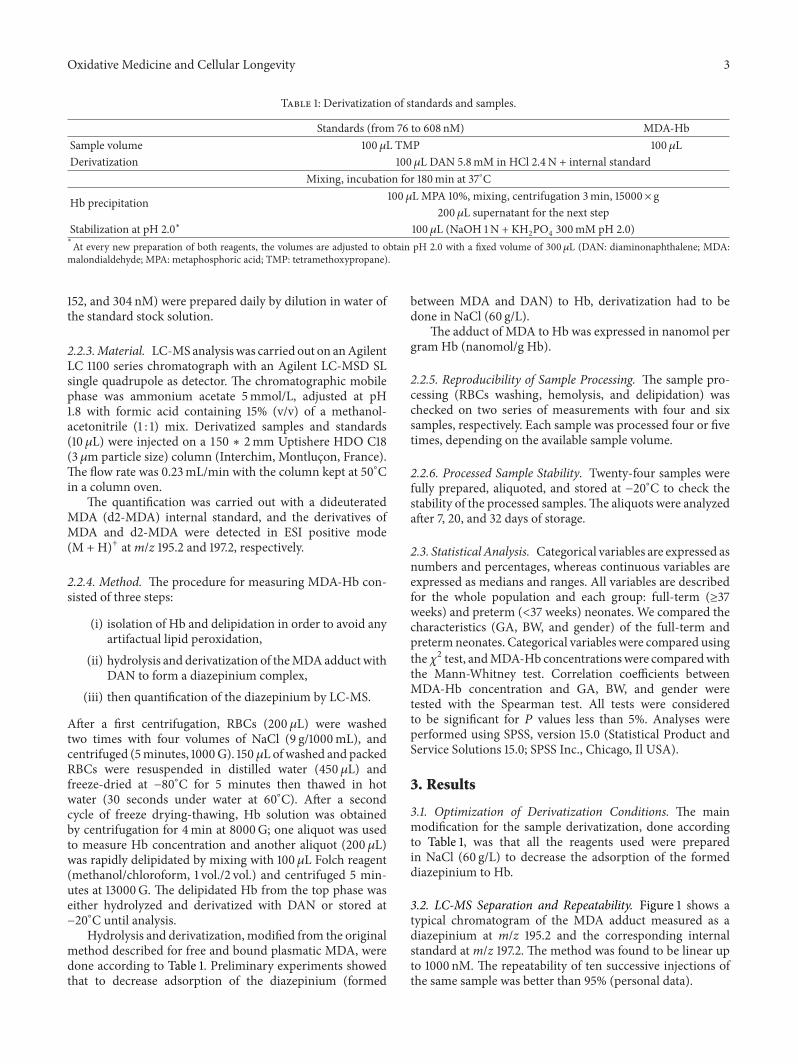
Table 1: Derivatization of standards and samples.
Standards (from 76 to 608 nM) MDA-HbSample volume 100𝜇L TMP 100𝜇LDerivatization 100𝜇L DAN 5.8mM in HCl 2.4N + internal standard
Mixing, incubation for 180min at 37∘C
Hb precipitation 100𝜇L MPA 10%, mixing, centrifugation 3min, 15000× g200 𝜇L supernatant for the next step
Stabilization at pH 2.0∗ 100𝜇L (NaOH 1N + KH2PO4 300mM pH 2.0)∗
At every new preparation of both reagents, the volumes are adjusted to obtain pH 2.0 with a fixed volume of 300𝜇L (DAN: diaminonaphthalene; MDA:malondialdehyde; MPA: metaphosphoric acid; TMP: tetramethoxypropane).
152, and 304 nM) were prepared daily by dilution in water ofthe standard stock solution.
2.2.3.Material. LC-MS analysiswas carried out on anAgilentLC 1100 series chromatograph with an Agilent LC-MSD SLsingle quadrupole as detector. The chromatographic mobilephase was ammonium acetate 5mmol/L, adjusted at pH1.8 with formic acid containing 15% (v/v) of a methanol-acetonitrile (1 : 1) mix. Derivatized samples and standards(10 𝜇L) were injected on a 150 ∗ 2mm Uptishere HDO C18(3 𝜇m particle size) column (Interchim, Montlucon, France).The flow rate was 0.23mL/min with the column kept at 50∘Cin a column oven.
The quantification was carried out with a dideuteratedMDA (d2-MDA) internal standard, and the derivatives ofMDA and d2-MDA were detected in ESI positive mode(M +H)+ atm/z 195.2 and 197.2, respectively.
2.2.4. Method. The procedure for measuring MDA-Hb con-sisted of three steps:
(i) isolation of Hb and delipidation in order to avoid anyartifactual lipid peroxidation,
(ii) hydrolysis and derivatization of theMDAadduct withDAN to form a diazepinium complex,
(iii) then quantification of the diazepinium by LC-MS.
After a first centrifugation, RBCs (200𝜇L) were washedtwo times with four volumes of NaCl (9 g/1000mL), andcentrifuged (5minutes, 1000G). 150 𝜇L of washed and packedRBCs were resuspended in distilled water (450𝜇L) andfreeze-dried at −80∘C for 5 minutes then thawed in hotwater (30 seconds under water at 60∘C). After a secondcycle of freeze drying-thawing, Hb solution was obtainedby centrifugation for 4min at 8000G; one aliquot was usedto measure Hb concentration and another aliquot (200𝜇L)was rapidly delipidated by mixing with 100𝜇L Folch reagent(methanol/chloroform, 1 vol./2 vol.) and centrifuged 5 min-utes at 13000G. The delipidated Hb from the top phase waseither hydrolyzed and derivatized with DAN or stored at−20∘C until analysis.
Hydrolysis and derivatization, modified from the originalmethod described for free and bound plasmatic MDA, weredone according to Table 1. Preliminary experiments showedthat to decrease adsorption of the diazepinium (formed
between MDA and DAN) to Hb, derivatization had to bedone in NaCl (60 g/L).
The adduct of MDA to Hb was expressed in nanomol pergram Hb (nanomol/g Hb).
2.2.5. Reproducibility of Sample Processing. The sample pro-cessing (RBCs washing, hemolysis, and delipidation) waschecked on two series of measurements with four and sixsamples, respectively. Each sample was processed four or fivetimes, depending on the available sample volume.
2.2.6. Processed Sample Stability. Twenty-four samples werefully prepared, aliquoted, and stored at −20∘C to check thestability of the processed samples.The aliquots were analyzedafter 7, 20, and 32 days of storage.
2.3. Statistical Analysis. Categorical variables are expressed asnumbers and percentages, whereas continuous variables areexpressed as medians and ranges. All variables are describedfor the whole population and each group: full-term (≥37weeks) and preterm (<37 weeks) neonates. We compared thecharacteristics (GA, BW, and gender) of the full-term andpretermneonates. Categorical variables were compared usingthe𝜒2 test, andMDA-Hb concentrationswere comparedwiththe Mann-Whitney test. Correlation coefficients betweenMDA-Hb concentration and GA, BW, and gender weretested with the Spearman test. All tests were consideredto be significant for 𝑃 values less than 5%. Analyses wereperformed using SPSS, version 15.0 (Statistical Product andService Solutions 15.0; SPSS Inc., Chicago, Il USA).
3. Results
3.1. Optimization of Derivatization Conditions. The mainmodification for the sample derivatization, done accordingto Table 1, was that all the reagents used were preparedin NaCl (60 g/L) to decrease the adsorption of the formeddiazepinium to Hb.
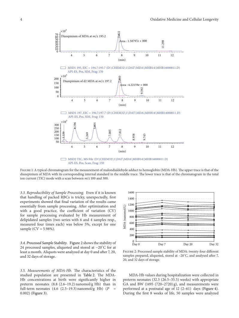
3.2. LC-MS Separation and Repeatability. Figure 1 shows atypical chromatogram of the MDA adduct measured as adiazepinium at m/z 195.2 and the corresponding internalstandard at m/z 197.2. The method was found to be linear upto 1000 nM. The repeatability of ten successive injections ofthe same sample was better than 95% (personal data).
4 Oxidative Medicine and Cellular Longevity
Diazepinium of MDA at m/z 195.2 7.82
2
11.2
98Area : 1.58797e + 000
4 5 6 7 8 9 10 11 12(min)
MSD1 195, EIC = 194.7:195.7 (D:\CHEM32\1\DAT\MDA\MHB14\MHB14000011.D)API-ES, Pos, SIM, Frag: 150
70605040302010
0
×103
4 5 6 7 8 9 10 11 12(min)
MSD1 197, EIC = 196.7:197.7 (D:\CHEM32\1\DAT\MDA\MHB14\MHB14000011.D)API-ES, Pos, SIM, Frag: 150
Diazepinium of d2 MDA at m/z 197.2 7.81
0
9.59
2
200150100
500
Area : 4.22159e + 000
×103
4 5 6 7 8 9 10 11 12(min)
MSD2 TIC, MS File (D:\CHEM32\1\DAT\MDA\MHB14\MHB14000011.D)API-ES, Pos, Scan, Frag: 150
3.33
9
4.19
6
5.26
9
5.80
1
7.00
5
7.81
2
8.95
8
9.74
3
200250300
150100
50
×103
Figure 1: A typical chromatogram for the measurement of malondialdehyde adduct to hemoglobin (MDA-Hb).The upper trace is that of thediazepinium of MDA with its corresponding internal standard in the middle trace. The lower trace is that of the chromatogram in the totalion current (TIC) mode with a scan betweenm/z 100 and 300.
3.3. Reproducibility of Sample Processing. Even if it is knownthat handling of packed RBCs is tricky, unexpectedly, firstexperiments showed that final variation of the results cameessentially from sample processing. After optimization andwith a good practice, the coefficient of variation (CV)for sample processing evaluated by Hb measurement ofdelipidated samples (two series with 6 and 4 samples resp.,measured four times each) was below 5%, except for onesample (CV = 5.98%).
3.4. Processed Sample Stability. Figure 2 shows the stability of24 processed samples, aliquoted and stored at −20∘C for atleast a month. Aliquots were analyzed at day 0 and after 7, 20,and 32 days of storage.
3.5. Measurements of MDA-Hb. The characteristics of thestudied population are presented in Table 2. The MDA-Hb concentrations at birth were significantly higher inpreterm neonates (8.8 (2.6–19.2) nanomol/g Hb) than infull-term neonates (4.6 (2.5–19.3) nanomol/g Hb) (𝑃 =0.002) (Figure 3).
Day 0 Day 7 Day 20 Day 32
MD
A (n
anom
ol/L
)
1600
1400
1200
1000
800
600
400
200
0
Figure 2: Processed sample stability of MDA: twenty-four differentsamples prepared, aliquoted, stored at –20∘C, and analysed after 7,20, and 32 days of storage.
MDA-Hb values during hospitalization were collected inpreterm neonates (32.3 (26.5–35.5) weeks) with appropriateGA and BW (1495 (720–2720) g), and measurements wereperformed at a postnatal age of 12 (2–61) days (Figure 4).During the first 8 weeks of life, 50 samples were analyzed

Oxidative Medicine and Cellular Longevity 5
Table 2: Clinical characteristics (mean (range unless specified 𝑛(%))) of 60 full-term neonates (GA ≥ 37 weeks, 𝑛 = 29) or preterm(GA < 37 weeks, 𝑛 = 31).
Full-termneonates
Pretermneonates P value
Gestational age atbirth, weeks 39.4 (37.4–41.6) 31.7 (28.1–35.7) <0.001
Birth weight, grams 3380 (2830–4270) 1580 (730–2580) <0.001Male, 𝑛 (%) 14 (48.3) 15 (48.4) 0.993
15
10
5
0
Healthy full-term neonates Healthy preterm neonates
MD
A-H
b (n
anom
ol/g
Hb)
∗
Figure 3: Concentrations of malondialdehyde adduct tohemoglobin (MDA-Hb) at birth in healthy full-term (𝑛 = 29)and healthy preterm neonates (𝑛 = 31). Values shown are medianlevels (25th/75th box; 10th/90th error bars). ∗Mann-Whitney test:significantly different from full-term neonates (𝑃 = 0.002).
in 50 uncomplicated preterm neonates (only one sample perpreterm neonate) as previously defined. MDA-Hb concen-trations were 9.4 (2.4–26.3) nanomol/g Hb. No correlationwas found between BW or patient gender and the MDA-Hbconcentration.
At birth, GA was significantly and negatively correlatedwith MDA-Hb concentration (𝑟 = −0.31, 𝑃 = 0.019).
4. Discussion
We report a convenient method to determine the concentra-tion of MDA adduct to Hb in neonates, which is sensitiveenough to detect low concentrations ofMDA-Hb and specificenough to assess lipid peroxidation.
The method to assess MDA-Hb is an adaptation of areliable and validatedmethod [10] used to evaluate the impactof parenteral nutrition composition on lipid peroxidation[18–20]. MDA-Hb was measured by LC-MS using a veryspecificmethod based on diaminonaphthalene derivatization[10].The advantages of themethod are its high sensitivity andspecificity which rely on the use of DAN. When reacted withMDA, DAN forms a diazepinium with a mass of 194 Daltonhigher than that of native MDA (72 Dalton) which makes itsdetection easier, because of an improved affinity for reversephase column and this higher mass outside the background
0 5
10 15 20 25 30
0 5 10 15 20 25 30 35 40 45 50 55 60 65 Postnatal age (days)
MD
A-H
b(n
anom
ol/g
Hb)
Birth
Figure 4: Relationship between concentrations of malondialdehydeadduct to hemoglobin (MDA-Hb) and postnatal age in healthypreterm neonates (𝑛 = 50).
noise. A direct consequence of this high sensitivity is the lowvolume of sample needed for measurement (only 200𝜇L).This is particularly interesting for neonatal care. Typicalintra-assay variability is good (5%), and good reproducibilitywas observed. Although this new method requires rigorousconditions for sample preparation andmeasurement andmaylead to the formation of artifacts, it has the advantage of beingrelatively simple, which is important for clinical practice. Ourresults thus suggest that this method is a useful, sensitive, andspecific assay for lipid peroxidation in neonates.
Furthermore, this assay has several advantages from aclinical point of view, not the least of them being the simplic-ity of collecting a component of blood (erythrocytes), that is,usually discarded after serum has been taken for electrolyteassessment from routine blood sampling. The relatively longand well-controlled life span of Hb was an important reasonfor choosing this protein as a dosemonitor for electrolyticallyreactive compounds. Hb has a predetermined life span equalto that of red blood cells. MDA-Hb could therefore be a long-term indicator of oxidative stress. Moreover, since proteinssuch as Hb are present in blood in much larger amounts,measurement of protein adducts favors a high capacity ofdetection [11].
We observed that themethod tomeasure Hb adducts wassensitive enough to detect variations in MDA-Hb concen-trations at different GAs at birth. As expected, we observedsignificantly higherMDA-Hb levels in preterm neonates thanin full-term neonates. Indeed, increased oxidative stress inpreterm neonates at birth has been described using otheroxidative stress markers [4, 6, 21, 22].
Based on these preliminary results, it appears that thismarker might be useful in a variety of pathological condi-tions, such as VLBWneonates, and thus, its clinical relevanceshould be validated in a larger population. The ability todetect lipid peroxidation noninvasively is of particular impor-tance in preterm neonatal care. A noninvasive techniquewould be a great aid in improving knowledge about oxidativestress in these patients and evaluating future improvementsin neonatal care (assisted ventilation, oxygen therapy, andparenteral nutrition) for diseases related to reactive oxygenspecies.
In summary, our results suggest that in vivo assessment ofMDA-Hb in neonates is a feasible, blood-sparing, and simplemethod to determine oxidative stress, notably in pretermneonates.
6 Oxidative Medicine and Cellular Longevity
Abbreviations
BW: Birth weightDAN: DiaminonaphthaleneGA: Gestational ageHb: HemoglobinLC-MS: Liquid chromatography-mass spectrometryMDA: MalondialdehydeMDA-Hb: Malondialdehyde adduct to hemoglobinRBCs: Red blood cellsROS: Reactive oxygen speciesTMP: TetramethoxypropaneOS: Oxidative stressVLBW: Very low birth weight.
Acknowledgments
The authors thank Brigitte Guy and Blandine Vallet-Sandrefor their help in collecting samples, Christine Delore, Cather-ine Castrichini, and Sophie Vasseur for their help in process-ing sample analysis, and C. Carmeni for editing the paper.
References
[1] O. D. Saugstad, “Update on oxygen radical disease in neonatol-ogy,” Current Opinion in Obstetrics and Gynecology, vol. 13, pp.147–153, 2001.
[2] E. Granot and R. Kohen, “Oxidative stress in childhood—inhealth and disease states,” Clinical Nutrition, vol. 23, no. 1, pp.3–11, 2004.
[3] O. D. Saugstad, “Mechanisms of tissue injury by oxygen rad-icals: implications for neonatal disease,” Acta Paediatrica, vol.85, no. 1, pp. 1–4, 1996.
[4] R. Robles, N. Palomino, and A. Robles, “Oxidative stress in theneonate,” Early Human Development, vol. 65, supplement 2, pp.S75–S81, 2001.
[5] S. Perrone, S. Negro, M. L. Tataranno, and G. Buonocore,“Oxidative stress and antioxidant strategies in newborns,” Jour-nal of Maternal-Fetal and Neonatal Medicine, vol. 23, no. 3, pp.63–65, 2010.
[6] J. M. Davis and R. L. Auten, “Maturation of the antioxidantsystem and the effects on preterm birth,” Seminars in Fetal andNeonatal Medicine, vol. 15, no. 4, pp. 191–195, 2010.
[7] H. Esterbauer, “Estimation of peroxidative damage. A criticalreview,” Pathologie Biologie, vol. 44, no. 1, pp. 25–28, 1996.
[8] D. del Rio, A. J. Stewart, and N. Pellegrini, “A review of recentstudies on malondialdehyde as toxic molecule and biologicalmarker of oxidative stress,” Nutrition, Metabolism and Cardio-vascular Diseases, vol. 15, no. 4, pp. 316–328, 2005.
[9] I. F. F. Benzie, “Lipid peroxidation: a review of causes, con-sequences, measurement and dietary influences,” InternationalJournal of Food Sciences andNutrition, vol. 47, no. 3, pp. 233–261,1996.
[10] J.-P. Steghens, A. L. van Kappel, I. Denis, and C. Collombel,“Diaminonaphtalene, a new highly specific reagent for HPLC-UV measurement of total and free malondialdehyde in humanplasma or serum,” Free Radical Biology andMedicine, vol. 15, no.31, pp. 242–249, 2001.
[11] M. Tornqvist, C. Fred, J. Haglund, H. Helleberg, B. Paulsson,and P. Rydberg, “Protein adducts: quantitative and qualitative
aspects of their formation, analysis and applications,” Journal ofChromatography B, vol. 778, no. 1-2, pp. 279–308, 2002.
[12] U. Groth and H.-G. Neumann, “The relevance of chemico-biological interactions for the toxic and carcinogenic effects ofaromatic amines V. the pharmacokinetics of related aromaticamines in blood,” Chemico-Biological Interactions, vol. 4, no. 6,pp. 409–419, 1972.
[13] A. Kautiainen, M. Tornqvist, K. Svensson, and S. Osterman-Golkar, “Adducts of malonaldehyde and a few other aldehydesto hemoglobin,” Carcinogenesis, vol. 10, no. 11, pp. 2123–2130,1989.
[14] M. Tornqvist and A. Kautiainen, “Adducted proteins for iden-tification of endogenous electrophiles,” Environmental HealthPerspectives, vol. 99, pp. 39–44, 1993.
[15] A. Kautiainen, M. Tornqvist, B. Anderstam, and C. E. Vaca, “Invivo hemoglobin dosimetry of malonaldehyde and ethene inmice after induction of lipid peroxidation. Effects of membranelipid fatty acid composition,” Carcinogenesis, vol. 12, no. 6, pp.1097–1102, 1991.
[16] J. L. Naiman and E. A. Oski, Hematologic Problems in theNewborn, WB Saunders, Philadelphia, Pa, USA, 3rd edition,1982.
[17] R. Usher and F. McLean, “Intrauterine growth of live-bornCaucasian infants at sea level: standards obtained from mea-surements in 7 dimensions of infants born between 25 and 44weeks,”The Journal of Pediatrics, vol. 74, no. 6, pp. 901–910, 1969.
[18] J. C. Picaud, J. P. Steghens, C. Auxenfans, A. Barbieux, S.Laborie, and O. Claris, “Lipid peroxidation assessment by mal-ondialdehyde measurement in parenteral nutrition solutionsfor newborn infants: a pilot study,” Acta Paediatrica, vol. 93, no.2, pp. 241–245, 2004.
[19] A. Grand, A. Jalabert, G. Mercier et al., “Influence of vitamins,trace elements, and iron on lipid peroxidation reactions in all-in-one admixtures for neonatal parenteral nutrition,” Journal ofParenteral and Enteral Nutrition, vol. 35, no. 4, pp. 505–510, 2011.
[20] A. Jalabert, A. Grand, J. P. Steghens, E. Barbotte, C. Pigue,and J. Picaud, “Lipid peroxidation in all-in-one admixturesfor preterm neonates: impact of amount of lipid, type of lipidemulsion and delivery condition,”Acta Paediatrica, vol. 100, no.9, pp. 1200–1205, 2011.
[21] G. Buonocore, S. Perrone, M. Longini et al., “Oxidative stressin preterm neonates at birth and on the seventh day of life,”Pediatric Research, vol. 52, no. 1, pp. 46–49, 2002.
[22] J. J. Ochoa, M. C. Ramirez-Tortosa, J. L. Quiles et al., “Oxidativestress in erythrocytes from premature and full-term infantsduring their first 72 h of life,” Free Radical Research, vol. 37, no.3, pp. 317–322, 2003.

Submit your manuscripts athttp://www.hindawi.com
Stem CellsInternational
Hindawi Publishing Corporationhttp://www.hindawi.com Volume 2014
Hindawi Publishing Corporationhttp://www.hindawi.com Volume 2014
MEDIATORSINFLAMMATION
of
Hindawi Publishing Corporationhttp://www.hindawi.com Volume 2014
Behavioural Neurology
EndocrinologyInternational Journal of
Hindawi Publishing Corporationhttp://www.hindawi.com Volume 2014
Hindawi Publishing Corporationhttp://www.hindawi.com Volume 2014
Disease Markers
Hindawi Publishing Corporationhttp://www.hindawi.com Volume 2014
BioMed Research International
OncologyJournal of
Hindawi Publishing Corporationhttp://www.hindawi.com Volume 2014
Hindawi Publishing Corporationhttp://www.hindawi.com Volume 2014
Oxidative Medicine and Cellular Longevity
Hindawi Publishing Corporationhttp://www.hindawi.com Volume 2014
PPAR Research
The Scientific World JournalHindawi Publishing Corporation http://www.hindawi.com Volume 2014
Immunology ResearchHindawi Publishing Corporationhttp://www.hindawi.com Volume 2014
Journal of
ObesityJournal of
Hindawi Publishing Corporationhttp://www.hindawi.com Volume 2014
Hindawi Publishing Corporationhttp://www.hindawi.com Volume 2014
Computational and Mathematical Methods in Medicine
OphthalmologyJournal of
Hindawi Publishing Corporationhttp://www.hindawi.com Volume 2014
Diabetes ResearchJournal of
Hindawi Publishing Corporationhttp://www.hindawi.com Volume 2014
Hindawi Publishing Corporationhttp://www.hindawi.com Volume 2014
Research and TreatmentAIDS
Hindawi Publishing Corporationhttp://www.hindawi.com Volume 2014
Gastroenterology Research and Practice
Hindawi Publishing Corporationhttp://www.hindawi.com Volume 2014
Parkinson’s Disease
Evidence-Based Complementary and Alternative Medicine
Volume 2014Hindawi Publishing Corporationhttp://www.hindawi.com