management of barrett's esophagus: screening to newer
TRANSCRIPT

Revista de Gastroenterología de México. 2016;81(2):91---102
www.elsevier.es/rgmx
REVISTA DE
GASTROENTEROLOGIA
DE MEXICO
´
´
REVIEW ARTICLE
Management of Barrett’s esophagus: Screening to
newer treatments�
V. Thoguluva Chandrasekar a, P. Vennalagantib, P. Sharmab,∗
a Department of Internal Medicine, SUNY Upstate Medical University, Syracuse, New York, USAb Department of Gastroenterology, Hepatology and Motility, University of Kansas Medical Center, Kansas city, Missouri, USA
Received 16 June 2015; accepted 15 July 2015
Available online 4 April 2016
KEYWORDSBarrett;Esophagus;Screening;Endoscopy
Abstract Barrett’s esophagus is a premalignant condition of the esophagus in which the squa-
mous epithelium of the lower end of the esophagus is replaced with columnar epithelium. Since
the incidence of esophageal adenocarcinoma is on the rise, the major gastroenterology soci-
eties have come up with their recommendations for screening and surveillance. Specific factors
like obesity, white race, age over 50 years, early age of onset of GERD, smoking and hiatal
hernia have been identified as increasing the risk of Barrett’s esophagus and adenocarcinoma.
The diagnosis requires both endoscopic identification of columnar-lined mucosa and histolog-
ical confirmation with biopsy. Most medical societies recommend screening people with GERD
and other risk factors with endoscopy, but other alternatives employing less invasive methods
are currently being studied. Surveillance strategies vary depending on the endoscopic findings
and the Seattle biopsy protocol with random 4-quadrant sampling is recommended. Biomarkers
have shown promising results, but more studies are needed in the future. White light endoscopy
is the standard practice, but other advanced imaging modalities have shown variable results
and hence more studies are awaited for further validation. Endoscopic eradication techniques,
including both resection and ablation, have shown good but variable results for treating dysplas-
tic lesions confined to the mucosa. Resection procedures to remove visible lesions followed by
ablation of the dysplastic mucosa have shown the best results with higher eradication rates and
lower recurrence rates. Surgical management is reserved for lesions with sub-mucosal invasion
and lymph node spread with increased risk of metastasis.
© 2016 Asociación Mexicana de Gastroenterología. Published by Masson Doyma México S.A.
This is an open access article under the CC BY-NC-ND license (http://creativecommons.org/
licenses/by-nc-nd/4.0/).
� Please cite this article as: Thoguluva Chandrasekar V, Vennalaganti P, Sharma P. Manejo del esófago de Barrett: del tamizaje a los nuevos
tratamientos. Revista de Gastroenterología de México. 2016;82:91---102.∗ Corresponding author. University of Kansas Medical Center, Departmento: Gastroenterology, Hepatology and Motility, 3901 Rainbow
Boulevard, Kansas City, KS 66160. Phone: +816 861 4700.
E-mail address: [email protected] (P. Sharma).
2255-534X/© 2016 Asociación Mexicana de Gastroenterología. Published by Masson Doyma México S.A. This is an open access article under
the CC BY-NC-ND license (http://creativecommons.org/licenses/by-nc-nd/4.0/).

92 V. Thoguluva Chandrasekar et al.
PALABRAS CLAVEBarrett;Esófago;Tamizaje;Endoscopia
Manejo del esófago de Barrett: del tamizaje a los nuevos tratamientos
Resumen El esófago de Barrett es un trastorno premaligno del esófago en el cual el epitelio
escamoso de la porción distal del esófago es reemplazado por epitelio columnar. Debido a que
la incidencia de adenocarcinoma esofágico se encuentra al alza, la mayoría de las sociedades
de Gastroenterología han emitido sus propias recomendaciones para el tamizaje y la vigilancia.
Factores específicos como la obesidad, la raza blanca, la edad por encima de los 50 anos, el
inicio del ERGE a edad temprana, el tabaquismo y la hernia hiatal han sido identificados como
factores que incrementan el riesgo de esófago de Barrett y adenocarcinoma. El diagnóstico
requiere tanto de la identificación endoscópica de mucosa con revestimiento columnar como
de la confirmación histológica con biopsia. La mayoría de las sociedades médicas recomiendan
tamizar a todas las personas con ERGE, así como aquellos con otros factores de riesgo con
endoscopia; sin embargo, otras alternativas que utilizan métodos menos invasivos se encuen-
tran bajo estudio en la actualidad. Las estrategias de vigilancia varían dependiendo de los
hallazgos endoscópicos y se recomienda el protocolo de biopsias de Seattle con un muestreo
de 4 cuadrantes aleatorizado. Algunos biomarcadores han mostrado resultados prometedores,
aunque se requieren de más estudios en el futuro. La endoscopia de luz blanca es el estándar en
la práctica, sin embargo, otras modalidades de imagen más avanzadas han mostrado resultados
variables y, por lo tanto, se esperan más estudios para obtener validación adicional. Las técni-
cas de erradicación endoscópica, incluyendo tanto la resección como la ablación, han mostrado
buenos resultados, aunque variables, en el tratamiento de lesiones displásicas confinadas a
la mucosa. Los procedimientos de resección para remover las lesiones visibles seguida por la
ablación de la mucosa displásica han mostrado los mejores resultados, con tasas de erradicación
más altas y menores tasas de recurrencia. El manejo quirúrgico está reservado para lesiones
con invasión de la submucosa y propagación a ganglios linfáticos con un riesgo incrementado
de metástasis.
© 2016 Asociación Mexicana de Gastroenterología. Publicado por Masson Doyma México S.A.
Este es un artículo Open Access bajo la licencia CC BY-NC-ND (http://creativecommons.org/
licenses/by-nc-nd/4.0/).
Introduction
Barrett’s esophagus (BE) is a pre-malignant condition of theesophagus in which the squamous epithelium of the lowerend of the esophagus is replaced with columnar epithelium.It is generally due to chronic mucosal damage caused bygastroesophageal reflux disease (GERD). The incidence of BEin the United States has been estimated at about 5.6% of thegeneral population.1 In recent years, BE has become a focusof studies as the incidence of esophageal adenocarcinoma(EAC) is on the rise in the western world and is currentlythe fifth leading cause of cancer-related deaths among menworldwide.2 The sequence of GERD leading to BE, which ispremalignant and eventually leads to EAC, has gained theattention of physicians around the world, resulting in theelaboration of guidelines for screening and surveillance.
Epidemiology
The prevalence of BE has been difficult to estimate, as mostof the patients are asymptomatic and remain undiagnosed.Various rates have been reported from different parts ofthe world. In a prospective study reported by Rex et al.based on upper endoscopy (EGD) offered to patients under-going colonoscopy, the prevalence of BE was 6.8% with ashort-segment BE rate of 5.5%.3 A similar study with a
smaller cohort conducted by Ward et al. revealed short-segment BE in 15% and long-segment Barrett’s esophagusin 4%, but this cohort had a significantly older population.4
Ronkainen et al. published a study from Sweden based onEGD done on 1,000 random individuals and reported a preva-lence of BE of 1.6% with a short-segment BE of 1.1% anda long-segment BE of 0.5%.5 Zagari et al. from Italy pub-lished a study with BE prevalence of 1.3% and long-segmentBE of 0.2%,6 whereas Zou et al. from China reported BE of1.9% and long-segment BE of 0.5%.7 Published studies havereported an increasing incidence and prevalence of BE inthe male population with a ratio of almost 2:1, also asso-ciated with earlier presentation in males than in females.8
This may partly be due to the protective effect of estro-gens in females,9 which may be lost as they age, and to thedevelopment of obesity, leading to reflux esophagitis10 andconsequent BE.
There are several other risk factors for BE and EAC whichhave been identified in clinical studies. Obesity, white race,older age, chronic heartburn, early age of onset of GERD,hiatal hernia, smoking, a family history of GERD or familialforms of Barrett’s esophagus, and obstructive sleep apneahave been recognized as significant risk factors.11 The useof nonsteroidal anti-inflammatory drugs, statins, Helicobac-
ter pylori (H. pylori) infection and a diet rich in fruits andvegetables have been found to protect against BE. H. pylori
infection causes gastritis, which leads to decreased gastric
Management of Barrett’s esophagus: Screening to newer treatments 93
acid production and hence decreased acid reflux, thus offer-ing a protective influence against BE.11
There has been increasing incidence in both BE and EACin the developed countries in the past few decades thathas been attributed to several factors. The increasing inci-dence of obesity, especially truncal obesity, which promotesGERD and hence carcinogenesis, has been a major risk fac-tor. It has been shown to increase GERD by 1.5-2% and risk ofEAC by 2-2.5%.11 Abdominal circumference (waist-hip ratio)has been identified as an independent risk factor.12 Vis-ceral obesity also leads to a pro-inflammatory state with theincrease of several cytokines, such as interleukins, tumornecrosis factor alpha, C-reactive protein, and leptin, lead-ing to elevated cell proliferation and reduced apoptosis,and eventually to EAC.13,14 Decrease in the incidence ofH. pylori infection in these countries leading to increasedacid secretion and GERD has also been postulated.15 Dietarymodifications involving more nitrates in both the food andthe fertilizers used in growing them, together with low lev-els of antioxidants in food, could also be a contributingfactor.16
Diagnostic criteria
The diagnosis of BE requires both endoscopic identificationof columnar-lined mucosa and the histologic presence ofintestinal-type metaplasia.17 The mucosa of the esophagusis normally lined with stratified squamous epithelium andit changes to columnar epithelium at the level of the gas-troesophageal (GE) junction, which is identified by the endof the proximal gastric mucosal folds. The squamous epithe-lium is pale and glossy in architecture, whereas the columnarepithelium is salmon-colored. Normally the squamocolum-nar junction coincides with the GE junction, but when it isproximal to the GE junction, there is a columnar epithelium-lined esophagus, which is considered BE. If the segmentis < 3 cm, it is called short-segment BE and if ≥ 3 cm it islong-segment BE.18 Short-segment BE was not widely rec-ognized until 1994 and earlier studies generally reportedlong-segment BE.19 More recent studies have found varyingproportions of both, and thus they could influence symptomsand complications. The endoscopic extent of Barrett’s seg-ment should be reported using the Prague criteria, whichincludes both the circumferential extent (C) and the maxi-mum extent (M) of the endoscopically visible columnar-linedesophagus and separate islands above the main segmentnoted in centimeters from the GE junction.20 Several soci-eties including the American societies require specializedcolumnar cells with secretory cells called goblet cells, oth-erwise known as intestinal metaplasia, to be present in thebiopsy samples of the esophagus to diagnose BE.21---23 Thereis a controversy as to whether to accept cardiac type colum-nar cells (without goblet cells) as a criterion, but the BritishSociety of Gastroenterology accepts them according to theirrecently updated guidelines.24
Screening
The traditional strategy has been to screen patients withGERD with an endoscopy and to identify columnar meta-plastic epithelium, obtain biopsy specimens to confirm BE,
identify dysplasia, and then treat them. There are severalpitfalls associated with this strategy. Despite the rising inci-dence of EAC, the annual cancer incidence of EAC from BEhas been shown to be only 0.1 to 0.3%, which is still rela-tively low.25---28 Nearly 40% of the patients with EAC have noprior history of GERD and only 10% of the patients with EAChave a prior diagnosis of BE.29---31
Most medical societies recommend endoscopic screeningin patients with GERD, along with other risk factors for BE,such as age > 50 years, male sex, white race, intra-abdominalfat distribution with truncal obesity, tobacco use, elevatedbody mass index (BMI), and hiatal hernia.17,22,23,32
Newer less invasive screening modalities such as unse-dated trans-nasal endoscopy and video capsule endoscopyhave been studied. They had better participation rates thansedated endoscopy33 and also proved to be cost-effective.Capsule endoscopy has shown a high diagnostic yield in afew pilot studies,34,35 whereas other studies have reporteda low sensitivity and specificity36 with higher cost, making ita disadvantage. The cytosponge test, an ingestible samp-ling device that allows cytology samples to be retrievedfrom the esophagus to run immunohistochemical assays hasshown promising results37,38 with reduction in mortality com-pared with no screening.39 Currently none of these havereplaced the traditional endoscopy with sedation due to lackof evidence suggesting that they are superior to standardscreening methods.
Surveillance
The goal of endoscopic surveillance is to identify precancer-ous lesions at an early stage and intervene with a curativeintent. EAC had a very poor survival rate of only 13% atthe end of 5 years.40 Patients with BE were enrolled inendoscopic surveillance programs and were risk-stratifiedbased on the presence of different grades of dysplasia afterhistopathologic study. There are no prospective randomizedcontrolled trials that demonstrate the efficacy and superior-ity of these surveillance strategies in identifying the patientsat risk, as the annual incidence of EAC is still low and a sig-nificant proportion of those patients do not have BE. In aDutch cohort study, only 5.6% of the patients with BE dieddue to EAC.41
The biopsy protocol that is currently recommended dur-ing endoscopy is the Seattle protocol. It involves targetedsampling of the endoscopically visible lesions followed byrandom 4-quadrant biopsy sampling every 1-2 cm startingfrom the proximal gastric folds to the uppermost part of thesquamocolumnar junction. The biopsy should proceed in adistal to proximal fashion. This has been proven to increasethe yield with regards to diagnosis of dysplasia in BE.42 Evenwith adherence to this protocol, it is possible to sample onlyup to 6% of the BE area.43 Studies have shown that this rig-orous protocol is not followed completely in many cases,especially in patients with the highest risk of dysplasia, lead-ing to decreased rates of detection.44,45 Furthermore, it isdifficult to adhere to this protocol in patients with small-segment BE. Hence the need for better imaging and visualmodalities, as described below, for better identification ofdysplasia and those at the highest risk for EAC.
94 V. Thoguluva Chandrasekar et al.
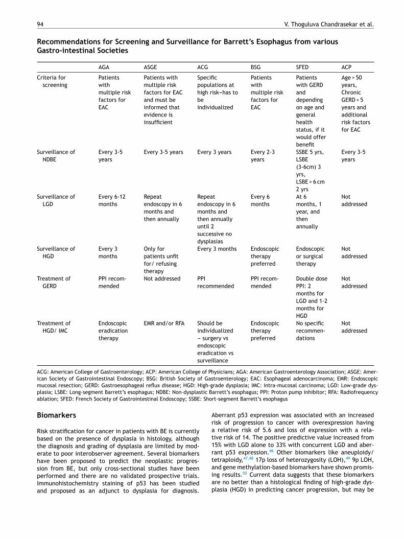
Recommendations for Screening and Surveillance for Barrett’s Esophagus from variousGastro-intestinal Societies
AGA ASGE ACG BSG SFED ACP
Criteria for
screening
Patients
with
multiple risk
factors for
EAC
Patients with
multiple risk
factors for EAC
and must be
informed that
evidence is
insufficient
Specific
populations at
high risk---has to
be
individualized
Patients
with
multiple risk
factors for
EAC
Patients
with GERD
and
depending
on age and
general
health
status, if it
would offer
benefit
Age > 50
years,
Chronic
GERD > 5
years and
additional
risk factors
for EAC
Surveillance of
NDBE
Every 3-5
years
Every 3-5 years Every 3 years Every 2-3
years
SSBE 5 yrs,
LSBE
(3-6cm) 3
yrs,
LSBE > 6 cm
2 yrs
Every 3-5
years
Surveillance of
LGD
Every 6-12
months
Repeat
endoscopy in 6
months and
then annually
Repeat
endoscopy in 6
months and
then annually
until 2
successive no
dysplasias
Every 6
months
At 6
months, 1
year, and
then
annually
Not
addressed
Surveillance of
HGD
Every 3
months
Only for
patients unfit
for/ refusing
therapy
Every 3 months Endoscopic
therapy
preferred
Endoscopic
or surgical
therapy
Not
addressed
Treatment of
GERD
PPI recom-
mended
Not addressed PPI
recommended
PPI recom-
mended
Double dose
PPI: 2
months for
LGD and 1-2
months for
HGD
Not
addressed
Treatment of
HGD/ IMC
Endoscopic
eradication
therapy
EMR and/or RFA Should be
individualized
--- surgery vs
endoscopic
eradication vs
surveillance
Endoscopic
therapy
preferred
No specific
recommen-
dations
Not
addressed
ACG: American College of Gastroenterology; ACP: American College of Physicians; AGA: American Gastroenterology Association; ASGE: Amer-
ican Society of Gastrointestinal Endoscopy; BSG: British Society of Gastroenterology; EAC: Esophageal adenocarcinoma; EMR: Endoscopic
mucosal resection; GERD: Gastroesophageal reflux disease; HGD: High-grade dysplasia; IMC: Intra-mucosal carcinoma; LGD: Low-grade dys-
plasia; LSBE: Long-segment Barrett’s esophagus; NDBE: Non-dysplastic Barrett’s esophagus; PPI: Proton pump inhibitor; RFA: Radiofrequency
ablation; SFED: French Society of Gastrointestinal Endoscopy; SSBE: Short-segment Barrett’s esophagus
Biomarkers
Risk stratification for cancer in patients with BE is currentlybased on the presence of dysplasia in histology, althoughthe diagnosis and grading of dysplasia are limited by mod-erate to poor interobserver agreement. Several biomarkershave been proposed to predict the neoplastic progres-sion from BE, but only cross-sectional studies have beenperformed and there are no validated prospective trials.Immunohistochemistry staining of p53 has been studiedand proposed as an adjunct to dysplasia for diagnosis.
Aberrant p53 expression was associated with an increasedrisk of progression to cancer with overexpression havinga relative risk of 5.6 and loss of expression with a rela-tive risk of 14. The positive predictive value increased from15% with LGD alone to 33% with concurrent LGD and aber-rant p53 expression.46 Other biomarkers like aneuploidy/tetraploidy,47,48 17p loss of heterozygosity (LOH),49 9p LOH,and gene methylation-based biomarkers have shown promis-ing results.50 Current data suggests that these biomarkersare no better than a histological finding of high-grade dys-plasia (HGD) in predicting cancer progression, but may be

Management of Barrett’s esophagus: Screening to newer treatments 95
superior in prediction compared with non-dysplastic Bar-rett’s esophagus (NDBE), indeterminate dysplasia, or LGD.The combination of biomarkers and histology can provideeven better results. More studies may be available in thefuture and hence these biomarkers can be used to predictwhich patients would benefit from surveillance vs ablation.
Advanced imaging modalities
Apart from high resolution white light endoscopy, otheradvanced techniques, such as narrow band imaging (NBI),chromo-endoscopy, autofluorescence imaging (AFI), confo-cal laser micro-endoscopy, diffuse reflectance spectroscopy,light scattering spectroscopy, and optical coherence tomo-graphy (OCT) have recently emerged. Currently white lightinspection of the esophagus using high resolution endoscopyis the standard of practice and guidelines do not recom-mend routine use of advanced endoscopic imaging eitherfor screening or surveillance of patients with BE. Withthe advances in technology, combining normal white lightendoscopy with magnification devices and high resolutiontelevision systems has enabled the production of higherquality images that have been shown to have a higher sen-sitivity for the detection of dysplasia and early neoplasticlesions in BE.51,52
Virtual chromo-endoscopy involves the application ofchemical agents and dyes that highlight specific areas of theesophageal mucosa and therefore aid in targeted biopsies.Several agents, such as methylene blue, Lugol’s iodine solu-tion, indigo carmine dye, and acetic acid have been usedfor this purpose. Lugol’s iodine stains the squamous cellsof the epithelium which contains glycogen and thus enablesthe identification of Barrett’s islands with columnar mucosaafter eradication therapy for BE.53 The studies using methy-lene blue to stain the non-dysplastic areas in the BE havereported mixed results. Horwhat et al. conducted a ran-domized, prospective, cross-over trial in 48 patients anddemonstrated that the yield of detecting BE or dysplasiawas similar among patients who received targeted biopsiesusing methylene blue and in those patients who had random4-quadrant biopsies, but required significantly fewer biop-sies compared with random 4-quadrant biopsies (9.23 ± 2.89and 18.92 ± 6.36, respectively) (p < 0.001).54 Lim et al. con-cluded a randomized cross-over study with 30 patients witha mean length of Barrett’s segment of 5 cm. Overall, 17 outof the 18 patients with dysplasia were identified by ran-dom 4-quadrant biopsies, whereas only 9 were identifiedusing methylene blue (p = 0.02).55 Ngamruengphong et al.conducted a meta-analysis reporting the diagnostic yieldof using methylene blue chromoendoscopy. There was nosignificant incremental yield (IY) over random 4-quadrantbiopsies for metaplasia (IY 4%), dysplasia (IY 9%), and earlycancer (IY 5%).56 Olliver et al. claimed that it could lead toDNA damage in Barrett’s epithelium, and thus could poten-tially accelerate carcinogenesis.57 Four specific pit patternshave been identified in studies using application of bothacetic acid and indigo carmine---round, reticular, ridged, andvillous. Guelred et al. described that the ridged and villouspatterns were associated with intestinal metaplasia usingacetic acid, whereas another study used indigo carmine in80 patients and reported that the ridged/villous pattern
had a sensitivity of 97% and specificity of 76% for intestinalmetaplasia.53
NBI is a form of electronic chromo-endoscopy that usesspectral narrow band optical filters, optical band imaging,and I-scan to highlight the vascular patterns or to dif-ferentiate the contrast between squamous and columnarepithelium.58 Prospective trials comparing NBI with high res-olution white light endoscopy have been published. In thestudy conducted by Wolfsen et al. with 65 patients knownto have dysplasia in BE, NBI identified more patients withdysplasia and also higher grades of dysplasia compared withwhite light endoscopy.59 In another randomized, multicen-ter cross-over trial which compared both, NBI detected thesame number of patients with metaplasia, but a higher pro-portion of areas with dysplasia (30% vs 21%, p = 0.01).60
Regular-appearing mucosa and vessels with NBI did notharbor any high-grade dysplasias. Moreover, they requiredsignificantly fewer biopsies than with white light endoscopy(3.6 vs. 7.6, p < 0.0001)
AFI endoscopy is based on the principle that when alaser light is emitted by endoscopy, cells would re-emita fluorescent light with distinct spectroscopic characteris-tics. Kara et al. found that it is a study with very goodsensitivity for identifying HGD, but with poor specificityand a high false positive rate.61 Mannath et al. pub-lished a recent report studying the interobserver agreementbetween experts and non-experts for AFI.62 They concludedthat there was fair-to-moderate agreement for AFI alone,which improved when it was combined with high resolu-tion white light endoscopy. Curvers et al. published a reportthat utilized a ‘‘trimodal imaging’’ technique in which theesophagus was first examined using the normal high res-olution white light followed by AFI to highlight abnormalareas not seen with white light, followed by NBI to confirmthe abnormal areas in AFI.63 AFI diagnosed a significantlyhigher proportion of abnormal areas than white light, butwith a higher false positive rate of 81%, which was reducedto 26% with NBI. It could still not replace the traditional4-quadrant biopsy, as 10% of the cases were missed by all3 modalities.
Confocal micro-endoscopy uses the technique of trans-lation of reflected endoscopy-derived laser light onto thecomputer into a cross-sectional image of the mucosalarchitecture. Real time analysis of the blood vessels andthe crypts can be performed. Reports published so farhave shown good accuracy rates (85%-94%) of dysplasiadetection.64,65 Pohl et al. conducted a prospective 2-centertrial and reported that the confocal laser microscopy crite-ria for BE were more frequently detected in HGD and earlycancer than in low-grade dysplasia, with good interobserveragreement with a kappa value of 0.6.66 The spectroscopictechniques are based on the analysis of the light scatteredfrom the tissue. Diffuse reflectance spectroscopy analy-ses the light scattered multiple times within the tissue,whereas light scattering spectroscopy uses light reflectedonly once. These techniques have been reported to dis-tinguish higher grades from lower grades of dysplasia andalso from no dysplasia up to 88% with a sensitivity andspecificity approaching 90%. OCT uses near-infrared lightto provide high resolution images and initial studies havereported higher sensitivity and specificity rates of > 90% inthe detection of metaplasia.67---69
96 V. Thoguluva Chandrasekar et al.
Despite having promising results, more studies areneeded to validate these techniques, optimizing the associ-ated costs and developing ideal teaching methods to get thistechnology to gastroenterologists beyond the tertiary cen-ters. Currently high resolution endoscopy with white lightwith random 4-quadrant biopsies has been the most widelyaccepted modality for diagnosis of metaplasia and dysplasiain BE.
Endoscopic eradication techniques
Eradication using endoscopic techniques involves eithera resection procedure like endoscopic mucosal resection(EMR) and endoscopic sub-mucosal dissection (ESD) or abla-tive procedures of the BE mucosa with several techniques.The advantage with the resection procedures is that they areboth diagnostic and therapeutic as we get a tissue sample,whereas the ablative procedures are only therapeutic. Allendoscopic procedures should be followed by acid suppres-sion therapy to allow for healing and re-epithelialization ofthe esophageal mucosa with squamous epithelium.
Surgical procedures used to be the treatment of choiceeven for early pre-malignant lesions, but endoscopic meth-ods, thanks to the advances made, are now being used morewidely. One important criterion to be considered beforeselecting endoscopic therapy is to assess the extent of theinvolvement of dysplasia in BE. It must be confined only tothe mucosa (T1a stage) and there should be no involvementof the submucosa, in which case surgical management is thestandard of care. It has been proven that early neoplasmsinvolving only the mucosa have a 1-2% risk of lymph nodemetastasis,70 whereas those with sub-mucosal invasion havea 10% risk of lymphadenopathy,71 and a few reports havesuggested up to 20%.72,73 Therefore, an accurate T-staging isessential before deciding on therapy. Endoscopic ultrasound(EUS) is the most accurate imaging modality for T-stagingof neoplasms, but they can predict the depth of invasionin only 50-60% of the cases. On the other hand, studies onEMR with biopsy of the specimen have been shown to havea better prediction rate of T-staging and to be superior toEUS studies.74,75
Retrospective studies comparing surgery to endoscopictherapy for early neoplasms have shown that surgery hashigher short-term mortality,76---80 whereas endoscopy haslower morbidity, better cost-effectiveness,81 and a lowerrisk of complications.82,83
Endoscopic resection
EMR and ESD are the 2 resection procedures. They involvea systematic resection of a specific area of the esopha-gus with an endoscopic knife or a diathermy snare. Theyare used especially with visible nodular dysplasia and short-segment dysplastic BE, for which they are preferred overthe ablative techniques. The BSG recommends 2 basic tech-niques that are the principle behind this technique. Theband ligation technique involves suction of the desired areaand deployment of a band that creates a pseudo-polyp,which can be removed by a diathermy snare.24 The cap andsnare technique involves lifting of the desired area with asub-mucosal injection and placing a cap over the mucosa,
which can then be snared.24 EMR for HGD and EAC T1astages has been successful in 91-98% of the cases.84---86 More-over, cohort studies have shown that EMR eradicates BE in75-100% of patients and eradicates dysplasia in 86-100% ofthe cases.87---92 Water jet-assisted ESD has been attemptedwith an en-bloc resection rate of around 90%.93 Not manydirect studies comparing EMR and ESD are available, butthe study by Ishihara et al. comparing EMR with ESD foresophageal squamous cell carcinoma revealed that EMR hada higher local recurrence rate (23.91% vs 3.13%), suggest-ing that ESD is the preferred technique when available,even though it is technically more demanding.94 Their studyresults also suggest that despite the resection of the dysplas-tic mucosa, the remaining non-dysplastic BE is still at riskand hence has to be eradicated by further resection or abla-tion. Wani et al. reported in their multicenter cohort studythat EMR could change the histological diagnosis in up to 30%of the patients, which could potentially change the course ofmanagement.95 Complications of EMR are immediate bleed-ing up to 10%,76,85,96 perforation in 3-7% of patients,83,86,97
and delayed stricture formation in 17-37% of patients.98
Endoscopic ablative techniques
Ablative techniques can be thermal, radiofrequencyablation (RFA), photodynamic therapy (PDT), multipolarelectro-coagulation (MPEC), cryotherapy, and argon plasmacoagulation (APC). These techniques are generally per-formed when there are no visible lesions in the dysplasticepithelium, in which case resection techniques have a highersuccess rate.
PDT involves the use of a photosensitive chemical like5-aminolevulinic acid or porfimer sodium to sensitize the tis-sues and destroy them with endoscopically delivered laserlight. The laser light causes free radical generation uponexposure to the sensitized cells, thus leading to damage.Success rates of eradication of HGD up to 77% in 5 yearshave been achieved.99,100 In a retrospective cohort study byOverholt et al. involving 103 patients presenting with BEwith dysplasia and IMC that had a mean follow-up of 50months, intent-to-treat success and eradication rates were92.2% for low-grade dysplasia, 77.5% for HGD, and 44.4% forIMC.99 Complications included stricture formation rates upto 30%, photosensitivity of the skin in up to 33% (p < 0.05),and buried BE epithelium beneath the squamous epitheliumthat was significantly higher in up to 48% of patients and mayeventually become malignant.99---102 Pain during the immedi-ate post-operative period has been reported as a significantside effect in most of the patients, with studies reportingup to as high as 86% of patients.103,104 Due to comparativelyhigher complication rates, PDT is not a highly favored optionfor BE therapy.
Cryoablation therapy is a non-contact technique thatinvolves the spraying of liquid nitrogen or carbon dioxide tofreeze and destroy the Barrett’s mucosa.105,106 Liquid nitro-gen or carbon dioxide is sprayed to freeze the tissue for10 to 20 s, then thawed for one minute, with 3-4 cyclesper session. Another session may be repeated in a couple ofmonths if needed. It has been shown to cause eradication ofintestinal metaplasia and dysplasia in 46-78% and 79-87% ofthe cases, respectively.106,107 Dumot et al. demonstrated in
Management of Barrett’s esophagus: Screening to newer treatments 97
their non-randomized cohort trial of 30 patients that 68% ofthe patients with HGD and 80% with IMC had downstaging ofHGD or elimination of cancer.108
MPEC and APC are other endoscopic eradication tech-niques which have not been extensively studied for thetreatment of BE, although there are several prospective caseseries that describe their use and success. Montes et al.reported an eradication rate of 100% of cases of NDBE inhis series with 14 patients with a mean follow-up of 21.6months,109 whereas Sampliner et al. reported only a 78%eradication rate in a 6-month follow-up.110 For APC, Madischet al. reported an eradication rate of 98% and a recur-rence rate of 12% in his series with a mean follow-up of51 months,111 whereas another study reported an eradica-tion rate of only 84% and a recurrence rate of up to 66% ina follow-up of 30 months.112 Randomized controlled studiescomparing MPEC with APC found no significant differencebetween eradication rates, but both needed multiple treat-ment sessions.
RFA involves an assembly of closely placed electrodes todeliver radiofrequency energy to the esophageal mucosa,thus causing ablation. This generates uniform thermalenergy in a circumferential fashion, while the power, dura-tion, and density of thermal energy can be varied. Severalstudies have compared RFA with other ablative therapiesand have shown that RFA has a better success rate, alower recurrence rate of dysplasia, and is comparativelysafer than other techniques. A prospective multicenterstudy demonstrated a 70% remission in BE in circumferentialtreatment,113 but Ganz et al. published a subsequent reportthat described a 98% eradication rate when a focal abla-tion technique was used after circumferential ablation.114
In a multi-center sham-controlled trial with 127 patientswith dysplastic BE, there was complete eradication of LGD in90.5% (p < 0.001) of the patients and in 81% of the patientswith HGD (p < 0.001) during a 12-month follow-up.115 In a
multicenter, randomized, controlled trial conducted by Phoaet al., comparing RFA with endoscopic surveillance for BEwith LGD, it was shown that RFA reduced the risk of pro-gression to HGD by 25% (p < 0.001) and adenocarcinoma by7.6% (p = 0.03) during a 3-year follow-up.116 There was com-plete resolution of the dysplasia in 92.6% of the patients.In a United Kingdom-based registry involving 335 patientswith BE, with 72% of them having HGD, RFA ablation ledto 86% resolution and clearance of HGD during a 12-monthfollow-up.117 Shorter segment Barrett’s responded betterand reversal of dysplasia was 15% less likely with everyincremented centimeter in the length of the BE segment.117
A systematic review comparing sub-squamous metaplasiain RFA and PDT-treated patients showed that 0.9% of RFA-treated patients and 14.2% of PDT-treated patients hadmetaplasia, thus demonstrating the superior efficacy ofRFA.118 In a systematic review and meta-analysis by Ormanet al., the most frequent complication was stricture forma-tion (5%), followed by pain (3%) and bleeding (1%), whichreflects the results in most of the other published articlesand retrospective studies.119 RFA has also been shown tohave a lower stenosis rate than stepwise radical endoscopicresection (14% vs 88%, p < 0.001).120 Despite the positiveresults of ablation procedures in eliminating dysplasia andreducing neoplastic progression, their role in NDBE has beenquestioned by studies in relation to the recurrence of meta-plasia during follow-up due to sub-squamous metaplasia,durability of response, and in some analyses, a recurrencerate up to 33%.121 Hence, ablation procedures might be auseful approach in LGD treatment, given the reduced rateof progression to cancer. However, the unnecessary subjec-tion of NDBE patients to these endoscopic procedures withthe consequent increase in complication rates demand thepublication of long-term data. Under the present circum-stances, ablation procedures are not recommended by themedical societies.
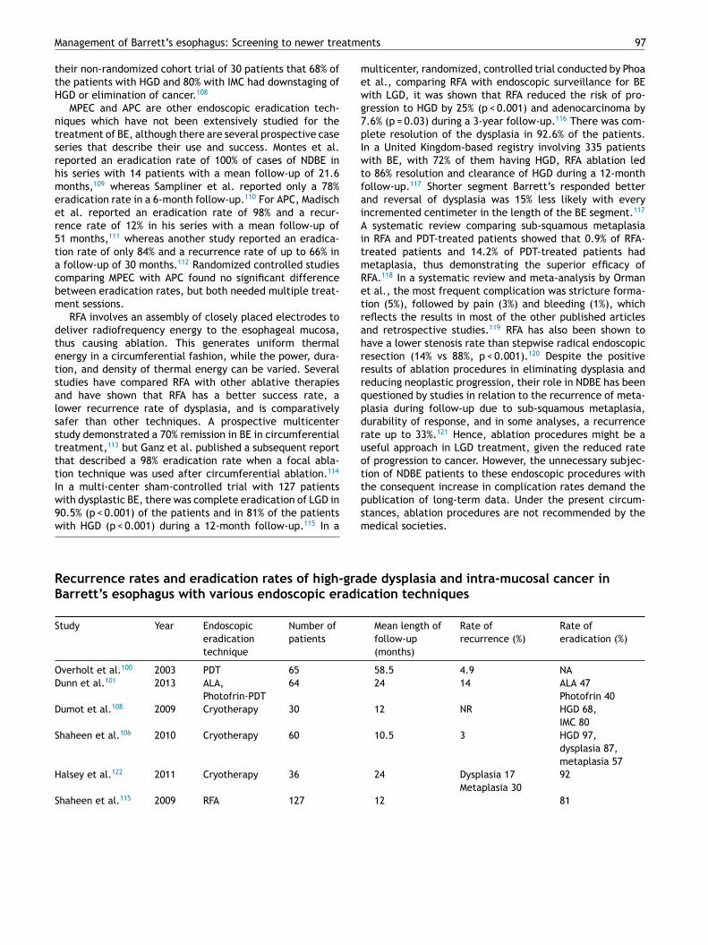
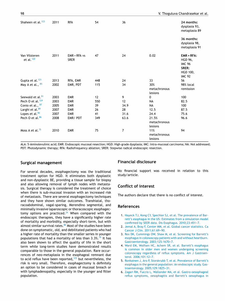
Recurrence rates and eradication rates of high-grade dysplasia and intra-mucosal cancer inBarrett’s esophagus with various endoscopic eradication techniques
Study Year Endoscopic
eradication
technique
Number of
patients
Mean length of
follow-up
(months)
Rate of
recurrence (%)
Rate of
eradication (%)
Overholt et al.100 2003 PDT 65 58.5 4.9 NA
Dunn et al.101 2013 ALA,
Photofrin-PDT
64 24 14 ALA 47
Photofrin 40
Dumot et al.108 2009 Cryotherapy 30 12 NR HGD 68,
IMC 80
Shaheen et al.106 2010 Cryotherapy 60 10.5 3 HGD 97,
dysplasia 87,
metaplasia 57
Halsey et al.122 2011 Cryotherapy 36 24 Dysplasia 17
Metaplasia 30
92
Shaheen et al.115 2009 RFA 127 12 81
98 V. Thoguluva Chandrasekar et al.
Shaheen et al.123 2011 RFA 54 36 24 months:
dysplasia 93,
metaplasia 89
36 months:
dysplasia 98,
metaplasia 91
Van Vilsteren
et al.120
2011 EMR + RFA vs
SRER
47 24 0.02 EMR + RFA:
HGD 96,
IMC 96
SRER:
HGD 100,
IMC 92
Gupta et al.121 2013 RFA, EMR 448 24 33 56
May A et al., 85 2002 EMR, PDT 115 34 30%
metachronous
lesions
98% local
remission
Seewald et al.91 2003 EMR 12 9 0 100
Pech O et al.124 2003 EMR 550 12 NA 82.5
Conio et al., 87 2005 EMR 39 34.9 NA 100
Larghi et al.89 2007 EMR 26 28 12.5 87.5
Lopes et al.90 2007 EMR 41 31.6 24.4 75.6
Pech O et al.86 2008 EMR/ PDT 349 63.6 21.5%
metachronous
lesions
96.6
Moss A et al.75 2010 EMR 75 7 11%
metachronous
lesions
94
ALA: 5-Aminolevulinic acid; EMR: Endoscopic mucosal resection; HGD: High-grade dysplasia; IMC: Intra-mucosal carcinoma; NA: Not addressed;
PDT: Photodynamic therapy; RFA: Radiofrequency ablation; SRER: Stepwise radical endoscopic resection.
Surgical management
For several decades, esophagectomy was the traditionaltreatment option for HGD. It eliminates both dysplasticand non-dysplastic BE, providing a tissue sample for biopsyand also allowing removal of lymph nodes with metasta-sis. Surgical therapy is considered the treatment of choicewhen there is sub-mucosal invasion with an increased riskof metastasis. There are several esophagectomy techniquesand they have shown similar outcomes. Transhiatal, tho-racoabdominal, vagal-sparing, Merendino segmental, andminimally invasive laparoscopic or thoracoscopic esophagec-tomy options are practiced.24 When compared with theendoscopic therapies, they have a significantly higher rateof mortality and morbidity, especially short-term, but withalmost similar survival rates.80 Most of the studies have beendone on symptomatic, old, and debilitated patients who hada higher rate of mortality than the smaller series in youngerpopulations that had a mortality of less than 3.3%.79 It hasalso been shown to affect the quality of life in the shortterm while long-term studies have demonstrated resultscomparable to those in the general population. Rare occur-rences of neo-metaplasia in the esophageal remnant dueto acid reflux have been reported,125 but nevertheless, therisk is very small. Therefore, esophagectomy is definitelyan option to be considered in cases of mucosal breach orwith lymphadenopathy, especially in the younger and fitterpopulation.
Financial disclosure
No financial support was received in relation to thisstudy/article.
Conflict of interest
The authors declare that there is no conflict of interest.
References
1. Hayeck TJ, Kong CY, Spechler SJ, et al. The prevalence of Bar-
rett’s esophagus in the US: Estimates from a simulation model
confirmed by SEER data. Dis Esophagus. 2010;23:451---7.
2. Jemal A, Bray F, Center MM, et al. Global cancer statistics. Ca
Cancer J Clin. 2011;61:69---90.
3. Rex DK, Cummings OW, Shaw M, et al. Screening for Barrett’s
esophagus in colonoscopy patients with and without heartburn.
Gastroenterology. 2003;125:1670---7.
4. Ward EM, Wolfsen HC, Achem SR, et al. Barrett’s esophagus
is common in older men and women undergoing screening
colonoscopy regardless of reflux symptoms. Am J Gastroen-
terol. 2006;101:12---7.
5. Ronkainen J, Aro P, Storskrubb T, et al. Prevalence of Barrett’s
esophagus in the general population: An endoscopic study. Gas-
troenterology. 2005;129:1825---31.
6. Zagari RM, Fuccio L, Wallander MA, et al. Gastro-oesophageal
reflux symptoms, oesophagitis and Barrett’s oesophagus in
Management of Barrett’s esophagus: Screening to newer treatments 99
the general population: The Loiano-Monghidoro study. Gut.
2008;57:1354---9.
7. Zou D, He J, Ma X, et al. Epidemiology of symptom-defined
gastroesophageal reflux disease and reflux esophagitis: The
systematic investigation of gastrointestinal diseases in China
(SILC). Scand J Gastroenterol. 2011;46:133---41.
8. Van Blankenstein M, Looman CW, Johnston BJ, et al. Age and
sex distribution of the prevalence of Barrett’s esophagus found
in a primary referral endoscopy center. Am J Gastroenterol.
2005;100:568---76.
9. Harnish DC. Estrogen receptor ligands in the control of
pathogenic inflammation. Current Opinion in Investigational
Drugs (London, England: 2000). 2006;7:997---1001.
10. Nilsson M, Lundegardh G, Carling L, et al. Body mass and reflux
oesophagitis: An oestrogen-dependent association. Scand J
Gastroenterol. 2002;37:626---30.
11. Spechler SJ, Souza RF. Barrett’s esophagus. New Engl J Med.
2014;371:836---45.
12. El-Serag HB, Kvapil P, Hacken-Bitar J, et al. Abdominal obe-
sity and the risk of Barrett’s esophagus. Am J Gastroenterol.
2005;100:2151---6.
13. Ryan AM, Healy LA, Power DG, et al. Barrett esophagus:
Prevalence of central adiposity, metabolic syndrome, and a
proinflammatory state. Ann Surg. 2008;247:909---15.
14. Beasley LE, Koster A, Newman AB, et al. Inflammation and
race and gender differences in computerized tomography-
measured adipose depots. Obesity (Silver Spring, Md).
2009;17:1062---9.
15. Parsonnet J. The incidence of Helicobacter pylori infection.
Aliment Pharmacol Therp. 1995;9 Suppl 2:45---51.
16. Iijima K, Henry E, Moriya A, et al. Dietary nitrate gener-
ates potentially mutagenic concentrations of nitric oxide at
the gastroesophageal junction. Gastroenterology. 2002;122:
1248---57.
17. Spechler SJ. Barrett esophagus and risk of esophageal cancer:
A clinical review. JAMA. 2013;310:627---36.
18. Sharma P, Morales TG, Sampliner RE. Short segment Barrett’s
esophagus ----the need for standardization of the definition and
of endoscopic criteria. Am J Gastroenterol. 1998;93:1033---6.
19. Spechler SJ, Zeroogian JM, Antonioli DA, et al. Prevalence
of metaplasia at the gastro-oesophageal junction. Lancet.
1994;344:1533---6.
20. Sharma P, Dent J, Armstrong D, et al. The development
and validation of an endoscopic grading system for Barrett’s
esophagus: The Prague C & M criteria. Gastroenterology.
2006;131:1392---9.
21. Wang KK, Sampliner RE. Updated guidelines 2008 for the diag-
nosis, surveillance and therapy of Barrett’s esophagus. Am J
Gastroenterol. 2008;103:788---97.
22. Spechler SJ, Sharma P, Souza RF, et al. American Gastroen-
terological Association technical review on the management of
Barrett’s esophagus. Gastroenterology. 2011;140:e18---52 [quiz
e13].
23. Evans JA, Early DS, Fukami N, et al. The role of endoscopy in
Barrett’s esophagus and other premalignant conditions of the
esophagus. Gastrointest Endosc. 2012;76:1087---94.
24. Fitzgerald RC, di Pietro M, Ragunath K, et al. British Society of
Gastroenterology guidelines on the diagnosis and management
of Barrett’s oesophagus. Gut. 2014;63:7---42.
25. Desai TK, Krishnan K, Samala N, et al. The incidence
of oesophageal adenocarcinoma in non-dysplastic Barrett’s
oesophagus: A meta-analysis. Gut. 2012;61:970---6.
26. Hvid-Jensen F, Pedersen L, Drewes AM, et al. Incidence of ade-
nocarcinoma among patients with Barrett’s esophagus. New
Engl J Med. 2011;365:1375---83.
27. Bhat S, Coleman HG, Yousef F, et al. Risk of malignant pro-
gression in Barrett’s esophagus patients: Results from a large
population-based study. J Nat Cancer Inst. 2011;103:1049---57.
28. Wani S, Falk G, Hall M, et al. Patients with nondysplastic
Barrett’s esophagus have low risks for developing dysplasia
or esophageal adenocarcinoma. Clin Gastroenterol Hepatol.
2011;9:220---7 [quiz e26].
29. Chak A, Faulx A, Eng C, et al. Gastroesophageal reflux symp-
toms in patients with adenocarcinoma of the esophagus or
cardia. Cancer. 2006;107:2160---6.
30. Bhat SK, McManus DT, Coleman HG, et al. Oesophageal ade-
nocarcinoma and prior diagnosis of Barrett’s oesophagus: A
population-based study. Gut. 2015;64:20---5.
31. Dulai GS, Guha S, Kahn KL, et al. Preoperative prevalence of
Barrett’s esophagus in esophageal adenocarcinoma: A system-
atic review. Gastroenterology. 2002;122:26---33.
32. Corley DA, Levin TR, Habel LA, et al. Surveillance and sur-
vival in Barrett’s adenocarcinomas: A population-based study.
Gastroenterology. 2002;122:633---40.
33. Fountoulakis A, Zafirellis KD, Dolan K, et al. Effect of surveil-
lance of Barrett’s oesophagus on the clinical outcome of
oesophageal cancer. Br J surg. 2004;91:997---1003.
34. Shaheen NJ, Weinberg DS, Denberg TD, et al. Upper endoscopy
for gastroesophageal reflux disease: Best practice advice from
the clinical guidelines committee of the American College of
Physicians. Annals of Internal Medicine. 2012;157:808---16.
35. Chang JY, Talley NJ, Locke GR, et al. Population screening for
barrett esophagus: A prospective randomized pilot study. Mayo
Clin Proc. 2011;86:1174---80.
36. Eliakim R, Sharma VK, Yassin K, et al. A prospective study of
the diagnostic accuracy of PillCam ESO esophageal capsule
endoscopy versus conventional upper endoscopy in patients
with chronic gastroesophageal reflux diseases. J Clin Gastroen-
terol. 2005;39:572---8.
37. Eliakim R, Yassin K, Shlomi I, et al. A novel diagnostic tool for
detecting oesophageal pathology: The PillCam oesophageal
video capsule. Aliment Pharmacol Therp. 2004;20:
1083---9.
38. Bhardwaj A, Hollenbeak CS, Pooran N, et al. A meta-analysis of
the diagnostic accuracy of esophageal capsule endoscopy for
Barrett’s esophagus in patients with gastroesophageal reflux
disease. Am J Gastroenterol. 2009;104:1533---9.
39. Lao-Sirieix P, Boussioutas A, Kadri SR, et al. Non-endoscopic
screening biomarkers for Barrett’s oesophagus: From microar-
ray analysis to the clinic. Gut. 2009;58:1451---9.
40. Kadri SR, Lao-Sirieix P, O’Donovan M, et al. Acceptabil-
ity and accuracy of a non-endoscopic screening test for
Barrett’s oesophagus in primary care: cohort study. BMJ.
2010;341:c4372.
41. Benaglia T, Sharples LD, Fitzgerald RC, et al. Health bene-
fits and cost effectiveness of endoscopic and nonendoscopic
cytosponge screening for Barrett’s esophagus. Gastroenter-
ology. 2013;144, 62-73.e6.
42. Eloubeidi MA, Mason AC, Desmond RA, et al. Temporal trends
(1973-1997) in survival of patients with esophageal adeno-
carcinoma in the United States: a glimmer of hope. Am J
Gastroenterol. 2003;98:1627---33.
43. Van der Burgh A, Dees J, Hop WC, et al. Oesophageal can-
cer is an uncommon cause of death in patients with Barrett’s
oesophagus. Gut. 1996;39:5---8.
44. Fitzgerald RC, Saeed IT, Khoo D, et al. Rigorous surveillance
protocol increases detection of curable cancers associated
with Barrett’s esophagus. Dig Dis Sci. 2001;46:1892---8.
45. Harrison R, Perry I, Haddadin W, et al. Detection of intestinal
metaplasia in Barrett’s esophagus: An observational compara-
tor study suggests the need for a minimum of eight biopsies.
Am J Gastroenterol. 2007;102:1154---61.
46. Kastelein F, Biermann K, Steyerberg EW, et al. Aberrant p53
protein expression is associated with an increased risk of neo-
plastic progression in patients with Barrett’s oesophagus. Gut.
2013;62:1676---83.
100 V. Thoguluva Chandrasekar et al.
47. Rabinovitch PS, Longton G, Blount PL, et al. Predictors of pro-
gression in Barrett’s esophagus iii: Baseline flow cytometric
variables. Am J Gastroenterol. 2001;96:3071---83.
48. Reid BJ, Levine DS, Longton G, et al. Predictors of progression
to cancer in Barrett’s esophagus: Baseline histology and flow
cytometry identify low- and high-risk patient subsets. Am J
Gastroenterol. 2000;95:1669---76.
49. Reid BJ, Prevo LJ, Galipeau PC, et al. Predictors of pro-
gression in Barrett’s esophagus ii: Baseline 17p (p53) loss of
heterozygosity identifies a patient subset at increased risk for
neoplastic progression. Am J Gastroenterol. 2001;96:2839---48.
50. Schulmann K, Sterian A, Berki A, et al. Inactivation of p16,
RUNX3, and HPP1 occurs early in Barrett’s-associated neo-
plastic progression and predicts progression risk. Oncogene.
2005;24:4138---48.
51. Kara MA, Peters FP, Rosmolen WD, et al. High-resolution
endoscopy plus chromoendoscopy or narrow-band imaging
in Barrett’s esophagus: A prospective randomized crossover
study. Endoscopy. 2005;37:929---36.
52. Kara MA, Smits ME, Rosmolen WD, et al. A random-
ized crossover study comparing light-induced fluorescence
endoscopy with standard videoendoscopy for the detection of
early neoplasia in Barrett’s esophagus. Gastrointest Endosc.
2005;61:671---8.
53. Guelrud M, Herrera I, Essenfeld H, et al. Enhanced magni-
fication endoscopy: A new technique to identify specialized
intestinal metaplasia in Barrett’s esophagus. Gastrointest
Endosc. 2001;53:559---65.
54. Horwhat JD, Maydonovitch CL, Ramos F, et al. A randomized
comparison of methylene blue-directed biopsy versus conven-
tional four-quadrant biopsy for the detection of intestinal
metaplasia and dysplasia in patients with long-segment Bar-
rett’s esophagus. Am J Gastroenterol. 2008;103:546---54.
55. Lim CH, Rotimi O, Dexter SP, et al. Randomized crossover study
that used methylene blue or random 4-quadrant biopsy for
the diagnosis of dysplasia in Barrett’s esophagus. Gastrointest
Endosc. 2006;64:195---9.
56. Ngamruengphong S, Sharma VK, Das A. Diagnostic yield of
methylene blue chromoendoscopy for detecting specialized
intestinal metaplasia and dysplasia in Barrett’s esophagus: A
meta-analysis. Gastrointest Endosc. 2009;69:1021---8.
57. Olliver JR, Wild CP, Sahay P, et al. Chromoendoscopy with
methylene blue and associated DNA damage in Barrett’s
oesophagus. Lancet. 2003;362:373---4.
58. Gono K, Obi T, Yamaguchi M, et al. Appearance of enhanced
tissue features in narrow-band endoscopic imaging. J Biomed
Op. 2004;9:568---77.
59. Wolfsen HC, Crook JE, Krishna M, et al. Prospective, con-
trolled tandem endoscopy study of narrow band imaging for
dysplasia detection in Barrett’s Esophagus. Gastroenterology.
2008;135:24---31.
60. Sharma P, Hawes RH, Bansal A, et al. Standard endoscopy
with random biopsies versus narrow band imaging targeted
biopsies in Barrett’s oesophagus: A prospective, international,
randomised controlled trial. Gut. 2013;62:15---21.
61. Kara MA, Peters FP, Ten Kate FJ, et al. Endoscopic video
autofluorescence imaging may improve the detection of early
neoplasia in patients with Barrett’s esophagus. Gastrointest
Endosc. 2005;61:679---85.
62. Mannath J, Subramanian V, Telakis E, et al. An inter-observer
agreement study of autofluorescence endoscopy in Barrett’s
esophagus among expert and non-expert endoscopists. Dig Dis
Sci. 2013;58:465---70.
63. Curvers WL, Singh R, Song LM, et al. Endoscopic tri-modal
imaging for detection of early neoplasia in Barrett’s oesoph-
agus: A multi-centre feasibility study using high-resolution
endoscopy, autofluorescence imaging and narrow band imaging
incorporated in one endoscopy system. Gut. 2008;57:167---72.
64. Kiesslich R, Burg J, Vieth M, et al. Confocal laser endoscopy
for diagnosing intraepithelial neoplasias and colorectal cancer
in vivo. Gastroenterology. 2004;127:706---13.
65. Kiesslich R, Gossner L, Goetz M, et al. In vivo histology of Bar-
rett’s esophagus and associated neoplasia by confocal laser
endomicroscopy. Clin Gastroenterol Hepatol. 2006;4:979---87.
66. Pohl H, Rosch T, Vieth M, et al. Miniprobe confocal laser
microscopy for the detection of invisible neoplasia in patients
with Barrett’s oesophagus. Gut. 2008;57:1648---53.
67. Ortner MA, Ebert B, Hein E, et al. Time gated fluorescence
spectroscopy in Barrett’s oesophagus. Gut. 2003;52:28---33.
68. Georgakoudi I, Jacobson BC, van Dam J, et al. Fluorescence,
reflectance, and light-scattering spectroscopy for evaluating
dysplasia in patients with Barrett’s esophagus. Gastroenter-
ology. 2001;120:1620---9.
69. Wallace MB, Perelman LT, Backman V, et al. Endoscopic
detection of dysplasia in patients with Barrett’s esoph-
agus using light-scattering spectroscopy. Gastroenterology.
2000;119:677---82.
70. Dunbar KB, Spechler SJ. The risk of lymph-node metastases in
patients with high-grade dysplasia or intramucosal carcinoma
in Barrett’s esophagus: A systematic review. Am J Gastroen-
terol. 2012;107:850---62 [quiz 63].
71. Leers JM, DeMeester SR, Oezcelik A, et al. The prevalence
of lymph node metastases in patients with T1 esophageal
adenocarcinoma a retrospective review of esophagectomy
specimens. Ann Surg. 2011;253:271---8.
72. Feith M, Stein HJ, Siewert JR. Pattern of lymphatic spread of
Barrett’s cancer. Wolr J Surg. 2003;27:1052---7.
73. Rice TW, Zuccaro G Jr, Adelstein DJ, et al. Esophageal carci-
noma: Depth of tumor invasion is predictive of regional lymph
node status. Ann Thorac Surg. 1998;65:787---92.
74. Larghi A, Lightdale CJ, Memeo L, et al. EUS followed by EMR
for staging of high-grade dysplasia and early cancer in Barrett’s
esophagus. Gastrointest Endosc. 2005;62:16---23.
75. Moss A, Bourke MJ, Hourigan LF, et al. Endoscopic resection
for Barrett’s high-grade dysplasia and early esophageal ade-
nocarcinoma: An essential staging procedure with long-term
therapeutic benefit. Am J Gastroenterol. 2010;105:1276---83.
76. Ell C, May A, Pech O, et al. Curative endoscopic resection
of early esophageal adenocarcinomas (Barrett’s cancer). Gas-
trointest Endosc. 2007;65:3---10.
77. Menon D, Stafinski T, Wu H, et al. Endoscopic treatments for
Barrett’s esophagus: A systematic review of safety and effec-
tiveness compared to esophagectomy. BMC Gastroenterology.
2010;10:111.
78. Prasad GA, Wu TT, Wigle DA, et al. Endoscopic and surgical
treatment of mucosal (T1a) esophageal adenocarcinoma in
Barrett’s esophagus. Gastroenterology. 2009;137:815---23.
79. Wang VS, Hornick JL, Sepulveda JA, et al. Low preva-
lence of submucosal invasive carcinoma at esophagectomy for
high-grade dysplasia or intramucosal adenocarcinoma in Bar-
rett’s esophagus: A 20-year experience. Gastrointest Endosc.
2009;69:777---83.
80. Zehetner J, DeMeester SR, Hagen JA, et al. Endoscopic
resection and ablation versus esophagectomy for high-grade
dysplasia and intramucosal adenocarcinoma. J Thor Cardiovas
Surg. 2011;141:39---47.
81. Boger PC, Turner D, Roderick P, et al. A UK-based cost-utility
analysis of radiofrequency ablation or oesophagectomy for the
management of high-grade dysplasia in Barrett’s oesophagus.
Aliment Pharmacol Therp. 2010;32:1332---42.
82. Peters FP, Brakenhoff KP, Curvers WL, et al. Endoscopic cap
resection for treatment of early Barrett’s neoplasia is safe:
A prospective analysis of acute and early complications in
216 procedures. Diseases of the Esophagus: Official Journal of
the International Society for Diseases of the Esophagus/ISDE.
2007;20:510---5.
Management of Barrett’s esophagus: Screening to newer treatments 101
83. Pouw RE, van Vilsteren FG, Peters FP, et al. Randomized trial on
endoscopic resection-cap versus multiband mucosectomy for
piecemeal endoscopic resection of early Barrett’s neoplasia.
Gastrointest Endosc. 2011;74:35---43.
84. Ciocirlan M, Lapalus MG, Hervieu V, et al. Endoscopic mucosal
resection for squamous premalignant and early malignant
lesions of the esophagus. Endoscopy. 2007;39:24---9.
85. May A, Gossner L, Pech O, et al. Local endoscopic therapy for
intraepithelial high-grade neoplasia and early adenocarcinoma
in Barrett’s oesophagus: Acute-phase and intermediate results
of a new treatment approach. European Journal of Gastroen-
terology & Hepatology. 2002;14:1085---91.
86. Pech O, Behrens A, May A, et al. Long-term results and risk
factor analysis for recurrence after curative endoscopic ther-
apy in 349 patients with high-grade intraepithelial neoplasia
and mucosal adenocarcinoma in Barrett’s oesophagus. Gut.
2008;57:1200---6.
87. Conio M, Repici A, Cestari R, et al. Endoscopic mucosal
resection for high-grade dysplasia and intramucosal carcinoma
in Barrett’s esophagus: An Italian experience. Wolr J Gastroen-
terol. 2005;11:6650---5.
88. Giovannini M, Bories E, Pesenti C, et al. Circumferential
endoscopic mucosal resection in Barrett’s esophagus with high-
grade intraepithelial neoplasia or mucosal cancer. Preliminary
results in 21 patients. Endoscopy. 2004;36:782---7.
89. Larghi A, Lightdale CJ, Ross AS, et al. Long-term follow-up of
complete Barrett’s eradication endoscopic mucosal resection
(CBE-EMR) for the treatment of high grade dysplasia and intra-
mucosal carcinoma. Endoscopy. 2007;39:1086---91.
90. Lopes CV, Hela M, Pesenti C, et al. Circumferential endoscopic
resection of Barrett’s esophagus with high-grade dysplasia or
early adenocarcinoma. Surg Endosc. 2007;21:820---4.
91. Seewald S, Akaraviputh T, Seitz U, et al. Circumferential EMR
and complete removal of Barrett’s epithelium: A new approach
to management of Barrett’s esophagus containing high-grade
intraepithelial neoplasia and intramucosal carcinoma. Gas-
trointest Endosc. 2003;57:854---9.
92. Soehendra N, Seewald S, Groth S, et al. Use of modified
multiband ligator facilitates circumferential EMR in Barrett’s
esophagus (with video). Gastrointest Endosc. 2006;63:847---52.
93. Neuhaus H, Terheggen G, Rutz EM, et al. Endoscopic sub-
mucosal dissection plus radiofrequency ablation of neoplastic
Barrett’s esophagus. Endoscopy. 2012;44:1105---13.
94. Ishihara R, Iishi H, Takeuchi Y, et al. Local recurrence of large
squamous-cell carcinoma of the esophagus after endoscopic
resection. Gastrointest Endosc. 2008;67:799---804.
95. Wani S, Abrams J, Edmundowicz SA, et al. Endoscopic mucosal
resection results in change of histologic diagnosis in Barrett’s
esophagus patients with visible and flat neoplasia: A multicen-
ter cohort study. Dig Dis Sci. 2013;58:1703---9.
96. Shami VM, Villaverde A, Stearns L, et al. Clinical impact
of conventional endosonography and endoscopic ultrasound-
guided fine-needle aspiration in the assessment of patients
with Barrett’s esophagus and high-grade dysplasia or intra-
mucosal carcinoma who have been referred for endoscopic
ablation therapy. Endoscopy. 2006;38:157---61.
97. Esaki M, Matsumoto T, Hirakawa K, et al. Risk factors for
local recurrence of superficial esophageal cancer after treat-
ment by endoscopic mucosal resection. Endoscopy. 2007;39:
41---5.
98. Chennat J, Konda VJ, Ross AS, et al. Complete Barrett’s erad-
ication endoscopic mucosal resection: An effective treatment
modality for high-grade dysplasia and intramucosal carcinoma–
an American single-center experience. Am J Gastroenterol.
2009;104:2684---92.
99. Overholt BF, Wang KK, Burdick JS, et al. Five-year efficacy
and safety of photodynamic therapy with Photofrin in Barrett’s
high-grade dysplasia. Gastrointest Endosc. 2007;66:460---8.
100. Overholt BF, Panjehpour M, Halberg DL. Photodynamic ther-
apy for Barrett’s esophagus with dysplasia and/or early stage
carcinoma: Long-term results. Gastrointest Endosc. 2003;58:
183---8.
101. Dunn JM, Mackenzie GD, Banks MR, et al. A randomised con-
trolled trial of ALA vs. Photofrin photodynamic therapy for
high-grade dysplasia arising in Barrett’s oesophagus. Laser Med
Sci. 2013;28:707---15.
102. Heuberger D, Manner H, Ell C, et al. How is early Barrett’s
cancer currently diagnosed and treated in Western Europe?
Results of a survey at 52 university hospitals in eight Western
European countries. Z Gastroenterol. 2012;50:670---6.
103. Hage M, Siersema PD, van Dekken H, et al. 5-aminolevulinic
acid photodynamic therapy versus argon plasma coagulation
for ablation of Barrett’s oesophagus: A randomised trial. Gut.
2004;53:785---90.
104. Nava HR, Allamaneni SS, Dougherty TJ, et al. Photodynamic
therapy (PDT) using HPPH for the treatment of precancerous
lesions associated with Barrett’s esophagus. Laser Med Sci.
2011;43:705---12.
105. Greenwald BD, Dumot JA, Abrams JA, et al. Endoscopic spray
cryotherapy for esophageal cancer: Safety and efficacy. Gas-
trointest Endosc. 2010;71:686---93.
106. Shaheen NJ, Greenwald BD, Peery AF, et al. Safety and efficacy
of endoscopic spray cryotherapy for Barrett’s esophagus with
high-grade dysplasia. Gastrointest Endosc. 2010;71:680---5.
107. Johnston MH, Eastone JA, Horwhat JD, et al. Cryoablation
of Barrett’s esophagus: A pilot study. Gastrointest Endosc.
2005;62:842---8.
108. Dumot JA, Vargo JJ 2nd, Falk GW, et al. An open-label,
prospective trial of cryospray ablation for Barrett’s esophagus
high-grade dysplasia and early esophageal cancer in high-risk
patients. Gastrointest Endosc. 2009;70:635---44.
109. Montes CG, Brandalise NA, Deliza R, et al. Antireflux
surgery followed by bipolar electrocoagulation in the treat-
ment of Barrett’s esophagus. Gastrointest Endosc. 1999;50:
173---7.
110. Sampliner RE, Faigel D, Fennerty MB, et al. Effective and
safe endoscopic reversal of nondysplastic Barrett’s esopha-
gus with thermal electrocoagulation combined with high-dose
acid inhibition: A multicenter study. Gastrointest Endosc.
2001;53:554---8.
111. Madisch A, Miehlke S, Bayerdorffer E, et al. Long-term follow-
up after complete ablation of Barrett’s esophagus with argon
plasma coagulation. Wolr J Gastroenterol. 2005;11:1182---6.
112. Mork H, Al-Taie O, Berlin F, et al. High recurrence rate of
Barrett’s epithelium during long-term follow-up after argon
plasma coagulation. Scand J Gastroenterol. 2007;42:23---7.
113. Sharma VK, Kim HJ, Das A, et al. A prospective pilot trial of
ablation of Barrett’s esophagus with low-grade dysplasia using
stepwise circumferential and focal ablation (HALO system).
Endoscopy. 2008;40:380---7.
114. Ganz RA, Overholt BF, Sharma VK, et al. Circumferential
ablation of Barrett’s esophagus that contains high-grade dys-
plasia: A U. S. Multicenter Registry. Gastrointest Endosc.
2008;68:35---40.
115. Shaheen NJ, Sharma P, Overholt BF, et al. Radiofrequency abla-
tion in Barrett’s esophagus with dysplasia. New Engl J Med.
2009;360:2277---88.
116. Phoa KN, van Vilsteren FG, Weusten BL, et al. Radiofrequency
ablation vs endoscopic surveillance for patients with Bar-
rett esophagus and low-grade dysplasia: A randomized clinical
trial. JAMA. 2014;311:1209---17.
117. Haidry RJ, Dunn JM, Butt MA, et al. Radiofrequency abla-
tion and endoscopic mucosal resection for dysplastic barrett’s
esophagus and early esophageal adenocarcinoma: Outcomes
of the UK National Halo RFA Registry. Gastroenterology.
2013;145:87---95.
102 V. Thoguluva Chandrasekar et al.
118. Gray NA, Odze RD, Spechler SJ. Buried metaplasia after endo-
scopic ablation of Barrett’s esophagus: A systematic review.
Am J Gastroenterol. 2011;106:1899---908 [quiz 909].
119. Orman ES, Li N, Shaheen NJ. Efficacy and durability of
radiofrequency ablation for Barrett’s Esophagus: System-
atic review and meta-analysis. Clin Gastroenterol Hepatol.
2013;11:1245---55.
120. Van Vilsteren FG, Pouw RE, Seewald S, et al. Stepwise radical
endoscopic resection versus radiofrequency ablation for Bar-
rett’s oesophagus with high-grade dysplasia or early cancer: A
multicentre randomised trial. Gut. 2011;60:765---73.
121. Gupta M, Iyer PG, Lutzke L, et al. Recurrence of
esophageal intestinal metaplasia after endoscopic mucosal
resection and radiofrequency ablation of Barrett’s esophagus:
Results from a US Multicenter Consortium. Gastroenterology.
2013;145:79---86.e1.
122. Halsey KD, Chang JW, Waldt A, et al. Recurrent disease follow-
ing endoscopic ablation of Barrett’s high-grade dysplasia with
spray cryotherapy. Endoscopy. 2011;43:844---8.
123. Shaheen NJ, Overholt BF, Sampliner RE, et al. Durability of
radiofrequency ablation in Barrett’s esophagus with dysplasia.
Gastroenterology. 2011;141:460---8.
124. Pech O, May A, Gossner L, et al. Barrett’s esopha-
gus: Endoscopic resection. Gastrointes endosc Clin N Am.
2003;13:505---12.
125. Da Rocha JR, Ribeiro U Jr, Sallum RA, et al. Barrett’s esoph-
agus (BE) and carcinoma in the esophageal stump (ES) after
esophagectomy with gastric pull-up in achalasia patients:
A study based on 10 years follow-up. Ann Surg Oncol.
2008;15:2903---9.