management of craniocerebral gunshot injuries: a … gunshot injuries (cgi) are increasingly...
TRANSCRIPT

Copyright © 2015 Korean Neurotraumatology Society 35
Introduction
Craniocerebral gunshot injuries (CGI), initially described and managed in military settings, are now increasingly en-countered by neurosurgeons in civilian and urban settings, been on the rise especially in developing countries. Al-
though less prevalent than closed head trauma, penetrating brain injury carries a worse prognosis.52) CGI are the most lethal of all firearm injuries, with reported survival rates of only 7% to 15%.61)
About 90% of the time, the victims die before arriving at the hospital, and for those who survive and make it to the hospital, about 50% die in the emergency room.11,62) Peak mortality from CGI happens at the scene or within 3 hours of injury.11,40) This reality requires that management of gun-shot (missile) wounds (injuries) of head (craniocerebrum) due to bullets, shotguns, blasts, explosion of grenades and mines must be a routine experience in any Neurosurgical centre in countries with civil armed conflict. Since patient epidemiology is a multifactorial phenomenon and influ-enced by psychological, socioeconomic, as well as cultural factors, the characteristics of CGI patients might be funda-mentally different between all the continents.
Management of Craniocerebral Gunshot Injuries: A Review
Hernando Raphael Alvis-Miranda, MD1, Roberto Adie Villafañe, MD1, Alejandro Rojas, MD2, Gabriel Alcala-Cerra, MD3, and Luis Rafael Moscote-Salazar, MD3
1Department of Medicine, University of Cartagena, Cartagena, Colombia 2Department of Neurosurgery, FUSC, Hospital San Jose, Bogota, Colombia 3Department of Neurosurgery, University of Cartagena, Cartagena, Colombia
Craniocerebral gunshot injuries (CGI) are increasingly encountered by neurosurgeons in civilian and urban settings. Un-fortunately this is a prevalent condition in developing countries, with major armed conflicts which is not very likely to achieve a high rate of prevention. Management goals should focus on early aggressive, vigorous resuscitation and correc-tion of coagulopathy; those with stable vital signs undergo brain computed tomography scan. Neuroimaging is vital for surgical purposes, especially for determine type surgery, size and location of the approach, route of extraction of the for-eign body; however not always surgical management is indicated, there is also the not uncommon decision to choose non-surgical management. The treatment consist of immediate life salvage, through control of persistent bleeding and cerebral decompression; prevention of infection, through extensive debridement of all contaminated, macerated or ischemic tis-sues; preservation of nervous tissue, through preventing meningocerebral scars; and restoration of anatomic structures through the hermetic seal of dura and scalp. There have been few recent studies involving penetrating craniocerebral inju-ries, and most studies have been restricted to small numbers of patients; classic studies in military and civil environment have identified that this is a highly lethal or devastating violent condition, able to leave marked consequences for the af-fected individual, the family and the health system itself. Various measures have been aimed to lower the incidence of CGI, especially in civilians. It is necessarily urgent to promote research in a neurocritical topic such as CGI, looking im-pact positively the quality of life for those who survive. (Korean J Neurotrauma 2015;11(2):35-43)
KEY WORDS: Brain injuries ㆍCraniocerebral trauma ㆍWounds ㆍGunshot ㆍNeurons.
REVIEW ARTICLEKorean J Neurotrauma 2015;11(2):35-43
pISSN 2234-8999 / eISSN 2288-2243
http://dx.doi.org/10.13004/kjnt.2015.11.2.35
Received: June 23, 2014 / Revised: April 1, 2015Accepted: April 10, 2015Address for correspondence: Luis Rafael Moscote-Salazar, MDDepartment of Neurological Research, Health Sciences and Neu-rosciences (CISNEURO) Research Group;Department of Neurosurgery, University of Cartagena, Avenida del Consulado #Calle 30 No. 48-152, Cartagena, ColombiaTel: +57-31-2687-6610, Fax: +57-31-2687-6610E-mail: [email protected] cc This is an Open Access article distributed under the terms of Cre-ative Attributions Non-Commercial License (http://creativecommons.org/licenses/by-nc/3.0/) which permits unrestricted noncommercial use, distribution, and reproduction in any medium, provided the original work is properly cited.

36 Korean J Neurotrauma 2015;11(2):35-43
Craniocerebral Gunshot Injuries
There are many lessons to be learnt. This review consid-ers current ballistic aspects and aspects based on the patho-physiology and related to the diagnosis and management.
Ballistics and Pathophysiology
Projectiles are pellets fired from a shotgun, bullets from rifles, machine-guns, carbines, automatic guns and shrap-nel and splinters by exploding bombs, mines and grenades. A missile is a projectile of either a high velocity (muzzle ve-locity >600 m/sec) or a low velocity (muzzle velocity <300 m/sec).6)
BallisticsAs most of the penetrating brain injuries are caused by
missiles or projectiles, an understanding of ballistics (i.e., the study of the dynamics of projectiles) is imperative.52)
Firearms can be classified in many ways; the simplest is according to their speed:
1) Low velocity: less than 1,000 feet per second (<300 m/sec) as handguns or hand sizes 22, 38-caliber and 9 mm.
2) Medium velocity: speeds of 1,000 to 2,000 feet per second (300 to 600 m/sec) as the submachine guns.
3) High velocity: more than 2,000 feet per second (600 to 1,000 m/sec) as the AK-47, G-3 or Galil. These have proper-ties such as rotation, oscillation and fragmentation that make them much more lethal.
PhysiopathologyThe ability of any penetrating object to penetrate the
brain, thus is, the primary injury to the brain is dependent on the ballistic properties (kinetic energy, mass, velocity, shape, angle of approach, the characteristics of interven-ing tissues, etc.) of the projectile and any secondary pro-jectiles, such as bone or metallic fragments.17,52)
It is important to consider the energy delivered and the location in the brain to which that energy is imparted. The kinetic energy (wounding energy) is defined by the rela-tionship: E=1/2 mv2, velocity can be represented as E=1/2 m (Vi2-Vr2) where m is the mass of the projectile, Vi is the impact velocity and Vr is the residual velocity in the case of perforating wound.27,42) This implies that the veloc-ity of the projectile has a greater influence than the mass of the projectile alone, meaning that the bullet of an AK-47 assault rifle, which weighs 7.9 g and has an initial veloc-ity of 720 m/s, has a kinetic energy of 2,635 ft/lb (3,573 J). Projectile velocity from firearms in handguns is less than that of rifles, varying usually from 180 m/s to 450 m/s.
When the velocity exceeds 700 m/s, the wounding ener-
gy of the projectile is significantly increased, causing more severe brain damage, more bone fragmentation, and en-hanced secondary brain cavitation.1,12) Higher velocity pro-jectiles will also impart an additional temporary cavitation effect in their trail,17) which is a velocity-related phenome-non, a high-pressure sonic wave, lasting for microseconds (2 µsec; insignificant), radiates outwards from the point of primary missile impact.6,9) Expanding (dumdum) and dev-astator bullets transmit most kinetic energy preferentially at the impact site.64) The missile deposits its kinetic energy on the skull, fragmenting and fracturing the bone, gener-ating small bone pieces (secondary missiles) into brain tissue furthering damage. Adjacent and distant to track, is a low pressure, long (milliseconds) lasting wave which displaces and crushes the brain tissue radially due to mov-ing missile in the brain, thus rapidly compressing it tan-gentially from the primary track, leading to temporary cavitation and suction of air, skin, hairs and debris into brain parenchyma. Such a phenomenon leads to a large exit wound with a perforating injury.18)
Posteriorly, this temporary cavity collapses upon itself only to re-expand in progressively smaller undulating wave-like patterns. Every cycle of temporary expansion and col-lapse creates significant surrounding tissue injury to the brain. This can result in shear-like injury of the neurons or can result in epidural hematomas, subdural hematomas, or parenchymal contusions.50) However, a bullet does not need to penetrate the skull to cause intracranial damage; the mechanism of injury in these cases is either blunt force or sending bone fragments into the brain.58) The velocity of the missile is important in tangential wounds, having the ability to release sufficient energy to cause intracranial damage without skull bone damage.24)
A projectile loses its kinetic energy rapidly as it travels through the air because of its resistance.50) This loss of ki-netic energy is related to the decrement in its velocity which in turn is dependent on the shape of the projectile. Bullets can be blunt-nosed, half or fully jacketed and hollow tipped to increase mushrooming (deformity), to ensure more dam-age to the tissue of target. The sharper the nose of a bullet, the less the velocity will be decreased by air resistance. The rounder the nose of a bullet or more irregular the shape (as in shrapnel), the quicker the velocity slows and kinetic ener-gy decreases.
Classification
Craniocerebral missile wounds have been classified by Cushing (Table 1), based in his experience on World War I14)

Hernando Raphael Alvis-Miranda, et al.
http://www.kjnt.org 37
and refined by Matson (Table 2) in World War II,38) which is the most currently used. Missile wounds are tangential, penetrating and perforating.5,38) Penetrating injury is de-scribed as gunshot penetrating skull and dura without any exit wound, whereas perforating (transfixing) injury enters the skull and dura and then exits creating both entry and exit wounds. Tangential wounds occurs when a missile grazes the skull at an oblique angle, only lacerating the scalp or stays under scalp causing depressed or elevated fractures and indriving bone fragments into brain parenchyma caus-ing dural tears, cortical contusions, extradural or subdural hematomas.
The preponderance of low muzzle velocity weapons seen in clinical practice and the availability of computed tomographic (CT) evaluation within minutes after presen-tation has altered the range of prognostic indicators avail-able to the neurosurgeon and the amount of relative impor-tance placed on each factor.62) Raimondi and Samuelson54) in
1970 noted this difference in wound ballistics and offered a classification scheme based on initial neurologic assessment.
Neuroimaging
The diverse modalities of neuroimaging used in patients with CGI lies on treatment decision making and for prog-nostic implications. Neuroimaging is vital for surgical pur-poses, especially for determine type surgery, size and lo-cation of the approach, route of extraction of the foreign body; however not always surgical management is indicat-ed, there is also the not uncommon decision to choose non-surgical management, as will be discussed later. Basic find-ings that needs determination in CGI include: exit and entry sites; intracranial fragments; missile track and its relation-ship to both vessels and air-containing skull base structures; intracranial air; transventricular injury; missile track cross-ing the midline; multilobar injury; basal cistern effacement; brain parenchymal herniation (i.e., fungus cerebri) and asso-ciated mass effect.49)
In case of survival, CT scan and magnetic resonance imaging (MRI) can be used to monitor progress and any possible complications, in particular vascular or infectious complications which are specific to this type of injury.20)
Plain filmsThe availability of CT-scan precludes the use of plain ra-
diography, thus it is not routinely recommended.35,59) How-ever, post-mortem radiography is routine and invaluable, particularly when death has occurred prior to the instigation of any emergent medical management and imaging. It has proved invaluable in forensic investigation of gunshot wounds used to locate the bullet, identify the type of am-munition, document the path of the bullet, and assist in re-trieval.49)
Brain CT-scanBrain CT-scan is widely recommended to evaluate pa-
tients with penetrating head trauma, in addition to the stan-dard axial view with window bone and brain parenchyma, coronal cuts may be helpful to evaluate lesions of the base and convexity of skull. The literature shows many advan-tages from CT-scan with respect to skull radiography, in-cluding increased capacity to identify missile and bone fragments, characterization of the projectile trajectory, evaluation of the extent of brain injury, and detection of in-tracranial hematomas (Figure 1). All patients with CGI should be imaged emergently by unenhanced CT whether or not there is evidence of penetration on clinical examina-
TABLE 2. Matson’s classification of gunshot wound on head (World War II)38)
Grade Description
I Scalp laceration
II Fractured skull, dura intact
III Skull fracture with dural/brain penetrationA. Tangential: no projectile fragments in the brainB. Penetrating: projectile fragments in the brain C. Perforating: transfixing (side to side)
IV Aggravating factors:A. Ventricular penetration B. Air sinuses or orbits fractureC. Injury of dural sinusesD. Intracerebral hematoma
TABLE 1. Cushing’s classification of gunshot wound on head (World War I)14)
Grade Description
I Scalp laceration, skull intact
II Fractured skull, dura intact
III Depressed skull fracture and dural laceration
IV Bone fragments in the path
V Penetrating injury with projectile hosted
VI Penetrating wounds to the ventricles: A. Bone fragmentsB. Projectile
VII Injuries that compromise: A. Orbito-nasal region B. Auro-petrous region
VIII Perforating injuries
IX Comminuted fracture plus extensive cerebral contusion

38 Korean J Neurotrauma 2015;11(2):35-43
Craniocerebral Gunshot Injuries
tion.49) Volume acquisition, is the protocol of choice on cur-rent multi-detector helical systems as scanning time is rap-id and the volume dataset obtained allows retro-formatting of images to variable section thickness (e.g., for skull-base assessment) as well as three-dimensional surface-rendered fracture depiction.49) CT findings of multilobar injury and intraventricular hemorrhage correlate with poor outcome, as will be discussed in the mortality and prognosis section.28,37)
MRIIn the acute setting of CGI, MRI is generally not recom-
mended, because it is time consuming and carries the po-tential danger when there are retained ferromagnetic ob-jects because of possible movement of the object in response to the magnetic torque,17) this consideration is even more important because the majority of military and paramilitary ammunition contains ferromagnetic materials, usually in the jacket covering the lead or antimony core. It has been suggested that bullets showing less deformation on CT or plain film imaging more likely have a hard steel ferromag-netic component compared with the more easily deform-ing non-ferromagnetic bullets.29) Nevertheless, owing to the uncertainty of bullet construct in the vast majority of civil-ian shootings, the use of MRI would seem imprudent, and CT should continue to be the primary imaging mode.49)
Cerebral angiographyAngiography (either CT or catheter) may be required in
those patients where there is increased risk of vascular inju-ry, this include those cases where the wound trajectory is through or near the Sylvian fissure and, therefore, M1 and M2 segments of the middle cerebral artery, peripheral branch-es of the anterior cerebral artery, the supraclinoid carotid ar-tery, the vertebrobasilar vessels, the cavernous sinus region or the major dural venous sinuses.48,65)
Angiography has a significant role to play in delayed vas-
cular complications occurring following CGI, most notably traumatic aneurysm formation. CT angiography (CTA) has many advantages over conventional catheter angiography. CTA is a rapid, non-invasive, and relatively inexpensive mo-dality; also reveals the trajectory of the wound track and non-vascular injuries. However, there are some limitations of CTA. Streak artifact from shoulders, retained metallic frag-ments, and dental fillings can prevent adequate visualization of vessels. Also, suboptimal timing of contrast or failed in-travenous injection may lead to decreased opacification of vessels, which can impair the detection of vascular injury.60)
Treatment
General considerationsManagement goals should focus on early aggressive, vig-
orous resuscitation and correction of coagulopathy; those with stable vital signs undergo brain CT scan.33)
If aggressive therapy results in a high chance of severe disability or persistent vegetative state in survivors with only a very small chance of a good outcome most neurosurgeons would be discouraged from aggressively treating the Glasgow Coma Scale (GCS) 3-5 group.59) Grahm et al.22) recommend in these patients (GCS of 3-5) that after resuscitation should not be treated unless there was an operable hematoma, the economic and psychosocial burden of caring for these dis-abled survivors is immense.59) Patients with a GCS of ≥8 should be treated aggressively.22) To consider, organ dona-tion after fatal CGI is a legitimate goal.
The treatment can be summarized in 4 steps: 1) Immediate life salvage, through control of persistent
bleeding and cerebral decompression. 2) Prevention of infection, through extensive debridement
of all contaminated, macerated or ischemic tissues. 3) Preservation of nervous tissue, through preventing me-
ningocerebral scars. 4) Restoration of anatomic structures through the hermet-
ic seal of the dura and scalp.
Intracranial hypertensionIntracranial hypertension (ICH) is the leading cause of
death in patients with traumatic brain injury (TBI) and con-tributes to secondary brain injury if not properly handled. The Monroe-Kelly doctrine suggests that the rigid skull is occupied by three volumes: blood, brain and cerebrospinal fluid (CSF), at least any additional volume, such as hemato- (CSF), at least any additional volume, such as hemato-, at least any additional volume, such as hemato-mas, cerebral edema or hydrocephalus result in increased intracranial pressure (ICP) when compensatory movements of the primary volumes have been exceeded. It has been
FIGURE 1. Simple brain computed tomography (CT) scan in a case of craniocerebral gunshot injuries. A: Multiple shrapnel from the left region to the right parieto-occipital region, accompanied by subdural hematoma, cerebral edema and ventricular collapse. B: CT bone window, right frontal fracture, accompanied by multiple intracranial shrapnel.
A B

Hernando Raphael Alvis-Miranda, et al.
http://www.kjnt.org 39
shown in clinical studies that TBI patients with ICP greater than 20 mm Hg, particularly when refractory to treatment, have a worse prognosis and are more likely to have cerebral herniation syndromes. Cerebral perfusion pressure below 60-70 mm Hg, is associated with decreased cerebral paren-chymal oxygenation altered metabolism and prognosis. The goal of neuromonitoring and treatment is at least maintain-ing cerebral perfusion, oxygenation and metabolism suit-able, but also to limit the progression of elevated ICP, desat-ICP, desat-, desat-uration events.
ICP monitoring has been well documented to be an im-portant predictor of prognosis in severe non-penetrating TBI as ICH is clearly associated with worse recovery and optimum control of elevated ICP leads to a better outcome. The available data suggest a higher frequency of raised ICP in CGI patients, and when present, raised ICP is document-ed to be a predictor of worse prognosis.35) In cases, where ICP is monitored and ICH is present, treatment measures are the same, which are used in non-penetrating TBI, i.e., hyperventilation, mannitol, CSF drainage, high-dose barbi-turates, and more recently, decompressive craniectomy.2,45)
Surgical managementSurgical management of these patients is controversial.
Some neurosurgeons favor a surgical approach consisting of minimal local debridement while preserving as much ce-rebral tissue as possible. Other neurosurgeons are more ag-gressive and try to remove all bone and any metallic frag-ments that are reasonably accessible.39) In theory, intracranial bone and metallic fragments that are not removed might be associated with a higher rate of infection, however in a small group of 13 patients, there was no correlation be-tween the presence of retained fragments and the subse-quent development of intracranial infection or epilepsy.37) In making a management decision, the neurosurgeon must take into account the type of weapon used and the distance from which it was fired, the patient’s age and clinical condi-tion and the CT scan findings. It is reasonable for the neu-rosurgeon to decide against active therapy for the patients in poor condition with multiple poor prognostic variables.59)
Tsuei et al.64) suggested an algorithm for penetrating gun-shot injuries of the brain, in which according to GCS and pupillary reactivity, the decision to perform or not surgery is taken. Patients with a GCS 3 to 5 following resuscitation who have responsive pupils and are not hypotensive should have a CT scan;59) but those with GCS ≥3 and/or reactive pupils can undergo to head CT-scan, and based on findings, is decided to perform surgery or not (Figure 2).
What to do on surgery?Surgical procedures mainly included irrigation, debride-
ment of devitalized tissues, and removal of space-occupying hematomas, in-driven bone, and accessible bullet fragments. For the treatment of CGI with small inlets the recommenda-tion is local wound care and closure in patients without de-vitalized scalp and without significant intracranial pathologic findings. The term “significant” has not a clear definition, however, the volume and location of the hematoma, evi-dence of mass effect (midline shift >5 mm) or compression of the basal cisterns by edema or hematoma and the clinical condition of the patient, all belong to the term “significant”.
The treatment of most extensive wounds with nonviable scalp or bone (significant fragmentation of the skull) is a large debridement with craniectomy or craniotomy before primary closure. In the presence of significant mass effect, debridement of necrotic tissue and secure access to the bone fragments is the recommendation, also hematoma evacuation (Figure 3). In the absence of significant mass effect, surgical debridement of projectile trajectory is not recommended on the basis of evidence Class III.52) Repair-ing of open sinuses with dural sealing is recommended, the clinical circumstances dictate the time of repair. The dural graft technique and the material used for closure are discre-
FIGURE 2. Management guideline for craniocerebral gunshot injury. Modified from Tsuei YS, Sun MH, Lee HD, Chiang MZ, Leu CH, Cheng WY, et al. Civilian gunshot wounds to the brain. J Chin Med Assoc 68:126-130, 2005.64) Copyright 2005 by the Elsevier. Reprinted with permission. GCS: Glasgow Coma Scale.
Craniocerebral gunshot injury
GCS 3 and fixed pupils
No surgery
GCS≤8 GCS>8
No surgery Surgery
Surgery
Surgery
Extensive
brain injury
Limited
brain injury
No hematoma
GCS 3 and reactive pupils OR GCS >3
Brain CT-scan
Space-occupying hematoma

40 Korean J Neurotrauma 2015;11(2):35-43
Craniocerebral Gunshot Injuries
tionary to the neurosurgeon.Regarding surgical management techniques, these vary
whether civil literature and military literature were sepa-rately analyzed, especially for the following reasons:
1) The majority of injuries by gun fire in skull on the bat-tlefield are produced by high-speed projectiles unlike most penetrating brain injuries in civilians.
2) Wounds are much more contaminated in the battlefield. 3) Early surgical treatment in the military is limited in com-
parison with the facilities of treatment on civilians. 4) The technical difficulties for adequate brain and cardio-
pulmonary resuscitation on the battlefield compared to the technical facilities in the civil situation.
Until the end of Vietnam War, was recommended the com-plete removal of metal and bone fragments, vigorous de-bridement and performing many surgeries as necessary to prevent complications such as infections, epilepsy and ce-rebral edema, but was demonstrated in prospective studies during wartime, that repeated craniotomies to remove re-tained fragments and vigorous exploration in the initial surgery exponentially increased the morbidity and mortal-ity of these patients and their ineffectiveness in preventing seizures or infections, so the current trend is the realization of a less aggressive debridement.
The management of CSF fistulas should be early to pre-vent infections; regarding the choice of closure techniques and the material used is within the discretion of the neurosur-geon, although preferably autologous fascia lata graft can be recommended.
Regarding to performing craniotomy or craniectomy in patients with military injuries, the study of Rish et al.56,57) is the most important, the standard protocol includes exposing the surgical field from the skull defect by craniectomy to the periphery, removing devitalized tissue, however, in this study there is no difference between the two groups in terms of morbidity (including postoperative infection) and mortal-ity, in addition there was no ability to control the factors
which initially led to the decision to perform craniotomy be-fore craniectomy.
In terms of time to perform cranioplasty in military wounds the most representative study is from Hammon and Kem-pe,26) the findings of the study were:
1) The incidence of post-cranioplasty complications was 56% in patients who had complications during the initial care (infection or CSF fistula).
2) Cranioplasty after 1 year of injury is recommended (post-cranioplasty complication 4% after 1 year vs. 20% within 1 year).
3) In patients without baseline complications there is no difference in infection rate or time of cranioplasty.
Management of Complications
Vascular complications of CGIAs mentioned previously, angio-CT and/or conventional
angiography may be considered to identify traumatic intra-cranial aneurysms (TICA) and arteriovenous fistulas in pa-tients with SHW in orbitofacial or pterional region. Between 0.4% and 0.7% of all aneurysms are caused by trauma, ap-proximately 20% of traumatic aneurysms are from penetrat-ing traumas. The branches of the middle cerebral artery and the anterior cerebral artery are the most vulnerable to penetrating trauma, followed by the internal carotid artery. TICA should be suspected in patients with CGI, presenting with secondary neurological deterioration;23) are rare occur-rences; the majority of these lesions develop in the anterior and middle cerebral artery distributions.30) TICA may ap-pear as early as 2 hours post-injury, but also appear in a de--injury, but also appear in a de-injury, but also appear in a de-layed fashion. TICA may regress, resolve, or grow with time; they are associated with intracranial hemorrhage in 80% of cases and subdural hematomas in 26% of cases; whenever possible, TICAs should be trapped and excised.30) The evolu-tion of diagnostic neuroangiographic techniques provides opportunities for endovascular therapy of traumatic vascular
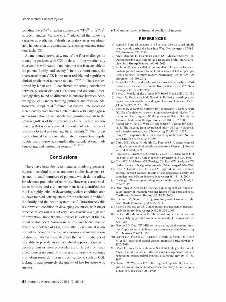
FIGURE 3. Adult male victim of craniocerebral gunshot injury (CGI) during assault. He was transferred promptly to our emergency service, received vigorous resuscitation despite Glasgow Coma Scale of 5 (E1V2M2) and emergent damage control neurosurgery. A: Image showing the inlet hole of CGI in left parietal region with perilesional tissue devitalization. B: Comminuted left skull associated to dural tear, brain laceration and bulging of macerated brain parenchyma. C: Postoperative image of subtotal left fronto-parietal lo-bectomy with drain of left intraparenchymal hemorrhage; hemisphere shows blunt damage and congestive feature. D: Suturing of operation site and inlet wound.
A B C D
Hernando Raphael Alvis-Miranda, et al.
http://www.kjnt.org 41
lesions of the head and neck that are minimally invasive, at-tractive options in selected cases.15)
Management of CSF fistulasDuring primary surgery all efforts should be directed to
seal the dura to prevent CSF fistulas. Surgical correction is recommended for CSF fistulas do not close spontaneously or refractory to medical management. The management of fistulas in the inlet and outlet of the projectile require the closure of the dura mater, fascia and skin. Infection is the most common complication of penetrating brain injuries and is directly associated with increased morbidity and mortali-ty,21) so prevention is essential to optimize their prognosis regardless of the initial management of the injury.
Factors considered determinants of infection include:13,21,44) 1) Retained fragments of bone or metal.2) Time of surgery.3) Use of antibiotics.4) CSF fistulas.In the study of Meirowsky et al.43) only 50% of the fistulas
were at the site of entry or exit of the projectile, 72% occurred in the first 2 weeks of trauma and 44% closed spontaneously. The conclusion is that the more early CSF fistulas are treated less is the risk of infectious complications, morbidity and mortality.
Antibiotic prophylaxis in CGIThe use of broad spectrum antibiotics is recommended
in patients with penetrating brain trauma.7) The risk of in-tracranial infection in patients with penetrating brain trau-ma is high due to the presence of foreign bodies, contami-nated skin, hair and bone fragments in the path of the projectile. In this review we only make reference to antibi-otic prophylaxis after trauma and before any clinical evi-dence of infection, management of established infection (wound infection, meningitis, brain abscess, etc.) are not discussed here.
The infection rate in the pre-antibiotic era during World War I as reported by Whitaker66) was 58.8% in the Second World War in the Post-Antibiotic Era in Slemon study com-pared the use of Sulfa Local and/or parenteral sulfonamide with an infection rate of 21% to 31%, but when added peni-cillin the infection rate dropped from 5.7% to 13%.63)
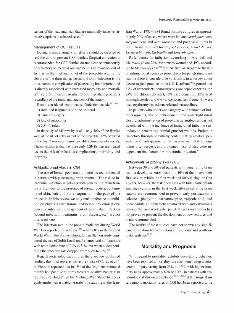
Regard bacteriological cultures there are few published studies, the most representative are those of Carey et al.10) in Vietnam reported that in 45% of the fragments removed mainly had positive cultures for gram-positive bacteria; in the study of Hagan25) in the Vietnam War Staphylococcus epidermidis was isolated; Aarabi1) in studying in the Iran-
Iraq War of 1983-1984 found positive cultures in approxi-mately 20% of cases, where were isolated staphylococcus, streptococcus and acinetobacter, and positive cultures in brain tissue removed for Staphylococcus, Acinetobacter, Escherichia coli, Klebsiella and Enterobacter.
Risk factors for infection, according to Arendall and Meirowsky4) are 29% for sinuses wound and 49% accord-ing to Meirowsky et al.43) for CSF fistulas. Regard to the use of antimicrobial agents as prophylaxis for penetrating brain trauma there is considerable variability, in a survey about Neurosurgical practice in the U.S. Kaufman34) reported that 87% of respondents neurosurgeons use cephalosporins, the 24% use chloramphenicol, 16% used penicillin, 12% used aminoglycosides and 6% vancomycin, less frequently were used erythromycin, miconazole and tetracyclines.
In patients who underwent surgery with removal of bul-let fragments, wound debridement, and watertight dural closure, administration of prophylactic antibiotics was not associated with the incidence of intracranial infection sec-ondary to penetrating cranial gunshot wounds. Projectile trajectory through potentially contaminating cavities, per-sistence of intraparenchymal osseous or metallic frag-ments after surgery, and prolonged hospital stay were in-dependent risk factors for intracranial infection.31)
Anticonvulsive prophylaxis in CGIBetween 30 and 50% of patients with penetrating brain
trauma develop seizures from 4 to 10% of them have their first seizure within the first week and 80% during the first 2 years, however, the risk decreases with time. Anticonvul-sant medications in the first week after penetrating brain trauma are recommended to prevent early posttraumatic seizures (phenytoin, carbamazepine, valproic acid, and phenobarbital). Prophylactic treatment with anticonvulsants beyond the first week after penetrating brain trauma has not proven to prevent the development of new seizures and is not recommended.
The results of most studies have not shown any signifi-cant correlation between retained fragments and posttrau-matic epilepsy.19,52)
Mortality and Prognosis
With regard to mortality, exhibits devastating behavior, have been reported a mortality rate after penetrating cranio-cerebral injury varing from 23% to 92%, with higher mor-tality rates, approximately 87% to 100% in patients with low neurologic status on presentation.1,22,34,47,53) After surgical in-terventions mortality rates of CGI has been reported to be
42 Korean J Neurotrauma 2015;11(2):35-43
Craniocerebral Gunshot Injuries
rounding the 20%8) in earlier studies and 7.4%41) to 18.7%51) in recent studies. Murano et al.46) identified the following variables as predictors of death: respiratory arrest on admis-sion, hypotension on admission, transhemispheric and trans-ventricular CGI.
As mentioned previously, one of the first challenges in managing patients with CGI is determining whether any intervention will result in an outcome that is acceptable to the patient, family, and society,16) in this circumstance, the postresuscitation GCS is the most reliable and significant clinical predictor of outcome to date.3,28,40,51,55) The series re-ported by Khan et al.36) confirmed the strong correlation between postresuscitation GCS score and outcome. Inter-estingly, they found no difference in outcome between pene-trating (no exit) and perforating (entrance and exit) wounds. However, Joseph et al.32) found that survival rate increased incrementally over time to a rate of 46% with early aggres-sive resuscitation of all patients with gunshot wounds to the brain regardless of their presenting clinical picture, recom-mending that notion of low GCS should not deter the use of resources to treat and manage these patients.32) Other prog-nostic clinical factors include dilated, nonreactive pupils, hypotension, hypoxia, coagulopathy, suicide attempt, ad-vanced age, and perforating wounds.1,3,35,52,55)
Conclusions
There have been few recent studies involving penetrat-ing craniocerebral injuries, and most studies have been re-stricted to small numbers of patients, which do not allow for adequate prediction of mortality. However, classic stud-ies in military and civil environment have identified that this is a highly lethal or devastating violent condition, able to leave marked consequences for the affected individual, the family and the health system itself. Unfortunately this is a prevalent condition in developing countries, with major armed conflicts which is not very likely to achieve a high rate of prevention, since the main trigger is violence at the na-tional or state level. Various measures have been aimed to lower the incidence of CGI, especially in civilians. It is im-portant to recognize the role of vigorous and intense resus-citation but always evaluated together with predictors of mortality, to provide an individualized approach, especially because injuries from projectiles are different from each other, there is no equal. It is necessarily urgent to continue promoting research in a neurocritical topic such as CGI, looking impact positively the quality of life for those who survive.
■ The authors have no financial conflicts of interest.
REFERENCES1) Aarabi B. Surgical outcome in 435 patients who sustained missile
head wounds during the Iran-Iraq War. Neurosurgery 27:692-695; discussion 695, 1990
2) Alvis Miranda H, Castellar-Leones SM, Moscote-Salazar LR. Decompressive craniectomy and traumatic brain injury: a re-view. Bull Emerg Trauma 1:60-68, 2013
3) Ambrosi PB, Valença MM, Azevedo-Filho H. Prognostic factors in civilian gunshot wounds to the head: a series of 110 surgical pa-tients and brief literature review. Neurosurg Rev 35:429-435; discussion 435-436, 2012
4) Arendall RE, Meirowsky AM. Air sinus wounds: an analysis of 163 consecutive cases incurred in the Korean War, 1950-1952. Neu-rosurgery 13:377-380, 1983
5) Bakay L. Missile injuries of brain. N Y State J Med 82:313-319, 19826) Barach E, Tomlanovich M, Nowak R. Ballistics: a pathophysio-
logic examination of the wounding mechanisms of firearms: Part I. J Trauma 26:225-235, 1986
7) Bayston R, de Louvois J, Brown EM, Johnston RA, Lees P, Pople IK. Use of antibiotics in penetrating craniocerebral injuries. “In-fection in Neurosurgery” Working Party of British Society for Antimicrobial Chemotherapy. Lancet 355:1813-1817, 2000
8) Becker DP, Miller JD, Ward JD, Greenberg RP, Young HF, Saka-las R. The outcome from severe head injury with early diagnosis and intensive management. J Neurosurg 47:491-502, 1977
9) Carey ME. Experimental missile wounding of the brain. Neuro-surg Clin N Am 6:629-642, 1995
10) Carey ME, Young H, Mathis JL, Forsythe J. A bacteriological study of craniocerebral missile wounds from Vietnam. J Neuro-surg 34:145-154, 1971
11) Cavaliere R, Cavenago L, Siccardi D, Viale GL. Gunshot wounds of the brain in civilians. Acta Neurochir (Wien) 94:133-136, 1988
12) Clark WC, Muhlbauer MS, Watridge CB, Ray MW. Analysis of 76 civilian craniocerebral gunshot wounds. J Neurosurg 65:9-14, 1986
13) Coşar A, Gönül E, Kurt E, Gönül M, Taşar M, Yetişer S. Cranio-cerebral gunshot wounds: results of less aggressive surgery and complications. Minim Invasive Neurosurg 48:113-118, 2005
14) Cushing H. Notes on penetrating wounds of the brain. Br Med J 1: 221-226, 1918
15) Diaz-Daza O, Arraiza FJ, Barkley JM, Whigham CJ. Endovas-cular therapy of traumatic vascular lesions of the head and neck. Cardiovasc Intervent Radiol 26:213-221, 2003
16) Ecklund JM, Sioutos P. Prognosis for gunshot wounds to the head. World Neurosurg 82:27-29, 2014
17) Esposito DP, Walker JB. Contemporary management of penetrat-ing brain injury. Neurosurg Q 19:249-254, 2009
18) Fackler ML, Malinowski JA. The wound profile: a visual method for quantifying gunshot wound components. J Trauma 25:522-529, 1985
19) George ED, Dagi TF. Military penetrating craniocerebral inju-ries. Applications to civilian triage and management. Neurosurg Clin N Am 6:753-759, 1995
20) Gervaise A, Foscolo S, Rivierre A, Derelle A, Schmitt E, Braun M, et al. [Imaging of cranial gunshot traumas]. J Radiol 91:1113-1120, 2010
21) Gönül E, Baysefer A, Kahraman S, Ciklatekerlioğlu O, Gezen F, Yayla O, et al. Causes of infections and management results in penetrating craniocerebral injuries. Neurosurg Rev 20:177-181, 1997
22) Grahm TW, Williams FC Jr, Harrington T, Spetzler RF. Civilian gunshot wounds to the head: a prospective study. Neurosurgery 27:696-700; discussion 700, 1990

Hernando Raphael Alvis-Miranda, et al.
http://www.kjnt.org 43
23) Hachemi M, Jourdan C, Di Roio C, Turjman F, Ricci-Franchi A, Mottolese C, et al. Delayed rupture of traumatic aneurysm after civilian craniocerebral gunshot injury in children. Childs Nerv Syst 23:283-287, 2007
24) Hadas N, Schiffer J, Rogev M, Shperber Y. Tangential low-ve-locity missile wound of the head with acute subdural hematoma: case report. J Trauma 30:358-359, 1990
25) Hagan RE. Early complications following penetrating wounds of the brain. J Neurosurg 34:132-141, 1971
26) Hammon WM, Kempe LG. Methyl methacrylate cranioplasty. 13 years experience with 417 patients. Acta Neurochir (Wien) 25:69-77, 1971
27) Harvey EN, McMillen JH. An Experimental Study of Shock Waves Resulting from the Impact of High Velocity Missiles on Animal Tissues. J Exp Med 85:321-328, 1947
28) Hofbauer M, Kdolsky R, Figl M, Grünauer J, Aldrian S, Oster-mann RC, et al. Predictive factors influencing the outcome after gunshot injuries to the head-a retrospective cohort study. J Trau-ma 69:770-775, 2010
29) Hollerman JJ, Fackler ML, Coldwell DM, Ben-Menachem Y. Gun-shot wounds: 2. Radiology. AJR Am J Roentgenol 155:691-702, 1990
30) Horowitz MB, Kopitnik TA, Landreneau F, Ramnani DM, Rush-ing EJ, George E, et al. Multidisciplinary approach to traumatic intracranial aneurysms secondary to shotgun and handgun wounds. Surg Neurol 51:31-41; discussion 41-42, 1999
31) Jimenez CM, Polo J, España JA. Risk factors for intracranial in-fection secondary to penetrating craniocerebral gunshot wounds in civilian practice. World Neurosurg 79:749-755, 2013
32) Joseph B, Aziz H, Pandit V, Kulvatunyou N, O’Keeffe T, Wynne J, et al. Improving survival rates after civilian gunshot wounds to the brain. J Am Coll Surg 218:58-65, 2014
33) Joseph B, Aziz H, Sadoun M, Kulvatunyou N, Pandit V, Tang A, et al. Fatal gunshot wound to the head: the impact of aggressive management. Am J Surg 207:89-94, 2014
34) Kaufman HH. Civilian gunshot wounds to the head. Neurosur-gery 32:962-964; discussion 964, 1993
35) Kazim SF, Shamim MS, Tahir MZ, Enam SA, Waheed S. Man-agement of penetrating brain injury. J Emerg Trauma Shock 4:395-402, 2011
36) Khan MB, Kumar R, Irfan FB, Irfan AB, Bari ME. Civilian cra-niocerebral gunshot injuries in a developing country: presenta-tion, injury characteristics, prognostic indicators, and complica-tions. World Neurosurg 82:14-19, 2014
37) Kim TW, Lee JK, Moon KS, Kwak HJ, Joo SP, Kim JH, et al. Penetrating gunshot injuries to the brain. J Trauma 62:1446-1451, 2007
38) Knightly JJ, Pulliam MW. Military head injuries in Narayan RK, Wilberger JE, Povlishock JT (eds): Neurotrauma. New York: McGraw-Hill, pp891-902, 1996
39) Kubal WS. Updated imaging of traumatic brain injury. Radiol Clin North Am 50:15-41, 2012
40) Levy ML, Masri LS, Lavine S, Apuzzo ML. Outcome prediction after penetrating craniocerebral injury in a civilian population: ag-gressive surgical management in patients with admission Glasgow Coma Scale scores of 3, 4, or 5. Neurosurgery 35:77-84; discussion 84-85, 1994
41) Liebenberg WA, Demetriades AK, Hankins M, Hardwidge C, Hartzenberg BH. Penetrating civilian craniocerebral gunshot wounds: a protocol of delayed surgery. Neurosurgery 57:293-299; discussion 293-299, 2005
42) Liker MA, Aarabi B, Levy ML. Missile wounds to the head: bal-listics and forensics in Aarabi B, Kaufman HH (eds): Missile wounds of the head and neck. Park Ridge: Thieme, Vol 2, pp35-56, 1999
43) Meirowsky AM, Caveness WF, Dillon JD, Rish BL, Mohr JP, Kistler JP, et al. Cerebrospinal fluid fistulas complicating missile wounds of the brain. J Neurosurg 54:44-48, 1981
44) Melada A, Marcikić M, Mrak G, Stimac D, Sćap M. Cerebrospi-nal fluid fistula as a consequence of war head injury. Mil Med 167:666-670, 2002
45) Moscote-Salazar LR, Alvis-Miranda HR, Palencia C, Rubiano AM. Emergent decompressive craniectomy in patients with fixed dilated pupils; a single center experience. Bull Emerg Trauma 1:175-178, 2013
46) Murano T, Mohr AM, Lavery RF, Lynch C, Homnick AT, Liv-ingston DH. Civilian craniocerebral gunshot wounds: an update in predicting outcomes. Am Surg 71:1009-1014, 2005
47) Nagib MG, Rockswold GL, Sherman RS, Lagaard MW. Civilian gunshot wounds to the brain: prognosis and management. Neu-rosurgery 18:533-537, 1986
48) Neuroimaging in the management of penetrating brain injury. J Trauma 51:S7-S11, 2001
49) Offiah C, Twigg S. Imaging assessment of penetrating cranioce-rebral and spinal trauma. Clin Radiol 64:1146-1157, 2009
50) Ordog GJ, Wasserberger J, Balasubramanium S. Wound ballis-tics: theory and practice. Ann Emerg Med 13:1113-1122, 1984
51) Ozkan U, Kemaloğlu S, Ozateş M, Aydin MD. Analysis of 107 civilian craniocerebral gunshot wounds. Neurosurg Rev 25:231-236, 2002
52) Part 1: Guidelines for the management of penetrating brain inju-ry. Introduction and methodology. J Trauma 51:S3-S6, 2001
53) Part 2: Prognosis in penetrating brain injury. J Trauma 51:S44-S86, 2001
54) Raimondi AJ, Samuelson GH. Craniocerebral gunshot wounds in civilian practice. J Neurosurg 32:647-653, 1970
55) Rish BL, Caveness WF, Dillon JD, Kistler JP, Mohr JP, Weiss GH. Analysis of brain abscess after penetrating craniocerebral injuries in Vietnam. Neurosurgery 9:535-541, 1981
56) Rish BL, Dillon JD, Caveness WF, Mohr JP, Kistler JP, Weiss GH. Evolution of craniotomy as a debridement technique for penetrating craniocerebral injuries. J Neurosurg 53:772-775, 1980
57) Rish BL, Dillon JD, Meirowsky AM, Caveness WF, Mohr JP, Kistler JP, et al. Cranioplasty: a review of 1030 cases of penetrat-ing head injury. Neurosurgery 4:381-385, 1979
58) Robles LA. High-velocity gunshot to the head presenting as initial minor head injury: things are not what they seem. Am J Emerg Med 30:2089.e5-2089.e7, 2012
59) Rosenfeld JV. Gunshot injury to the head and spine. J Clin Neuro-sci 9:9-16, 2002
60) Saito N, Hito R, Burke PA, Sakai O. Imaging of penetrating inju-ries of the head and neck: current practice at a level I trauma cen-ter in the United States. Keio J Med 63:23-33, 2014
61) Selden BS, Goodman JM, Cordell W, Rodman GH Jr, Schnitzer PG. Outcome of self-inflicted gunshot wounds of the brain. Ann Emerg Med 17:247-253, 1988
62) Shaffrey ME, Polin RS, Phillips CD, Germanson T, Shaffrey CI, Jane JA. Classification of civilian craniocerebral gunshot wounds: a multivariate analysis predictive of mortality. J Neurotrauma 9 Suppl 1:S279-S285, 1992
63) Slemon HV. Forward neurosurgery in Italy. J Neurosurg 2:332-339, 1945
64) Tsuei YS, Sun MH, Lee HD, Chiang MZ, Leu CH, Cheng WY, et al. Civilian gunshot wounds to the brain. J Chin Med Assoc 68:126- 130, 2005
65) Vascular complications of penetrating brain injury. J Trauma 51: S26-S28, 2001
66) Whitaker R. Gunshot wounds of the cranium: with special reference to those of the brain. Br J Surg 3:708-735, 1915