management of diabetic macular edema
TRANSCRIPT
This continuing medical education activity is supported through anunrestricted educational grant from Allergan.
This continuing medical education activity is jointly provided by new York eye and ear infirmary of Mount sinai and MedEdicus LLC.
The Real-World Discussion Series™
CMe MoNoGraPH
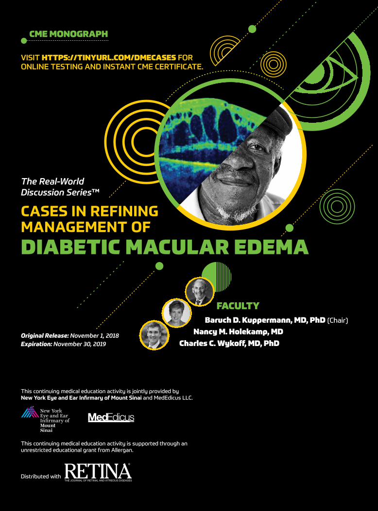
Cases in RefiningManageMent ofDiaBetiC MaCular eDeMa
Distributed with
Visit HttPs://tiNyurl.CoM/DMeCases foronline testing and instant CMe CertifiCate.
Original Release: November 1, 2018Expiration: November 30, 2019
FaCulty
Charles C. Wykoff, MD, PhD
Baruch D. Kuppermann, MD, PhD (Chair)
Nancy M. Holekamp, MD
learNiNG MetHoD aND MeDiuMThis educational activity consists of a supplement andseven (7) study questions. The participant should, in order,read the learning objectives contained at the beginning ofthis supplement, read the supplement, answer all questionsin the post test, and complete the Activity Evaluation/CreditRequest form. To receive credit for this activity, pleasefollow the instructions provided on the post test andActivity Evaluation/Credit Request form. This educationalactivity should take a maximum of 1.5 hours to complete.
aCtiVity DesCriPtioNThe prevalence of diabetic retinopathy (DR) and diabeticmacular edema (DME) is on the rise. Although antivascularendothelial growth factor therapy effectively treats DMEand DR, it does not target the inflammatory aspect of DME.As such, a significant proportion of patients might notexperience an improvement in their visual acuity or mightcontinue to have persistent DME that threatens long-termpotential for visual acuity gains. Using steroids as acomplementary or alternative therapy can be useful inthese patients. However, intraocular pressure elevationshave been observed with the use of intravitreal steroidimplants. Intraocular pressure elevation followingintravitreal steroid administration follows a predictablecourse, and, in most cases, can be safely managed in theretina practice. The desired results of this activity are toupdate retina specialists and other ophthalmologists oncurrent and new approaches to treating DR and DME.
tarGet auDieNCeThis educational activity is intended for retina specialistsand other ophthalmologists.
learNiNG oBJeCtiVesUpon completion of this activity, participants will be betterable to:• Recognize the different mechanisms of disease that drive
treatment selection for patients with DME• Explain the implications of persistent edema for selecting
treatment for patients with DME• Discuss management of IOP elevations due to intravitreal
steroid implants• Develop long-term treatment plans for patients with DME
aCCreDitatioN stateMeNtThis activity has been planned and implemented inaccordance with the accreditation requirements and policiesof the Accreditation Council for Continuing MedicalEducation (ACCME) through the joint providership of new York eye and ear infirmary of Mount sinai andMedEdicus LLC. The new York eye and ear infirmary ofMount sinai is accredited by the ACCME to providecontinuing medical education for physicians.
In July 2013, the Accreditation Council for ContinuingMedical Education (ACCME) awarded New York Eyeand Ear Infirmary of Mount Sinai “Accreditation withCommendation,” for six years as a provider ofcontinuing medical education for physicians, thehighest accreditation status awarded by the ACCME.
aMa CreDit DesiGNatioN stateMeNtThe new York eye and ear infirmary of Mount sinaidesignates this enduring material for a maximum of 1.5 AMA PRA Category 1 Credits™. Physicians should claimonly the credit commensurate with the extent of theirparticipation in the activity.
GraNtor stateMeNtThis continuing medical education activity is supportedthrough an unrestricted educational grant from Allergan.
DisClosure PoliCy stateMeNtIt is the policy of new York eye and ear infirmary of Mount sinai that the faculty and anyone in a position tocontrol activity content disclose any real or apparent conflictsof interest relating to the topics of this educational activity,and also disclose discussions of unlabeled/unapproved usesof drugs or devices during their presentation(s). new Yorkeye and ear infirmary of Mount sinai has established policiesin place that will identify and resolve all conflicts of interestprior to this educational activity. Full disclosure of faculty/planners and their commercial relationships, if any, follows.
DisClosuresnancy M. Holekamp, MD, had a financial agreement oraffiliation during the past year with the following commercialinterests in the form of Receipt of Intellectual Rights/PatentHolder: Katalyst; Consultant/Advisory Board: Allergan;BioTime, Inc; Genentech, Inc; Novartis PharmaceuticalsCorporation; and Regeneron Pharmaceuticals, Inc;Contracted Research: Alimera Sciences; Allergan; Genentech,Inc; Ohr Pharmaceutical; and Ophthotech Corporation;Honoraria from promotional, advertising or non-CME servicesreceived directly from commercial interests or their Agents(eg, Speakers Bureaus): Alimera Sciences; Allergan;Genentech, Inc; and Regeneron Pharmaceuticals, Inc.
Baruch D. Kuppermann, MD, PhD, had a financialagreement or affiliation during the past year with thefollowing commercial interests in the form ofConsultant/Advisory Board: Aerpio Therapeutics; Alcon;Alimera Sciences; Allegro Ophthalmics, LLC; Allergan; AmpioPharmaceuticals Inc; Catalyst Pharma; Dose MedicalCorporation; Eleven Biotherapeutics; Genentech, Inc;Glaukos Corporation; Lumenis; Neurotech Pharmaceuticals;Novartis Pharmaceuticals Corporation; OphthotechCorporation; Regeneron Pharmaceuticals, Inc; and SciFluorLife Sciences, Inc; Contracted Research: Alcon; AlimeraSciences; Allegro Ophthalmics, LLC; Allergan; ApellisPharmaceuticals; Genentech, Inc; GlaxoSmithKline; jCyte;Neurotech Pharmaceuticals; Ohr Pharmaceutical;Ophthotech Corporation; Oxurion NV; and RegeneronPharmaceuticals, Inc; Honoraria from promotional,advertising or non-CME services received directly fromcommercial interests or their Agents (eg, Speakers Bureaus):Allergan; and Optovue, Incorporated.
Charles C. Wykoff, MD, PhD, had a financial agreement oraffiliation during the past year with the following commercialinterests in the form of Consultant/Advisory Board: Alcon;Alimera Sciences; Allergan; Bayer Corporation; ClearsideBiomedical, Inc; D.O.R.C.; F. Hoffmann-La Roche Ltd;Genentech, Inc; and Regeneron Pharmaceuticals, Inc;Contracted Research: Aerpio Therapeutics; Alcon; Allergan;Apellis Pharmaceuticals; Clearside Biomedical, Inc; F.Hoffmann-La Roche Ltd; Genentech, Inc; HeidelbergEngineering GmbH; Novartis Pharmaceuticals Corporation;Ophthotech Corporation; and Regeneron Pharmaceuticals, Inc;Honoraria from promotional, advertising or non-CME servicesreceived directly from commercial interests or their Agents (eg, Speakers Bureaus): Allergan; Bayer Corporation; andRegeneron Pharmaceuticals, Inc.
NeW yorK eye aND ear iNFirMary oF MouNt siNai Peer reVieW DisClosure gennady Landa, MD, has no relevant commercialrelationships to disclose.
eDitorial suPPort DisClosurestony Realini, MD, had a financial agreement or affiliationduring the past year with the following commercialinterests in the form of Consultant/Advisory Board: AeriePharmaceuticals, Inc; Inotek Pharmaceuticals Corporation;New World Medical, Inc; and Reichert, Inc.
erika Langsfeld, PhD; Diane Mcardle, PhD; Cynthiatornallyay, RD, MBa, CHCP; Kimberly Corbin, CHCP;Barbara aubel; and Michelle ong have no relevantcommercial relationships to disclose.
DisClosure attestatioNThe contributing physicians listed above have attested tothe following:1) that the relationships/affiliations noted will not bias or
otherwise influence their involvement in this activity;2) that practice recommendations given relevant to the
companies with whom they have relationships/affiliations will be supported by the best availableevidence or, absent evidence, will be consistent withgenerally accepted medical practice; and
3) that all reasonable clinical alternatives will be discussedwhen making practice recommendations.
oFF-laBel DisCussioNThis CME activity includes discussion of unlabeled and/orinvestigative uses of drugs. Please refer to the officialprescribing information for each drug discussed in thisactivity for FDA-approved dosing, indications, andwarnings.
NeW yorK eye aND ear iNFirMary oF MouNtsiNai PriVaCy & CoNFiDeNtiality PoliCieshttp://www.nyee.edu/health-professionals/cme/enduring-activities
CMe ProViDer CoNtaCt iNForMatioNFor questions about this activity, call 212-870-8127.
to oBtaiN AMA PRA CATEGORY 1 CREDIT™To obtain AMA PRA Category 1 Credit™ for this activity,read the material in its entirety and consult referencedsources as necessary. Please take this post test andevaluation online by going to https://tinyurl.com/dmecases.Upon passing, you will receive your certificate immediately.You must score 70% or higher to receive credit for thisactivity, and may take the test up to 2 times. Uponregistering and successfully completing the post test, yourcertificate will be made available online and you can print itor file it.
DisClaiMerThe views and opinions expressed in this educationalactivity are those of the faculty and do not necessarilyrepresent the views of new York eye and ear infirmary ofMount sinai, MedEdicus LLC, Allergan, or Retina. 2
Baruch D. Kuppermann, MD, PhDRoger F. Steinert Endowed Professor Chair, Department of Ophthalmology Director, Gavin Herbert Eye Institute University of California, IrvineIrvine, California
CHair
Gennady landa, MDAssociate Professor of OphthalmologyIcahn School of Medicine at Mount SinaiVitreoretinal Specialist and
Attending SurgeonDepartment of OphthalmologyMedical Director of Tribeca
and Williamsburg OfficesNew York Eye and Ear Infirmary
of Mount SinaiNew York, New York
CMe reVieWer For NeW yorK eye aND eariNFirMary oF MouNt siNai
This CME activity is copyrighted to MedEdicus LLC ©2018. All rights reserved. 166
Nancy M. Holekamp, MDProfessor of Clinical OphthalmologyWashington University School of MedicineDirector, Retina ServicesPepose Vision InstituteSt Louis, Missouri
Charles C. Wykoff, MD, PhDDirector of ResearchRetina Consultants of HoustonClinical Associate Professor
of OphthalmologyWeill Cornell Medical College,
Houston Methodist HospitalHouston, Texas
FaCulty
3foR instant PRoCessing, CoMPLete tHe CMe Post test onLine at HttPs://tiNyurl.CoM/DMeCases
iNtroDuCtioNThe prevalence of diabetes mellitus (DM) and diabetic retinopathy(DR), including diabetic macular edema (DME), is on the rise. In thenext 12 years alone, DM prevalence is projected to increase by morethan 50%, with the southern United States seeing the greatestincreases.1 Because the risk of DR and its progression rises withincreasing levels of hemoglobin A1c,2,3 the burden of DR willincrease among poorly controlled patients with DM. Therapeuticoptions for the management of DR and DME have expandedsignificantly in recent years, and clinical trials offer insights intooptimal treatment approaches. In this review, an experienced panelof retina specialists will put the management of DME into a moderncontext through the discussion of a series of cases. Our objectivesare to review the underlying mechanisms of DME, the significanceof persistent edema, and their implications for the selection oftherapy. We will discuss options for treating eyes with DME,including the management of complications of therapy, such ascataract and glaucoma associated with steroid implants. At thecompletion of this activity, retina specialists and otherophthalmologists who treat DME will be better able to developlong-term strategies for the management of eyes with DME.
—Baruch D. Kuppermann, MD, PhD (Chair)
loNG-terM FluiD MaNaGeMeNt iN DiaBetiC MaCular eDeMa Charles C. Wykoff, MD, PhDPharmacologic inhibition of vascular endothelial growth factor(VEGF) works very well in DME, improving macular fluid in manytreated eyes. There are, however, at least 2 key limitations of currentanti-VEGF therapy. One limitation is durability: the therapeutic effectdoes not last indefinitely, and repeated retreatments are oennecessary. A second limitation is efficacy: many eyes never achieveoptimal visual function even if their DME resolves.
Limited durability of anti-VEGF therapy imposes a substantialtreatment burden in many eyes. In the DRCRnet’s (DiabeticRetinopathy Clinical Research Network’s) Protocol T studyevaluating aflibercept, bevacizumab, or ranibizumab for DME, amedian of 23 visits was required through 2 years, and the meannumber of injections given during that time was 15 to 16, dependingon the agent.4 Treatment burden appears to diminish over time inmany eyes. In the open-label extension following the pivotal RISEand RIDE trials of ranibizumab for DME, the annualized rate ofranibizumab injections in years 4 to 5 was fewer than 4, withapproximately 25% of eyes requiring no additional injections duringthis period (n = 121 at month 54).5 In the Protocol I study ofranibizumab for DME, by the fih year of treatment, the annualnumber of clinical visits was only 4 to 5 and the ranibizumabinjection rate was very low.6 In the ENDURANCE open-labelextension following the VIVID and VISTA trials of aflibercept forDME, the mean number of aflibercept injections given in years 4and 5 was 4.5 and 3.4, respectively.7,8
Among the lessons learned from these long-term studies is thatthe visual gains achieved with initial anti-VEGF treatment for DMEcan persist for years. Although the treatment burden does diminishwith time, it does not disappear completely. Many eyes will requireongoing retreatment on an as-needed basis.
A clinically relevant question that we have not yet answered fromthese trials is when to re-treat. At what point is the next injectionwarranted? There are several issues to consider. Should we holdinjections as long as the visual acuity is stable? Should we inject ifwe see fluid recurring on optical coherence tomography (OCT)imaging, even if visual acuity is preserved? If fluid does notcompletely resolve with anti-VEGF therapy, should the treatment
regimen be expanded to include other modalities, such as steroids,which might target other mechanisms of persistent edema? Thereis also the issue of DR. Anti-VEGF therapy not only treats DME, italso slows the progression of DR and can improve DR in manycases.9 If the DME clears and further injections are withheld, butthen the DR progresses to proliferative DR with associated visualloss, we might have missed an opportunity for disease modificationby not continuing treatment once the DME had resolved. A casecould be made for some frequency of long-term maintenancetherapy in eyes with both DME and DR—including in eyes withgood visual acuity and a dry macula—to preserve the central visualgains while holding off the DR.
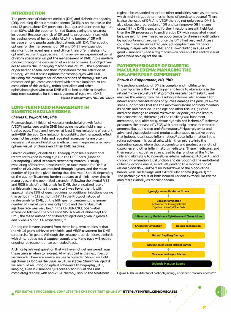
PatHoPHysioloGy oF DiaBetiC MaCular eDeMa: MaNaGiNG tHeiNFlaMMatory CoMPoNeNtBaruch D. Kuppermann, MD, PhDThe pathophysiology of DME is complex and multifactorial.Hyperglycemia is the initial trigger and leads to alterations in theretinal microvasculature that promote vascular permeability andmacular thickening from the resulting extravascular edema. Highintravascular concentrations of glucose damage the pericytes—thesmall support cells that line the microvasculature and help maintainits health and function. In the eye and other tissues, glucose-mediated damage to retinal microvascular pericytes can lead tovasoconstriction, thickening of the capillary wall basementmembrane, and, ultimately, tissue hypoxia and ischemia.10 Ischemiapromotes the release of VEGF, which not only increases vascularpermeability, but is also proinflammatory.11 Hyperglycemia andadvanced glycosylation end products also cause oxidative stressthat induces local tissue inflammation.12 Local inflammation in theretina activates microglial cells, which then migrate into thesubretinal space, where they accumulate and produce a variety ofcytokines and other inflammatory mediators. These mediators, andtheir resulting oxidative stress, lead to dysfunction of the Müllercells and ultimately to intracellular edema, retinal excitotoxicity, andchronic inflammation. Dysfunction and disruption of the endothelialcellular junctions ensue, eventually leading to a modification ofretinal blood flow, leukostasis, breakdown of the blood-retinabarrier, vascular leakage, and extracellular edema (figure 1).12-14
The pathologic result of both intracellular and extracellular edemamanifests clinically as macular edema.
Hyperglycemia - Oxidative Stress
Local InflammationActivation of Microglial CellsDysfunction of Müller Cells
Inflammatory Mediators - Cytokines and Chemokines
Chronic Inflammation Neurodegeneration
Retinal Capillary Damage
Disruption of Blood-Retinal Barrier
Vascular Leakage - Edema
Diabetic Macular Edema
figure 1. The multifactorial pathophysiology of diabetic macular edema12-14
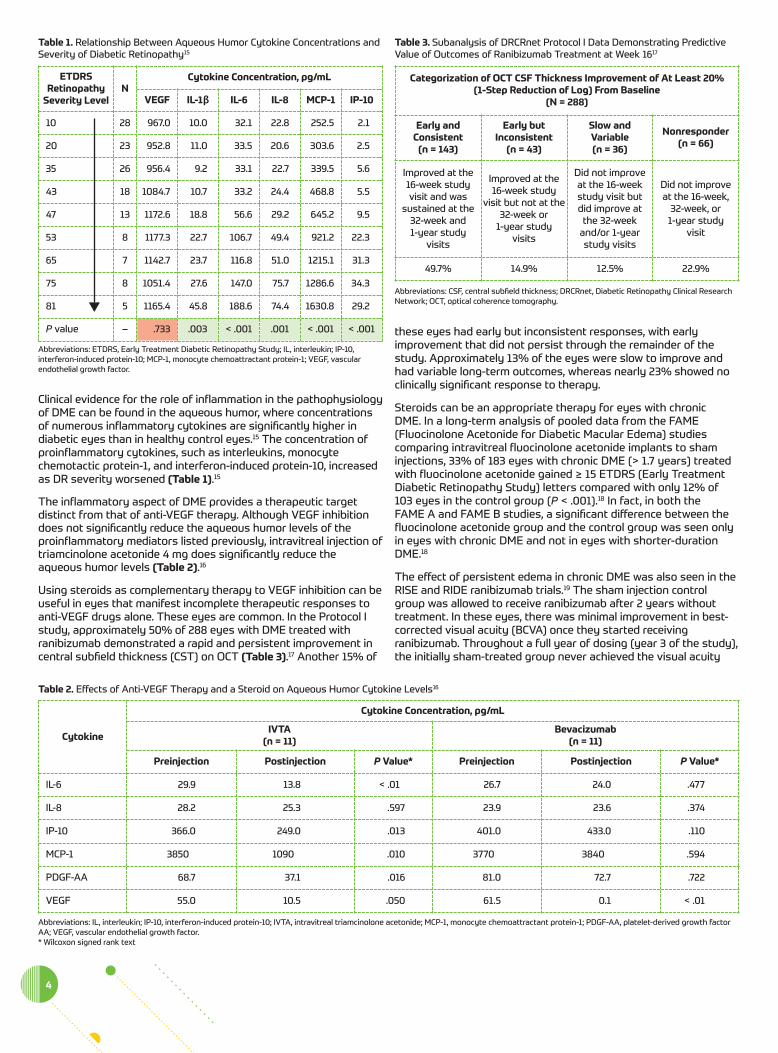
Clinical evidence for the role of inflammation in the pathophysiologyof DME can be found in the aqueous humor, where concentrationsof numerous inflammatory cytokines are significantly higher indiabetic eyes than in healthy control eyes.15 The concentration ofproinflammatory cytokines, such as interleukins, monocytechemotactic protein-1, and interferon-induced protein-10, increased as DR severity worsened (table 1).15
The inflammatory aspect of DME provides a therapeutic targetdistinct from that of anti-VEGF therapy. Although VEGF inhibitiondoes not significantly reduce the aqueous humor levels of theproinflammatory mediators listed previously, intravitreal injection oftriamcinolone acetonide 4 mg does significantly reduce theaqueous humor levels (table 2).16
Using steroids as complementary therapy to VEGF inhibition can beuseful in eyes that manifest incomplete therapeutic responses toanti-VEGF drugs alone. These eyes are common. In the Protocol Istudy, approximately 50% of 288 eyes with DME treated withranibizumab demonstrated a rapid and persistent improvement incentral subfield thickness (CST) on OCT (table 3).17 Another 15% of
these eyes had early but inconsistent responses, with earlyimprovement that did not persist through the remainder of thestudy. Approximately 13% of the eyes were slow to improve andhad variable long-term outcomes, whereas nearly 23% showed noclinically significant response to therapy.
Steroids can be an appropriate therapy for eyes with chronic DME. In a long-term analysis of pooled data from the FAME(Fluocinolone Acetonide for Diabetic Macular Edema) studiescomparing intravitreal fluocinolone acetonide implants to shaminjections, 33% of 183 eyes with chronic DME (> 1.7 years) treatedwith fluocinolone acetonide gained ≥ 15 ETDRS (Early TreatmentDiabetic Retinopathy Study) letters compared with only 12% of103 eyes in the control group (P < .001).18 In fact, in both the FAME A and FAME B studies, a significant difference between thefluocinolone acetonide group and the control group was seen onlyin eyes with chronic DME and not in eyes with shorter-durationDME.18
The effect of persistent edema in chronic DME was also seen in theRISE and RIDE ranibizumab trials.19 The sham injection controlgroup was allowed to receive ranibizumab aer 2 years withouttreatment. In these eyes, there was minimal improvement in best-corrected visual acuity (BCVA) once they started receivingranibizumab. Throughout a full year of dosing (year 3 of the study),the initially sham-treated group never achieved the visual acuity
4
etDRsRetinopathy
severity Leveln
Cytokine Concentration, pg/mL
Vegf iL-1β iL-6 iL-8 MCP-1 iP-10
10 28 967.0 10.0 32.1 22.8 252.5 2.1
20 23 952.8 11.0 33.5 20.6 303.6 2.5
35 26 956.4 9.2 33.1 22.7 339.5 5.6
43 18 1084.7 10.7 33.2 24.4 468.8 5.5
47 13 1172.6 18.8 56.6 29.2 645.2 9.5
53 8 1177.3 22.7 106.7 49.4 921.2 22.3
65 7 1142.7 23.7 116.8 51.0 1215.1 31.3
75 8 1051.4 27.6 147.0 75.7 1286.6 34.3
81 5 1165.4 45.8 188.6 74.4 1630.8 29.2
P value – .733 .003 < .001 .001 < .001 < .001
table 1. Relationship Between Aqueous Humor Cytokine Concentrations andSeverity of Diabetic Retinopathy15
Abbreviations: ETDRS, Early Treatment Diabetic Retinopathy Study; IL, interleukin; IP-10,interferon-induced protein-10; MCP-1, monocyte chemoattractant protein-1; VEGF, vascularendothelial growth factor.
Cytokine
Cytokine Concentration, pg/mL
iVta(n = 11)
Bevacizumab(n = 11)
Preinjection Postinjection P Value* Preinjection Postinjection P Value*
IL-6 29.9 13.8 < .01 26.7 24.0 .477
IL-8 28.2 25.3 .597 23.9 23.6 .374
IP-10 366.0 249.0 .013 401.0 433.0 .110
MCP-1 3850 1090 .010 3770 3840 .594
PDGF-AA 68.7 37.1 .016 81.0 72.7 .722
VEGF 55.0 10.5 .050 61.5 0.1 < .01
table 2. Effects of Anti-VEGF Therapy and a Steroid on Aqueous Humor Cytokine Levels16
Abbreviations: IL, interleukin; IP-10, interferon-induced protein-10; IVTA, intravitreal triamcinolone acetonide; MCP-1, monocyte chemoattractant protein-1; PDGF-AA, platelet-derived growth factorAA; VEGF, vascular endothelial growth factor.* Wilcoxon signed rank text
Categorization of oCt Csf thickness improvement of at Least 20% (1-step Reduction of Log) from Baseline
(n = 288)
early andConsistent
(n = 143)
early butinconsistent
(n = 43)
slow and Variable(n = 36)
nonresponder(n = 66)
Improved at the16-week studyvisit and was
sustained at the32-week and 1-year study
visits
Improved at the16-week study
visit but not at the32-week or
1-year study visits
Did not improve at the 16-weekstudy visit but did improve at
the 32-weekand/or 1-yearstudy visits
Did not improve at the 16-week,
32-week, or 1-year study
visit
49.7% 14.9% 12.5% 22.9%
table 3. Subanalysis of DRCRnet Protocol I Data Demonstrating PredictiveValue of Outcomes of Ranibizumab Treatment at Week 1617
Abbreviations: CSF, central subfield thickness; DRCRnet, Diabetic Retinopathy Clinical ResearchNetwork; OCT, optical coherence tomography.
gains that were seen in the group treated with ranibizumab fromthe start. Similarly, in the RESTORE study comparing ranibizumab,macular laser, and a combination of the 2 for DME, eyes receivingonly laser for the first 12 months and then crossed over toranibizumab therapy required 2 years of treatment to achieve thevisual gains achieved within less than 1 year in eyes receivingranibizumab from the start.20 These results suggest that perhapsthere are inflammatory mediators that accrue during earlyuntreated DME, so when anti-VEGF therapy is applied, it affectsonly 1 of the causes of DME in these eyes, leaving theinflammatory triggers untreated.
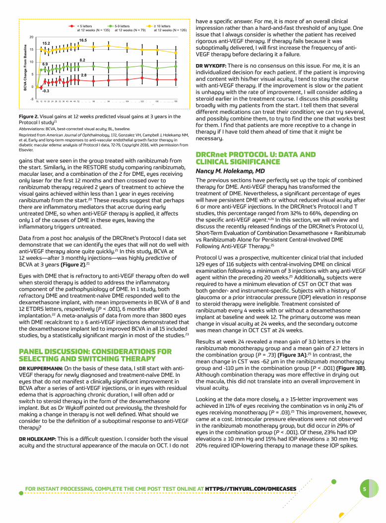
Data from a post hoc analysis of the DRCRnet’s Protocol I data setdemonstrate that we can identify the eyes that will not do well withanti-VEGF therapy alone quite quickly.21 In this study, BCVA at 12 weeks—aer 3 monthly injections—was highly predictive ofBCVA at 3 years (figure 2).21
Eyes with DME that is refractory to anti-VEGF therapy oen do wellwhen steroid therapy is added to address the inflammatorycomponent of the pathophysiology of DME. In 1 study, bothrefractory DME and treatment-naïve DME responded well to thedexamethasone implant, with mean improvements in BCVA of 8 and12 ETDRS letters, respectively (P < .001), 6 months aerimplantation.22 A meta-analysis of data from more than 3800 eyeswith DME recalcitrant to ≥ 6 anti-VEGF injections demonstrated thatthe dexamethasone implant led to improved BCVA in all 15 includedstudies, by a statistically significant margin in most of the studies.23
PaNel DisCussioN: CoNsiDeratioNs ForseleCtiNG aND sWitCHiNG tHeraPyDR KUPPERMANN: On the basis of these data, I still start with anti-VEGF therapy for newly diagnosed and treatment-naïve DME. Ineyes that do not manifest a clinically significant improvement inBCVA aer a series of anti-VEGF injections, or in eyes with residualedema that is approaching chronic duration, I will oen add orswitch to steroid therapy in the form of the dexamethasoneimplant. But as Dr Wykoff pointed out previously, the threshold formaking a change in therapy is not well defined. What should weconsider to be the definition of a suboptimal response to anti-VEGFtherapy?
DR HOLEKAMP: This is a difficult question. I consider both the visualacuity and the structural appearance of the macula on OCT. I do not
have a specific answer. For me, it is more of an overall clinicalimpression rather than a hard-and-fast threshold of any type. Oneissue that I always consider is whether the patient has receivedrigorous anti-VEGF therapy. If therapy fails because it wassuboptimally delivered, I will first increase the frequency of anti-VEGF therapy before declaring it a failure.
DR WYKOFF: There is no consensus on this issue. For me, it is anindividualized decision for each patient. If the patient is improvingand content with his/her visual acuity, I tend to stay the coursewith anti-VEGF therapy. If the improvement is slow or the patientis unhappy with the rate of improvement, I will consider adding asteroid earlier in the treatment course. I discuss this possibilitybroadly with my patients from the start. I tell them that severaldifferent medications can treat their condition; we can try several,and possibly combine them, to try to find the one that works bestfor them. I find that patients are more receptive to a change intherapy if I have told them ahead of time that it might benecessary.
DrCrnet ProtoCol u: Data aND CliNiCal siGNiFiCaNCeNancy M. Holekamp, MDThe previous sections have perfectly set up the topic of combinedtherapy for DME. Anti-VEGF therapy has transformed thetreatment of DME. Nevertheless, a significant percentage of eyeswill have persistent DME with or without reduced visual acuity aer6 or more anti-VEGF injections. In the DRCRnet’s Protocol I and Tstudies, this percentage ranged from 32% to 66%, depending onthe specific anti-VEGF agent.4,24 In this section, we will review anddiscuss the recently released findings of the DRCRnet’s Protocol U,Short-Term Evaluation of Combination Dexamethasone + Ranibizumabvs Ranibizumab Alone for Persistent Central-Involved DMEFollowing Anti-VEGF Therapy.25
Protocol U was a prospective, multicenter clinical trial that included129 eyes of 116 subjects with central-involving DME on clinicalexamination following a minimum of 3 injections with any anti-VEGFagent within the preceding 20 weeks.25 Additionally, subjects wererequired to have a minimum elevation of CST on OCT that was both gender- and instrument-specific. Subjects with a history ofglaucoma or a prior intraocular pressure (IOP) elevation in responseto steroid therapy were ineligible. Treatment consisted ofranibizumab every 4 weeks with or without a dexamethasoneimplant at baseline and week 12. The primary outcome was meanchange in visual acuity at 24 weeks, and the secondary outcomewas mean change in OCT CST at 24 weeks.
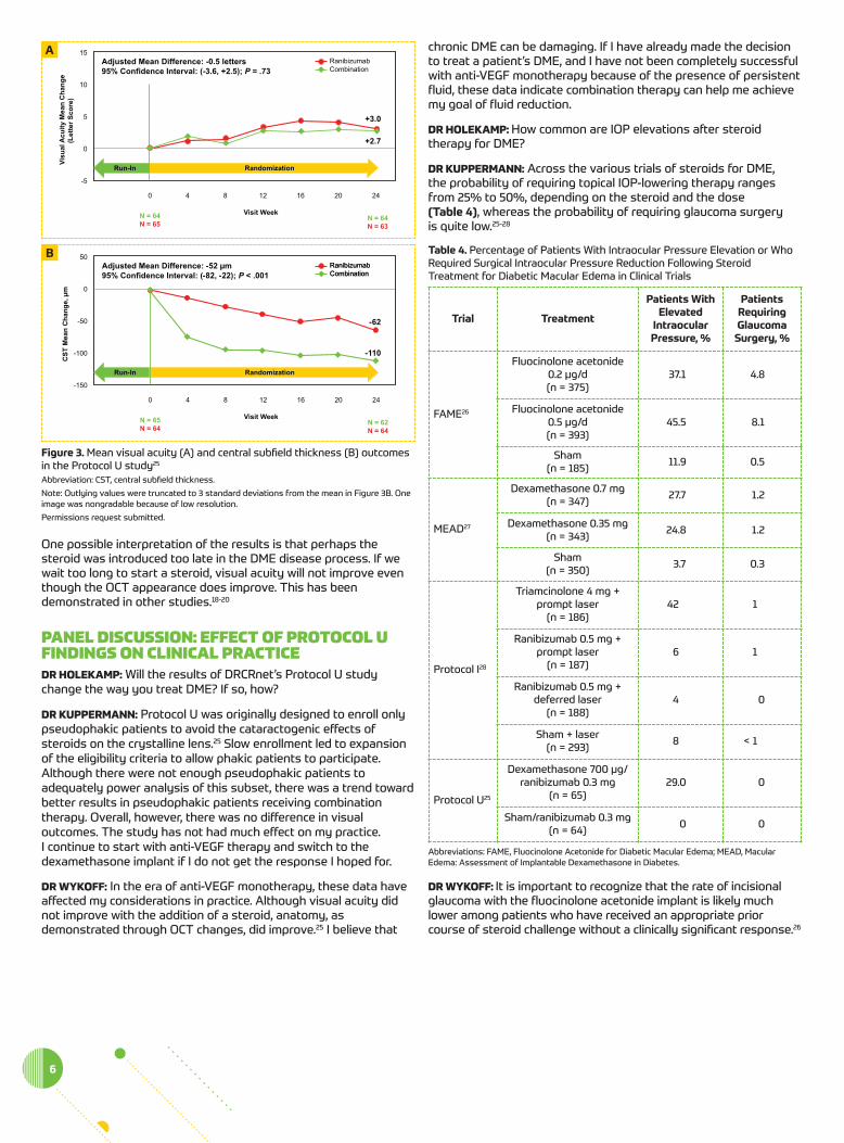
Results at week 24 revealed a mean gain of 3.0 letters in theranibizumab monotherapy group and a mean gain of 2.7 letters inthe combination group (P = .73) (figure 3a).25 In contrast, themean change in CST was -62 µm in the ranibizumab monotherapygroup and -110 µm in the combination group (P < .001) (figure 3B).Although combination therapy was more effective in drying outthe macula, this did not translate into an overall improvement invisual acuity.
Looking at the data more closely, a ≥ 15-letter improvement wasachieved in 11% of eyes receiving the combination vs in only 2% ofeyes receiving monotherapy (P = .03).25 This improvement, however,came at a cost. Intraocular pressure elevations were not observedin the ranibizumab monotherapy group, but did occur in 29% ofeyes in the combination group (P < .001). Of these, 23% had IOPelevations ≥ 10 mm Hg and 15% had IOP elevations ≥ 30 mm Hg;20% required IOP-lowering therapy to manage these IOP spikes.
5foR instant PRoCessing, CoMPLete tHe CMe Post test onLine at HttPs://tiNyurl.CoM/DMeCases
BL 12 16 20 24 28 32 36 40 44 48 52 68 84 104 120 136 156
< 5 lettersat 12 weeks (N = 135)
20
15
10
5
0
-5
16.5
8.2
2.8
-0.3
6.9
15.2
BC
VA C
hang
e Fr
om B
asel
ine
5-9 lettersat 12 weeks (N = 79)
≥ 10 lettersat 12 weeks (N = 126)
figure 2. Visual gains at 12 weeks predicted visual gains at 3 years in theProtocol I study21
Abbreviations: BCVA, best-corrected visual acuity; BL, baseline.
Reprinted from American Journal of Ophthalmology, 172, Gonzalez VH, Campbell J, Holekamp NM,et al, Early and long-term responses to anti-vascular endothelial growth factor therapy indiabetic macular edema: analysis of Protocol I data, 72-79, Copyright 2016, with permission fromElsevier.
One possible interpretation of the results is that perhaps thesteroid was introduced too late in the DME disease process. If wewait too long to start a steroid, visual acuity will not improve eventhough the OCT appearance does improve. This has beendemonstrated in other studies.18-20
PaNel DisCussioN: eFFeCt oF ProtoCol uFiNDiNGs oN CliNiCal PraCtiCeDR HOLEKAMP: Will the results of DRCRnet’s Protocol U studychange the way you treat DME? If so, how?
DR KUPPERMANN: Protocol U was originally designed to enroll onlypseudophakic patients to avoid the cataractogenic effects ofsteroids on the crystalline lens.25 Slow enrollment led to expansionof the eligibility criteria to allow phakic patients to participate.Although there were not enough pseudophakic patients toadequately power analysis of this subset, there was a trend towardbetter results in pseudophakic patients receiving combinationtherapy. Overall, however, there was no difference in visualoutcomes. The study has not had much effect on my practice. I continue to start with anti-VEGF therapy and switch to thedexamethasone implant if I do not get the response I hoped for.
DR WYKOFF: In the era of anti-VEGF monotherapy, these data haveaffected my considerations in practice. Although visual acuity didnot improve with the addition of a steroid, anatomy, asdemonstrated through OCT changes, did improve.25 I believe that
chronic DME can be damaging. If I have already made the decisionto treat a patient’s DME, and I have not been completely successfulwith anti-VEGF monotherapy because of the presence of persistentfluid, these data indicate combination therapy can help me achievemy goal of fluid reduction.
DR HOLEKAMP: How common are IOP elevations aer steroidtherapy for DME?
DR KUPPERMANN: Across the various trials of steroids for DME, the probability of requiring topical IOP-lowering therapy rangesfrom 25% to 50%, depending on the steroid and the dose (table 4), whereas the probability of requiring glaucoma surgery is quite low.25-28
DR WYKOFF: It is important to recognize that the rate of incisionalglaucoma with the fluocinolone acetonide implant is likely muchlower among patients who have received an appropriate priorcourse of steroid challenge without a clinically significant response.26
6
trial treatment
Patients Withelevated
intraocularPressure, %
PatientsRequiringglaucoma
surgery, %
FAME26
Fluocinolone acetonide0.2 µg/d(n = 375)
37.1 4.8
Fluocinolone acetonide0.5 µg/d(n = 393)
45.5 8.1
Sham(n = 185) 11.9 0.5
MEAD27
Dexamethasone 0.7 mg(n = 347) 27.7 1.2
Dexamethasone 0.35 mg(n = 343) 24.8 1.2
Sham(n = 350) 3.7 0.3
Protocol I28
Triamcinolone 4 mg +prompt laser
(n = 186)42 1
Ranibizumab 0.5 mg +prompt laser
(n = 187)6 1
Ranibizumab 0.5 mg +deferred laser
(n = 188)4 0
Sham + laser(n = 293) 8 < 1
Protocol U25
Dexamethasone 700 µg/ranibizumab 0.3 mg
(n = 65)29.0 0
Sham/ranibizumab 0.3 mg(n = 64) 0 0
table 4. Percentage of Patients With Intraocular Pressure Elevation or WhoRequired Surgical Intraocular Pressure Reduction Following SteroidTreatment for Diabetic Macular Edema in Clinical Trials
Abbreviations: FAME, Fluocinolone Acetonide for Diabetic Macular Edema; MEAD, MacularEdema: Assessment of Implantable Dexamethasone in Diabetes.
Visu
al A
cuity
Mea
n C
hang
e(L
ette
r Sco
re)
15
10
5
0
0 4 8 12 16 20 24
-5
Visit WeekN = 64N = 65
N = 64N = 63
RanibizumabCombination
Adjusted Mean Difference: -0.5 letters95% Confidence Interval: (-3.6, +2.5); P = .73
Run-In Randomization
+3.0
+2.7
figure 3. Mean visual acuity (A) and central subfield thickness (B) outcomesin the Protocol U study25
Abbreviation: CST, central subfield thickness.
Note: Outlying values were truncated to 3 standard deviations from the mean in Figure 3B. Oneimage was nongradable because of low resolution.
Permissions request submitted.
CST
Mea
n C
hang
e, µ
m
50
0
-50
-100
0 4 8 12 16 20 24
-150
Visit WeekN = 65N = 64
N = 62N = 64
RanibizumabCombinationRanibizumabCombination
Adjusted Mean Difference: -52 µm95% Confidence Interval: (-82, -22); P < .001
Run-In Randomization
-62
-110
a
B
Case 1. DiaBetiC MaCular eDeMaNoNresPoNsiVe to aNti–VasCulareNDotHelial GroWtH FaCtor tHeraPyFrom the Files of Nancy M. Holekamp, MDA 59-year-old woman presented with a 2-year history of DME inboth eyes, for which she received a number of anti-VEGF injectionsin both eyes from 4 different physicians. The most recent injectionoccurred approximately 6 weeks ago. She stated she has had DMfor only 3 years, and it is managed with oral hypoglycemic agents.She was uninsured and did not obtain routine health maintenance,so it is likely she has had DM for far longer than 3 years.
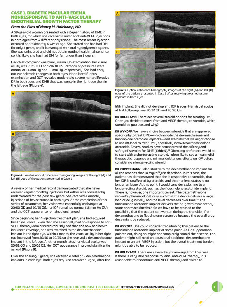
Her chief complaint was blurry vision. On examination, her visualacuity was 20/50 OD and 20/30 OS. Intraocular pressures werenormal at 14 mm Hg and 13 mm Hg, respectively. She had earlynuclear sclerotic changes in both eyes. Her dilated fundusexamination and OCT revealed moderately severe nonproliferativeDR in both eyes and DME that was worse in the right eye than inthe le eye (figure 4).
A review of her medical record demonstrated that she neverreceived regular monthly injections, but rather was consistentlyundertreated for the past few years. She received 4 monthlyinjections of bevacizumab in both eyes. At the completion of thisseries of treatments, her vision was essentially unchanged at20/50 OD and 20/25 OS, her IOP remained normal (16 mm Hg OU),and the OCT appearance remained unchanged.
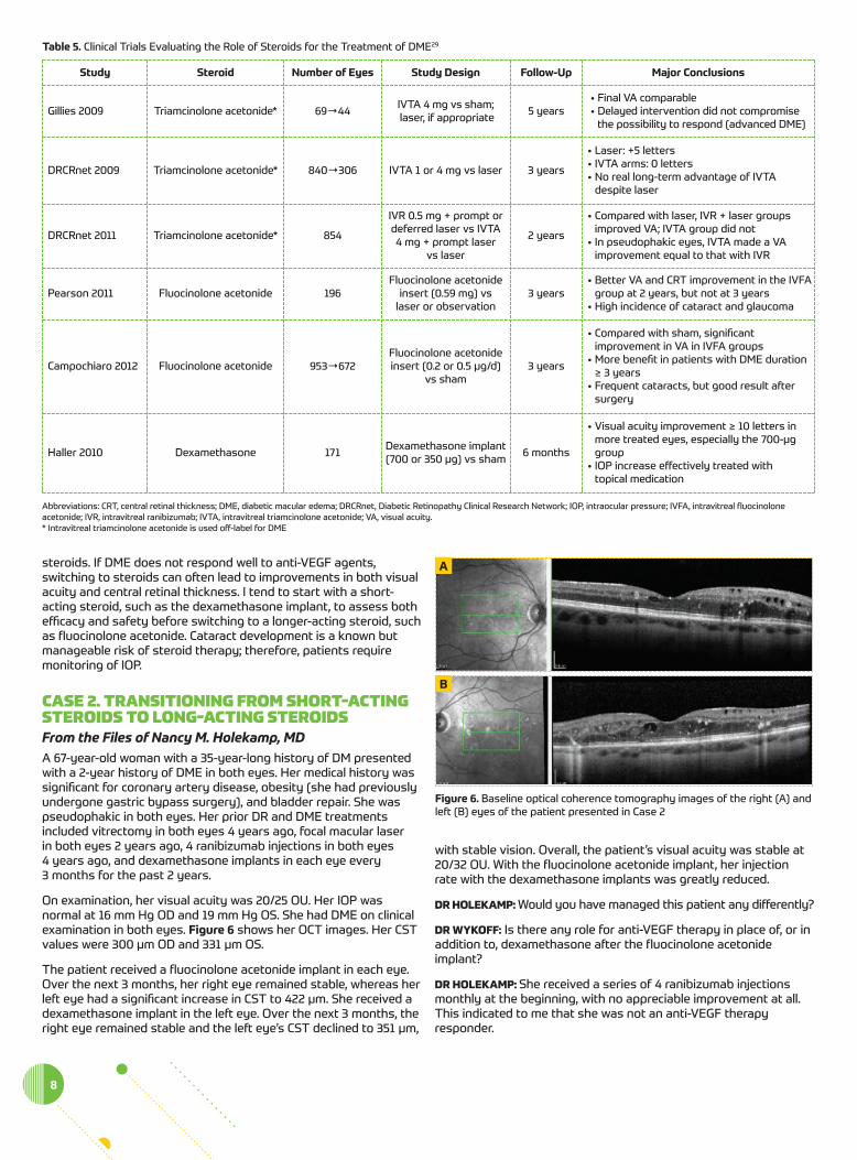
Since beginning her 4-injection treatment plan, she had acquiredhealth insurance. Given that she essentially had no response to anti-VEGF therapy administered robustly and that she now had healthinsurance coverage, she was switched to the dexamethasoneimplant in the right eye. Within 1 month, the visual acuity in her righteye improved from 20/50 to 20/25, so she received a dexamethasoneimplant in the le eye. Another month later, her visual acuity was20/32 OD and 20/16 OS. Her OCT appearance improved significantlyas well (figure 5).
Over the ensuing 2 years, she received a total of 9 dexamethasoneimplants in each eye. Both eyes required cataract surgery aer the
fih implant. She did not develop any IOP issues. Her visual acuityat last follow-up was 20/32 OD and 20/20 OS.
DR HOLEKAMP: There are several steroid options for treating DME.Once you decide to move from anti-VEGF therapy to steroids, whichsteroid do you use, and why?
DR WYKOFF: We have a choice between steroids that are approvedspecifically to treat DME—which include the dexamethasone andfluocinolone acetonide implants—and steroids that we might chooseto use off-label to treat DME, specifically intravitreal triamcinoloneacetonide. Several studies have demonstrated the efficacy andsafety of steroids for DME (table 5).29 Oen, my preference would beto start with a shorter-acting steroid. I oen like to see a meaningfultherapeutic response and minimal deleterious effects on IOP beforeconsidering a longer-acting steroid.
DR KUPPERMANN: I also start with the dexamethasone implant forall the reasons that Dr Wykoff just described. In this case, thepatient has demonstrated that she is responsive to steroids, thather IOP is unaffected by steroids, and that her lens status is nolonger an issue. At this point, I would consider switching to alonger-acting steroid, such as the fluocinolone acetonide implant.There is, however, one important caveat. The dexamethasoneimplant’s pharmacokinetics is such that the device delivers a largeload of drug initially, and the level decreases over time.30 Thefluocinolone acetonide implant delivers the drug with more steady-state pharmacokinetics.31 So we have to be attuned to thepossibility that the patient can worsen during the transition fromdexamethasone to fluocinolone acetonide because the overall drugdose might be reduced.
DR WYKOFF: One could consider transitioning this patient to thefluocinolone acetonide implant at some point. As Dr Kuppermannpointed out, doing so might not completely control the disease. Thepatient might still need an occasional additional dexamethasoneimplant or an anti-VEGF injection, but the overall treatment burdenmight be able to be reduced.
DR HOLEKAMP: There are several key takeaways from this case. If there is very little response to initial anti-VEGF therapy, it isreasonable to discontinue anti-VEGF therapy and switch to
7foR instant PRoCessing, CoMPLete tHe CMe Post test onLine at HttPs://tiNyurl.CoM/DMeCases
figure 5. Optical coherence tomography images of the right (A) and le (B)eyes of the patient presented in Case 1 aer receiving dexamethasoneimplants in both eyes
a
B
a
B
figure 4. Baseline optical coherence tomography images of the right (A) andle (B) eyes of the patient presented in Case 1
steroids. If DME does not respond well to anti-VEGF agents,switching to steroids can oen lead to improvements in both visualacuity and central retinal thickness. I tend to start with a short-acting steroid, such as the dexamethasone implant, to assess bothefficacy and safety before switching to a longer-acting steroid, suchas fluocinolone acetonide. Cataract development is a known butmanageable risk of steroid therapy; therefore, patients requiremonitoring of IOP.
Case 2. traNsitioNiNG FroM sHort-aCtiNGsteroiDs to loNG-aCtiNG steroiDsFrom the Files of Nancy M. Holekamp, MDA 67-year-old woman with a 35-year-long history of DM presentedwith a 2-year history of DME in both eyes. Her medical history wassignificant for coronary artery disease, obesity (she had previouslyundergone gastric bypass surgery), and bladder repair. She waspseudophakic in both eyes. Her prior DR and DME treatmentsincluded vitrectomy in both eyes 4 years ago, focal macular laser in both eyes 2 years ago, 4 ranibizumab injections in both eyes 4 years ago, and dexamethasone implants in each eye every 3 months for the past 2 years.
On examination, her visual acuity was 20/25 OU. Her IOP wasnormal at 16 mm Hg OD and 19 mm Hg OS. She had DME on clinicalexamination in both eyes. figure 6 shows her OCT images. Her CSTvalues were 300 µm OD and 331 µm OS.
The patient received a fluocinolone acetonide implant in each eye.Over the next 3 months, her right eye remained stable, whereas herle eye had a significant increase in CST to 422 µm. She received adexamethasone implant in the le eye. Over the next 3 months, theright eye remained stable and the le eye’s CST declined to 351 µm,
with stable vision. Overall, the patient’s visual acuity was stable at20/32 OU. With the fluocinolone acetonide implant, her injectionrate with the dexamethasone implants was greatly reduced.
DR HOLEKAMP: Would you have managed this patient any differently?
DR WYKOFF: Is there any role for anti-VEGF therapy in place of, or inaddition to, dexamethasone aer the fluocinolone acetonideimplant?
DR HOLEKAMP: She received a series of 4 ranibizumab injectionsmonthly at the beginning, with no appreciable improvement at all.This indicated to me that she was not an anti-VEGF therapyresponder.
8
figure 6. Baseline optical coherence tomography images of the right (A) andle (B) eyes of the patient presented in Case 2
a
B
study steroid number of eyes study Design follow-Up Major Conclusions
Gillies 2009 Triamcinolone acetonide* 69→44IVTA 4 mg vs sham;laser, if appropriate 5 years
• Final VA comparable • Delayed intervention did not compromise
the possibility to respond (advanced DME)
DRCRnet 2009 Triamcinolone acetonide* 840→306 IVTA 1 or 4 mg vs laser 3 years
• Laser: +5 letters • IVTA arms: 0 letters• No real long-term advantage of IVTA
despite laser
DRCRnet 2011 Triamcinolone acetonide* 854
IVR 0.5 mg + prompt ordeferred laser vs IVTA 4 mg + prompt laser
vs laser
2 years
• Compared with laser, IVR + laser groupsimproved VA; IVTA group did not
• In pseudophakic eyes, IVTA made a VAimprovement equal to that with IVR
Pearson 2011 Fluocinolone acetonide 196Fluocinolone acetonide
insert (0.59 mg) vs laser or observation
3 years• Better VA and CRT improvement in the IVFA
group at 2 years, but not at 3 years• High incidence of cataract and glaucoma
Campochiaro 2012 Fluocinolone acetonide 953→672Fluocinolone acetonideinsert (0.2 or 0.5 µg/d)
vs sham3 years
• Compared with sham, significantimprovement in VA in IVFA groups
• More benefit in patients with DME duration≥ 3 years
• Frequent cataracts, but good result aersurgery
Haller 2010 Dexamethasone 171 Dexamethasone implant(700 or 350 µg) vs sham 6 months
• Visual acuity improvement ≥ 10 letters inmore treated eyes, especially the 700-µggroup
• IOP increase effectively treated with topical medication
table 5. Clinical Trials Evaluating the Role of Steroids for the Treatment of DME29
Abbreviations: CRT, central retinal thickness; DME, diabetic macular edema; DRCRnet, Diabetic Retinopathy Clinical Research Network; IOP, intraocular pressure; IVFA, intravitreal fluocinoloneacetonide; IVR, intravitreal ranibizumab; IVTA, intravitreal triamcinolone acetonide; VA, visual acuity.* Intravitreal triamcinolone acetonide is used off-label for DME

DR KUPPERMANN: Because she had undergone vitrectomy, the half-life of any drug—including anti-VEGF agents—will be considerablyshortened. Even if you had tried more frequent injections and beensuccessful, the treatment burden would still have supported themove to steroid therapy. Fortunately, the determinant of drug half-life with steroid implants is the device itself and not the presence orabsence of vitreous humor.
DR WYKOFF: This case raises the issue of switching vs addingtreatment. I might have added dexamethasone to the anti-VEGFtherapy aer those 4 injections to see if the combination providedadequate control with reasonable treatment burden.
DR HOLEKAMP: That is an excellent point. I prefer to usemonotherapy whenever possible for both cost and safety reasons.I will monitor the DR and can always give an anti-VEGF injectionlater if needed. I will add that at the last follow-up, this patient’s IOPin the le eye was 22 mm Hg aer receiving the dexamethasoneimplant. Does this concern anyone?
DR KUPPERMANN: Elevations of IOP are common aer steroidimplants. In the pivotal MEAD (Macular Edema: Assessment ofImplantable Dexamethasone in Diabetes) study of thedexamethasone implant for DME, approximately 30% of eyes hadIOP elevations requiring the use of topical IOP-lowering therapy byyear 3, and only 3 of the 690 implanted eyes required atrabeculectomy.32 With the fluocinolone acetonide implant, thepercentage of eyes needing IOP-lowering medications was similar,on the order of 35% to 40%, but approximately 5% of eyesrequired glaucoma surgery.18 In general, because dexamethasone isa short-acting drug compared with fluocinolone acetonide ortriamcinolone acetonide, the IOP elevations are easier to treat andwill oen resolve as the implant’s drug level is depleted.
DR WYKOFF: You dosed the dexamethasone implant every 3 monthsbefore switching to the fluocinolone acetonide implant. When usingthe dexamethasone implant for chronic management, do youreinject every 90 days regardless of clinical status, do you injectbased on clinical status, or do you wait until the current implant hascompletely dissolved?
DR HOLEKAMP: I typically dose every 3 months when usingdexamethasone chronically. I do not wait until it dissolves becauseremnants can linger for months, and I do not wait until the vision oredema gets worse because it is better to keep it under control thanto constantly play catch-up.
Case 3. reCalCitraNt DiaBetiC MaCular eDeMaFrom the Files of Charles C. Wykoff, MD, PhDA 45-year-old man with type 1 DM had incurred a substantialtreatment burden to manage DME in his le eye. Overapproximately 2.5 years, he received 5 bevacizumab injections, focaland panretinal laser treatments, and 21 ranibizumab injections.When he received monthly injections, his visual acuity remained inthe 20/25 range, his edema was well controlled, and he was happy.Every attempt to reduce the frequency of ranibizumab injectionsresulted in worsening vision and edema—even stretching to 5 to 6 weeks. Unlike the prior cases, this patient is not a suboptimalresponder; rather, this is a patient in whom the beneficial effect oftherapy has had a very short endurance period that is not gettinglonger over time.
Several options were considered. One was to switch to a differentanti-VEGF agent, another was to switch to a steroid, and a thirdwas to add a steroid and continue the anti-VEGF injections, hopingto extend the endurance and thereby reduce the frequency of
injections. A switch to aflibercept was chosen. Aer 4 monthlyaflibercept injections, the injections began to be spaced out. For ashort while, the patient remained stable with an injection every 5 to7 weeks until injection number 10, when worsening vision (now20/40) and edema forced him back to a monthly injection schedule.
It was clear that anti-VEGF therapy alone, while effective whenrepeated monthly, posed an enormous treatment burden on thispatient. He simply did not want to come in monthly because ofemployment commitments. To address this, a dexamethasoneimplant was injected. He responded well to the dexamethasoneimplant, but durability was still less than ideal, so combinationdosing with aflibercept injections was attempted. During this time,he also developed a visible epiretinal membrane. When dry,however, his vision was still excellent at 20/25. With steroids, hisIOP rose from the mid-teens to 23 mm Hg. Within 3 months, hisedema was significantly worse, and his visual acuity dropped to20/50. At this point, he was enrolled in a prospective clinical trialand received a suprachoroidal injection of triamcinolone acetonide,which is currently not approved for ophthalmic use and was usedoff-label. Triamcinolone acetonide treatment is in clinicaldevelopment for a number of conditions, including noninfectiousposterior uveitis and macular edema due to retinal vein occlusion,as well as for DME.33,34 Aer completion of the clinical trial, he againhad recurrence of DME, and dexamethasone implants (his third)were reinitiated, which held the visual gains and edema for 2 months.He then received another dexamethasone implant (his fourth),which restored visual acuity and reduced edema for another 2 months.The effect was again lost aer just 2 months, so he received a fihdexamethasone implant. His visual acuity at his last visit was 20/25in his le eye.
DR WYKOFF: Any insight into how I might have managed this casedifferently?
DR KUPPERMANN: This case perfectly illustrates the fact that wemust individualize treatment. This is an extreme case, but it is clearthat this patient’s treatment burden will remain significant. Giventhe presence and progression of the epiretinal membrane, did youconsider vitrectomy for this patient?
DR WYKOFF: Vitrectomy is certainly an option in this case.
DR KUPPERMANN: I am not certain I would operate yet. Before goingto surgery, I would try more aggressive combination therapy—perhaps a dexamethasone implant and an aflibercept injection atthe same time. I would follow the implant with the injection a weeklater so that the aflibercept goes on board at the time the implantis releasing high levels of dexamethasone. Then, there will be highlevels of both drugs active for approximately a month. Perhaps thepatient would do well if both drugs were at peak activitysimultaneously.
DR WYKOFF: At this point, he is being maintained on adexamethasone implant approximately every 2 months.
DR HOLEKAMP: Are you having any difficulty getting reimbursed forthis so oen?
DR WYKOFF: I have not had that problem in my practice. Have youhad issues with reimbursement for delivering these treatmentsmore oen than labeled?
DR KUPPERMANN: I have had no issues with reimbursement whenusing the dexamethasone implant more oen than every 12 weeksor with anti-VEGF therapy more oen than every 4 weeks. I havenot, however, repeated a fluocinolone acetonide implant within 3 years of the first implant.
9foR instant PRoCessing, CoMPLete tHe CMe Post test onLine at HttPs://tiNyurl.CoM/DMeCases
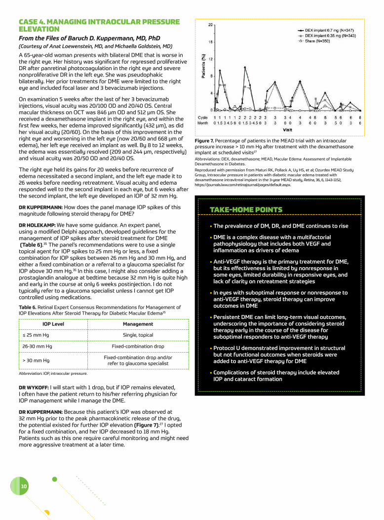
Case 4. MaNaGiNG iNtraoCular PressureeleVatioN From the Files of Baruch D. Kuppermann, MD, PhD(Courtesy of Anat Loewenstein, MD, and Michaella Goldstein, MD)
A 65-year-old woman presents with bilateral DME that is worse inthe right eye. Her history was significant for regressed proliferativeDR aer panretinal photocoagulation in the right eye and severenonproliferative DR in the le eye. She was pseudophakicbilaterally. Her prior treatments for DME were limited to the righteye and included focal laser and 3 bevacizumab injections.
On examination 5 weeks aer the last of her 3 bevacizumabinjections, visual acuity was 20/100 OD and 20/40 OS. Centralmacular thickness on OCT was 846 µm OD and 512 µm OS. Shereceived a dexamethasone implant in the right eye, and within thefirst few weeks, her edema improved significantly (432 µm), as didher visual acuity (20/60). On the basis of this improvement in theright eye and worsening in the le eye (now 20/60 and 668 µm ofedema), her le eye received an implant as well. By 8 to 12 weeks,the edema was essentially resolved (209 and 244 µm, respectively)and visual acuity was 20/50 OD and 20/40 OS.
The right eye held its gains for 20 weeks before recurrence ofedema necessitated a second implant, and the le eye made it to26 weeks before needing retreatment. Visual acuity and edemaresponded well to the second implant in each eye, but 6 weeks aerthe second implant, the le eye developed an IOP of 32 mm Hg.
DR KUPPERMANN: How does the panel manage IOP spikes of thismagnitude following steroid therapy for DME?
DR HOLEKAMP: We have some guidance. An expert panel, using a modified Delphi approach, developed guidelines for themanagement of IOP spikes aer steroid treatment for DME(table 6).35 The panel’s recommendations were to use a singletopical agent for IOP spikes to 25 mm Hg or less, a fixedcombination for IOP spikes between 26 mm Hg and 30 mm Hg, andeither a fixed combination or a referral to a glaucoma specialist forIOP above 30 mm Hg.35 In this case, I might also consider adding aprostaglandin analogue at bedtime because 32 mm Hg is quite highand early in the course at only 6 weeks postinjection. I do nottypically refer to a glaucoma specialist unless I cannot get IOPcontrolled using medications.
DR WYKOFF: I will start with 1 drop, but if IOP remains elevated, I oen have the patient return to his/her referring physician for IOP management while I manage the DME.
DR KUPPERMANN: Because this patient’s IOP was observed at 32 mm Hg prior to the peak pharmacokinetic release of the drug,the potential existed for further IOP elevation (figure 7).27 I optedfor a fixed combination, and her IOP decreased to 18 mm Hg.Patients such as this one require careful monitoring and might needmore aggressive treatment at a later time.
10
ioP Level Management
≤ 25 mm Hg Single, topical
26-30 mm Hg Fixed-combination drop
> 30 mm Hg Fixed-combination drop and/or refer to glaucoma specialist
table 6. Retinal Expert Consensus Recommendations for Management ofIOP Elevations Aer Steroid Therapy for Diabetic Macular Edema35
Abbreviation: IOP, intraocular pressure.
figure 7. Percentage of patients in the MEAD trial with an intraocularpressure increase > 10 mm Hg aer treatment with the dexamethasoneimplant at scheduled visits27
Abbreviations: DEX, dexamethasone; MEAD, Macular Edema: Assessment of ImplantableDexamethasone in Diabetes.
Reproduced with permission from Maturi RK, Pollack A, Uy HS, et al; Ozurdex MEAD StudyGroup, Intraocular pressure in patients with diabetic macular edema treated withdexamethasone intravitreal implant in the 3-year MEAD study, Retina, 36, 6, 1143-1152,https://journals.lww.com/retinajournal/pages/default.aspx.
taKe-HoMe PoiNts
• the prevalence of dM, dr, and dMe continues to rise
• dMe is a complex disease with a multifactorialpathophysiology that includes both Vegf andinflammation as drivers of edema
• anti-Vegf therapy is the primary treatment for dMe,but its effectiveness is limited by nonresponse insome eyes, limited durability in responsive eyes, andlack of clarity on retreatment strategies
• in eyes with suboptimal response or nonresponse to anti-Vegf therapy, steroid therapy can improveoutcomes in dMe
• Persistent dMe can limit long-term visual outcomes,underscoring the importance of considering steroidtherapy early in the course of the disease forsuboptimal responders to anti-Vegf therapy
• Protocol U demonstrated improvement in structuralbut not functional outcomes when steroids wereadded to anti-Vegf therapy for dMe
• Complications of steroid therapy include elevatedioP and cataract formation
1. Rowley WR, Bezold C, Arikan Y, Byrne E, Krohe S. Diabetes 2030: insightsfrom yesterday, today, and future trends. Popul Health Manag. 2017;20(1):6-12.
2. Nathan DM. Long-term complications of diabetes mellitus. N Engl J Med.1993;328(23):1676-1685.
3. The relationship of glycemic exposure (HbA1c) to the risk ofdevelopment and progression of retinopathy in the diabetes control andcomplications trial. Diabetes. 1995;44(8):968-983.
4. Wells JA, Glassman AR, Ayala AR, et al; Diabetic Retinopathy ClinicalResearch Network. Aflibercept, bevacizumab, or ranibizumab for diabeticmacular edema: two-year results from a comparative effectivenessrandomized clinical trial. Ophthalmology. 2016;123(6):1351-1359.
5. Boyer DS, Nguyen QD, Brown DM, Basu K, Ehrlich JS; RIDE and RISEResearch Group. Outcomes with as-needed ranibizumab aer initialmonthly therapy: long-term outcomes of the phase III RIDE and RISEtrials. Ophthalmology. 2015;122(12):2504-2513.e1.
6. Elman MJ, Ayala A, Bressler NM, et al; Diabetic Retinopathy ClinicalResearch Network. Intravitreal ranibizumab for diabetic macular edemawith prompt versus deferred laser treatment: 5-year randomized trialresults. Ophthalmology. 2015;122(2):375-381.
7. Wykoff CC, Ou WC, Khurana RN, Brown DM, Lloyd Clark W, Boyer DS;ENDURANCE Study Group. Long-term outcomes with as-neededaflibercept in diabetic macular oedema: 2-year outcomes of theENDURANCE extension study. Br J Ophthalmol. 2018;102(5):631-636.
8. Wykoff CC, Le RT, Khurana RN, et al; ENDURANCE Study Group. Outcomeswith as-needed aflibercept and macular laser following the phase IIIVISTA DME trial: ENDURANCE 12-month extension study. Am JOphthalmol. 2017;173:56-63.
9. Gross JG, Glassman AR, Jampol LM, et al; Writing Committee for theDiabetic Retinopathy Clinical Research Network. Panretinalphotocoagulation vs intravitreous ranibizumab for proliferative diabeticretinopathy: a randomized clinical trial. JAMA. 2015;314(20):2137-2146.
10. Warmke N, Griffin KJ, Cubbon RM. Pericytes in diabetes-associatedvascular disease. J Diabetes Complications. 2016;30(8):1643-1650.
11. Usui T, Ishida S, Yamashiro K, et al. VEGF164(165) as the pathologicalisoform: differential leukocyte and endothelial responses through VEGFR1and VEGFR2. Invest Ophthalmol Vis Sci. 2004;45(2):368-374.
12. Bhagat N, Grigorian RA, Tutela A, Zarbin MA. Diabetic macular edema:pathogenesis and treatment. Surv Ophthalmol. 2009;54(1):1-32.
13. Ehrlich R, Harris A, Ciulla TA, Kheradiya N, Winston DM, Wirostko B.Diabetic macular oedema: physical, physiological and molecular factorscontribute to this pathological process. Acta Ophthalmol. 2010;88(3):279-291.
14. Jain A, Varshney N, Smith C. The evolving treatment options for diabeticmacular edema. Int J Inflam. 2013;2013:689276.
15. Dong N, Xu B, Wang B, Chu L. Study of 27 aqueous humor cytokines inpatients with type 2 diabetes with or without retinopathy. Mol Vis.2013;19:1734-1746.
16. Sohn HJ, Han DH, Kim IT, et al. Changes in aqueous concentrations ofvarious cytokines aer intravitreal triamcinolone versus bevacizumab fordiabetic macular edema. Am J Ophthalmol. 2011;152(4):686-694.
17. Bressler SB, Qin H, Beck RW, et al; Diabetic Retinopathy Clinical ResearchNetwork. Factors associated with changes in visual acuity and centralsubfield thickness at 1 year aer treatment for diabetic macular edemawith ranibizumab. Arch Ophthalmol. 2012;130(9):1153-1161.
18. Cunha-Vaz J, Ashton P, Iezzi R, et al; FAME Study Group. Sustaineddelivery fluocinolone acetonide vitreous implants: long-term benefit inpatients with chronic diabetic macular edema. Ophthalmology. 2014;12(10):1892-1903.
19. Brown DM, Nguyen QD, Marcus DM, et al; RIDE and RISE Research Group.Long-term outcomes of ranibizumab therapy for diabetic macular edema:the 36-month results from two phase III trials: RISE and RIDE.Ophthalmology. 2013;120(10):2013-2022.
20. Schmidt-Erfurth U, Lang GE, Holz FG, et al; RESTORE Extension StudyGroup. Three-year outcomes of individualized ranibizumab treatment inpatients with diabetic macular edema: the RESTORE extension study.Ophthalmology. 2014;121(5):1045-1053.
21. Gonzalez VH, Campbell J, Holekamp NM, et al. Early and long-termresponses to anti-vascular endothelial growth factor therapy in diabeticmacular edema: analysis of Protocol I data. Am J Ophthalmol. 2016;172:72-79.
22. Escobar-Barranco JJ, Pina-Marín B, Fernández-Bonet M. Dexamethasoneimplants in patients with naïve or refractory diffuse diabetic macularedema. Ophthalmologica. 2015;233(3-4):176-185.
23. Khan Z, Kuriakose RK, Khan M, Chin EK, Almeida DR. Efficacy of theintravitreal sustained-release dexamethasone implant for diabeticmacular edema refractory to anti-vascular endothelial growth factortherapy: meta-analysis and clinical implications. Ophthalmic Surg LasersImaging Retina. 2017;48(2):160-166.
24. Elman MJ, Qin H, Aiello LP, et al. Intravitreal ranibizumab for diabeticmacular edema with prompt versus deferred laser treatment: three-yearrandomized trial results. Ophthalmology. 2012;119(11):2312-2318.
25. Maturi RK, Glassman AR, Liu D, et al; Diabetic Retinopathy ClinicalResearch Network. Effect of adding dexamethasone to continuedranibizumab treatment in patients with persistent diabetic macularedema: a DRCR Network phase 2 randomized clinical trial. JAMAOphthalmol. 2018;136(1):29-38.
26. Campochiaro PA, Brown DM, Pearson A, et al. Sustained deliveryfluocinolone acetonide vitreous inserts provide benefit for at least 3 years in patients with diabetic macular edema. Ophthalmology.2012;119(10):2125-2132.
27. Maturi RK, Pollack A, Uy HS, et al; Ozurdex MEAD Study Group. Intraocularpressure in patients with diabetic macular edema treated withdexamethasone intravitreal implant in the 3-year MEAD study. Retina.2016;36(6):1143-1152.
28. Elman MJ, Aiello LP, Beck RW, et al; Diabetic Retinopathy Clinical ResearchNetwork. Randomized trial evaluating ranibizumab plus prompt ordeferred laser or triamcinolone plus prompt laser for diabetic macularedema. Ophthalmology. 2010;117(6):1064-1077.e35.
29. Bandello F, Preziosa C, Querques G, Lattanzio R. Update of intravitrealsteroids for the treatment of diabetic macular edema. Ophthalmic Res.2014;52(2):89-96.
30. Chang-Lin JE, Attar M, Acheampong AA, et al. Pharmacokinetics andpharmacodynamics of a sustained-release dexamethasone intravitrealimplant. Invest Ophthalmol Vis Sci. 2011;52(1):80-86.
31. Campochiaro PA, Hafiz G, Shah SM, et al. Sustained ocular delivery offluocinolone acetonide by an intravitreal insert. Ophthalmology.2010;117(7):1393-1399.e3.
32. Boyer DS, Yoon YH, Belfort R Jr, et al; Ozurdex MEAD Study Group. Three-year, randomized, sham-controlled trial of dexamethasoneintravitreal implant in patients with diabetic macular edema.Ophthalmology. 2014;121(10):1904-1914.
33. Yeh S, Kurup SK, Wang RC, et al; DOGWOOD Study Team. Suprachoroidalinjection of triamcinolone acetonide, CLS-TA, for macular edema due tononinfectious uveitis: a randomized, phase 2 study (DOGWOOD)[published online ahead of print August 15, 2018]. Retina.doi:10.1097/IAE.0000000000002279.
34. Takkar B, Saxena H, Sinha P, Sharma B. Choroidal changes aersuprachoroidal injection of triamcinolone in eyes with macular edemasecondary to retinal vein occlusion. Am J Ophthalmol. 2018;189:177-178.
35. Regillo CD, Callanan DG, Do DV, et al. Use of corticosteroids in thetreatment of patients with diabetic macular edema who have asuboptimal response to anti-VEGF: recommendations of an expert panel.Ophthalmic Surg Lasers Imaging Retin. 2017;48(4):291-301.
11foR instant PRoCessing, CoMPLete tHe CMe Post test onLine at HttPs://tiNyurl.CoM/DMeCases
reFereNCes
1. What are the key pathophysiologic triggers for DME? a. VEGF and IOP b. Hemoglobin A1c and VEGF c. Inflammation and VEGF d. Inflammation and hemoglobin A1c
2. Limitations of anti-VEGF therapy for DME include: a. Treatment burden due to the need for frequent injections b. The risk of IOP increase c. The risk of cataract formation d. Serious systemic side effects
3. A patient presents with a 3-year history of DME. She has seenseveral physicians and sporadically received only 1 to 2 anti-VEGFtherapy injections from each. Her visual acuity and edema requirefurther treatment. Which of the following best describes herclinical status?
a. She has received and failed robust treatment with anti-VEGFtherapy and should now receive steroids
b. Inflammation is likely the only relevant trigger for her DME c. Her edema is chronic and might be more difficult to treat d. Steroids will be ineffective in managing her disease
4. A patient with newly diagnosed DME presents with a visualacuity of 20/50 and CST of 500 µm. Following 6 monthlyinjections of an anti-VEGF agent, her visual acuity and edema are unchanged. What is the next best step for her?
a. Continue the injections as monotherapy b. Switch to a fluocinolone acetonide implant c. Add focal laser treatment d. Switch to a dexamethasone implant
5. A patient with DME has had 3 ranibizumab injections, with noappreciable effect on visual acuity or edema. Which of thefollowing statements is supported by the findings of theDRCRnet’s Protocol U study?
a. Adding the dexamethasone implant will improve her visualacuity significantly more than simply continuing the monthlyinjections alone
b. She has failed anti-VEGF therapy and her vision cannot beimproved
c. Her edema will likely improve if a dexamethasone implant isadded to her treatment
d. If she receives a dexamethasone implant, she has a 5% chanceof developing elevated IOP
6. Eight weeks aer receiving a dexamethasone implant, a patient’sIOP rises from 14 mm Hg to 28 mm Hg. Which is a reasonableintervention for this patient?
a. Observe IOP without treatment b. Start therapy with a fixed-combination IOP-lowering
medication c. Refer the patient for glaucoma surgery d. Switch to a fluocinolone acetonide implant when retreatment is
necessary
7. When counseling patients with DME about the long-term courseof their disease and treatment, which point should be included?
a. Everyone eventually requires steroids for DME treatment b. The treatment burden decreases over time for most patients c. Controlling blood glucose levels helps DR, but not DME d. There will always be some edema; therapy only reduces it, but
cannot eliminate it
CMe Post test QuestioNs
To obtain AMA PRA Category 1 Credit™ for this activity, complete the CME Post Test and course evaluation online athttps://tinyurl.com/dmecases. Upon successful completion of the post test and evaluation, you will be able to generate an instant certificate of credit.
See detailed instructions under to obtain AMA PRA Category 1 Credit™ on page 2.
HttPs://tinYURL.CoM/DMeCases
iNstaNt CMe CertiFiCate aVailaBle WitH oNliNe testiNGaND Course eValuatioN at