management of grossly decayed mandibular molar with different
TRANSCRIPT

Case ReportManagement of Grossly Decayed Mandibular Molar withDifferent Designs of Split Cast Post and Core
Rashmi Bansal, Nakul Mehrotra, Priyanka Chowdhary, and Anuraag Gurtu
Department of Conservative Dentistry & Endodontics, Institute of Dental Sciences, Bareilly, India
Correspondence should be addressed to Rashmi Bansal; [email protected]
Received 4 January 2016; Accepted 22 March 2016
Academic Editor: Tatiana Pereira-Cenci
Copyright © 2016 Rashmi Bansal et al.This is an open access article distributed under the Creative Commons Attribution License,which permits unrestricted use, distribution, and reproduction in any medium, provided the original work is properly cited.
Mandibular molar with extensive loss of tooth structure, especially where no cavity wall is remaining, and insertion of posts in boththe roots appear necessary so as to achieve proper retention for the core material. A single unit metal casting with two posts, onein the mesial root and the other in the distal divergent root, is difficult to fabricate due to difference in the path of insertion of thetwo posts. Multisection post and core or single cast post and core with auxiliary post can be an effective design to manage grosslydecayed mandibular molars.
1. Introduction
Comprehensive treatment plan is required before the startof endodontic treatment. When the decision is made totreat the teeth endodontically consideration must be given tothe placement of the subsequent restoration. Endodonticallytreated posterior teeth are often mutilated due to cariesand access requirement, sometimes to the extent that allthe walls of coronal structure are missing and only theradicular portion is present. In such cases if ferrule is availableand coronal retention core buildup is not sufficient thenintraradicular retention may be used by custom made postand core which replaces any lost coronal tooth structure [1].Relatively long postwith circular cross sections provides goodretention and support in anterior teeth but should be avoidedin posterior teeth,which oftenhave curved roots and ellipticalor ribbon shaped canals. For these teeth, retention is betterprovided by two or more relatively short posts in divergentcanals [2].This can be achieved bymultisection post and corewith each section having its own path of withdrawal or singlepiece post and core with a separate auxiliary post.
This paper presents two case reports on post endodonticmanagement of badlymutilatedmandibularmolars: one withtwo-section post and core and one with single post and corewith separate auxiliary post. A review of custompost and corein posterior teeth is listed in Table 1 [3–6].
2. Case Presentation
Case 1 (multisection post and core). A 21-year-old malepatient reported to the Department of Conservative Den-tistry and Endodontics, Institute of Dental Sciences Bareilly,with the chief complaint of pain in lower left toothregion. Extraoral examination revealed no significant find-ings. Intraoral examination revealed grossly decayed 36(Figure 1(a)). The tooth was not tender on percussion. Thetooth was not mobile and nonresponsive to any pulp sen-sitivity tests. Intraoral sinus tract was present on the buccalaspect in left quadrant. Path of this sinus tract was tracedwithgutta percha using periapical radiograph. On radiographicexamination, radiolucency was observed involving both themesial and distal root of 36 (Figure 1(b)).
Chronic periapical abscess was diagnosed. A compre-hensive treatment plan was made consisting of two phases:endodontic phase and restorative phase.
2.1. Endodontic Phase. After excavation of caries unsup-ported tooth structure was removed. Access cavity wasrefined and working length was established (distal: 13mm,mesiobuccal and mesiolingual: 11mm). Biomechanical prep-aration was completed by Mtwo files up to 6% taper number25. During preparation canals were irrigated with normalsaline (0.9% W/V) and metronidazole (0.5% W/V). A final
Hindawi Publishing CorporationCase Reports in DentistryVolume 2016, Article ID 2976941, 6 pageshttp://dx.doi.org/10.1155/2016/2976941

2 Case Reports in Dentistry
Table 1: Review of literature.
S number Author Technique Drawbacks
1 Bass 2002 [3]Single post and core with auxiliary postDistal-single cast postMesiolingual-prefabricated screw post
As the mesial canal had prefabricated postthe precision was less than what could havebeen achieved by the custom made cast post.
2 Gogna et al. 2009 [4]Single post and core with auxiliary postDistal-single cast postMesiobuccal-post with core
Since mesiobuccal canal was chosen to placethe post there are more chances of rootperforation.
3 Kumar et al. 2013 [5]Single post and core with auxiliary postDistal-single cast postMesiolingual-post with core
Core was not encasing the coronal toothstructure; instead it was wedged within thecoronal tooth structure.
4 Daguci et al. 2014 [6]Multisection post and core (lock and keyarrangement)Distobuccal-single post with corePalatal-single post with core
Technique sensitive procedure.
5 Mattoo et al. 2014 [9]Multisection post and core (lock and keyarrangement)Distolingual-single post with coreMesiolingual-single post with core Indirect pattern was used to prepare the
posts that will not be as accurate.
6 Deenadayalan et al. 2015 [10]Multisection post and core (lock and keyarrangement)Distolingual-single post with coreMesiolingual-single post with core
rinse with 2% chlorhexidine solution was done after comple-tion of biomechanical preparation. Triple antibiotic paste(ciprofloxacin 200mg, metronidazole 500mg, and minocy-cline 100mg) was placed in the canal and patient was recalledafter two weeks.
At second appointment patient was asymptomatic, andsinus tract was healed so obturation was completed with 6%taper number 25 single cone gutta percha using AH plus asa sealer. Patient was recalled after three weeks; there were noclinical signs and symptoms. Restorative phase was planned.
2.2. Restorative Phase. Using peeso reamer numbers 1–3(1.1mmdiameter) post space of length 5mm(leaving 6mmofgutta percha apically) was prepared in the mesiobuccal canaltaking care that at least minimum of 1mm of dentin remainsaround the canal. Similarly post space was prepared in thedistal canal using peeso reamer numbers 1–4 (1.3mm diame-ter) of length 6mm (leaving 7mm of gutta percha apically).
Following this ferrule preparation was completed. Waxpattern of post in mesiobuccal canal with part of its core wasprepared.Anyundercut adjacent to another half was removed(Figure 1(c)). Casting of the mesial portion was done and thefitwas checked.Directwax pattern of the distal section of postand core was prepared with the casting of mesial section ofpost and core in place (Figure 1(d)). Distal section wax pat-tern was casted and fit was checked by keeping mesial castingin place. Dovetail to interlock the mesial and distal sectionwas not prepared as final buildup was to be held together bythe fixed cast restoration. Both the castings were luted withtype II GIC (Figure 1(g)). Core preparation was finished forall metal crown. Rubber base impression was taken and wastemporized for 3 weeks. All metal crown was fabricated and
cemented in the next appointment. At 1-year clinical follow-up, the prosthesis exhibited no evidence of failure and thepatient was satisfied with the function and esthetics.
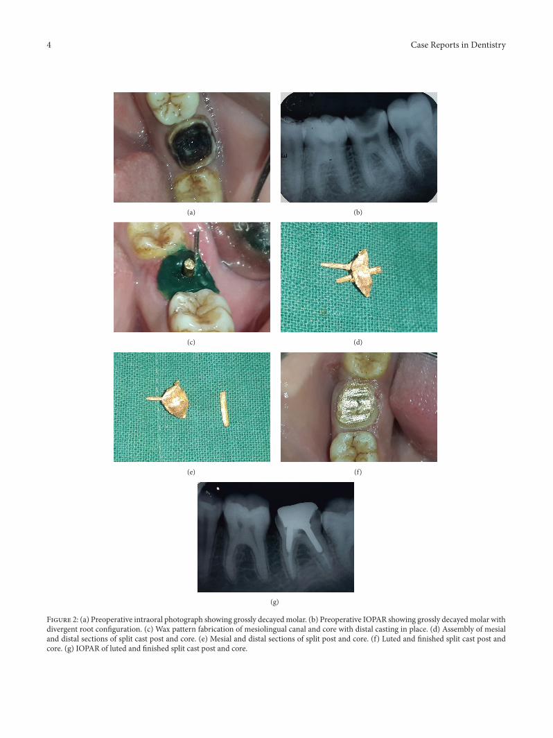
Case 2 (single piece core with auxiliary post). A 23-year-old male patient reported to the Department of ConservativeDentistry and Endodontics, with the chief complaint ofdecayed tooth in lower left tooth region. Extraoral exami-nation revealed no significant findings. On intraoral exam-ination 37 was grossly decayed (Figure 2(a)) and tender onpercussion. Intraoral periapical radiograph revealed deepcaries involving pulp space with no periapical changes(Figure 2(b)).Therewasmore divergence betweenmesial anddistal root as compared to Case 1. The tooth was nonrespon-sive to any pulp sensitivity tests. Endodontic therapy wasplanned for the tooth followed by post and core to rehabilitatethe occlusal portion.
2.3. Endodontic Phase. Since there was no periapical changea single sitting root canal treatment was planned which wascommenced by excavating all the caries and refining theaccess preparation. Working length was determined (distal:13mm, mesiobuccal and mesiolingual: 11mm). Biomechan-ical preparation with Mtwo rotary files was completed upto 6% taper number 25 with concomitant irrigation usingmetronidazole (0.5%W/V) and normal saline (0.9%W/V).Single cone obturation was completed with 6% gutta perchanumber 25 usingAHplus as a sealer. Patient was recalled after1 week for restorative phase.
2.4. Restorative Phase. Since the divergence between themesial and distal root was more a single core with small post

Case Reports in Dentistry 3
(a) (b)
(c) (d)
(e) (f)
(g) (h)
Figure 1: (a) Preoperative intraoral photograph showing grossly carious tooth. (b) Preoperative IOPAR showing grossly decayed molar withroots showing parallel configuration. (c) Fabrication of wax pattern in mesiolingual canal involving mesial half of the tooth. (d) Wax patternfabrication of the distal half with mesial post and core in place. (e) Mesial and distal sections of split cast post and core. (f) Assembly of mesialand distal sections of split cast post and core. (g) IOPAR of luted and finished split cast post and core. (h) Luted and finished split cast postand core.
4 Case Reports in Dentistry
(a) (b)
(c) (d)
(e) (f)
(g)
Figure 2: (a) Preoperative intraoral photograph showing grossly decayedmolar. (b) Preoperative IOPAR showing grossly decayedmolar withdivergent root configuration. (c) Wax pattern fabrication of mesiolingual canal and core with distal casting in place. (d) Assembly of mesialand distal sections of split cast post and core. (e) Mesial and distal sections of split post and core. (f) Luted and finished split cast post andcore. (g) IOPAR of luted and finished split cast post and core.

Case Reports in Dentistry 5
Divergent root
Path of insertion
M D
(a)
M D
Path of insertion
Divergent root
(b)
Less divergent root
M D
Path of insertion(c)
Figure 3: Line diagram showing different type of designs made according to the anatomy of root. In teeth with divergent roots the paths ofinsertion of the two posts (a) coincide with each other, so it is difficult to prepare the wax pattern. Hence in such cases the design in (b) issuggested, while in teeth with lesser divergence design in (c) is suggested.
in mesial root and a separate auxiliary post in distal root wasplanned.
Using pesso reamer numbers 1–3 (1.1mm diameter) postspace of length 4mm (leaving 7mm of gutta percha apically)was prepared in the mesiobuccal canal, taking care that atleast minimum of 1mm of dentin remains around the canal.Similarly post space was prepared in the distal canal usingpesso reamer numbers 1–4 (1.3 diameter) of length 6mm(leaving 7mm of gutta percha apically).
Ferrule preparation was completed followed by the auxil-iary post preparation in distal canal and single post with corepreparation in the mesial canal.
2.5. Auxiliary Post Preparation. K file number 40 was usedand its handlewas removed.Green inlaywaxwas added to thefile and impression of the distal canal was taken for auxiliarypost. Post length should extend coronally beyond the actualpreparation.Then impressionwas removed and reseated backinto the canal several times while it was still soft; it was theninvested and casted.
2.6. Single Core with Single Auxiliary Post. Wax pattern ofshort post in mesiobuccal canal and single core was preparedwith casted auxiliary distal post in place (Figure 2(c)).
Auxiliary post was gripped with forceps and removed.Wax pattern of single short post with core was removed andcasting was done. The hole for the auxiliary post was refinedwith the appropriate twist drill. The casting of core with postwas checked with the auxiliary post through the hole into thecanal. During luting of the castings, the single core and postof the mesial root was luted first followed by the immediatesliding of the distal auxiliary post through the hole in the corewhich was held by locking tweezer for fast and comfortable
insertion (Figure 2(f)). The finishing and refining of the axialwalls were done after 10mins so that the cement is fully set(Figure 2(f)).
3. Discussion
The mandibular molars in the two cases presented were nothaving sufficient coronal tooth structure to provide retentionfor crown. More conservative approach was planned insteadof extraction of the teeth followed by implant or fixed partialdenture. Decision for cast post and core was taken as fiber orprefabricated metal post with GIC, composite, or amalgamcore could have increased the chances of failure at theinterface of post and core. Single cast post and core in largestand straightest canal was also not considered as single postin the distal canal may lead to either rotation of the core orinadequate retention. Further a long post also increases thechances of perforation of the root leading to failure. Internalstresses aremore by placing one long post as compared to twoshort posts.
In both the cases, decision was made to place one longpost in distal canal and one short post in the mesiobuccalcanal.Mesiobuccal canal was selected instead ofmesiolingualcanal as more amount of dentin is present at the danger zonearea in mesiobuccal canal.
Divergence of mesial and distal root does not allowfabrication of the two posts with core as single unit sincepath of withdrawal for the two posts will be different. Soit was decided to prepare multisection post and core witheach section having separate path of withdrawal in Case 1(Figure 3). In Case 2 divergence between the roots was moreas compared to Case 1, so long axis of post and core in eachsection of multisection post and core will not be in a straight
6 Case Reports in Dentistry
line. This will cause interference during wax pattern fabrica-tion for both the castings due to the undercut formed. Furtherthe stresses will not be evenly distributed. Hence single postand core with auxiliary post was planned in Case 2. In boththe cases two units were fabricated in separate appointments.
Preparation of dowel space within 3–5mm of the apicalseal is not considered nowadays as post length equal to thelength of expected crown is thought to be sufficient. Whentwo posts are placed in divergent roots even this length isnot required and much shorter posts can provide adequateretention [7].
Multisection cast post and core reported in the literatureis one in which lock and key arrangement was provided inthe core of two sections [6]. This design is more techniquesensitive and may require more appointments. As both thesections were resting on ferrule and are encased by fullcoverage crowns interlock was not required. Success in suchcases was also reported in the literature previously [5].
Literature search reveals case reports in which customcast post and core with prefabricated auxiliary post was usedfor restoration of badly mutilated teeth [8]. Since custom castauxiliary post is better adapted according to canal anatomy itwas also fabricated in Case 2.
Advantages of Split Cast Post and Core. Consider the follow-ing:
(i) Preservation of more tooth structure.(ii) Provision of antirotation preparation.(iii) Core retention as it is an inherent part of at least one
post.(iv) Retention of core.
Disadvantages. Consider the following:
(i) Placing the custom cast post and core requires addi-tional operative and lab procedures. It is techniquesensitive.
(ii) Preparing the tooth to accommodate the post requiresremoval of additional tooth structure.
(iii) The post can complicate or prevent future endodonticretreatment if this becomes necessary.
4. Conclusion
Grossly decayed mandibular molars with all walls missingcan also be successfully restored by split cast post and core.Depending on the amount of divergence between mesial anddistal root which affects the straight line path of withdrawalof wax pattern, multisection split post and core or single postand core with auxiliary post can be fabricated for retentionof crown. Direct wax pattern technique results in precisecasting. Two short posts in divergent root are sufficient toprovide retention instead of one long post.
Competing Interests
The authors declare that they have no competing interests.
References
[1] I. Peroz, F. Blankenstein, K.-P. Lange, and M. Naumann,“Restoring endodontically treated teeth with posts and cores—a review,”Quintessence International, vol. 36, no. 9, pp. 737–746,2005.
[2] S. F. Rosensteil,M. F. Land, and J. Fujimoto,Contemporary FixedProsthodontics, Mosby, St. Louis, Mo, USA, 3rd edition, 2001.
[3] E. V. Bass, “Cast post and core foundation for the badly brokendown molar tooth,” Australian Dental Journal, vol. 47, no. 1, pp.57–62, 2002.
[4] R. Gogna, S. Jagadish, K. Shashikala, and K. Prasad, “Restora-tion of badly broken, endodontically treated posterior teeth,”Journal of Conservative Dentistry, vol. 12, no. 3, pp. 123–128,2009.
[5] L. Kumar, R. Gupta R, and A. Yadav, “A systematic approach torestore grossly decayed multirooted teeth: split cast post andcore,” International Journal of Prosthodontics and RestorativeDentistry, vol. 2, no. 1, pp. 16–18, 2013.
[6] C. Daguci, L. Daguci, M. Bataiosu et al., “Restoration of molarmorphology with a split cast post and core,” Romanian Journalof Morphology and Embryology, vol. 55, no. 2, pp. 401–405, 2014.
[7] A. Sadan, R. Elliot, and A. J. Raigrodski, “Treatment planningextensively broken-down mandibular molars for post and corefabrication,” Quintessence International, vol. 29, no. 6, pp. 351–355, 1998.
[8] M. R. Spector, “A cast core system with interlocking posts,”TheJournal of Prosthetic Dentistry, vol. 56, no. 1, pp. 16–19, 1986.
[9] K. Mattoo, R. Garg, and V. Arora, “Sliding cast post core sys-tem—prosthodontic solution for divergent root canal configu-ration,” Journal of Medical Science And Clical Research, vol. 2,no. 11, pp. 3001–3004, 2014.
[10] E. Deenadayalan, A. Kumar, R. K. Tewari, S. K. Mishra, andS. Alam, “Management of grossly destroyed endodonticallytreated teeth with lock and key custom modified cast post andcore design. A case series,”Contemporary Clinical Dentistry, vol.6, no. 1, pp. 88–93, 2015.

Submit your manuscripts athttp://www.hindawi.com
Hindawi Publishing Corporationhttp://www.hindawi.com Volume 2014
Oral OncologyJournal of
DentistryInternational Journal of
Hindawi Publishing Corporationhttp://www.hindawi.com Volume 2014
Hindawi Publishing Corporationhttp://www.hindawi.com Volume 2014
International Journal of
Biomaterials
Hindawi Publishing Corporationhttp://www.hindawi.com Volume 2014
BioMed Research International
Hindawi Publishing Corporationhttp://www.hindawi.com Volume 2014
Case Reports in Dentistry
Hindawi Publishing Corporationhttp://www.hindawi.com Volume 2014
Oral ImplantsJournal of
Hindawi Publishing Corporationhttp://www.hindawi.com Volume 2014
Anesthesiology Research and Practice
Hindawi Publishing Corporationhttp://www.hindawi.com Volume 2014
Radiology Research and Practice
Environmental and Public Health
Journal of
Hindawi Publishing Corporationhttp://www.hindawi.com Volume 2014
The Scientific World JournalHindawi Publishing Corporation http://www.hindawi.com Volume 2014
Hindawi Publishing Corporationhttp://www.hindawi.com Volume 2014
Dental SurgeryJournal of
Drug DeliveryJournal of
Hindawi Publishing Corporationhttp://www.hindawi.com Volume 2014
Hindawi Publishing Corporationhttp://www.hindawi.com Volume 2014
Oral DiseasesJournal of
Hindawi Publishing Corporationhttp://www.hindawi.com Volume 2014
Computational and Mathematical Methods in Medicine
ScientificaHindawi Publishing Corporationhttp://www.hindawi.com Volume 2014
PainResearch and TreatmentHindawi Publishing Corporationhttp://www.hindawi.com Volume 2014
Preventive MedicineAdvances in
Hindawi Publishing Corporationhttp://www.hindawi.com Volume 2014
EndocrinologyInternational Journal of
Hindawi Publishing Corporationhttp://www.hindawi.com Volume 2014
Hindawi Publishing Corporationhttp://www.hindawi.com Volume 2014
OrthopedicsAdvances in