management of metastatic spinal neoplasms - … rapidly progressing/far advanced. precautions for...
TRANSCRIPT
Management of Metastatic Spinal Neoplasms
Sanjay Yadla, MDJune 13, 2008Department of NeurosurgeryThomas Jefferson University
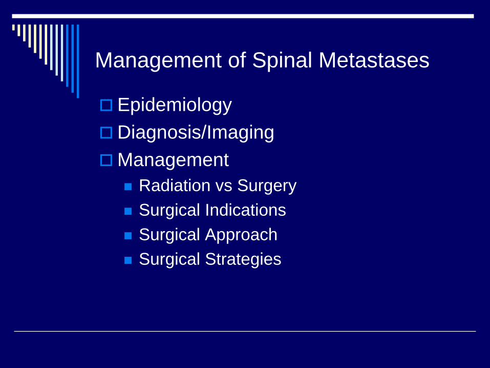
EpidemiologyDiagnosis/ImagingManagement
Radiation vs SurgerySurgical IndicationsSurgical ApproachSurgical Strategies
Management of Spinal Metastases
Metastatic Spinal Neoplasms
18,000 New Cases/YearMost frequent site of bone metastasis1 to 5% of all cancer patients will present with cord compression90% of patients will have spine metsat the time of death
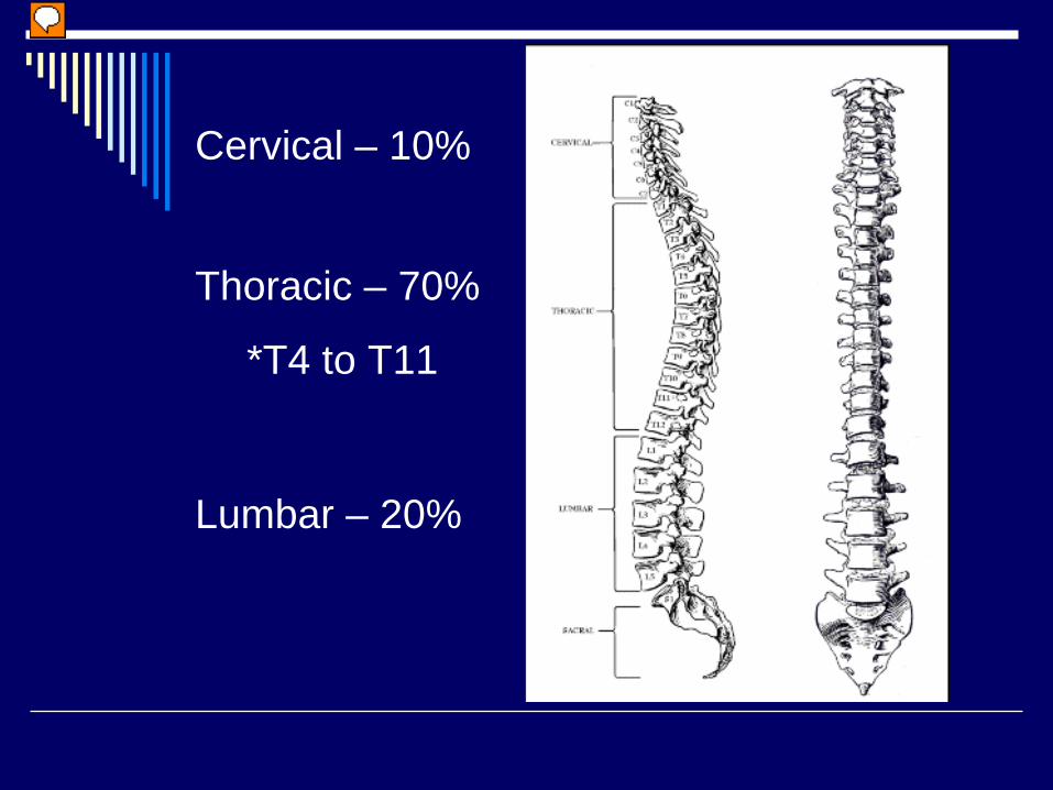
Cervical – 10%
Thoracic – 70%
*T4 to T11
Lumbar – 20%
Tumors That Disseminate to the Spine
BreastLung ProstateRenal CellMyeloma, Lymphoma, GI
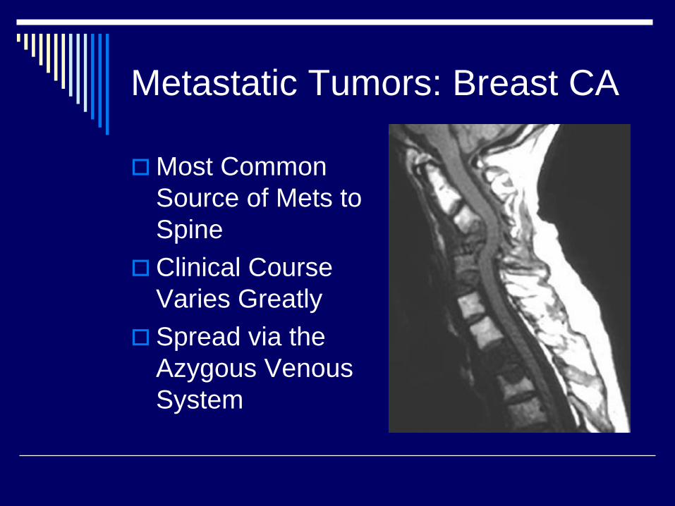
Metastatic Tumors: Breast CA
Most Common Source of Mets to SpineClinical Course Varies GreatlySpread via the Azygous Venous System
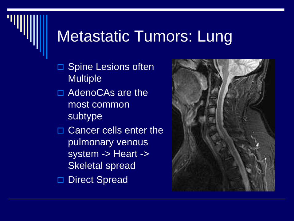
Metastatic Tumors: Lung
Spine Lesions often MultipleAdenoCAs are the most common subtypeCancer cells enter the pulmonary venous system -> Heart -> Skeletal spreadDirect Spread
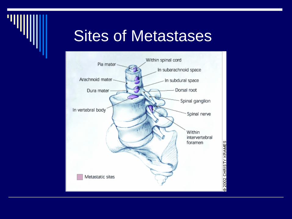
Sites of Metastases
Signs & Symptoms: Pain
Most Common Presenting SymptomOccurs in 83 to 95%Three Classic Syndromes
LocalMechanicalRadicular
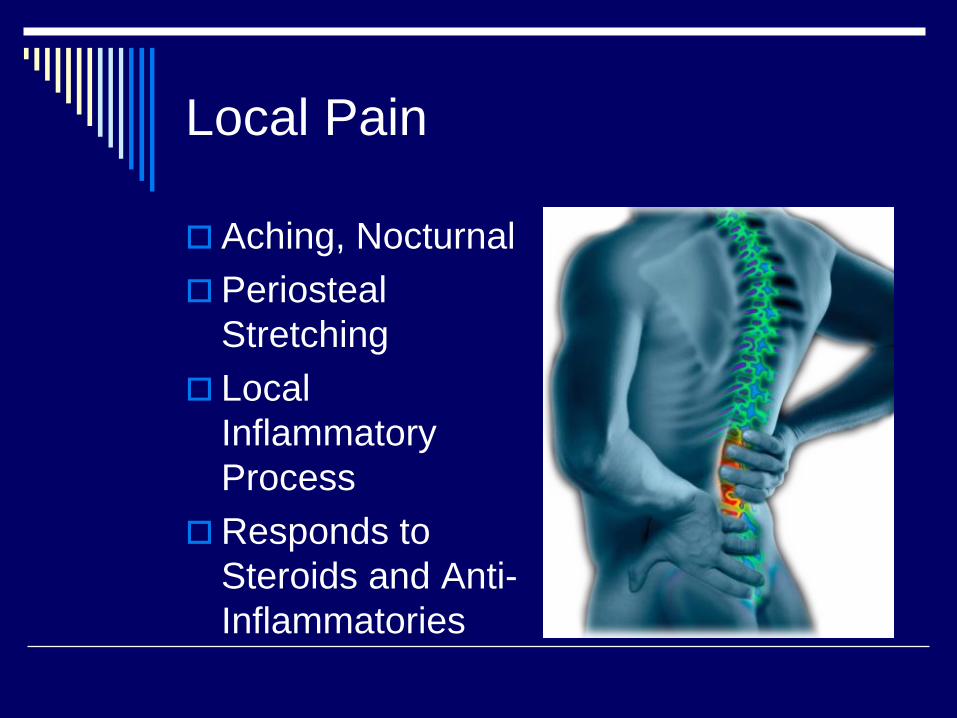
Local Pain
Aching, NocturnalPeriostealStretchingLocal Inflammatory ProcessResponds to Steroids and Anti-Inflammatories
Mechanical Back Pain
Instability of the Spinal ColumnPosturally RelatedWorsens as day progressesRelief with change in position or external bracingRefractory to narcotics, and anti-inflammatories
Radicular Back Pain
Compression or Irritation of Exiting Nerve RootDermatomal distributionStabbing, Shooting
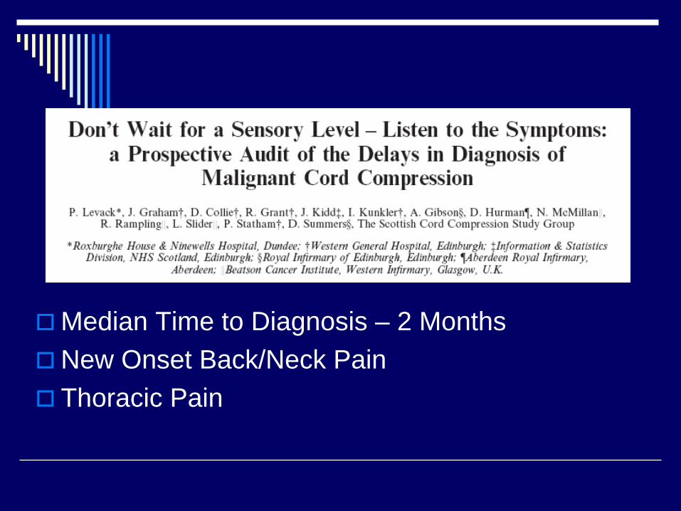
Median Time to Diagnosis – 2 MonthsNew Onset Back/Neck PainThoracic Pain
Signs & Symptoms
Anorexia, unexplained weight lossPalpable mass on examination
ParaspinalRectal
Myelopathy – poor coordination, Hoffman’s sign
Radiographic Studies: X-ray
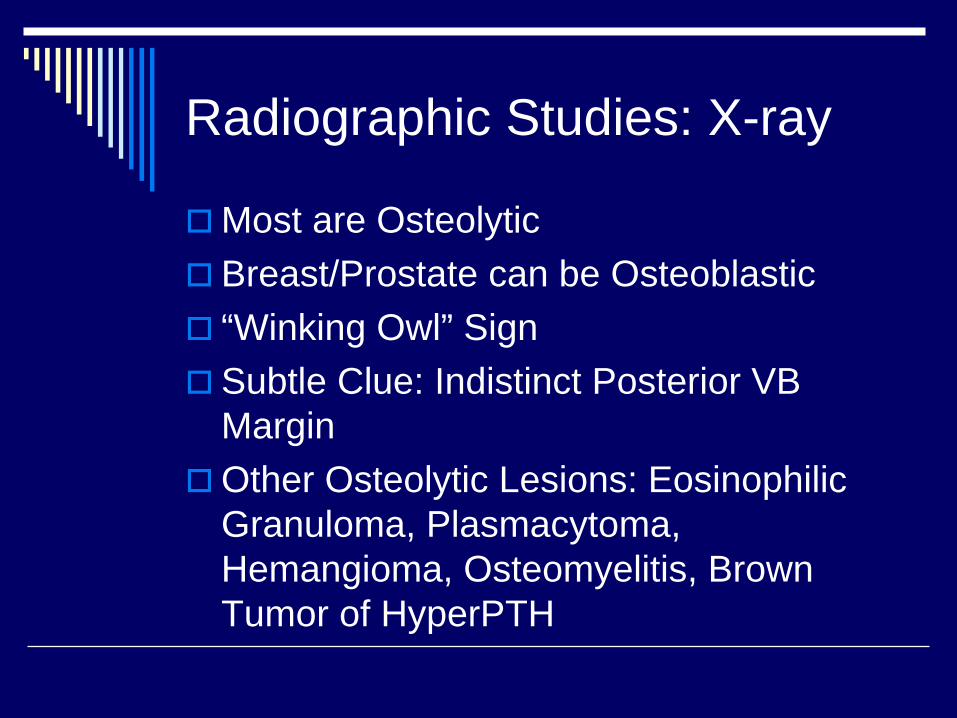
Most are OsteolyticBreast/Prostate can be Osteoblastic“Winking Owl” SignSubtle Clue: Indistinct Posterior VB MarginOther Osteolytic Lesions: EosinophilicGranuloma, Plasmacytoma, Hemangioma, Osteomyelitis, Brown Tumor of HyperPTH
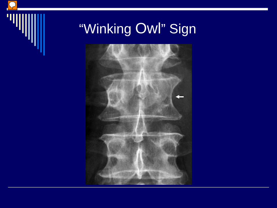
“Winking Owl” Sign
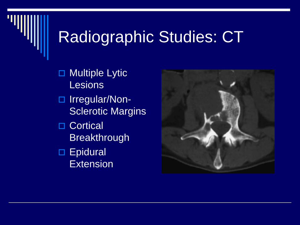
Radiographic Studies: CT
Multiple LyticLesionsIrregular/Non-Sclerotic MarginsCortical BreakthroughEpidural Extension
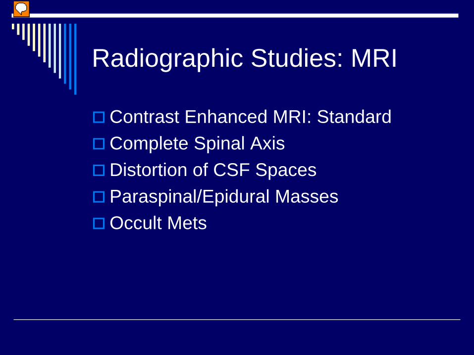
Radiographic Studies: MRI
Contrast Enhanced MRI: StandardComplete Spinal AxisDistortion of CSF SpacesParaspinal/Epidural MassesOccult Mets
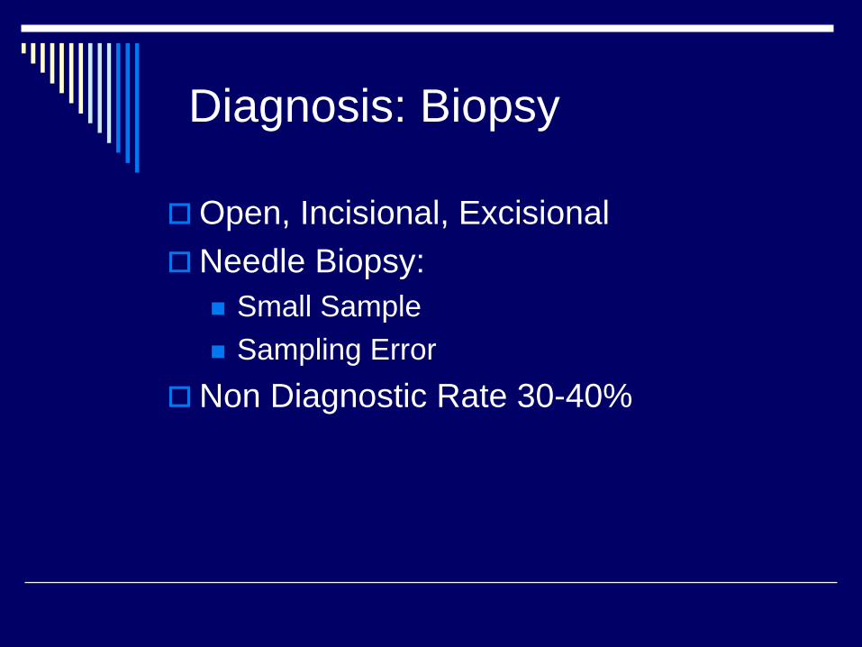
Diagnosis: Biopsy
Open, Incisional, ExcisionalNeedle Biopsy:
Small SampleSampling Error
Non Diagnostic Rate 30-40%
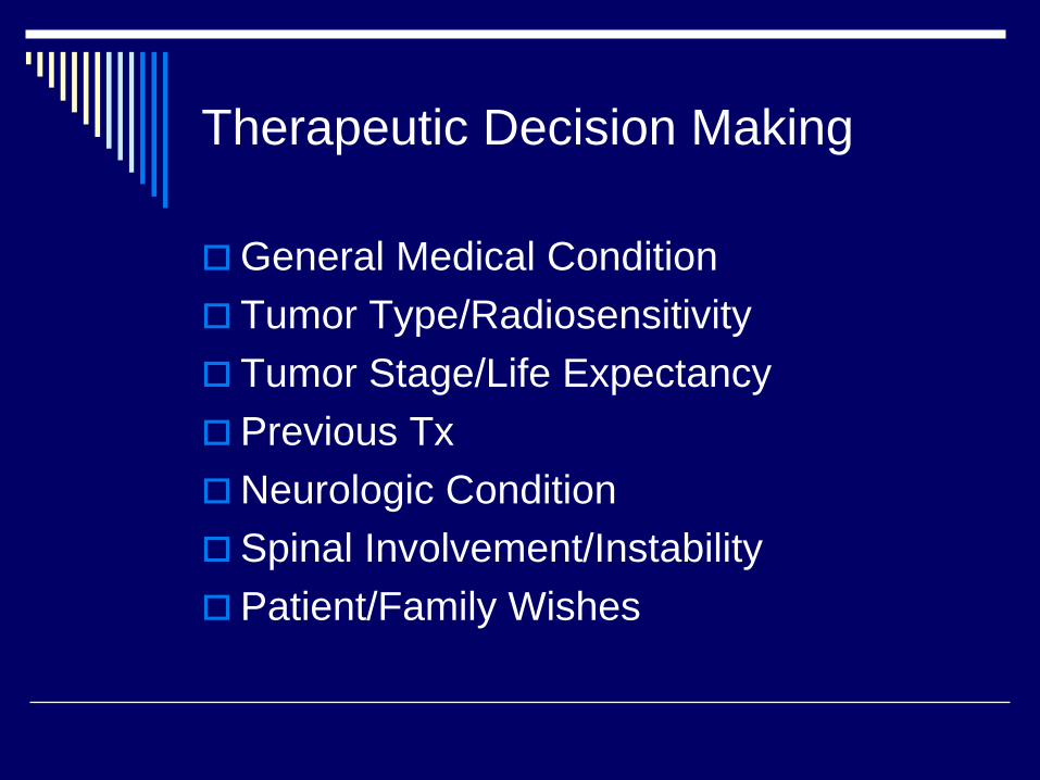
Therapeutic Decision Making
General Medical ConditionTumor Type/RadiosensitivityTumor Stage/Life ExpectancyPrevious TxNeurologic ConditionSpinal Involvement/InstabilityPatient/Family Wishes
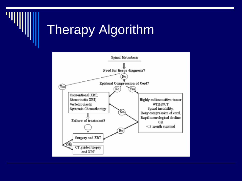
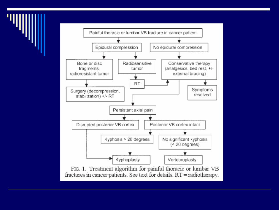
Therapy Algorithm
Treatment
Medical ManagementPreoperative EmbolizationRadiotherapySurgery
Treatment: Medical Mgmt
Steroids may improve pain relief and possibly neurological functionNo optimum dosing scheduleSorenson et al (1994 Euro J Cancer)
Randomized trial of 57 patientsHigh dose dexamethasoneAfter six months, 59 vs 33% ambulatory11% with significant side effects
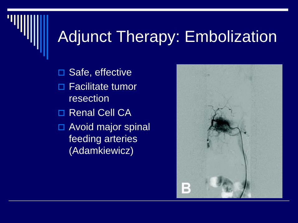
Adjunct Therapy: Embolization
Safe, effectiveFacilitate tumor resectionRenal Cell CAAvoid major spinal feeding arteries (Adamkiewicz)
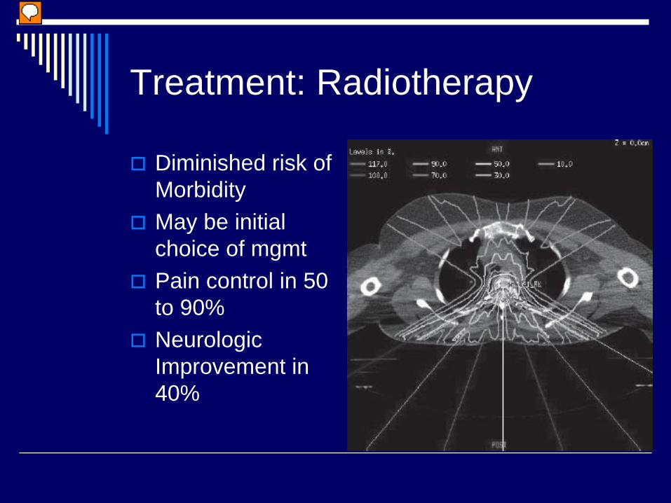
Treatment: Radiotherapy
Diminished risk of MorbidityMay be initial choice of mgmtPain control in 50 to 90%Neurologic Improvement in 40%
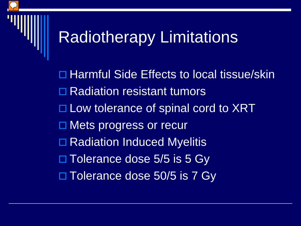
Radiotherapy Limitations
Harmful Side Effects to local tissue/skinRadiation resistant tumorsLow tolerance of spinal cord to XRTMets progress or recurRadiation Induced MyelitisTolerance dose 5/5 is 5 GyTolerance dose 50/5 is 7 Gy
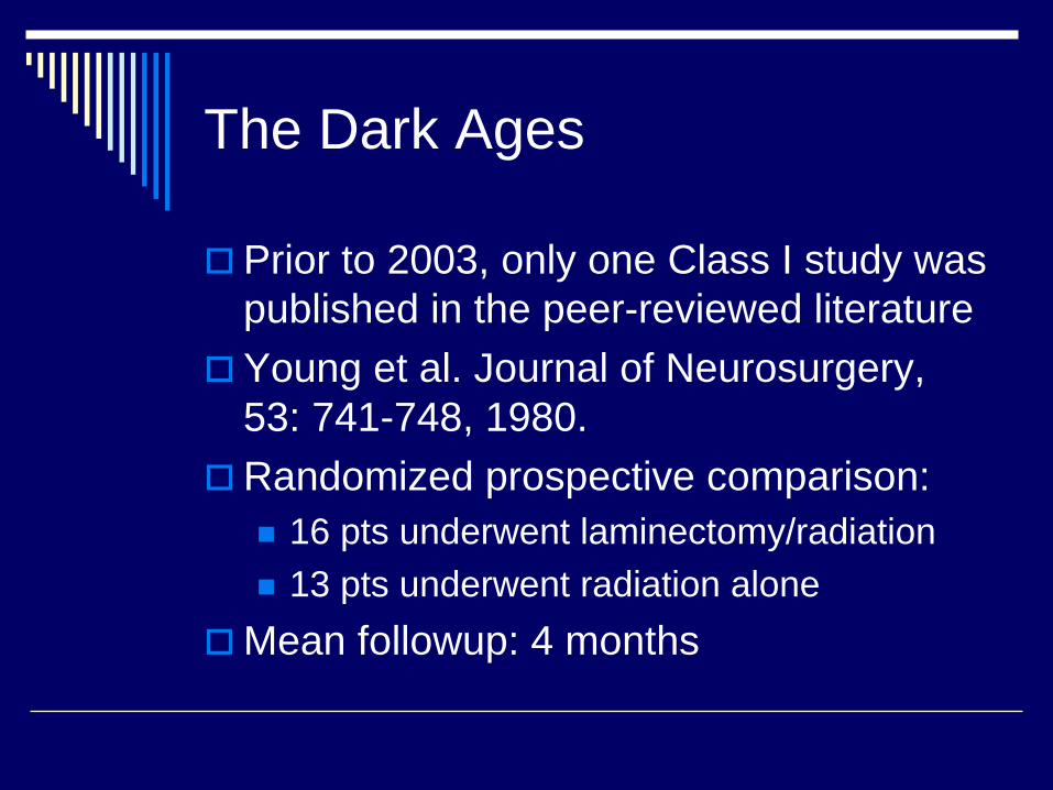
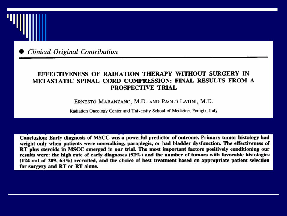
The Dark Ages
Prior to 2003, only one Class I study was published in the peer-reviewed literatureYoung et al. Journal of Neurosurgery, 53: 741-748, 1980.Randomized prospective comparison:
16 pts underwent laminectomy/radiation13 pts underwent radiation alone
Mean followup: 4 months
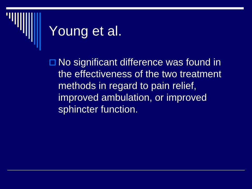
Young et al.
No significant difference was found in the effectiveness of the two treatment methods in regard to pain relief, improved ambulation, or improved sphincter function.
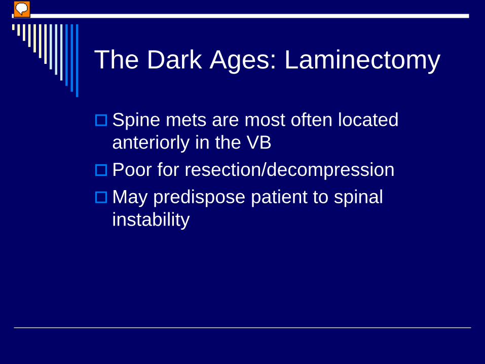
The Dark Ages: Laminectomy
Spine mets are most often located anteriorly in the VBPoor for resection/decompressionMay predispose patient to spinal instability
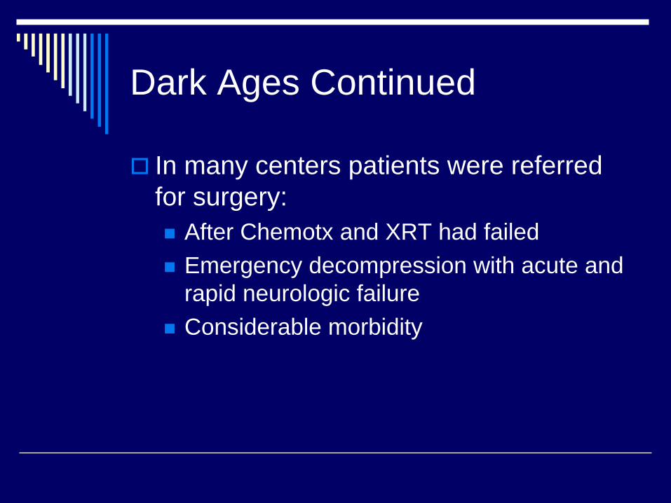
Dark Ages Continued
In many centers patients were referred for surgery:
After Chemotx and XRT had failedEmergency decompression with acute and rapid neurologic failureConsiderable morbidity
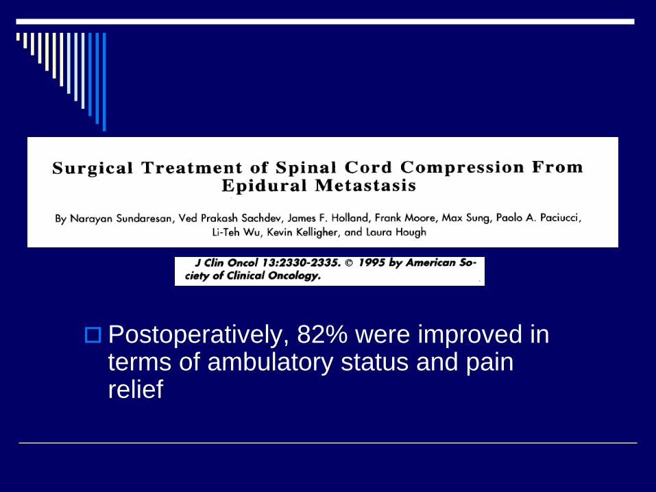
Postoperatively, 82% were improved in terms of ambulatory status and pain relief
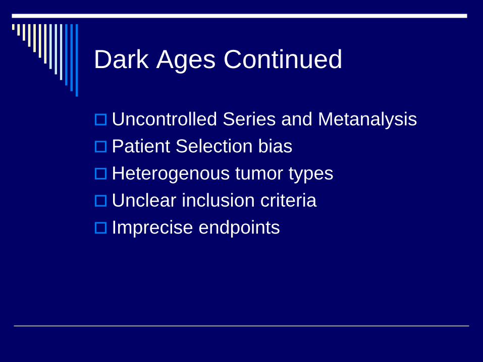
Dark Ages Continued
Uncontrolled Series and MetanalysisPatient Selection biasHeterogenous tumor typesUnclear inclusion criteriaImprecise endpoints
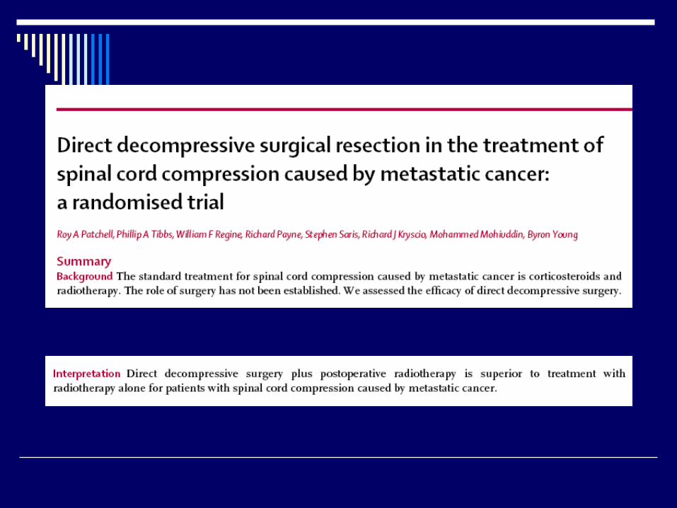
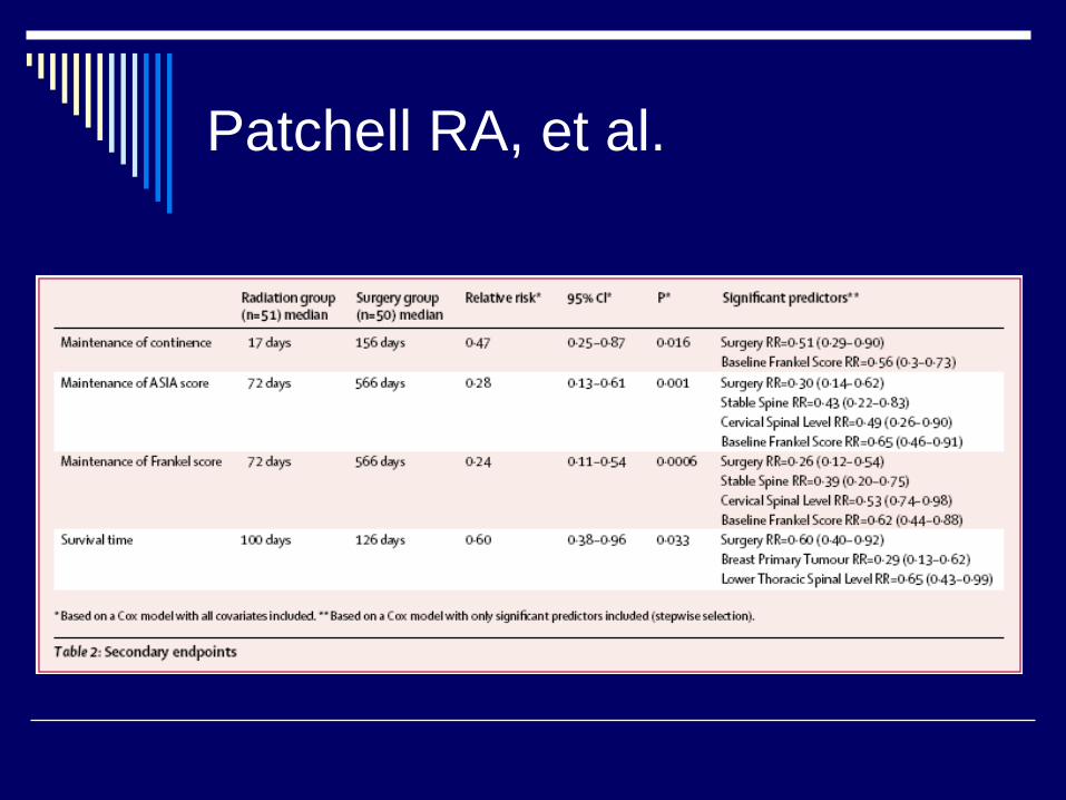
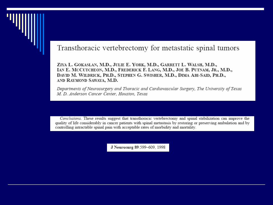
Patchell RA, et al.
Randomized, non-blinded prospective trial (n=123)Surgery and XRT vs XRT alonePrimary Endpoint: Ability to walkSecondary Endpoints: Urinary continence, muscle strength, functional status, survival time, need for steroids/opioids
Patchell RA, et al.
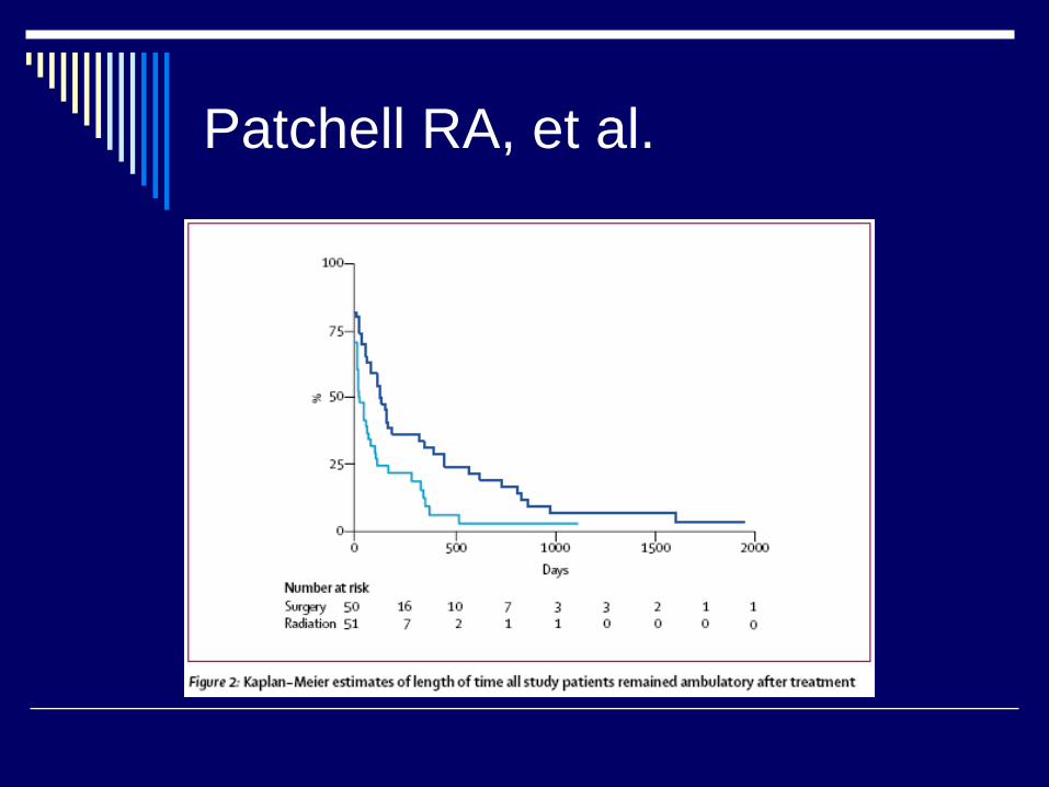
Patchell RA, et al.
Patchell RA, et al.
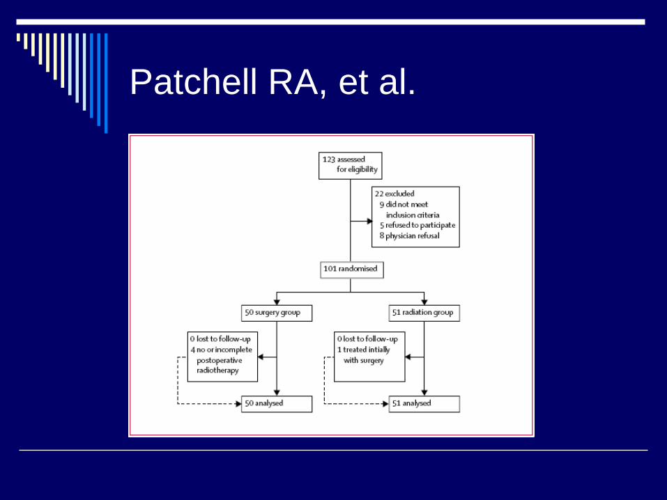
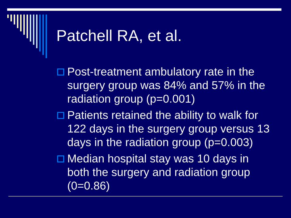
Post-treatment ambulatory rate in the surgery group was 84% and 57% in the radiation group (p=0.001)Patients retained the ability to walk for 122 days in the surgery group versus 13 days in the radiation group (p=0.003)Median hospital stay was 10 days in both the surgery and radiation group (0=0.86)
Patchell RA, et al.
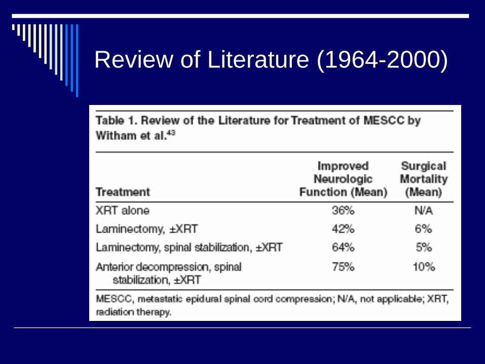
Review of Literature (1964-2000)
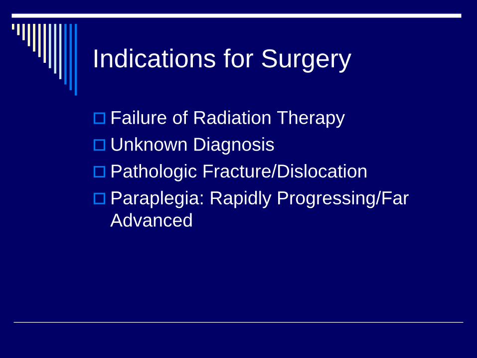
Indications for Surgery
Failure of Radiation TherapyUnknown DiagnosisPathologic Fracture/DislocationParaplegia: Rapidly Progressing/Far Advanced
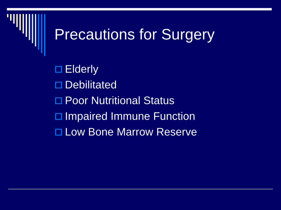
Precautions for Surgery
ElderlyDebilitatedPoor Nutritional StatusImpaired Immune FunctionLow Bone Marrow Reserve
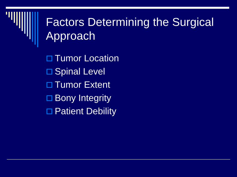
Factors Determining the Surgical Approach
Tumor LocationSpinal LevelTumor ExtentBony IntegrityPatient Debility
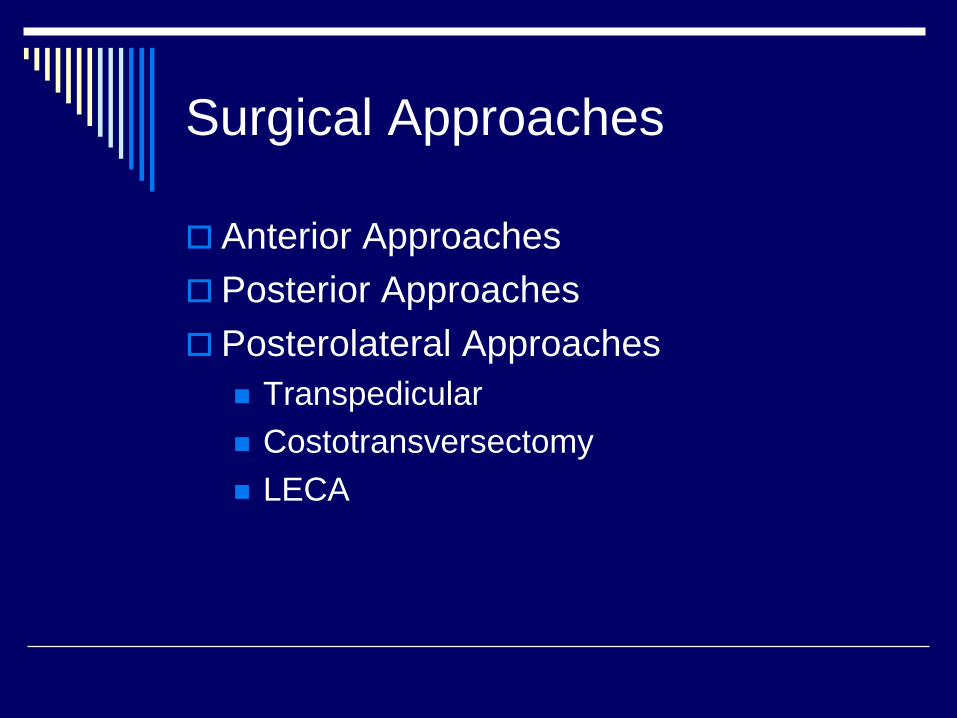
Surgical Approaches
Anterior ApproachesPosterior ApproachesPosterolateral Approaches
TranspedicularCostotransversectomyLECA
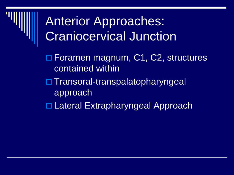
Anterior Approaches: Craniocervical Junction
Foramen magnum, C1, C2, structures contained withinTransoral-transpalatopharyngealapproachLateral Extrapharyngeal Approach

Transoral-transpharyngeal Approach
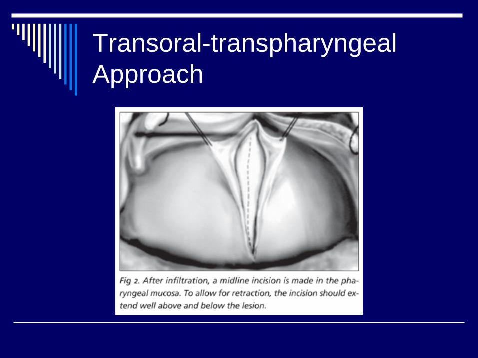
Transoral-transpharyngeal Approach
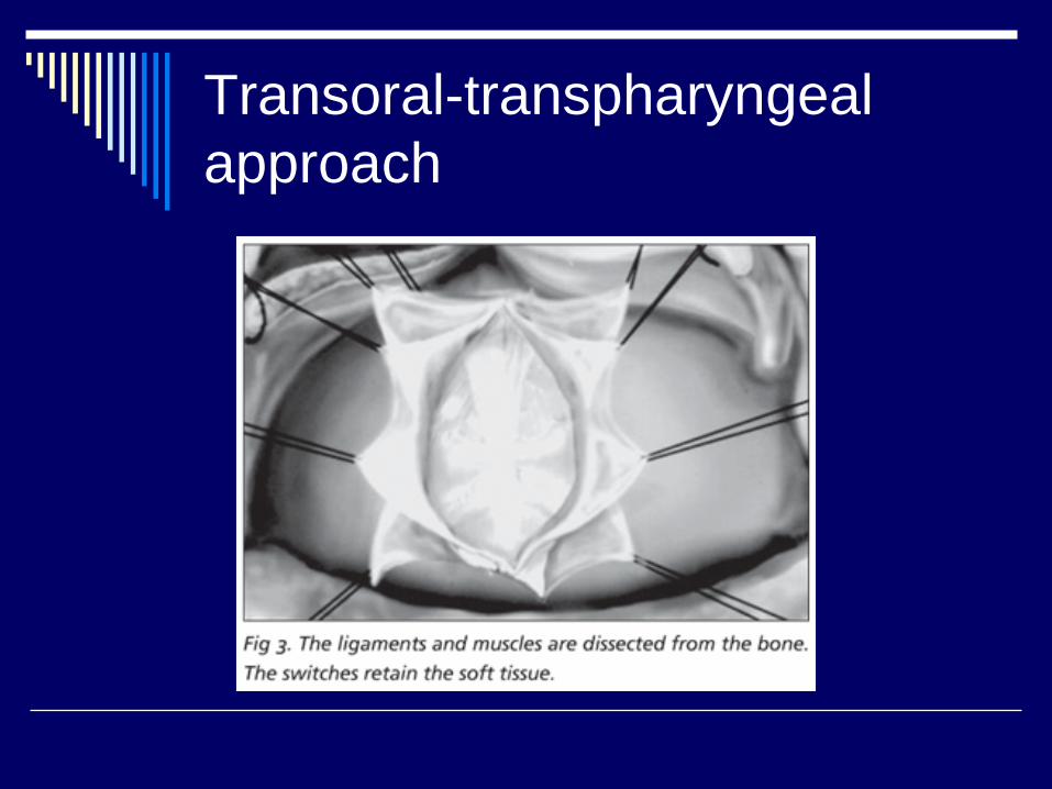
Transoral-transpharyngeal approach
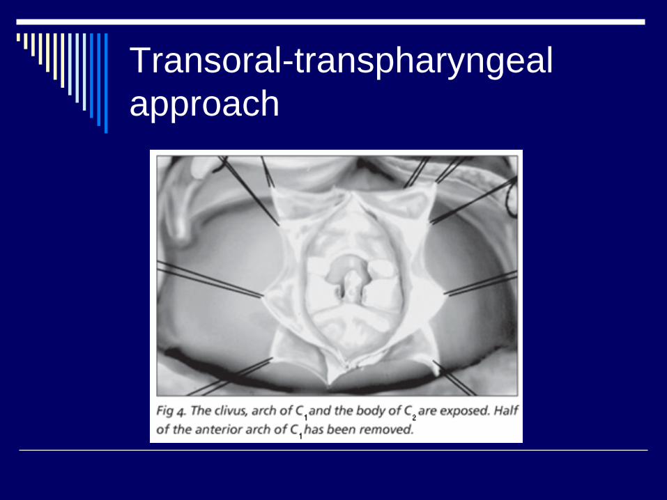
Transoral-transpharyngeal approach
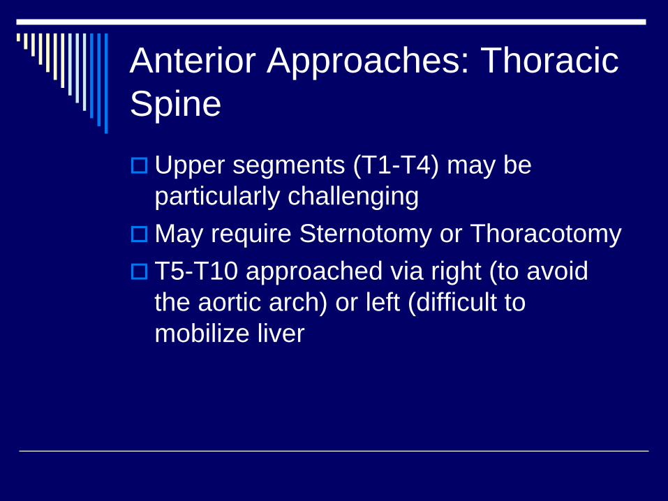
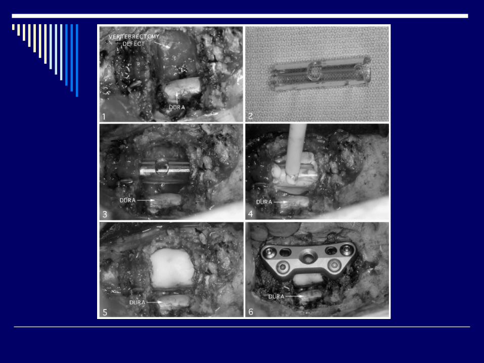
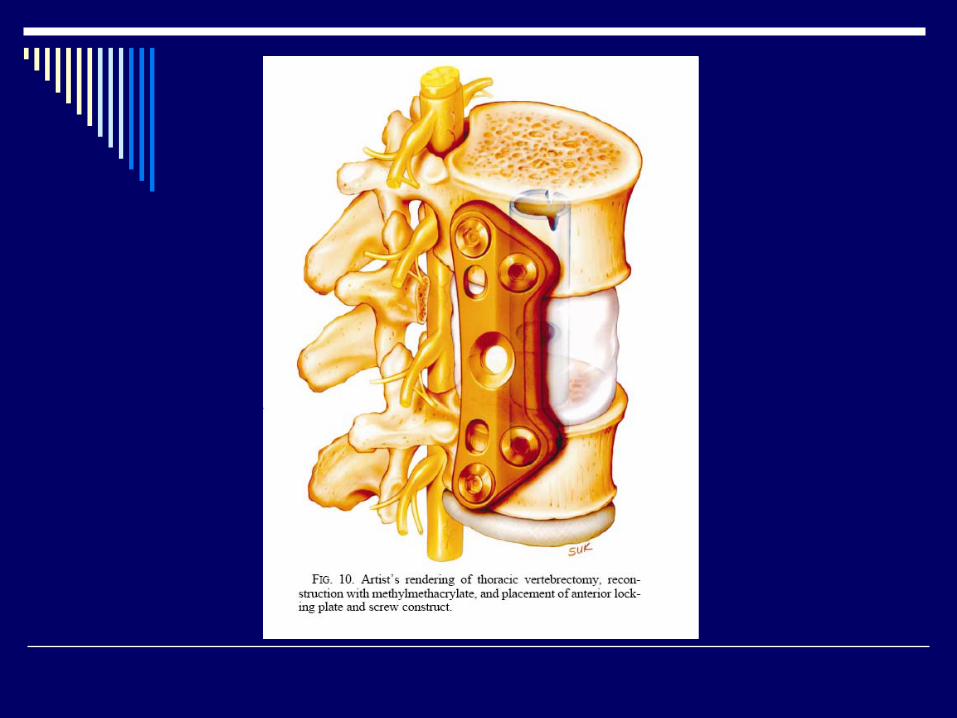
Anterior Approaches: Thoracic Spine
Upper segments (T1-T4) may be particularly challengingMay require Sternotomy or ThoracotomyT5-T10 approached via right (to avoid the aortic arch) or left (difficult to mobilize liver
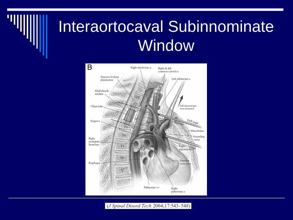
Interaortocaval Subinnominate Window
Anterior Approaches: Cont’d
Thoracolumbar Junction (T11-L1): Thoracotomy and Retroperitoneal ApproachLumbar (L2-L4): Retroperitoneal or Transabdominal ApproachIntra-abdominal contents at riskPatients should be expected to have post-op ileus
Posterior Approaches
Resultant Instability requires Instrumentation and FusionIn the upper thoracic spine the scapula must be mobilized.Working distance can be extensiveAt T11-12 the diaphragm limits the working space
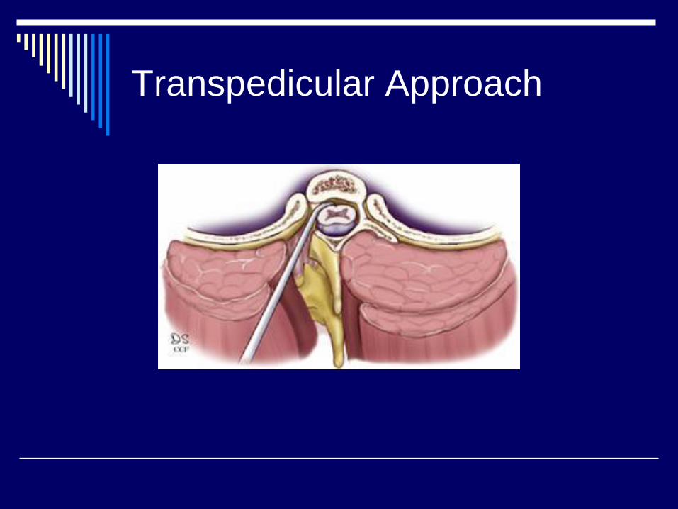
Transpedicular Approach
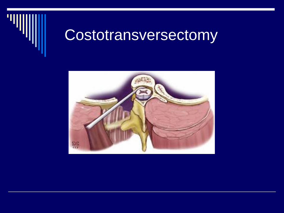
Costotransversectomy
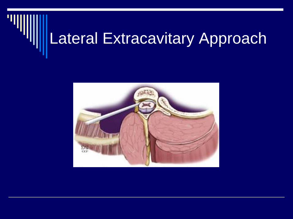
Lateral Extracavitary Approach
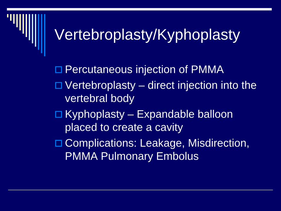
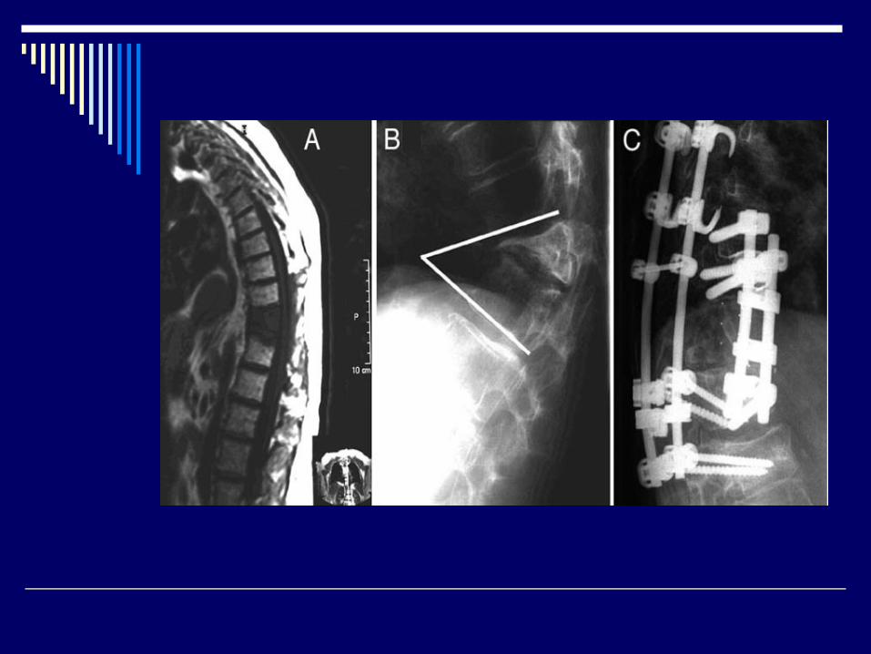
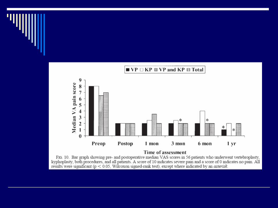
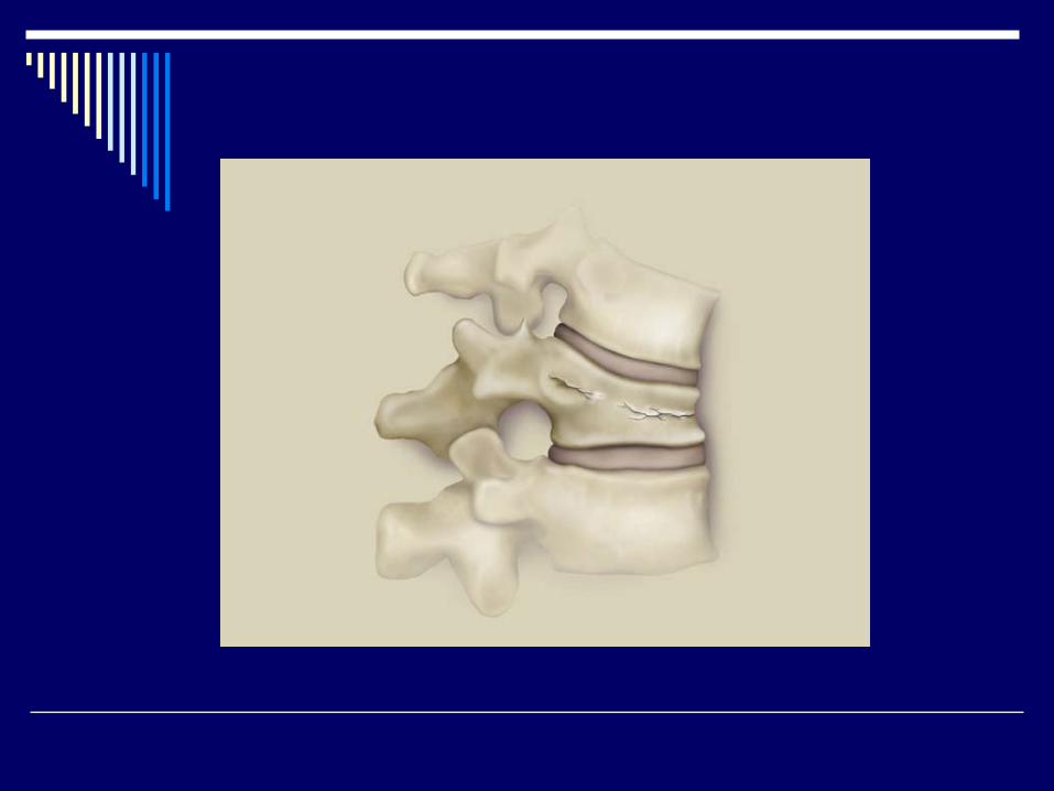
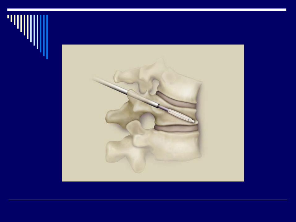
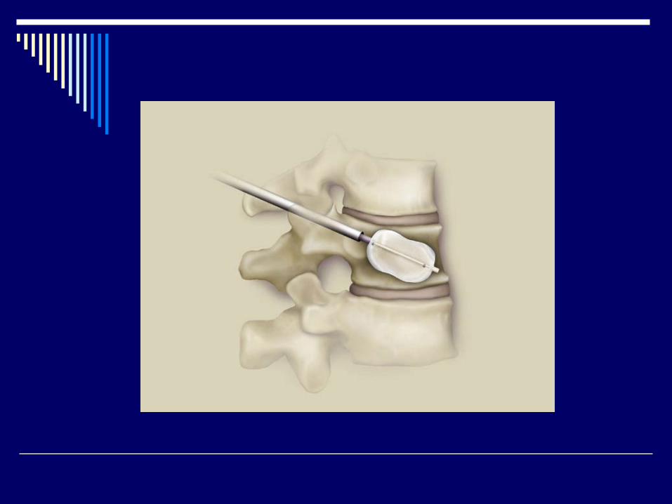
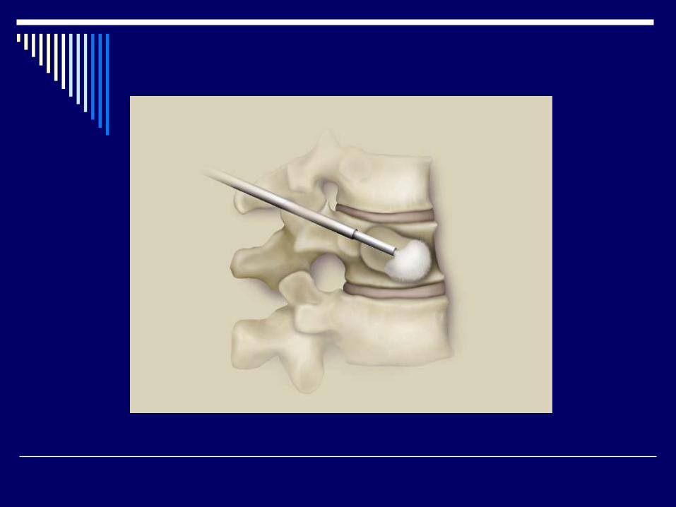
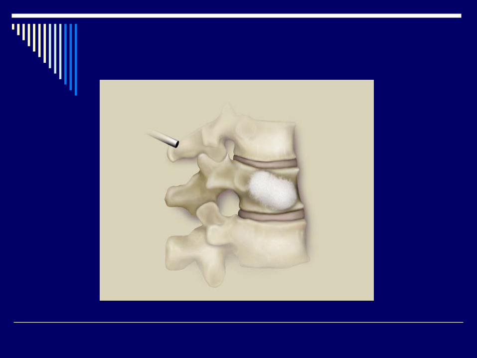
Vertebroplasty/Kyphoplasty
Percutaneous injection of PMMAVertebroplasty – direct injection into the vertebral bodyKyphoplasty – Expandable balloon placed to create a cavityComplications: Leakage, Misdirection, PMMA Pulmonary Embolus
Spine Metastasis: Summary
Spine Mets are not uncommon in patients with cancerSurgery and radiation therapy is superior to radiation therapy alone in selected patientsManagement of patients with spine metastases requires a multidisciplinary approach