management of the compressive multi-nodular goitre of the compressive multi-nodular goitre ......
TRANSCRIPT

Management of the Compressive Multi-Nodular Goitre
Doncaster 2012
Mr Mike Papesch FRACS
Whipps Cross Hospital Edvard Munch: The Seducer II 1913

What is a goitre?
►Normal thyroid gland: 10-25g
►Goitre: from Latin: guttur = throat
►WHO: a thyroid whose lobes have a vol. > terminal phalanges of the thumb of the person being examined
►Lifetime risk of nodule = 10%
in women, age, radiation exposure & iodine
deficiency
►Multinodular goitre: most common cause of thyroid enlargement

Multinodular Goitre
►Sporadic
most common cause of nontoxic goitre
►Diet
Iodine deficiency TSH
Brassica (cabbage, turnips, cauliflower, broccoli) block T4 formation
Cassava: contains thiocyanate
►Familial

Multinodular goitre (MNG)
►Prevalence in UK* Small goitre 9% (palpable not visible)
Obvious goitre 7% (palpable & visible)
F:M = 4:1 (UK)
* Tunbridge WM et al. The spectrum of thyroid disease in a community: the Whickam Survey. Clinical endocrinology, 1977; Vol 7, issue 6:481-493

Clinical features
► Functional
Hyper/Hypo thyroidism
► Cervical
► Retrosternal
► Non compressive
► Compressive
Laryngeal deviation
Tracheal compression /deviation
Oesophageal compression
SVC Obstruction

Retrosternal Goitre First described by Haller in 1749
►Definition1
Descends below the plane of the thoracic inlet > 50% below the plane of the thoracic inlet
Intrathoracic extension requiring reaching into the mediastinum for dissection
Anterior-superior mediastinum extension > 2cm
Reaching the 4th thoracic vertebra
► Female: Male 4:1
►Incidence ►0.02 to 0.5% (CXR1)
►0.05% of females > 40 years
►3-12% of mediastinal masses2
►1-20% of patients requiring thyroidectomy
1 Balasubramanian SP & Harrison BJ. Asymptomatic Retrosternal Goitre – A Case for Primum Non Nocere? Ann R Coll Surg Engl. 2009 January;
91(1): 10–11. 2 Khairy G, et al Large Retrosternal Goitre: A Diagnostic and Treatment Dilemma. OMJ. 25, (2010)

MNG-Symptoms/Signs
► Stridor, OSAS, Dyspnoea on lying flat
► Dysphagia
► Cosmesis
► Pemberton’s sign (facial plethora, cyanosis and distension of neck veins, when raising both arms simultaneously {Pembertons manoeuvre!}).
Seen in thoracic inlet obstruction
originally described in patients with retrosternal goitre
may also be seen in lung carcinoma, lymphomas, thymomas, dermoid cysts or aortic aneurysms
► SVC syndrome
► Ankle oedema
► Hypertension (reversed with surgery) M. P. Holden et al. Massive retrosternal goitre presenting with hypertension Thorax. 1972
November; 27(6): 772–774

Pathology
►Variably and irregularly enlarged gland

MNG Malignancy Risk
► Nodules of MNG undergo malignant change at similar rates to other thyroid tissue.
► Risk: 1 in 20 Wadström 1999 4-8%
Miccoli 1993 8%
Gandolfi 2004 14%
Yamashita 1997 31% (54% of those >1 cm)

Factors suggesting malignancy
► Moderate risk
Age <20, >60
Male
Solitary nodule
Irradiation
Compressive symptoms
Hashimoto’s Genetic factors
► High risk
Rapidly enlarging painless mass
Increases with age
Fixation
Hoarseness
Cystic Lymphadenopathy: levels 2/3/4
Previous Thyroid ca
Family history
Family history: MEN 2a/b
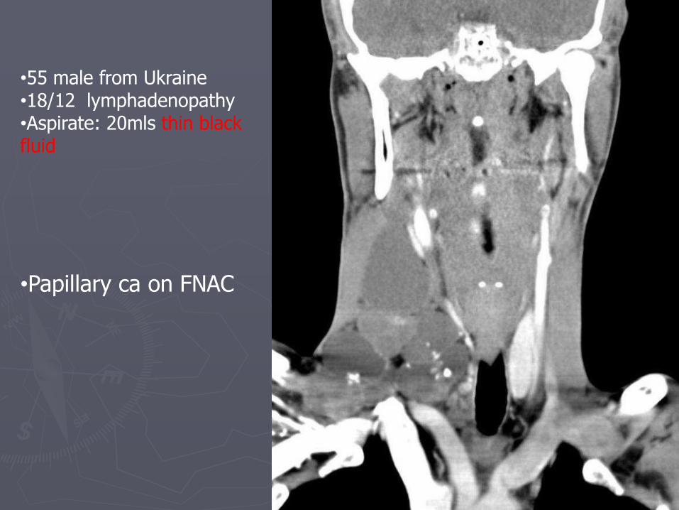
•55 male from Ukraine •18/12 lymphadenopathy •Aspirate: 20mls thin black fluid
•Papillary ca on FNAC

Investigation Compressive MNG
► US
► FNAC
► TFT’s ► Autoimmune antibodies
► CT (?Malignant – non contrast)
► Resp Function tests ►Flow volume loops
► FBC, ECG, Ca2+ etc
► Consider calcitonin
Not for screening
Family history
► RET proto oncogene
in up to 60% PTC
in up to 75% follicular variant PTC
in 33% sporadic MEN
in 95% MEN2A
► No role: Scintography
BAETS 2002
ATA 1996
Thomas GA et al. High prevalence of RET/PTC rearrangements in Ukrainian and Belarussian post-Chernobyl thyroid papillary carcinomas: A strong correlation between RET/PTC3 and the solid-follicular variant. Journal of clinical endocrinology and metabolism. 1999;84:4232-8

CT/MR
► Retrosternal extension
► Tracheal deviation (preop), intubation planning
► Tracheal compression
Note variation in tracheal diameter with respiration and patient position

Treatment of MN or Diffuse Goitre
Conservative
• Nil treatment • Cyst aspiration
Thyroid suppression Thyroxine
Anti Thyroids ►Propylthiouracil &
Methimazole
Radioiodine Ablation I-131
Surgery Hemi-thyroidectomy
Total Thyroidectomy Subtotal Thyroidectomy
Near Total Thyroidectomy

Non surgical treatment
►Elderly patients because of cardiac, pulmonary or other disabling disorders
►High risk patients
►Patients who decline surgery
►Smaller nodules
►Patients had previous surgery
►Recurrent laryngeal nerve injury
Howarth DM et al. Outpatient management of patients with large
multinodular goitres treated with fractionated radioiodine. European Journal of Nuclear Medicine and Molecular Imaging, Volume 24,
Number 12 / December, 1997

Thyroxine Suppression Thyroxine TSH gland stimulation
Recommendation: TSH 0.1-0.5mU/L Efficacy: Numerous small series suggest efficacy No additional benefit with carbimazole Less effective in larger goitres.
► Risks:
Growth of nodules resumes on ceasing suppression. Therefore treatment is life long Subclinical hyperthyroidism Reduced bone density and accelerated loss in Pre/Post MP women Increased LV Mass ?significant. AF Level of suppression not well defined
Berghout A et al. Comparison of placebo with L-thyroxine alone or with carbimazole for treatment of sporadic non-toxic goitre. Lancet. 336(8709):193-7, 1990 Jul 28.

Radio-Iodine
► Response rates 65-99%
► Improvement of dysphagia or dyspnoea in 70% - 90%
► Volume reduction 40% at 1 yr
50-60% at 3-5 yrs
Slowly produces tracheal widening and Respiratory Function
► Tracheal cross sectional area = 36%
► 18 months post treatment, 66% were hypothyroid.
► Excellent short- and medium-term safety, well tolerated
► Satisfactory alternative treatment to surgery
Nygaard B. et al. Radioiodine treatment of multinodular non-toxic goitre. BMJ.307(6908):828-32,1993 Oct 2
Huysmans DA et al. Large, compressive goiters treated with radioiodine. Annals of Internal Medicine. 121(10):757-62, 1994 Nov 15.
Verelst J. et al. Radioiodine therapy in voluminous multinodular non-toxic goitre. Acta Endocrinologica. 122(4):417-21, 1990 Apr.
Howarth DM et al. Outpatient management of patients with large multinodular goitres treated with fractionated radioiodine. Eur J Nuc Med & Mol Imag, 24 (12) 1997 Dec,

Huysmans, D. A.K.C. et. al. Ann Intern Med 1994;121:757-762
Thyroid volume (left), maximal deviation of the tracheal center from the midline (middle), and smallest cross-sectional area of the tracheal lumen (SCAT) (right) measured before and 1 year after radioiodine therapy in 19 patients with a large,
compressive multinodular goiter
Patient 17 before and 1 yr after treatment

Radio-Iodine Risks
► Acute thyroiditis (enlargement) further compressing the trachea
? prednisolone
► Induction of thyroid malignancy (lifetime risk 1.6%)
► Variable response
► High treatment failure
► Transient hyperthyroidism
► Post Ablation Hypothyroidism (up to 50% at 5 years)
Higher with rhTSH ► 23·5% group 1
► 64·0% group 2 at 12 months
► Local cervical pain
► Loss of taste
► Oesophagitis
► Sialoadenitis
► Transitory leukopaenia
► Weight loss
► Away from children for 2 weeks
► Autoimmune hyperthyroidism-Graves-like (5%)
► Recurrence 10% at three to five years
Second dose may be effective

Theodor Kocher 1841 - 1917
► The Nobel Prize in Physiology or Medicine 1909
► Trained by Demme, Lücke, Billroth, and Langenbeck
► His book Erkrankungen der Schilddrüse (Diseases of the thyroid gland ) discussed the aetiology, symptoms and treatment of goitres.
► 5000 career thyroidectomies ► Mortality rates decreased
40% in 1850 (pre-Kocher & Billroth) 12.6% in 1870’s (Kocher begins practice) 0.2% in 1898 (end of Kocher’s career) Attention to asepsis
► Many patients developed cretinism or myxoedema His conclusions in presentation to the German Surgical Congress in 1883 …
“ …the thyroid gland in fact had a function….”

Figure 21-19. The dramatic case of Maria Richsel, the first patient to have come to Kocher’s attention with postoperative myxedema following total thyroidectomy. A. The child and her younger sister before the operation. B. The changes nine years after the operation. The younger sister, now fully grown, contrasts vividly with the dwarfed and stunted patient. Also note Maria’s thickened face and fingers, which are typical of myxedema. Because of this and other patients with the same problem, Kocher stopped performing total thyroidectomies. For this work, demonstrating the physiological importance of the thyroid gland in humans, Professor Kocher was awarded the Nobel prize. From: Kocher T. Uber Kropfextirpation und ihre Folgen, Arch Klin Chir 29:254, 1883, with permission.

MNG-Surgery
►Indications
Cosmesis
Compressive Symptoms
OSA
Suspicion of Malignancy
Retrosternal Goitre
Thyrotoxicosis

Hemi-thyroidectomy
►For unilateral cosmesis (MNG), solitary nodule, Thy 3, U/S concern etc
Low risk of…
►Hypothyroidism
►Perm Hypoparathyroidism
►RLN injury
Risk of progressive contra-lateral disease (MNG)
Wadstrom C. Zedenius J. Guinea A. Reeve T. Delbridge L. Multinodular goitre presenting as a clinical single nodule: how effective is hemithyroidectomy? Australian & New Zealand Journal of Surgery. 69(1):34-6, 1999 Jan.

Total Thyroidectomy
►Complete excision of the gland bilaterally.
►Lifelong Thyroxine replacement.
►Nil risk of recurrence
►Recommended rather than subtotal/Near total thyroidectomy for MNG and Graves
Delbridge L et al. Total thyroidectomy for bilateral benign multinodular goiter: effect of changing practice. Archives of Surgery. 134(12):1389-93, 1999 Dec.
Khadra M et al. Total thyroidectomy: its role in the management of thyroid disease. Australian & New Zealand Journal of Surgery. 62(2):91-5, 1992 Feb.
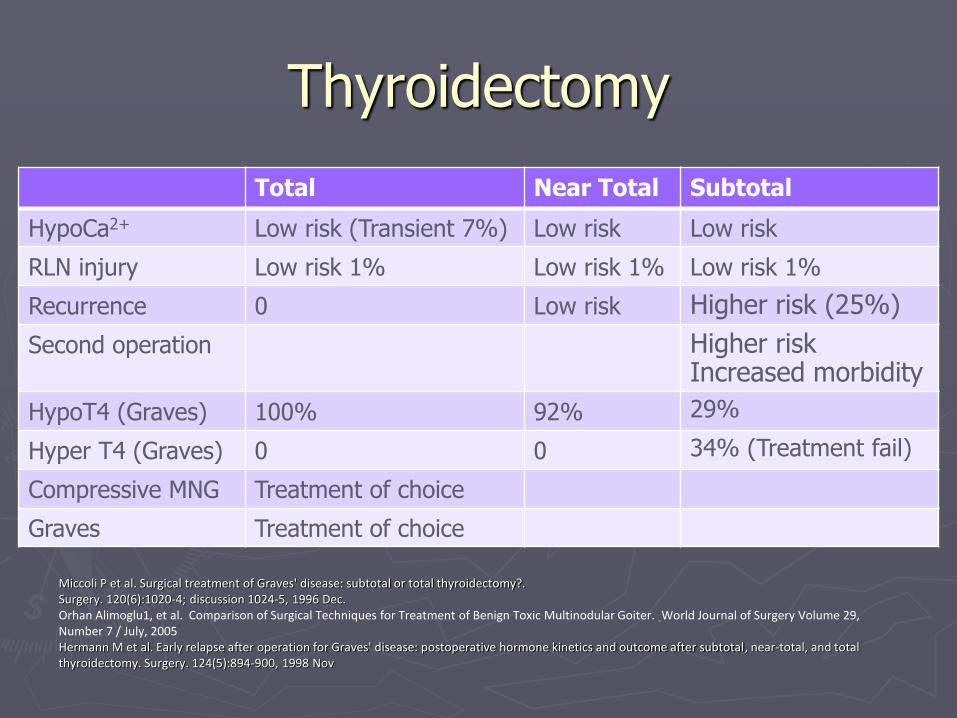
Thyroidectomy
Total Near Total Subtotal
HypoCa2+ Low risk (Transient 7%) Low risk Low risk
RLN injury Low risk 1% Low risk 1% Low risk 1%
Recurrence 0 Low risk Higher risk (25%)
Second operation Higher risk Increased morbidity
HypoT4 (Graves) 100% 92% 29%
Hyper T4 (Graves) 0 0 34% (Treatment fail)
Compressive MNG Treatment of choice
Graves Treatment of choice
Miccoli P et al. Surgical treatment of Graves' disease: subtotal or total thyroidectomy?.
Surgery. 120(6):1020-4; discussion 1024-5, 1996 Dec.
Orhan Alimoglu1, et al. Comparison of Surgical Techniques for Treatment of Benign Toxic Multinodular Goiter. World Journal of Surgery Volume 29,
Number 7 / July, 2005
Hermann M et al. Early relapse after operation for Graves' disease: postoperative hormone kinetics and outcome after subtotal, near-total, and total
thyroidectomy. Surgery. 124(5):894-900, 1998 Nov

Preop Assessment
►Airway issues
►Need for sternotomy: Rare
►Where is surgery best undertaken?

Aberrant Intrathoracic Thyroid
► Operative approach completely different
► Thoracotomy
► Blood supply intrathoracic Not cervical
Massive sub-sternal goiter causing partial obstruction to the innominate veins. Dye has
been injected into both arm veins. Hall TS, Caslowitz P, Popper C, Smith GW. Substernal
goiter versus intrathoracic aberrant thyroid: a critical difference. Ann Thorac Surg 1988;46:684-5.

Management of Differentiated Thyroid
Cancer
Mike Papesch
Whipps Cross 2013

Guidelines ► Revised ATA Guidelines for Patients with Thyroid
Nodules & DTC 2009 ► Guidelines for the management of thyroid cancer 2nd ed
2007 BTA & RCP ► European Association of Nuclear Medicine consensus guidelines for RAI therapy of
differentiated thyroid cancers 2008 ► ATA Statement on Essential Elements of Interdisciplinary Communication of
Perioperative Information for Patients Undergoing Thyroid Cancer Surgery 2012 ► Guidelines of the ATA for Diagnosis & Management of Thyroid Disease During
Pregnancy & Postpartum 2011 ► Radiation Safety in the Treatment of Patients with Thyroid Diseases by Radioiodine
131I: Practice Recommendations of the American Thyroid Association 2011 ► ATA Consensus Statement on Terminology & Classification of Central Neck
Dissection for Thyroid Cancer 2009 ► American Thyroid Association Design and Feasibility of a Prospective Randomized Controlled Trial of Prophylactic
Central Lymph Node Dissection for Papillary Thyroid Carcinoma 2012 ► UK Guidelines for the Use of Thyroid Function Tests 2006 ► Guidelines for the Surgical Management of Endocrine Disease and Training Requirements for Endocrine Surgery
2003 ► Paediatric Endocrine Tumour Guidelines

The Problem
►Nodule: Common palpable 5% ♀ 1% ♂
ultrasound 20-67%
►Ca: 10% ►Risks: age (<10 - >40), sex (male), radiation
(childhood/Chernobyl), FHx (ca & adenoma), endemic goitre, Cowden’s syndrome, familial adenomatous polyposis
►DTC: 90%: papillary (PTC)/follicular cancer (FTC)
Cowden’s syndrome: macrocephaly, mild learning difficulties, carpet-pile tongue, with benign or malignant breast disease

Incidence UK
► 1% of all malignancies, most common endocrine tumour ► Increasing incidence ► 1971–95
2.3/100,000 women 0.9 per 100,000 men 900 new cases/year 250 deaths/year
► 2001 3.5/100,000 women 1.3/100,000 men 1,200 new cases/year
► PTC due to use of US, FNAC and early treatment ► 50% of the in ca < 1cm ► 90% of the in ca < 2 cm

Diff Thyroid Ca Management
► Specialist multidisciplinary team (MDT) ► Regional cancer network ► Timeframe complying to DoH targets ► One stop clinic ► Surgeon, endocrinologist , oncologist (or nuclear
medicine physician), pathologist, dedicated radiologist, specialist nurse etc
► Combined clinic, Databases, BAETS audit ► Patients
seen by one or more members of the MDT have a key-worker Access to education, support etc

Aim of Management of DTC
► Accurate pre op staging ► Removal of primary: Total thyroidectomy (Hemi:
small/low risk) ► Treatment of locoregional disease (even in metastatic
disease) ► Minimal morbidity ► Post operative staging and prognosis
► Postoperative treatment with radioactive iodine ► Long-term surveillance for disease recurrence (RAI
whole-body scanning (WBS) and serum Tg) ► Education & Support (Key worker)

US Thyroid nodule & FNAC
► Need Dedicated Radiologist ► Suspicious features
Microcalcifications marked hypoechogenicity irregular margins absence of a hypoechoic halo around the nodule extracapsular extension local invasion of adjacent structures lymphadenopathy number, size, and interval growth of nodules
► US guided FNAC of suspicious thyroid nodules inadequate fine-needle aspirates

Microcalcification
► 30%–60% of all primary thyroid ca ► most commonly in PTC ► Also FTC & anaplastic ca as well as in benign
conditions (follicular adenoma & Hashimoto thyroiditis
► US: punctuate hyperechoic foci without acoustic shadowing
► psammoma bodies 10–100-μm round laminar crystalline calcific deposits specific feature of thyroid ca
►specificity of 85.8%–95% ►positive predictive value of 41.8%–94.2%

Papillary thyroid carcinoma in a 42-year-old man
Hoang J K et al. Radiographics 2007;27:847-860
©2007 by Radiological Society of North America
Transverse ultrasound of the right lobe of the thyroid demonstrates punctate echogenic foci without posterior acoustic shadowing, findings indicative of
microcalcifications (arrows).

Papillary thyroid carcinoma in a 42-year-old man
Hoang J K et al. Radiographics 2007;27:847-860
©2007 by Radiological Society of North America
Photomicrograph (original magnification, × 400; hematoxylineosin stain) shows a
psammoma body (arrow), a round laminar crystalline calcification

Papillary carcinoma in an 87-year-old man
Hoang J K et al. Radiographics 2007;27:847-860
©2007 by Radiological Society of North America
Papillary carcinoma in an 87-year-old man. Transverse sonogram of the thyroid
isthmus shows a poorly defined tumor with marked hypoechogenicity and irregular
margins (arrows) and without a hypoechoic halo.
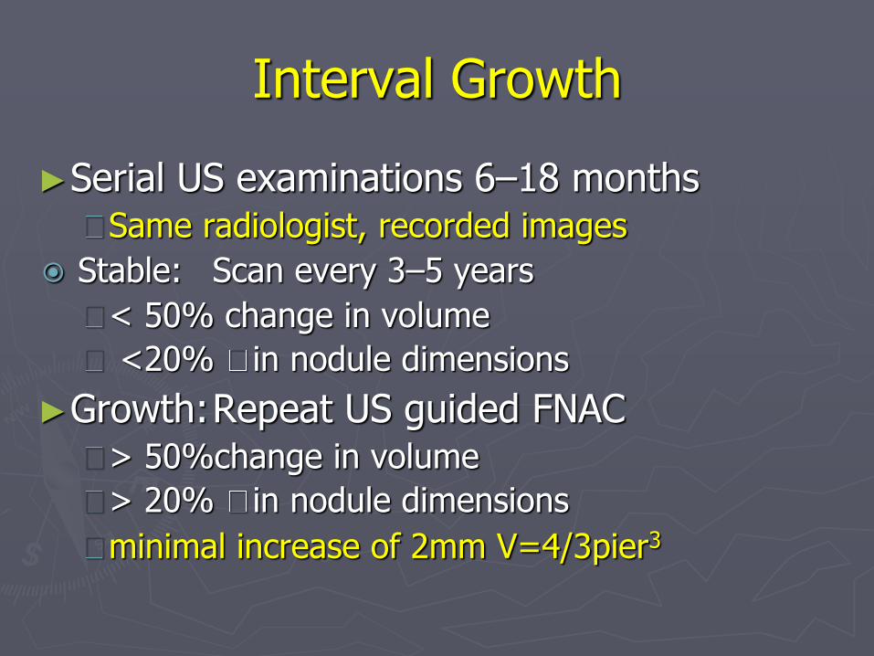
Interval Growth
►Serial US examinations 6–18 months Same radiologist, recorded images
Stable: Scan every 3–5 years
< 50% change in volume
<20% in nodule dimensions
►Growth: Repeat US guided FNAC > 50%change in volume
> 20% in nodule dimensions
minimal increase of 2mm V=4/3pier3

US central & lateral nodes & FNAC
US guided FNA of suspicious lymph nodes suspicious nodes: 20% (! ∆ in op)
► Loss of the fatty hilus: high sensitivity ► Peripheral vascularity (instead of just normal central hilar
vessels): high specificity ► Round bulging shape ► Increased size ► Cystic areas ► Heterogeneous echotexture ► Microcalcification ► Irregular margins
US guided lymph node FNAC with Tg washout Black cystic fluid: Papillary met!!! (send all fluid) Post op
► Histologically +ve nodes: 20-50% (papillary) ► Micromets: up to 90%

Papillary carcinoma and cystic lymph node metastasis in a 28-year-old woman
Hoang J K et al. Radiographics 2007;27:847-860
©2007 by Radiological Society of North America
(a)Longitudinal sonogram of the right lobe of the thyroid shows an irregular hypoechoic tumor with microcalcifications. (b) Longitudinal sonogram of the right neck shows a cystic level 5 nodal metastasis with internal septation and foci of calcification (arrows). (c) Axial contrast-enhanced CT image shows the metastasis (arrow).

Papillary carcinoma and cystic lymph node metastasis in a 28-year-old woman
Hoang J K et al. Radiographics 2007;27:847-860
©2007 by Radiological Society of North America
(a)Longitudinal sonogram of the right lobe of the thyroid shows an irregular
hypoechoic tumor with microcalcifications. (b) Longitudinal sonogram of the right neck shows a cystic level 5 nodal metastasis with internal septation and foci of
calcification (arrows). (c) Axial contrast-enhanced CT image shows the metastasis (arrow).
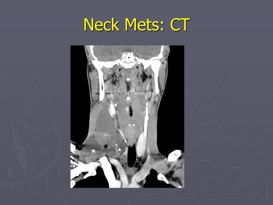
Neck Mets: CT

FNAC (Thy 1 – 5)
► Thy 1 Non-diagnostic repeat: US guided Cysts: colloid or histiocytes only, without epithelial cells, Aspirated to dryness with no residual mass, clinical/ultrasound follow-up
► Thy2 Non-neoplastic Two non-neoplastic results 3–6 months needed to exclude ca
High risk Lobectomy
► Thy3 Suspected follicular neoplasm: Lobectomy ► Thy4 Suspicious of malignancy (suspicious, but not diagnostic, of
papillary, medullary or anaplastic carcinoma, or lymphoma). ? Repeat (for Thy 5) MDT discussion Surgery
► Thy5 Diagnostic of malignancy MDT discussion Further investigation Surgery: Total thyroidectomy

Preop staging with diagnostic imaging & laboratory tests
►TFT & Ca
►CT (non contrast), MRI & PET
Not recommended routinely
Assessment of large, rapidly growing, retrosternal or invasive tumours
MRI can be useful for ? retropharyngeal nodes


What is the appropriate op for indeterminate thyroid nodules & DTC?
►Solitary thyroid nodules ca risk Thy 3: 20%
: tumour >4 cm, atypical cytology (eg cellular pleomorphism), FHx, radiation exposure, age, growth
: Repeatedly nondiagnostic FNAC: 5%
: No growth, interval scan
►MDT discussion Difficult histology, high risk
►Patient discussion of alternatives/risks

What is the appropriate op for Indeterminate thyroid nodules?
• Thyroid lobectomy (diagnositic)
solitary Thy 3 nodule
Thy 3 nodule >4 cm
marked atypia (?Thy 4)
FHx of thyroid carcinoma
Radiation exposure
► Total Thyroidectomy
Bilateral indeterminate nodules

What is the appropriate op for DTC?
►Papillary
<1 cm, low-risk, unifocal, intrathyroidal pap ca., no radiation exposure, no neck nodes
Lobectomy
>1 cm total thyroidectomy
Thyroid lobectomy ca completion
►Follicular
<1cm, minimal capsular invasion Lobectomy
<2cm low risk ? Lobectomy only
vascular invasion or > 4cm total
thyroidectomy

Neck Dissection for DTC
► Clinically or US involved nodes Anterior Neck (Level 6) dissection Lateral neck: lateral compartmental neck node dissection (favoured over “cherry picking” {lower mortality}) may reduce the risk of recurrence and mortality
► Prophylactic Level 6 Dissection Controversial Advanced T3/4 tumours, may improve survival Not for small (T1/2), noninvasive, clinically node-negative PTCs and most follicular ca higher morbidity, recurrent laryngeal nerve injury and transient hypoparathyroidism with no reduction in recurrence.

Surgery for Locally Advanced Disease
►If possible dissect the tumour from the recurrent laryngeal nerve
►unilateral nerve involvement & extensive extrathyroidal disease
Nerve sacrifice to achieve a curative procedure
►Bilateral nerve involvement A small residue of tumour may be left behind to protect the nerve/s
►131I ablation ►TSH suppression with T4 ►+/- external beam radiotherapy

Management of Aerodigestive Invasion
►Surgery
►131I RAI ablation
►Ext beam radiotherapy

Role of Postop Staging
1. To provide prognosis
2. To asses risk for disease recurrence and mortality
3. To tailor postoperative adjunctive therapy (RAI therapy and TSH suppression)
4. To determine frequency and intensity of follow-up
5. To enable comparative evaluation of results of treatments

Staging Systems
1. TNM staging cancer registries & epidemiologic studies
does not consider additional independent prognostic variables & risks misclassification of patients.
2. MACIS, AGES, AMES, CAEORTC, NTCTCS, U of C, OSU, MSKCC
more prognostic staging systems
most predictive factors: distant metastases, age & extent of tumour
AGES - Age, Grade, Extent of disease, Size AMES - Age, Metastasis, Extent of disease, Size CAEORTC - European Organisation for Research and Treatment of Cancer


MAICS (Mayo Clinic) Metastasis, Age, Invasion, Completeness of Resection,
Size ► Most reliable staging method to estimate prognosis of PTC
► Assigns scores to the main factors
► Sums score to calculate prognosis
► Children with multiple lung metastases and/or a miliary aspect have excellent long-term prognosis if given adequate treatment.
Factors Score
Distant Metastasis: spread of the cancer to areas outside the neck Yes 3
No 0
Age at the time the tumour was discovered <30 3.0
>40 0.08 x age
Invasion into surrounding areas of the neck as seen by the naked eye Yes 1
No 0
Completeness of surgical resection (or removal) of the tumour Incomplete 1
Complete 0
Size of the tumour 0.3x size (cm)

MACIS
Sum Of MACIS Score 20 year Survival
<6.0 99%
6.0-6.99 89%
7.0-7.99 56%
>8.0 24%

Prognostic Groups
► Low-risk:
no local or distant metastases
complete resection
no tumour invasion of locoregional tissues or structures
no aggressive histology (e.g., tall cell, insular, columnar cell carcinoma) or vascular invasion
no 131I uptake outside the thyroid bed on the first post treatment whole-body RAI
► Intermediate-risk:
microscopic invasion of tumour into the peri-thyroidal soft tissues
cervical lymph node metastases or 131I uptake outside the thyroid bed on the RxWBS after thyroid remnant ablation
tumour with aggressive histology or vascular invasion
► High-risk:
macroscopic tumour invasion
incomplete tumour resection
distant metastases
thyroglobulinemia out of proportion with post treatment scan

Postoperative RAI Remnant Ablation
1. Recommended Regional nodal disease, aerodigestive invasion or residual RAI avid disease known distant metastases gross extrathyroidal extension (regardless of size) Primary >4 cm
2. Recommended for selected patients 1–4cm thyroid cancers confined to the thyroid with lymph node mets or other risk features age, size, lymph node status & histology intermediate to high risk of recurrence/death
3. Not Recommended unifocal cancer <1 cm multifocal cancer when all foci are <1 cm without risk features

Remnant ablation requires TSH stimulation
►TSH >30mU/L RAI uptake in tumours
►Withdrawal of T3
►Exogenous TSH
Role of human recombinant TSH (thyrotropin) (rhTSH)
► for patients unable to tolerate hypothyroidism (underlying co-morbidities)
unable to generate an elevated TSH (pituitary disease)
in whom a delay in therapy might be deleterious
► equally effective , with QOL
► ablation rates with 50 mCi = 100 mCi rates when rhTSH used but with 33% in whole-body irradiation
► Used in the UK with the world wide shortage of T3
RAI Ablation Complications
►Salivary gland damage sialadenitis, dry mouth, dental caries, loss of taste
►Nasolacrimal duct obstruction/tearing ►Secondary malignancies (dose related)
risk of second malignancies: 1.19 Risk of leukaemia: 2.5 risk of breast cancer: ? screening bias, RAI therapy or other factors
► Reduce Laxatives radiation exposure of the bowel
Oral hydration will reduce exposure of the bladder and gonads

RAI: Gonadal fn & breast feeding
► Women
25% Temporary amenorrhea/oligomenorrhea
Should not be under go treatment if pregnant
Pregnancy should be postponed for 6-12 months
Radioactive iodine should not be given to breast feeding women.
Deferred until 6-8 week post breast-feeding
? Dopaminergic agents to breast exposure in recently lactating women
► Men
Temporary sperm counts
Permanent male infertility is unlikely with a single ablative activity of RAI
Sperm banking in men who receive > 2 RAI treatments
Fertility and risks of miscarriage or congenital abnormalities in subsequent pregnancies are not changed with moderate RAI activities

Adjunctive external beam irradiation or chemotherapy?
► External beam irradiation (EBR). palliative treatment for locally advanced, unresectable disease patients > 45 with
► grossly visible extrathyroidal extension at the time of surgery ► likelihood of microscopic residual disease,
patients with gross residual tumour in whom further surgery or RAI would be ineffective
► Chemotherapy no data to support the use of adjunctive chemotherapy in the management of DTC. Doxorubicin may act as a radiation sensitizer and could be considered for patients with locally advanced disease undergoing external beam radiation

Thyrotropin (TSH) suppression therapy?
• TSH suppression prevents major adverse events
• No benefit stage I disease • Total thyroidectomy, given in our MDT
• survival TSH 0.1 - 0.5 stage II disease
• survival TSH <0.1mU/L in patients with Stage III/IV
Adverse effects of TSH suppression subclinical thyrotoxicosis: exacerbation of angina, risk of atrial fib, risk of osteoporosis
McGriff NJ, Csako G, Gourgiotis L, Lori CG, Pucino F, Sarlis NJ 2002 Effects of thyroid hormone suppression therapy on adverse clinical outcomes in thyroid cancer. Ann Med 34:554–564.

Long-term Management
Accurate surveillance for possible recurrence Low risk of recurrence
► less aggressive follow up ► Normal life expectancy (with total thyroidectomy and 131I
ablation)
High risk of recurrence: monitor more closely ► life expectancy 60% of that in the general population (varies
depending upon tumour features)
Patients with persistent/recurrent disease ► offered treatment to cure or to delay future morbidity/ mortality.
Monitor thyroxine suppression or replacement
therapy, to avoid over aggressive therapy or under replacement

Serum thyroglobulin (Tg) assays
Monitor for residual/recurrent disease ► 6 – 12 monthly ► Low risk, total thyroidectomy, post op RAI, if –ve Tg, test yearly ► Monitor change in levels with time ► Highest sensitivity/specificity with T3/4 withdrawal or rhTSH stim ► Tg<0.5 ng/mL 99.5% disease free Pitfalls ► Use same lab: 2x variation in tests ► Fail to detect small amounts of residual ca ► anti-Tg antibodies false levels
► defective or absent Tg production ► Difficult to interpret if no post op RAI ablation
Better to use US in this group

Ultrasound Neck Post op
►6–12 months then periodically dependent patient risk & Tg status
►suspicious lymph nodes > 5–8mm FNAC
Tg measurement in the needle washout (?UK)
►suspicious lymph nodes < 5–8mm followed without biopsy
►consider if size
Diagnostic Whole body Radioactive Iodine Uptake Scan (Dx WBS)
(RAI = Radioactive Iodine Ablation)
(RxWBS = Post Treatment (3-6 days) Whole body Radioactive Iodine Uptake Scan)
(DxWBS = Diagnostic Whole Body Radioactive Iodine Uptake Scan )
1. Low-risk patients
undetectable Tg on T4 (-ve antiTg ab) and –ve US)
do not require routine DxWBS (after the first RxWBS)
2. Intermediate or high risk of persistent disease
DxWBS 6–12 months after remnant ablation

18FDG-PET (NOT IN LOW RISK PATIENTS!)
1. Disease localization in Tg-positive, RAI –ve patients
2. Initial staging & follow-up of
invasive or metastatic Hurthle cell ca
high-risk poorly diff. Ca
3. Measurement of response following external beam irradiation, surgical resection, embolisation or systemic therapy

Management of Metastatic Disease
► Surgery for locoregional disease (lymph nodes >0.8 cm) comprehensive or selective ipsilateral +/- Level VI dissection
► (preservation of XI, IJV & SCM) ► May be limited by previous neck dissection and external beam
radiotherapy ► NOT ‘‘berry picking’’
► 131I therapy for RAI-avid disease ► external beam radiation ► watchful waiting with patients with stable or slowly progressive
asymptomatic disease ► experimental trials (significantly progressive macroscopic
refractory disease) ► radiofrequency or ethanol ethanol ablation, chemo-
embolisation

Role of external beam radiation therapy?
►unresectable gross residual or recurrent cervical disease
►painful bone metastases
►metastatic lesions in critical locations, not amenable to surgery, likely to result in fracture, neurological, or compressive symptoms
►vertebral, CNS, Mediastinal or Pelvic metastases


US Reports
► There are multiple nodules in the thyroid gland
► There are multiple nodules with a dominant nodule
► There are multiple nodules with a 2 cm dominant nodule
► There are multiple nodules with a 3cm nodule with no suspicious features
► There is a solitary nodule with suspicious features (micro-calcification) and an FNAC was performed

US
►Nick Reading
►Curtis Offiah