management of the stridulous child - welcome to utmb ... · management of the stridulos child ......
TRANSCRIPT

Management of the
Stridulos Child
Ryan W. Ridley, MD
Faculty Advisors:Harold Pine, MD & Shraddha Mukerji, MD
The University of Texas Medical Branch,
Department of Otolaryngology
Grand Rounds Presentation
April 30, 2009

Definitions
• Stridor
– Harsh sound produced
by turbulent airflow
through a partial
obstruction
– May be soft and
tuneful/musical quality
– Characteristic of
certain pathology but
never diagnostic
• Stertor
– Snoring type of noise
often made by
nasopharyngeal or
oropharyngeal
obstruction
– May occassionally be
created by supraglottic
larynx

Pathophysiology of Stridor
• Based on Venturi
principle
– When a gas passes
through a narrowed
tube/trachea, the
lateral pressure that
has held the lumen
open can drop very
quickly causing the
tube/lumen to close.

Venturi Vuneralbility
(Pathophys cont’d)
• Pediatric airway more flexible
• Forces exerted by Venturi principle cause
the narrowed, flexible airway to be
momentarily closed during either
inspiration or expiration.
– Pattern of intermittent flow creates pattern of
vibrations yielding audible sounds
Anatomy
• Infant larynx situated high in the neck with
epiglottis located behind soft palate.
• Pharyngeal structures in closer proximity
compared to adult
• Hyoid bone higher
Anatomy
• Anatomic differences associated with infant airway create a separation between airway and digestive tract.
• Air movement is predominantly transnasal
• As child grows, larynx descends
– Larger pharynx to facilitate speech production
– Common conduit for food and air passage • Increases risk for foreign bodies, food, gastric
contents to enter airway

http://www.tracheostomy.com/images/drawings/index.htm

TABLE 78.1 SIGNS AND SYMPTOMS OF AIRWAY OBSTRUCTION BY LOCATION Region Voice Stridor Retractions Feeding Mouth Cough
Oropharyngeal obstruction Unaffected but can be throaty or full Inspiratory and coarse; increases during sleep Sternal and intercostal, increasing to total chest when severe Difficult to impossible, with drooling or saliva Open; jaw held forward None
Supraglottic laryngeal obstruction Muffled or throaty Snoring; inspiratory; fluttering None, until very late Difficult to impossible Open; jaw held forward None
Glottic obstruction Hoarse or aphonic Inspiratory early; expiratory also as obstruction increases Xiphoid early and intercostal later; suprasternal and supraclavicular Normal, except with severe obstruction May be closed; nares flared None
Subglottic obstruction Hoarse, but can be husky or normal Inspiratory early; expiratory also as obstruction increases Xiphoid early and intercostal later; suprasternal and supraclavicular Normal, except with severe obstruction May be closed; nares flared Barking
Tracheobronchial obstruction Normal Expiratory and wheezing; becoming to and fro with increasing obstruction None, except with severe obstruction; xiphoid and sternal Normal, except with severe airway obstruction or when extrinsic obstruction involves
esophagus
May be closed; nares flared Brassy
From Myer C III, Cotton RT. Pediatric airway and laryngeal problems. In: Lee K, ed. Textbook of otolaryngology and head and neck surgery. New York: Elsevier, 1989:658–673, with permission.


Evaluation • History
– Helpful pneumonic: SPECS-R
• Severity
• Progression
• Eating difficulties
• Cyanosis
• Sleep disturbance
• Radiologic findings
*Don’t forget to inquire about birth history,
maternal STD, history of intubation

Inspiratory stridor
Expiratory stridor
Biphasic stridor

Physical Assessment
First Things First
• Assess severity/need for emergent airway (ABC’s) – Noninvasive
inspection
– Indicators of severity • Respiratory rate
• Level of conciousness/mental status
• Accessory muscle use
– Signifies significant obstruction
• Auscultation – Lung fields
– Neck
– Mouth
– Nose

Distress
Immediately suction
Place oral airway
Mask ventilate
Obstruction persists Obstruction relieved
Congratulations! Use Laryngoscope to visualize larynx
Patent Airway?
yes No
Pathology readily seen by direct vision
Treat accordingly Small ETT placed
No improvement=cause in lower airway,lung
Further investigation
Respiratory Distress
in the Neonate…
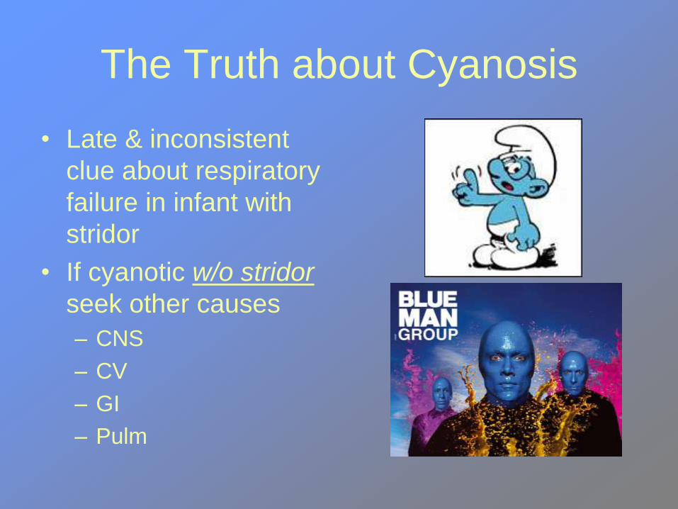
The Truth about Cyanosis
• Late & inconsistent
clue about respiratory
failure in infant with
stridor
• If cyanotic w/o stridor
seek other causes
– CNS
– CV
– GI
– Pulm

Further Assessment
• If child d/n have impending respiratory
failure, a more detailed physical exam
should be performed
– General (weight, growth percentile,
development)
– Nasal cavity, oral cavity and oropharynx more
thoroughly examined
– Flexible fiberoptic laryngoscopy

Endoscopy
• In unusual/difficult cases to determine etiology of stridor.
• Laryngoscope, Hopkins rod-lens telescopes, bronchoscope – Verify all equipment/light
sources work!
• Trach tray in room just in case.
• Good communication btwn endoscopist and anesthesiologist is a must.

Outline
I. Nose &
Nasopharynx
II. Oropharynx or
hypopharynx
III. Supraglottic larynx
IV. Glottic larynx
V. Subglottic larynx
VI. Tracheobronchial
• Congenital
• Infectious
• Traumatic
• Neoplastic
• Vascular
• Iatrogenic
• Toxic/metabolic

Nose
&
Nasopharynx
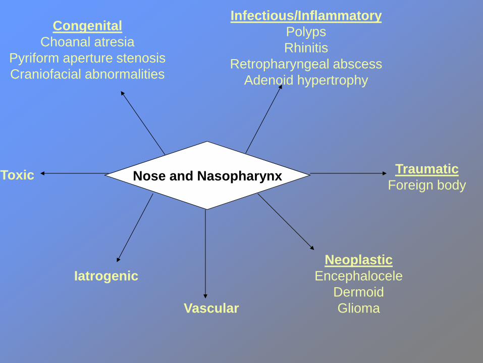
Nose and Nasopharynx
Congenital
Choanal atresia
Pyriform aperture stenosis
Craniofacial abnormalities
Infectious/Inflammatory
Polyps
Rhinitis
Retropharyngeal abscess
Adenoid hypertrophy
Traumatic
Foreign body
Neoplastic
Encephalocele
Dermoid
Glioma Vascular
Iatrogenic
Toxic

Choanal Atresia (CA)
• Epidemiology
– Rare: 1 in 10,000 births
– Females >males
– 50% unilateral, 50% bilateral
• 2 types: membranous or bony
– 29% bony
– 71% mixed bony-membranous (Brown et al, Laryngoscope
1996)
• Pathogenesis controversal

Choanal Atresia (CA)
• Clinical signs & sx
– Respiratory
distress/paradoxical
cyanosis
– Feeding difficulty
– Associated abnormalities
• C- Coloboma
• H- Heart anomaly
• A- Atresia of choana
• R- Retarded growth
• G- Genital hypoplasia
• E- Ear anomalies and/or
deafness
• Clues to diagnosis
– Inability to pass 8 Fr
catheter beyond 3.5 cm
from nasal vestibule
– Flexible scope hits a “brick
wall” during exam
– Mirror under nares fails to
fog on expiration
– Axial CT confirms
diagnosis


• Management
– InitialMcGovern nipple • Oral airway or McGovern nipple
– Surgical • Transpalatal
– Better visualization, high success rate
– Can damage palate growth plate=cross bite deformities
• Transnasal
– Less blood loss, faster procedure
– Increased CSF leak and meningitis risk
• Laser
– CO2, KTP, Holmium:YAG
– Good success with KTP + endoscopic techniques
– Operating microscope with CO2 laser also being employed
Choanal Atresia (CA)
Congenital Nasal Pyriform
Aperture Stenosis (CNPAS) • Pathogenesis
– Premature fusion and
overgrowth of medial
nasal processes
• May result in a central
megaincisor (60% of
cases)
• Could represent a
microform of
holoprosencephaly
– Concomitant
malfunction of
pituitary/adrenal axis
• Clinical picture – Very similar to CA
• Respiratory distress
• Feeding difficulty
• Cyclical cyanosis
– Exam reveals bony obstruction of vestibule
• Inability to pass catheter/scope into nose
• Thin cut CT with emphasis on pyriform aperture is image modality of choice

imaging.consult.com/.../S1933-0332(07)75454-X
Central Incisor

Rev. Bras. Otorrinolaringol. vol.71 no.2 São Paulo Mar./Apr. 2005
Congenital Nasal Pyriform
Aperture Stenosis • Management
– Initial approach is conservative
• McGovern nipple, topical decongestants, corticosteroids
– Surgical approach
• Aperture widened via superior gingivolabial
incision/premaxillary degloving.
• Mucosa preserved, stents left in place 1-4 weeks
• Prognosis
– Mild cases may resolve as the child grows
– Usually excellent long term results with surgery

Oropharynx
&
hypopharyn
x

Oropharynx/hypopharynx
Congenital
Glossoptosis/Macroglossia
Lingual thyroid
Vallecular cyst
Craniofacial abnormality
Infectious/Inflammatory
Retropharyngeal abscess
Tonsil hypertrophy
Traumatic
Foreign body
Neoplastic
Dermoid
Hemangioma
Lymphangioma Vascular
Iatrogenic
Toxic

Retropharyngeal Abscess
• Retropharyngeal space
– Boundaries
• Superior: skull base
• Inferior: As far as T6
• Posterior: prevertebral
fascia
• Anterior:
– Buccopharyngeal fascia
– Pharyngobasilar fascia
• Lateral: carotid sheath
*communicates with
parapharyngeal space!
Retropharyngeal Abscess
• Epidemiology – Most cases occur in
childhood
– 70% of cases in children < 6 yrs old
• Pathophysiology – Suppuration of
retropharyngeal space lymph nodes
• Clinical symptoms – Odynophagia
– Worsening dysphagia
• Physical exam – Asymmetrical,
posteriolateral pharyngeal swelling
– Torticollis
– Fever
– Stridor
– Drooling
• Labs/imaging – CBC w/ diff
– Lateral neck films • Retropharyngeal tissue
– At C2: <7mm
– At C6: <14mm
– CT neck w/ contrast • Distinguish cellulitis v.
phlegmon v. abscess
• Management
– Cellulitis
• Trial of IV antibiotics
– Clindamycin or ampicillin-sulbactam
– Repeat scan in 48hrs if no improvement
– Abscess
• Incision and drainage in OR
Retropharyngeal Abscess

Babl and Pascucci, N Engl J Med 337(7):472 August 14, 1997.
Enlarged prevertebral
soft tissue
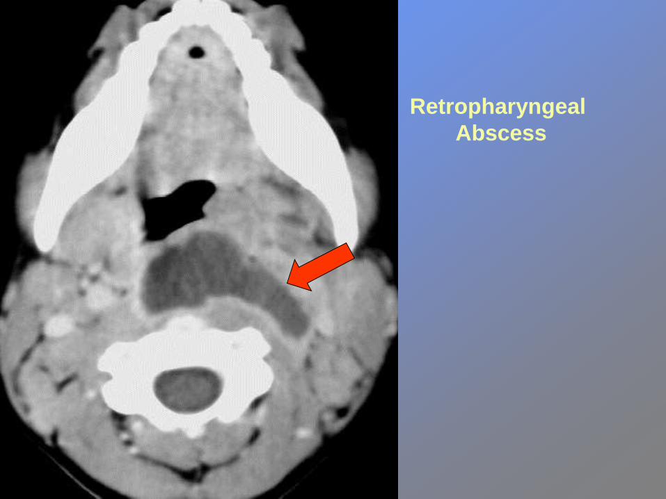
Retropharyngeal
Abscess

Supraglottic Larynx
Congenital
Laryngomalacia
Laryngocele/saccular cyst
Infectious/Inflammatory
Epiglottitis
Angioneurotic edema
Traumatic
Foreign body
Neoplastic
Hemangioma
Lymphangioma
Papilloma Vascular
Iatrogenic
Toxic

Laryngomalacia
• General
– Most common cause of congenital stridor
– May manifest days/weeks after birth
– Symptoms usually resolve by 12-18months
• Pathophysiology
– Stridor caused by prolapse of supraglottic
structures into laryngeal inlet

Laryngomalacia
• Signs/Symptoms
– low, pitched fluttering inspiratory stridor
• Peaks at 6-9months
• Positional variations
• Exacerbated by activity (i.e. feeding, exertion)
– Rarely produces cyanosis
• Cyanosis should prompt suspicion for other
pathology

Laryngomalacia
• Physical exam
– Fiberoptic
laryngoscopy while
child is awake
– Direct
laryngoscopy/broncho-
scopy sometimes
needed to rule out
synchronous lesions

Laryngomalacia
• Management
– Self-limited condition; majority of cases
resolve
– Surgical treatment (~10% of cases)
• Supraglottoplasty
– Indicated for cases with severe stridor, failure to thrive,
apneas, cor pulmonale, pulmonary HTN
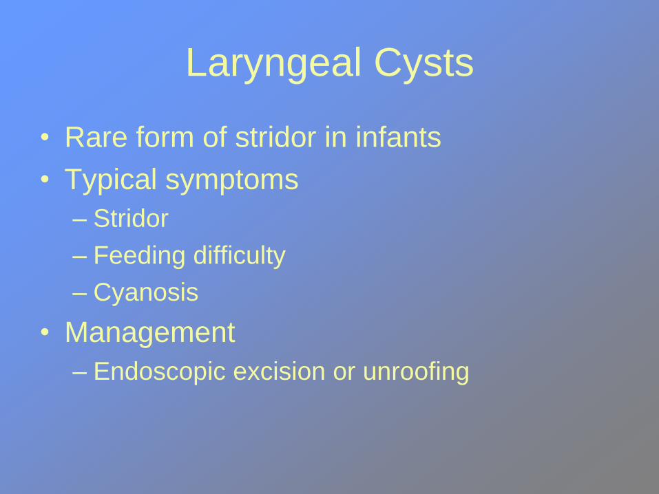
Laryngeal Cysts
• Rare form of stridor in infants
• Typical symptoms
– Stridor
– Feeding difficulty
– Cyanosis
• Management
– Endoscopic excision or unroofing

Laryngeal Cysts
• Ductal Cysts
– Most common type
– Etiology
• Obstruction of mucous
glands
– Location
• Anywhere in larynx but
most commonly in
supraglottis
• Saccular Cysts
– Least common
– Location
• Laryngeal ventricle
• Usually congenital in
infants
• No communication with
laryngeal lumen

A large cyst in the vestibule of the larynx
outgrowing the left glossoepiglottic fold Bielicka et al., New Medicine 3/2004, p. 71-73

Glottic
Larynx

Glottic Larynx
Congenital
Web/atresia
Laryngeal cleft
Stenosis
Vocal cord paralysis
Infectious/Inflammatory
Laryngitis
Traumatic
Hematoma
Fracture
Foreign body
Stenosis
Vocal cord paralysis Neoplastic
Hemangioma
Lymphangioma
Papilloma
Granuloma
Vascular
Iatrogenic
Vocal cord paralysis
Toxic

Congenital Laryngeal Web
• Pathogenesis – Arise from failure of recanalization of larynx in embryo
• Location – Predominantly in the anterior glottis
• Associated findings – Severe webbing assoc. with subglottic stenosis.
– Laryngeal atresia requires trach at birth
– Anterior glottic webs assoc. w/ velocardiofacial syndrome (22q11 deletion) (Oto Head & Neck 2004 130: 415-17)
• Symptoms – Present with abnormal cry, stridor

Congenital Laryngeal Web
• Diagnostic endoscopy
– Required for diagnosis
– Other abnormalities must be ruled out as well
• Treatment
– Simple insicion for small webs
– Laryngofissure with stenting for severe
webbing.
– Endoscopic laser treatment also an option

www.ferdawsfamily.com/.../index.php/t14103.html

Posterior Laryngeal Cleft
• Pathogenesis
– Failure of posterior larynx to fuse (may involve
trachea)
• Symptoms
– Aspiration and hoarseness
– Usually no stridor
• Classification
– Correlates with severity
• Type I-IV
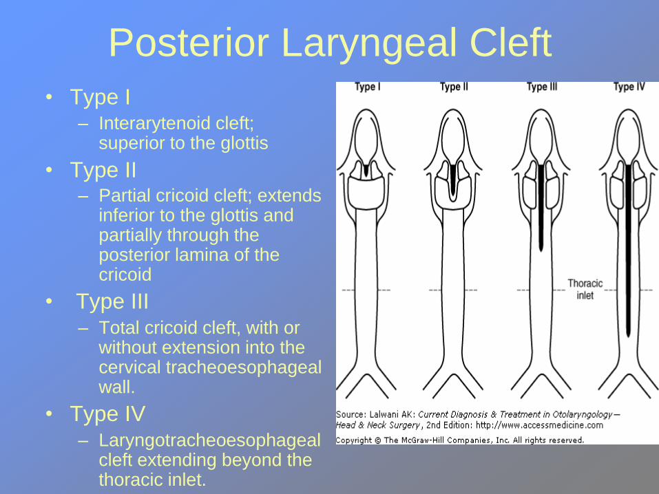
Posterior Laryngeal Cleft
• Type I – Interarytenoid cleft;
superior to the glottis
• Type II – Partial cricoid cleft; extends
inferior to the glottis and partially through the posterior lamina of the cricoid
• Type III – Total cricoid cleft, with or
without extension into the cervical tracheoesophageal wall.
• Type IV – Laryngotracheoesophageal
cleft extending beyond the thoracic inlet.

Type II Cleft

Vocal Cord Paralysis
• General – 10% of congenital
laryngeal lesions
– May be congenital or acquired
– Most often cause is idiopathic
• Etiologies – Traumatic/Iatrogenic
• Obstetric/birth trauma
• Cardiac surgery
• Esophageal surgery
– Other congenital abnormalities
• Cardiac anomalies
• CNS origin
– Chiari malformation
Chiari malformation

Vocal Cord Paralysis
• Unilateral
– Breathy voice/cry
– Mild stridor and/or
dyspnea
– Aspiration
– Treatment
• Speech therapy
• If tracheotomy needed,
decannulation is usually
possible as child/larynx
developes
• Bilateral – Severe stridor
– Aspiration
– Treatment • tracheotomy usually
required
• Serial endoscopies
• Surgery after at least 1 year after trach w/o improvement

Vocal Cord Paralysis
• Evaluation – Can be seen with FOL while pt is awake
– Laryngotracheobronchoscopy must be performed • Must palpate arytenoids
• Exclude synchronous lesions
– MRI brain, brain stem, neck and chest reasonable if cause not obvious (course of vagus)
– FEES/MBS may be utilized in cases of aspiration
• Management – VFP in infants usually resolves in 6-18mos
– Scheduled monitoring is reasonable for first 2 yrs
– Temporary tracheotomy may be necessary

Vocal Cord Paralysis
• Surgical methods
– CO2 transverse partial cordotomy
– Costal cartilage grafting
– Arytenoidopexy w/wo arytenoidectomy
• CO2 laser
• External approach

Recurrent Respiratory
Papillomatosis • General
– Rare, but most common neoplasm of larynx in children
• 4.3/100,000=incidence of newly diagnosed RRP in children<15yo
– Childhood and adult onset • Childhood onset
– Often dx 2-4 yrs old
– boys = girls
– No gender/ethnic difference regarding surgical frequency
– More aggressive
– 19.7 surgeries per child
» 4.4 per year

Recurrent Respiratory
Papillomatosis • Etiology
– HPV types 6 & 11 • Maternal-fetal transmission
• Clinical features – Hallmark triad:
• Progressive hoarseness
• Stridor
• Respiratory distress
– Most often present with dysphonia
– Stridor is usually 2nd symptom to manifest • Inspiratory biphasic
– 1 year = duration of sx prior to diagnosis
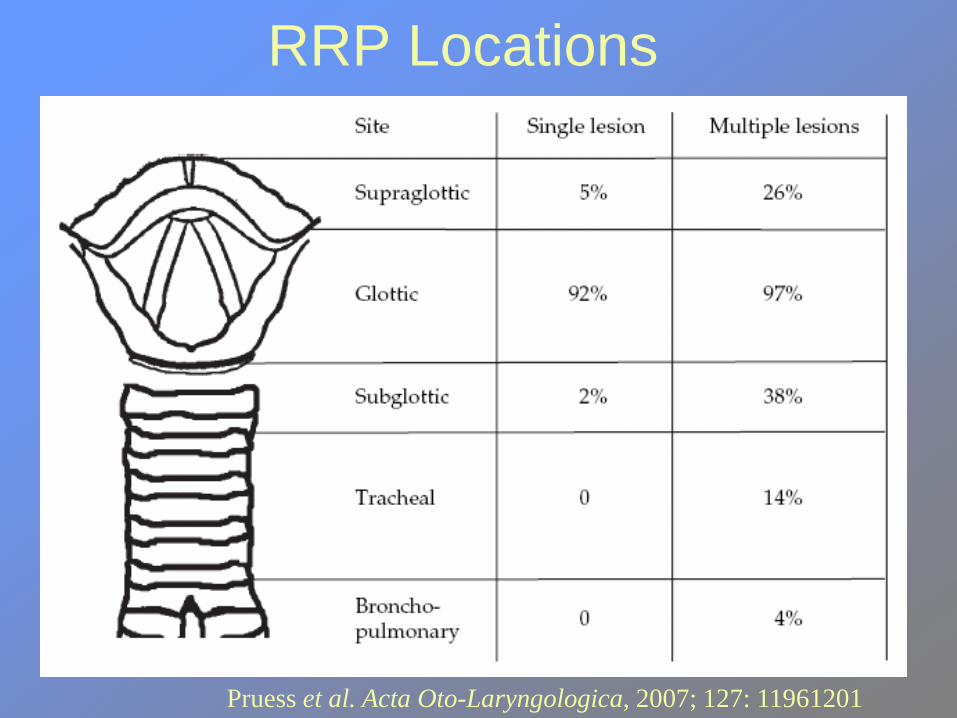
Pruess et al. Acta Oto-Laryngologica, 2007; 127: 11961201
RRP Locations

Papilloma

Recurrent Respiratory
Papillomatosis
• Surgical
– Microlaryngoscopy
with cups forceps
removal
– Microdebrider
– CO2 laser
– Phono-Microsurgical
– KTP/Nd:YAG laser
– Flash scan lasers
• Adjuvant
– α-Interferon
– Indole-3-carbinol
– Photodynamic therapy
– Cidofovir
– Acyclovir
– Ribavirin
– Retinoic acid
– Mumps vaccine
– Methotrexate
– Hsp E7
Treatment Modalities

Subglottic Larynx

Subglottic Larynx
Congenital
Stenosis
Cysts
Infectious/Inflammatory
Croup
Stenosis
Traumatic
Chondritis
Stenosis
Fracture
Foreign body
Neoplastic
Hemangioma
Papilloma
Vascular
Iatrogenic
Toxic

Subglottic Stenosis
• Congenital
– Dx made in absence
of factors causing
acquired stenosis
– Moderate-severe
stenosis=Stridor at
birth.
– Mild stenosis=
Intermittent stridor
• Acquired
– More common than
congenital
– Usually more severe
and difficult to manage
– Endotracheal
intubation
trauma=most
commom cause

Subglottic Stenosis
• Clinical signs/symptoms
– Degree of stenosis dictates symptoms
• Severe stenosis, infant may have stridor at birth
• Mild stenosis may not manifest until URI takes
place.
– In acquired SGS, a clue in neonates may be
failed extubation trial.
• Older children may successfully extubate but
present later with progressive worsening
respiratory distress
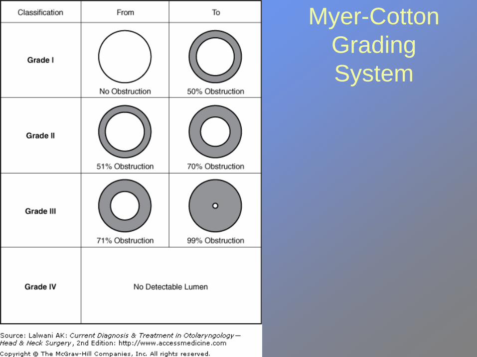
Myer-Cotton
Grading
System


Subglottic Stenosis
• Evaluation
– Stenosis may be visualized on plain films
– Direct laryngoscopy/tracheoscopy needed for confirmation and airway may be staged at this point.
• Prevention
– Use of uncuffed, polyvinylchloride ETT
– Smaller tubes
– Nasotracheal intubation

Subglottic Stenosis
• Treatment options
– Primary goal is to
achieve decannulation
(if tracheostomy
present) or prevent
tracheostomy
– Conservative
• Observation (grades I-
II)
– Temporizing measure
• Tracheostomy
• Definitive Surgical
Options
– Endoscopic methods
• Laser
– Anterior cricoid split
– Laryngotracheal
reconstruction
– Cricotracheal
resection

Subglottic Hemangioma
• General – 1.5% of all congenital laryngeal anomalies
– 2:1 female to male ratio
– Most common neoplasm of infant airway
• Clinical – Usually asymptomatic at birth.
– Biphasic stridor in first 6 months=presenting symptom
– Cutaneous hemangiomas in 50% at time of dx
– Lesion characterized by rapid growth that ceases at 12 months.
• May resolve by 5 yrs

Subglottic Stenosis Intubation
Pressure necrosis on subglottic mucosa
Edema & ulceration
Granulation tissue
Secondary infection & perichondritis
Fibrous tissue deposition
Stenosis!

Subglottic Hemangioma
• Diagnosis
– Biopsy unnecessary due to pathognomonic
appearance
• Compressible, submucosal mass
• Reddish or bluish hue
• Asymmetric
• Posterior left subglottis
– Laryngotracheobronchoscopy is method of
choice

www.chop.edu/ent/images/sub_hemagioma_thmb.gif

Subglottic Hemangioma
• Objectives of
treatment
– Preserve stable airway
while mitigating the
long term sequelae of
the treatment
• Current treatment
modalities
– Tracheotomy
• Temporary
– Steroids
– Laser excision
– Surgical excision
– Interferon

Tracheobronchial
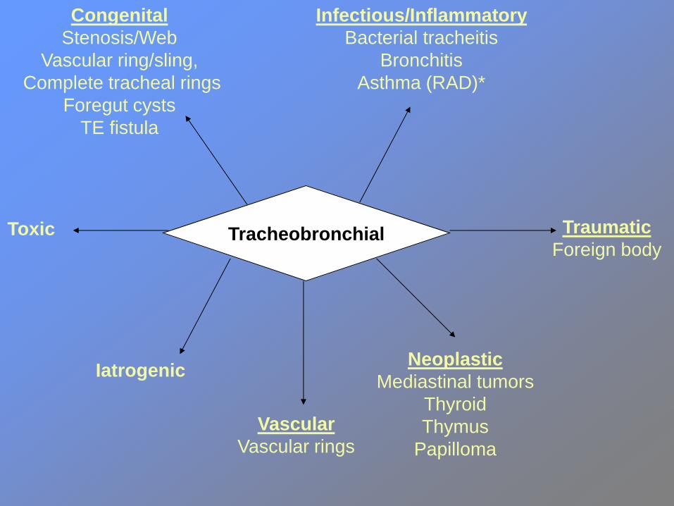
Tracheobronchial
Congenital
Stenosis/Web
Vascular ring/sling,
Complete tracheal rings
Foregut cysts
TE fistula
Infectious/Inflammatory
Bacterial tracheitis
Bronchitis
Asthma (RAD)*
Traumatic
Foreign body
Neoplastic
Mediastinal tumors
Thyroid
Thymus
Papilloma
Vascular
Vascular rings
Iatrogenic
Toxic

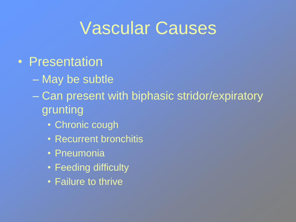
Vascular Causes
• General
– Congenital vascular anomalies = 5% of stridor cases
– Symptoms caused by tracheal/bronchial external
compression
– Main culprits:
• Innominate artery compression
• Vascular ring (double aortic arch)
• Pulmonary artery sling
• Aberrant right subclavian artery
– Most common anomaly in mediastinum

Vascular Causes
• Double aortic arch
– Persistance of fourth
branchial arch and
dorsal aortic root
bilaterally
– Most common
symptomatic vascular
ring
• Pulmonary artery
sling
– Most symptomatic of
noncircumferential
anomalies
– Right mainstem
bronchus affected in
majority of cases
– Associated with
presence of complete
tracheal rings

Double Aortic Arch Pulmonary Artery Sling
Vascular Causes
• Presentation
– May be subtle
– Can present with biphasic stridor/expiratory
grunting
• Chronic cough
• Recurrent bronchitis
• Pneumonia
• Feeding difficulty
• Failure to thrive

Vascular Causes
• Diagnostic imaging
– Plain films of limited value
– Barium esophagram may reveal characteristic
filling defects
– CT w/ contrast or MRI is modality of choice
• Endoscopy
– Allows greater assessment of degree of
compression

www.virtualpediatrichospital.org
Contrast CT scan showing vascular ring

Vascular Causes
• Surgery – Absolute indications:
• Reflex apnea
• 48 hrs of medical mgmt failure
• Prolonged intubation
– Relative indications • Recurrent infections
• Exercise intolerance
• Dysphagia causing failure to thrive
• Concomitant SGS
• Asthma
• CF

Tracheomalacia
• Congenital deformity of tracheal rings
• Expiratory stridor/respiratory distress – Depends on extent of lesion
• Diagnosis – Flexible bronchoscopy with awake patient
• Collapse of anterior tracheal wall against membranous posterior portion
• Treatment rarely needed as most cases are self limited – Some cases may need temporary tracheotomy
– In secondary tracheomalacia, treatment directed at underlying cause.

Tracheomalacia

Foreign Body Aspiration
• General
– Most pts < age 3
– Approx 150 pediatric deaths/year in US
– Choking=40% accidental deaths in children
<1yo
• Pathogenesis
– Kids being kids…

Foreign Body Aspiration
• Most common objects
– Coins most commonly
ingested
– Food most commonly
aspirated
• Nuts
• Seeds
– Fish/chicken bones in
older children

Foreign Body Aspiration
• Esophageal
– Drooling
– Dysphagia
– Emesis
– Chest pain
• Airway
– Cough
– Stridor
– Cyanosis
– Wheezing
– Asymmetric breath
sounds

Foreign Body Aspiration
• Imaging studies – PA and lateral CXR good for radio-opaque objects
• Still useful despite lack of obvious foreign body
• Rigid Endoscopy – Warranted when clinical suspicion is high despite
“innocent/negative” films
– Airway FB should be dealt with at time of presentation if pt is unstable
– It is possible to observe esophageal foreign body in hopes of spontaneous passage (mid/distal esophagus)
• Disc battery requires OR removal promptly

www.medicdirect.co.uk/images

Foreign Body Aspiration
• Prevention
– Consumer Products Safety Act 1979
• Objects must be > 3.17cm diameter and > 5.71 cm in length
– Poorly enforced
– Beware of school supplies

Croup Epiglottis
Onset 2yo 1-5yo
Etiology Parainfluenza
virus type 1
H. Influenza
Gram + bugs
Symptoms/Signs Barking cough,
inspiratory stridor
Odynophagia,
“sniff position”
with mouth open
Diagnostic AP neck
film=“steeple
sign”
Lateral neck
film=“thumb sign”
Treatment Racemic epi,
corticosteroid,
humidified O2
Airway
established in
OR, IV abx

“Steeple Sign” “Thumb Sign”

References
April MM, Ward RF. Choanal atresia repair: The use of powered instrumentation.
Oper Tech Otolaryngol Head Neck Surg 1996; 7:248-251
Bailey, B. Head and Neck Surgery-Otolaryngology. 4th
ed.
Brown OE, Pownell P, Manning SC. Choanal atresia: A new anatomic classification
and clinical management applications. Laryngoscope 1996; 106:97-101.
Coulthard M, Isaacs D: Retropharyngeal abscess. Arch Dis Child 1991; 66:1227
Cummings: Otolaryngology: Head & Neck Surgery, 4th ed.
De Jong AL, Kuppersmith RB, Sulek M, et al. Update on the pediatric airway: vocal
cord paralysis in infants and children. Otolaryngol Clin North Am 2000;33:131–149
Gonzalez Valdepena H, Wald ER, Rose E, et al. Epiglottitis and Haemophilus
influenza immunization. The Pittsburgh experience: a five year review. Pediatrics
1995;96:424–427
Harris J, Robert E, Kallen B. Epidemiology of choanal atresia with specific reference
to CHARGE association. Pediatrics 1997; 99:363-367
Holinger LD. Evaluation of stridor and wheezing. In: Holinger LD, Lusk RP, Green
CG, eds. Pediatric laryngology and bronchoesophagology. Philadelphia: Lippincott-
Raven, 1997:41–48
http://emedicine.medscape.com/article/995267-overview
Josephson GD, Vickery CL, GilesWC, et al. Transnasal endoscopic repair of
congential choanal atresia. Arch Otolaryngol Head Neck Surg 1998; 124:537-540.
Journal of Clinical Anesthesia Volume 12, Issue 5 August 2000, Pages 417-419
Kirse D, Roberson D: Surgical management of retropharyngeal space infections in
children. Laryngoscope 2001; 111:1413
Lusk RP, Kang D, Muntz HR. Auricular cartilage grafts in laryngotracheal
reconstruction. Ann Otol Rhinol Laryngol 1993;102: 247–254
Rahbar R, Nicollas R, Roger G, et al. The biology and management of subglottic
hemangioma: past, present, future. Laryngoscope 2004;114:1880–1891
Stern Y. Partial cricotracheal resection with primary anastomosis in the pediatric age
group. Ann Otol Rhinol Laryngol 1997; 106:891–896

Walker P, Crysdale W: Croup, epiglottitis, retropharyngeal abscess, and bacterial
tracheitis: evolving patterns of occurrence and care. Int Anesth Clin 1992; 30:57
Wetmore R.F., Muntz H.R., McGill T.J. Pediatric Otolaryngology. Thieme Medical
Publishers, Inc., New York 2000 pgs 453-63.
Yates Philip D, Anari Shahram, "Chapter 32. Stridor in Children" (Chapter). Lalwani
AK: CURRENT Diagnosis & Treatment in Otolaryngology—Head & Neck Surgery,
2nd Edition
Brigger MT, Hartnick CJ. Surgery for pediatric vocal cord paralysis:
a meta-analysis. Otolaryngol Head Neck Surg. 2002; 126(4):349.
Goldenberg D, Golz A, Joachims HZ: Retropharyngeal abscess: a clinical review.
J Larngol Otol 1997; 111:546