managing shaping the future of animal health chronic ... · managing shaping the future of animal...
TRANSCRIPT

Issue 02 | APRIL 2016 | 19
ACCREDITED CPD - DERMATOLOGY
Shaping the future of animal healthManaging
Chronic Otitis ExternaDr Martin Briggs B.Sc, B.V.Sc, M.Sc(Med), FRCVS, Registered Specialist In Veterinary Dermatology028 316 2297 [email protected] Reviewed by Dr Heidi Schroeder
Otitis externa (Fig 1), inflammation of the external ear canal, is one of the most commonly diagnosed skin conditions in dogs. There are a number of predispos-ing factors which render individual pets susceptible to chronic and recurrent otitis. Otitis media is inflamma-tion of the middle ear.
Otitis externa is usually easily recognised by the clini-cian, while otitis media may be less apparent. It is im-portant to manage otitis externa thoroughly in order to prevent the development of otitis media.
There is no recognised sex distribution for otitis exter-na. Young animals may be more commonly affected.
There are clear breed predispositions for otitis, which directly reflect the breed predispositions for skin dis-ease (e.g. allergic skin disease in retrievers and terriers, food allergies in German Shepherd dogs ). The most common historical findings are headshaking and aural pruritus.
Predisposing factors, primary causes and perpetuating factors are all implicated in the development of otitis.
Predisposing factorsThese include defective anatomical conformation, excessive moisture, irritant topical products, and sys-temic disease. Conformation defects in dogs include
April 2016 Vet360 for Madaleen Review.indd 19 2016/03/24 2:24 PM

vet360Issue 02 | APRIL 2016 | 20
ACCREDITED CPD - DERMATOLOGY
Figure 1. Typical appearance of otitis externa
Figure 2a. Schnauzers and Poodles typically have a lot of hair lining the ear canal.
a hirsute (Fig 2a,b) or stenotic meatus, and pendulous pinnae. Excessive moisture occurs in dogs that swim. Systemic disease such as immunosuppression, renal disease, hepatic disease, and in cats, FeLV and FIV in-fection, may predispose to otitis.
Primary causes These include parasites, foreign material, hypersensi-tivity, neoplasia, polyps, keratinization disorders, and endocrine disorders.
Hypersensitivity is a very common underlying cause of otits externa, even if only one ear is affected. Animals such as golden retreivers and German Shepherd dogs have the most perfect anatomy - wide open canals, minimal hair in the canal and in GSDs' upright ears - yet they are predisposed to otitis. It’s because they are predisposed to allergic disease: atopy or food allergic dermatitis. Often just managing this underlying cause will cause resolution of the otitis. Without manag-ing the allergic disease the otitis will be recurrent and chronic changes will develop in the ear canals which will perpetuate the problem. Parasites include mites (Otodectes, Demodex, Sarcoptes), ticks (Otobius) and biting flies (Stomoxys calcitrans). Otodectes cynotis (Fig 3) the ear mite of dogs and cats, spend their entire 3-week life-cycle deep in the ear canal of the host. Otoscopic examination reveals these long-legged mites. The mites produce a marked reaction which is probably allergic in origin. Dogs shake their heads, which may lead to the formation of othaematomas. The tympanic membrane may perforate, leading to otitis media and nervous symptoms.
A roll smear of ear canal debris often reveals path-ogenic organisms. Demodex infestation may in-tra- or peri-aural. Acaricides (e.g. benzyl benzoate and thiabendazole) can be inserted into the ear ca-nal. Spot-ons containing acaricides (e.g. moxidectin, milbemycin, eprinomectin or selamectin) are advised concurrently, since the mite will often be found in the peri-auricular regions. Otobius megnini, the spinose ear tick, may occasionally invade cats’ ears. Manage-
ment is as for O. cynotis.
Foreign material may be grass awns, grass seeds or ticks as well as certain irritant topical agents. Atopy, cutaneous adverse food reactions, and contact hy-persensitivities frequently play a mayor role.
Sensitivity to otic preparations may occur, although this is rare. Keratinization disorders include sebor-rhoea and endocrine disorders causing otitis include hypothyroidism, hyperadrenocorticism and sex hor-mone imbalance. Auditory polyps occur in both the canine and the feline, although they are extremely rare in dogs.
There is increased risk for tumours in pets with a his-tory of chronic otitis. Benign or malignant tumours can develop in the external ear canal of dogs and cats, and arise from the apocrine or ceruminous glands that line the ear canal. The most commonly encoun-tered tumour is the ceruminous gland adenocarcino-ma and is more commonly seen than adenomas in both dogs and cats.
Figure 2b. Otoscopic view of an ear canal with a lot of hair showing how plugs form within the ear canal.
April 2016 Vet360 for Madaleen Review.indd 20 2016/03/24 2:24 PM

Issue 02 | APRIL 2016 | 21
ACCREDITED CPD - DERMATOLOGY
Perpetuating factors Bacteria, yeasts, otitis media, swimming, sensitivity to ceruminolytics, and progressive pathological changes such as narrowing of the ear canal due to chronic changes in the epidermis.
membrane visualisation. In some cases, where there is severe inflammation, swelling and pain it may be ap-propriate to treat with glucocorticoid anti-inflammato-ries for a few days prior to performing and otoscopic examination, even if a GA will be given. This allows the swelling to decrease facilitating visulasation of struc-tures within the ear canal The epidermis is also less friable and bleeds and oozes less serum.
Radiographs are normal in many otitis media cases. CT or MRI, if available, should be performed for cases of severe, chronic otitis. Radiographic abnormalities include calcification of the walls of the ear canal, fluid opacity with in the bulla and thickening and sclerosis of the bulla wall.
Roll smears should be prepared using a cotton wool bud to retrieve debris and this applied to a microscope slide. Low power examination (parasites) and high power examination (stained, for pathogens) is neces-sary.
Selecting appropriate topical treatmentAntimicrobials should be narrow spectrum products to minimise antimicrobial resistance. Whereas cul-ture is necessary for specific identification of causative organisms, the key to determining successful antimi-crobial therapy is ear cytology. Repeated cytology is necessary to evaluate the response to therapy. Owner compliance requires careful instruction. Pets should be suitably restrained so that insertion of medication is successful and reaches the target organisms.
Accurate quantities of medication for insertion may be provided by the manufacturer, or correct amounts can be inserted using a syringe. This is especially rel-evant for ceruminolytics. Separate syringes for each ear prevent cross-contamination.
Since purulent material decreases the efficacy of anti-microbial therapy, ceruminolyitic therapy is an essen-tial part of successful management; purulent exudate is hyperosmolar, and results in a hypoxic, acidic envi-ronment within the ear canal.
Prophylactic use of ceruminolytics is indicated in chronic and recurring otitis. Ceruminolytics have sur-factant and detergent action and help soften, emulsify and dissolve cerumen and debris.
Dioctyl sodium succinate and triethanolamine poly-peptide oleate condensate are potent ceruminolytics. In the presence of a ruptured tympanic membrane sa-line solution only should be used. Irrigation with a bulb syringe under sedation or anaesthesia may be neces-sary prior to topical antibacterial therapy. Although irritation of the ear canal can occur, the author has found that diluting ceruminolytics with water can re-duce irritation to the ear canal.
Astringents (drying agents) assist in preventing mac-
Figure 3. Otodectes cynotis on low power magnification
Recognition of the predisposing, primary and perpetuating causes
allows the formulation of a regime for the management of chronic otitis
externa, tailored to the individual pet.
Management The first step in the management of chronic otitis is to determine the severity of pain. This can be done by gentle palpation or petting of the animal. If the ear is painful or the degree of discomfort is high, the animal should be sedated before performing any further di-agnostic testing. The second step is gentle palpation to determine the presence of swelling, pruritus, fibro-sis, or calcification since these findings will determine whether imaging is necessary.
The outside of the ear should be examined, noting erythema, oedema, crusts, scales, ulceration, licheni-fication, hyperpigmentation, or the presence of exu-date. The pinnae and peri-auricular regions should be examined for evidence of self-trauma and demodico-sis (Fig 4), erythema, and primary and secondary skin lesions. Pinnal deformities, hyperplastic tissue in the ear canal, and headshaking suggest a chronic condi-tion.
If the otitis is unilateral, the unaffected ear should be examined first to prevent iatrogenic contamination of the unaffected ear with organisms (e.g. Pseudomonas aeruginosa or Proteus mirabilis) that may be present in the diseased ear. The clinically unaffected ear may, in fact, be diseased, meaning that the differential diag-nosis list should also include causes of bilateral otitis.Otoscopic examination often requires sedation.
Hyperplasia of the ear canal may prevent tympanic
April 2016 Vet360 for Madaleen Review.indd 21 2016/03/24 2:24 PM

vet360Issue 02 | APRIL 2016 | 22
ACCREDITED CPD - DERMATOLOGY
eration of the ear canal, and include isopropyl alcohol and mild acids such as benzoic, acetic, boric, salicylic, malic and lactic acids. If the ear canal is ulcerated avoid the use of products containing alcohol or acid. Acetic, malic, boric, benzoic, salicylic and lactic acid also have antibacterial action, however a lower pH in-activates aminoglycosides and fluoroquinolones.
Fungal otitisMalassezia spp are a normal commensal inhabitant of the ear. Increases in numbers is a secondary changetreatment off the underlying cause will normally re-duce numbers to normal. In true infections Malasse-zia pachydermatitis is the species routinely involved and this usually responds to azole antifungals. Azoles include clotrimazole and miconazole. However, re-cently developed triazole antifungals such as vari-conazole (Vfend®) and posaconazole may be more effective. Candida, although a common cause of fun-gal disease in humans, is rarely encountered in com-panion animal otitis. In these cases, polyene mac-rolides, such as nystatin is advised
Otomax® contains clotrimazole, gentamycin and betamethasone, and Surolan® contains miconazole, polymixin and prednisolone. Fluoroquinolones are broadspectrum and Posatex® is a new product not yet available here which contains posaconazole and orbifloxacin. Panalog® contains neomycin, triamci-nolone and nystatin. Caution is necessary, since oto-toxicity can occur in the event of a ruptured tympanic. Chlorhexidine, polymixin, and aminoglycosides have been implicated
Bacterial otitisCoccoid organisms routinely incriminated in bacterial otitis externa include Staphylococcus, Streptococcus and Enterococcus. Rods (pseudomonads) are often encountered in chronic and recurrent otitis externa. Where Gram negative rods are found, culture and sensitivity should be performed. Generally, amino-
glycosides such as gentamycin and fluoroquinolones such as orbifloxacin and marbofloxacin are effective.
However, the selected antibiotic should be continued for a minimum of 6 weeks. TrizEDTA (eromethamine and disodium EDTA dehydrate) is an aqueous prepa-ration with efficacy against Pseudomonas spp. as is Flamazine ( see Vet 360 May 2015 page 32 for reci-pes or homemade solution) \(vet360/vetlink/publica-tions).
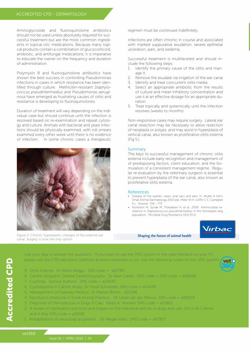
Glucocorticoids assist in reducing the inflammation and subsequent susceptibility to infection as well as secondary hyperplasia and proliferative changes (Fig 5). Prednisolone can be administered orally at 1 to 4 mg/kg once daily.
Approach to managing chronic otitisOwners should be informed of correct ear cleaning procedures. Frequency of cleaning usually decreas-es over time to once or twice weekly as a preventive maintenance procedure. Owners should be warned to keep the pet as dry as possible, and avoid swim-ming. Dogs with confirmation defects such as hirsute or pendulous ears may need to have regular clipping of the pinnae.
The ear canals should be kept dry and well ventilated. Topical astringents prevent water from entering the ear canals in dogs that swim frequently, minimizing maceration of the ear canal. Chronic maceration impairs the barrier function of the skin, which predis-poses to opportunistic infection. Preventative otic as-tringents may decrease the frequency of bacterial or fungal infections in moist ear canals.
Clipping hair from the inside of the pinna and around the external auditory meatus, and plucking it from hir-sute ear canals, improves ventilation and decreases humidity in the ears. Hair, however should not rou-tinely be removed from the ear canal if it is not caus-ing a problem, because doing so can induce an acute inflammatory reaction.
Owners will have to be instructed as to the correct procedure for instilling ceruminolytics and astringents. Since pets predisposed to chronic otitis may require life-long ceruminolytic application, minimising dis-comfort to the pet during this procedure is necessary. Warming the solution to body temperature in a water bath also minimises the discomfort felt by the patient and controlled "dribbling" administration rather than a rapid squirt may be better.
Control of inflammation minimises discomfort from ceruminolytics, and corticosteroids may be required in the early stages. Non-steroidal anti-inflammatories are not as effective otic analgesics as opiods and should not be used concurrently with corticosteroids. NSAIDS may have to be supplied to the owner for ad-ministration prior to instilling the ceruminolytic. Oc-
Figure 4. In many cases of otitis externa the skin of the pinna and face can also be severley affected and need specific treat-ment
April 2016 Vet360 for Madaleen Review.indd 22 2016/03/24 2:24 PM

Issue 02 | APRIL 2016 | 23
ACCREDITED CPD - DERMATOLOGY
casionally sedatives may be necessary until the pet has become accustomed to the procedure.
The pet should be suitably restrained, as this allows for speedy instillation of otic products. For dogs, re-straint may include a muzzle and cats may need to be wrapped in a towel. The author advises that dogs and cats should be accustomed to a collar, and dogs should have the lead attached while the ears are treat-ed. Dogs should be trained to sit during application of otic products.
A treat given afterwards encourages compliance. Pets may want to irritate (rub or scratch at) the ear and if a collar and lead has been applied they can be taken for a walk immediately afterwards as a distraction and reward for compliance.
Rinsing products should be inserted by holding the container vertical and upside down. The owner holds the ear pinnae with one hand, and the solution is in-stilled along the grooves of the inner pinnal surface into the canal. This avoids the container tip contact-ing an inflamed ear.
The owner maintains their hold on the ear pinna to prevent the pet shaking the product out of the ca-nal, and gently massages the base of the ear canal to promote the action. Cotton wool or tissue paper can be used to wipe away excess solution since cats, especially, are averse to topically applied liquids. The owner may not be 100% successful at first, but should be encouraged to persist on a regular basis.
Discuss with the owner the suspected cause of the otitis, emphasising that treatment should be long term or even lifelong. All primary and secondary causes and predisposing factors should be identified and managed. Pain or pruritus should be controlled. Otitis externa is one of the few dermatologic condi-tions in which glucocorticoids concurrently with an-timicrobial use are beneficial. Glucocorticoids such as prednisolone and triamcinolone decrease swelling of the ear canal - a key to successful treatment.
Duration depends on the severity. Ear hygiene is im-portant; in particular, the hair in the peri-auricular area should be clipped as well as hair on the inner surface of the pinnae. This facilitates cleaning and medica-tion. Plucking hair from the canal is controversial but may be necessary, under anaesthesia, to adequately resolve the infection.
The first ear cleaning should be performed in the clin-ic, and owners should refrain from topical administra-tion until rechecked in 5–7 days since they may be too aggressive, causing further damage. Owners can administer systemic drugs and then begin to clean the ears after the first recheck, provided the otitis is resolv-ing. Topical medications are inactivated by exudates,
and excessive cerumen may prevent medications from reaching the epithelium.
Thick, dry, or waxy material requires a ceruminolytic solution such as carbamide peroxide, dioctyl sodium sulfosuccinate (DSS) or combinations of weak acids. If the tympanic membrane is ruptured, detergents and DSS are contraindicated; milder cleansers (eg, saline, saline plus povidone iodine, the Triz EDTA formulation) should be used to flush the ear.
Effective treatment may require both topical and sys-temic antimicrobial therapy, along with pain medi-cations and glucocorticoids. In the management of acute bacterial otitis, the administration of corticoster-oids in combination with antibacterial agents causes as reduction in exudation, pain, swelling, and glandu-lar secretions. The least potent glucocorticoid at the lowest effective dose should be used.
Most topical products contain a combination of an an-tibiotic or antifungal and glucocorticoids. Most dogs require the instillation of at least one ml twice daily for effective therapy. Products with an aqueous base are preferable, however, vinegar dilutions and propyl-ene glycol should be used with caution since swelling of the lining of the ear canal and increased glandular secretions may result. Substances that do not usu-ally irritate the normal ear may result in irritation and inflammation.
Powders, such as those used after plucking hair from the canal, can form irritating concretions within the ear canal and should be avoided. It is important to demonstrate to the owner into which “hole” the oint-ment must be placed - holding the pinna upright and placing the tip into (not necessarily deeply) the most outer opening - which is the meatus of the ear canal. It is not necessary to fill the canal with ointment - a drop or two with a good ear massage, if possible, is required.
Systemic antibiotics should be used when neutrophils or rod-type bacteria are found on cytology, in cases of therapeutic failure with topical antimicrobial agents, in chronic recurring ear infections, and in all cases of oti-tis media. The most common cause of recurrent oti-tis externa is undiagnosed otitis media. Failure to use systemic antimicrobial therapy is an important cause of chronic ear disease in dogs.
Yeast infections can be treated with oral ketoconazole 5 mg/kg/day, per os, for 15–30 days. In cats, itracona-zole at 2–3 mg/kg/day for 15–30 days or one week on/one week off, is recommended.
The best treatment of chronic otitis is prevention. In addition to identifying the cause of acute otitis, topical and/or systemic medications should be chosen based on cytology or culture. They should have a narrow spectrum and be specific for the current condition.
April 2016 Vet360 for Madaleen Review.indd 23 2016/03/24 2:24 PM
vet360Issue 02 | APRIL 2016 | 24
ACCREDITED CPD - DERMATOLOGY
Aminoglycoside and fluoroquinolone antibiotics should not be used unless absolutely required for suc-cessful treatment but are the most common ingredi-ents in topical otic medications. Because many topi-cal products contain a combination of glucocorticoid, antibiotic, and antifungal medications, it is imperative to educate the owner on the frequency and duration of administration.
Polymyxin B and fluoroquinolone antibiotics have shown the best success in controlling Pseudomonas infections in cases in which resistance has been iden-tified through culture. Methicillin-resistant Staphylo-coccus pseudintermedius and Pseudomonas aerugi-nosa have emerged as frustrating causes of otitis and resistance is developing to fluoroquinolones.
Duration of treatment will vary depending on the indi-vidual case but should continue until the infection is resolved based on re-examination and repeat cytolo-gy and culture. Animals with bacterial and yeast infec-tions should be physically examined, with roll smears examined every other week until there is no evidence of infection. In some chronic cases a therapeutic
regimen must be continued indefinitely.
Infections are often chronic in course and associated with marked suppurative exudation, severe epithelial ulceration, pain, and oedema.
Successful treatment is multifaceted and should in-clude the following steps:1. Identify the primary cause of the otitis and man-
age it. 2. Remove the exudate via irrigation of the ear canal 3. Identify and treat concurrent otitis media. 4. Select an appropriate antibiotic from the results
of culture and mean inhibitory concentration and use it at an effective dosage for an appropriate du-ration.
5. Treat topically and systemically until the infection resolves (weeks to months).
Non-responsive cases may require surgery. Lateral ear canal resection may be necessary to allow resection of neoplasia or polyps, and may assist in hyperplasia of vertical canal, also known as proliferative otitis externa (Fig 5.).
SummaryThe keys to successful management of chronic otitis externa include early recognition and management of of predisposing factors, client education, and the for-mulation of a consistent management regime. Regu-lar re-evaluation by the veterinary surgeon is essential to prevent hyperplasia of the ear canal, also known as proliferative otitis externa.
References• Disease of the eyelids, claws, anal sacs and ears. In: Muller & Kirk’s
Small Animal Dermatology 2013 eds. Miller W H, Griffin C E, Campbell K L. Elsevier: 724 – 773
• Norström M, Sunde M, Tharaldsen H, et al 2009 Antimicrobial re-sistance in Staphylococcus pseudintermedius in the Norwegian dog population. Microbial Drug Resistance 15(1):55-9.
Shaping the future of animal healthFigure 5. Chronic hyperplastic changes of the external ear canal. Surgery is now the only option
ANSWER the questions on the Vet360 App. Available from the Itunes/Play store! Use your App to answer the questions. If you want to use the SMS system or the web interface on your PC,
please visit the CPD electronic platform at www.onlinevets.co.za Use the following codes for the SMS system:
9. Otitis Externa. Dr Martin Briggs. SMS code = a22785
8. Canine Idiopathic Dilated Cardiomyopathy. Dr Alain Carter. SMS code = SMS code = a68068
7. Cushings. Various Authors. SMS code = a20643
6. Cyclosporine in Canine Atopy. Dr Heidi Schroeder. SMS code = a24438
5. Management of Diabetes Mellitus Dr Marlies Bohm. a55046
4. Transfusion Medicine in Small Animal Practice. Dr Liesel van der Merwe SMS code = a90559
3. Diagnosis of Demodicosis in Dogs & Cats. Karen A. Moriello SMS code = a22801
2. A review of sterilisation practices and impact on the individual animal, in dogs and cats. Drs K de Cramer
and K May SMS code = a26581
1. Rehabilitation of neurological patients. Dr Megan Kelly, SMS code = a97907Acc
red
ited
CP
D
April 2016 Vet360 for Madaleen Review.indd 24 2016/03/24 2:24 PM

Issue 02 | APRIL 2016 | 25
ACCREDITED CPD - DERMATOLOGY
JOURNAL SCANCPD Questions AC/1473/16
1. Which one of the factors listed below is NOT a predisposing factor for otitis?
a. Defective anatomical conformation such as stenotic meatus and pendulous pinnae.
b. Excessive moisture in dogs which swim. c. Ear parasites. d. Irritant topical products. e. Systemic disease such as immunosuppression.
2. Which one of the factors listed below in NOT a primary cause of otitis?
a. Foreign material. b. Hypersensitivity. c. Neoplasia. d. Keratinization disorders. e. Excessive moisture.
3. Which one of the following statements is FALSE when applied to otitis caused by cutaneous hyper-sensitivity reactions?
a. It is a very common underlying cause. b. Both ears are generally affected. c. German Shepherds and Golden Retrievers
are predisposed. d. Dermatitis can be caused by food allergies or
atopy. e. Recurrent otitis will occur if the allergic
disease is not managed.
4. Which one of the following statements regarding bacteria seen on an ear smear is INCORRECT?
a. Best sampled using a cottonbud and rolled onto the slide.
b. Best seen under high power magnification on a slide stained with diffkwik.
c. Are secondary and not the primary cause of the otitis.
d. Are primary pathogens and need to be managed as such.
e. Are commonly seen together with Malassezia spp in most otitis cases.
5. Which one of the following statements regarding bacterial infection of the ear canal is INCORRECT?
a. Narrow spectrum antibiotics reduce the development of resistance.
b. Antibiogram is the key to successful antibiotic therapy. c. Antibiograms are required to evaluate
response to therapy. d. Ear cytology is required to evaluate response
to therapy. e. Purulent material decreases the efficacy of
antibiotics.
6. Which one of the following statements regarding bacterial otitis is INCORRECT?
a. Coccoid organisms often seen with bacterial otitis externa include Staphylococcus, Streptococcus and Enterococcus.
Please read both articles to answer MCQ: (1) Managing of Chronic Otitis Externa & (2) Total Ear Canal Ablation and a Lateral Bulla Osteotomy
b. Rods seen on an ear smear are generally Pseudomonas spp.
c. Where Gram negative rods are found, culture and sensitivity should be performed.
d. Glucocorticoids are contra-indicated when rod shaped bacteria are seen on a smear.
e. Generally, aminoglycosides such as gentamycin and fluoroquinolones such as orbifloxacin and marbofloxacin are effective against Pseudomonas spp.
7. Which one of the factors listed below is NOT an indications for performing a total ear canal abla-tion and bulla osteotomy?
a. Cases that have failed to respond to long term medical management.
b. Cases with severe calcification of the cartilage or sclerosis or fluid accumulation in the
tympanic bulla. c. Cases with severe soft tissue hyperplasia from
chronic inflammation extending past the vertical canal. d. Cases with severe trauma to the ear canal. e. Cases with neoplasia of the lateral wall of the
vertical ear canal.
8. An abnormal neurological examination may indi-cate severe progression of the otitis. Which one of the neurological deficits listed is NOT typical with otitis externa?
a. Ipsilateral facial nerve paresis or paralysis. b. Ipsilateral peripheral vestibular disease. c. Ipsilateral trigeminal disease. d. Ipsilateral horners syndrome. e. Ipsilateral deficits of the sympathetic supply
to the face.
9. Which one of the intra and post-operative com-plications listed below is the most common and serious?
a. Intra-operative haemorrhage is the most common complication
b. permanent facial nerve damage is 31% c. Draining tracts from remaining secretory tissue
in the bulla occur in 5-10% of cases d. There can be acute wound complications such
as wound breakdown and soft tissue swelling. These can often be controlled with good nursing and wound management
e. Necrosis of the pinna is seen from damage to the blood vessels that supply the pinna
10. Which one of the neurological deficits listed below
occurs IN peripheral vestibular syndrome? a. Vertical nystagmus b. Ipsilateral proprioceptive and motor deficits c. Horizontal and vertical nystagmus d. Disorientation and loss of balance e. Ipsilateral trigeminal nerve deficits (CN V) disease
April 2016 Vet360 for Madaleen Review.indd 25 2016/03/24 2:24 PM

vet360Issue 02 | APRIL 2016 | 26
Article sponsored by Petcam®ACCREDITED CPD - SURGERY
Total Ear Canal Ablation and
Lateral Bulla Osteotomy
Dr Ross Elliot BVSC MMedVet (Surg) Bryanston Veterinary Hospital, 011 706 6023
The surgical procedure for a TECA-BO entails removal of both the vertical and horizontal ear canal with all the secretory epithelial lining of the middle ear. This surgery has the potential for serious complications and should not be performed unless the surgeon is familiar with the anatomy of the ear and associated structures. A total ear canal ablation should never be performed without a lateral bulla osteotomy. If the bulla osteotomy is not performed all the secretory lining of the middle ear is left behind and this will increase the potential for complica-tions by as much as 82%.
There are very few cases in dogs where a bulla osteoto-my is performed without a total ear canal ablation. These
Total ear canal ablation and a lateral bulla osteotomy (TECA-BO) are two separate procedures which are usually combined as a surgical treatment for otitis externa and media.
are otitis media in the presence of an intact tympanic membrane. Neoplasia of the middle ear or polyps of the middle ear in cats that have recurred after previous re-moval, generally a ventral bulla osteotomy is performed in these cases as the ear canal cannot be removed and the ventral bulla osteotomy is easier to perform.
A lateral ear canal resection or Zepps procedure has lit-tle place in the treatment of otitis externa and media. A study on its effectiveness showed in all dogs it has a 45% success rate in the management of otitis externa and media. Better outcomes have been reported in Sharpei’s and Spaniels have a very poor outcome with a just a lat-eral ear canal resection. The only indication for it is neo-
BRYANSTON VETERINARY HOSPITAL
Photo courtesy: Julia van Draanen, Valley Farm Animal Hospital
April 2016 Vet360 for Madaleen Review.indd 26 2016/03/24 2:24 PM

Issue 02 | APRIL 2016 | 27
ACCREDITED CPD - SURGERY
plasia of the lateral wall of the vertical ear canal.
The indications for a TECA-BO is severe end stage otitis externa and media. All of these cases usually have severe narrowing of the ear canal, which makes topical medical treatment unsuccessful
These are cases that have failed to respond to appropriate long term medical management:
• Have severe calcification of the cartilage of the ear ca-nal.
• Have visible sclerosis or fluid accumulation in the tym-panic bulla.
• Have severe soft tissue hyperplasia from chronic in-flammation extending past the vertical canal.
• Have neoplasia of the medial vertical and horizontal ear canal.
Severe trauma to the ear canal, or congenital malforma-tions can often require a TECA BO when the integrity of the ear canal is severely damaged.
A high percentage of dogs with severe otitis have asso-ciated allergic skin disease. This skin disease should be managed medically prior to resorting to surgery. The otitis will often benefit greatly from management of the allergic skin disease. However if there is marked mineralisation of the ear canal, or secondary changes in the bulla then the ear disease will eventually require surgery.
The advantages of surgery include:• No need for continued medical treatment with suc-
cessful surgery (can often negatively affect the pet-owner relationship)
• Relief of pain and improved quality of life for the patient• Prevents further secondary change and damage to as-
sociated structures
The disadvantages of surgery include:• Surgical complications such as facial nerve paralysis, vestibular syndrome and Horner’s syndrome• Financial cost of surgery• Surgical complications
Complete ear work up for surgeryA complete physical examination is part of any work up to try ascertain if there are any concurrent disease processes that may complicate the healing of the patient or the abil-ity to tolerate the anaesthesia and surgery. A full neuro-logical exam should always be performed to detect any pre-existing facial nerve or peripheral vestibular involve-ment. The owners should be informed of these results as they may be present in a significant number of dogs. An abnormal neurological examination may indicate severe progression of the otitis or other differential diagnoses
Figure 1. A standard ventro-dorsal radiograph. In this patient the right ear canal shows soft tissues opacities (white arrow) which were diagnosed as severley inflammed tissue on biopsy. The left ear canal (blue arrow) is normal
Figure 2. Rostro-caudal skull radiograph. Arrow indicates thickening of right tympanic bulla wall suggestive of otitis media. Arrowhead indicates normal left tympanic bulla
Figure 3. The patient in position prior to surgery
April 2016 Vet360 for Madaleen Review.indd 27 2016/03/24 2:24 PM

vet360Issue 02 | APRIL 2016 | 28
Article sponsored by Petcam®ACCREDITED CPD - SURGERY
such as granulomatous menigioencephalitis or central vestibular disease. Detecting facial nerve damage prior to surgery prevents its being considered a surgical com-plication.
Owners will often be concerned about the ability of the animal to hear after the procedure especially when it has to be performed bilaterally. This can be assessed with a BAER test pre-operatively. However in reported owner perceptions it has been found that there is not a notice-able loss of hearing after the procedure. The reality is that the middle ear is already so damaged in the appro-priate surgical candidate for the surgery, that the ability to hear has already been damaged and the patients have already adapted.
Otoscopy and culture are essential in the work up. This is best done under sedation as the ears are often severely inflamed and painful. The patients should be admitted and once sedated, otoscopy can be performed and a culture taken from the middle ear, via a myringotomy if the tympanic membrane is still intact. The tympanic is often found to be perforated and otitis media is as-sumed. A culture can be taken from the ear canal.
The next step is to perform radiographs or CT/MRI. Plain film radiographs are most commonly used to evaluate the outer and middle ear, but they are not considered highly sensitive. They are however highly specific. CT shows a higher sensitivity especially when combined with plain film radiographs. MRI is once again highly sen-sitive and specific. In practice the first step is to perform a standard ventro-dorsal radiograph (Fig 1) to assess the ear canal and check for calcification of the ear canal. A frontal open mouth view (Fig 1) is then used to assess the tympanic bulla for fluid opacity and periosteal reaction, which would indicate chronic otitis media in most cases.
In cases where there is calcification of the ear canal, peri-osteal reaction or fluid in the bulla then a TECA BO is in-dicated. These cases are unlikely to respond to medical management as this can be considered an “end stage” ear.
Surgical procedureOtitis externa and media is usually bilateral and cases of-ten require surgery on both ears. It is possible to perform both surgeries at the same time but this has little advan-tage to the patient. It prolongs the anaesthetic time and doubles the pain experienced by the patient. Generally the disease process has been going on for many months to years and to wait 3 weeks between surgery is of little consequence to the patient.
The surgical site should be widely clipped and aseptically prepared. The ear canal should be thoroughly lavaged with 0.5% hibitane and water prior to surgery.
The patient is placed in lateral recumbency with a towel placed under the head to elevate the head (Fig 3). The entire ear canal is then sharply dissected out from the surrounding tissue, staying as close as possible to the cartilage of the ear canal without penetrating the carti-lage (Fig 4).
A recent report of a subtotal ear canal surgical method has been reported, which will maintain the ear carriage but this can leave a small amount of diseased tissue just distal to the pinna which may lead to recurrent dermato-logical disease.
The facial nerve courses caudo-ventrally to the horizon-tal canal and should be identified and gently retracted (Fig 5). Where the bulla connects with the horizontal ear canal is easily digitally palpated. Being careful not to transect the facial nerve the horizontal canal is sharply dissected off the bulla (Fig 6 and 7).
The external acoustic meatus can now be seen with a small rim of remaining cartilage of the horizontal ear ca-nal. This cartilage is gently removed with a small peri-osteal elevator. The meatus is then enlarged in a ventro-lateral direction with a sharp bone Rongeur being careful to avoid the branches of the internal carotid ventral to the bulla and the venous sinus caudal to the bulla. The secretory lining of the middle ear should now be visible.
April 2016 Vet360 for Madaleen Review.indd 28 2016/03/24 2:24 PM

Issue 02 | APRIL 2016 | 29
ACCREDITED CPD - SURGERY
A small curette is gently used to try peel off the membrane from the underlying bone. The dorso-medial section of the middle ear should be avoided with the curette as this will damage the structures of the internal ear. The membrane is usually a grey black colour, once removed there should be a shiny white appearance to the bone of the middle ear (Fig 7).
The surgical site should be irrigated with saline and the subcutaneous tissues and skin closed. The de-cision to place a drain is the choice of the surgeon. However if the ear has had a culture taken as a part of the work-up, an appropriate course of intra-op-erative antibiotics is sufficient. This varies from case to case and the amount of contamination during the surgery and the surgical time. I generally don’t place a drain after the surgery.
The use of postoperative antibiotics is controversial as if all tissue has been removed and thorough lav-age performed there should be no need for con-tinued antibiotics. It would be bad practice to rely on antibiotics to prevent infection from tissue left behind. If there is any secretory tissue left behind regardless of antibiotics used it is likely that the in-fection will return. A culture can be taken form the bulla once the secretory lining has been removed and lavage has been done.
Postoperative carePain control is essential in these patients. This should be titrated on a patient to patient basis. Basic pain control can be managed with injectable opi-oids, the pure agonists being the obvious choice. Generally I will start the patient on morphine every 4 hours at 0.5mg/kg. The next stage would be a fentanyl CRI. In patients that are still assessed to be in pain then a morphine, lignocaine and ketamine infusion is administered. Pain control is continued as long as is required.
These patients are often hypothermic from the ex-tended surgical time and this complication needs to be monitored and addressed. The best is to warm them up with warm air blankets to prevent thermal burns. I will often use ocular lubricants post-surgery as there can be a delayed blink reflex from neuropraxia of the facial nerve. This tends to return to normal in 24 to 48 hours.
In most cases when there has been no obvious contamination of the surgical site I will use antibi-otics based on a culture for 12 hours post-surgery. I generally do not continue them longer than this.
Figure 4. The vertical ear canal is seen with the initial dissection performed. The facial nerve is not visible at this point.
Figure 5. The vertical (VEC) and horizontal ear canal (HEC) have been dissected and the facial nerve can be seen in close proximity (FN)
Figure 6. The ear canal removed completely.
However if there is obvious evidence of bulla osteo-myelitis the antibiotics should be continued on the basis of a culture for 4-6 weeks.
April 2016 Vet360 for Madaleen Review.indd 29 2016/03/24 2:24 PM

vet360Issue 02 | APRIL 2016 | 30
Article sponsored by Petcam®ACCREDITED CPD - SURGERY
Once the patient is mobile, alert and preferably eating in hospital I will look to change them onto oral pain medi-cation and send them home. These patients have often been on corticosteroids and hence one should be care-ful with non-steroidals for pain. If there has been no cor-ticosteroid use in the last 5 days they can be discharged on NSAIDs and oral tramadol.
A buster collar is placed to stop the patient scratching at the wound. If a drain is placed then the head should be bandaged until the drain is removed. This bandage needs to be changed every 24 to 48 hours till the drain is removed.
ComplicationsThe complication rates with a TECA BO are high. Intra-operative haemorrhage is the most common complica-tion, which could be fatal if not managed. However the haemorrhage is usually insignificant and hampers the surgeon’s ability to see during surgery more than any-thing else.
The reported incidence of permanent facial nerve dam-age is 31% this can be from transection of the facial nerve as it exits the stylomastoid foramina, or severe neuron-tometesis (traction injury) from inappropriate retraction directly on the nerve. Signs of facial nerve paralysis are loss of the palpebral reflex and hemiparesis of that side of the face. The parasympathetic portion of the facial nerve innervates the lacrimal glands thus damage to the facial nerve will cause decreased tear production. Eye lubricants will be needed to lubricate the eye in the short term until the lacrimal function returns to normal. However they are not essential in the long term as the lacrimal function is normal, passive function of the third eyelid and retraction of the globe are enough to protect the globe.
Draining tracts from remaining secretory tissue in the bulla occur in 5-10% of cases. These will not respond to antibiotics and will require repeat surgery and curettage of the bulla. This can be performed through a repeat lat-eral approach or through a ventral bulla osteotomy. A lateral approach would have a higher incidence of facial nerve damage when compared to a ventral approach.
There can be acute wound complications such as wound breakdown and soft tissue swelling. These can often be controlled with good nursing and wound management.
Necrosis of the pinna is seen from damage to the blood vessels that supply the pinna. These vessels run under the cartilage on the proximal edge of the pinna margin and can be damaged when removing the vertical ear canal.
References1. Sylvestre AM. (1998) Potential factors affecting the outcome of
dogs with resection of the lateral wall of the vertical ear canal.
Can Vet J 3,157-160.
2. Seward C., Blackmore W., Ott R. (1958) Treatment of chronic
canine otitis externa by ablation of the ear canal. J Am Vet Med
Assoc 133, 417-419.
3. Mason LK., Harvey CE., Orsher RJ. (1988) Total ear canal abla-
tion combined with lateral bulla osteotomy for end stage otitis
in dogs. Results in thirty dogs. Vet Surg 17, 263-268.
4. Beckman SL., Henry WB., Cechner P. (1990) Total ear canal ab-
lation combining bulla osteotomy and curettage in dogs with
chronic otitis externa and media. J Am Vet Med Assoc 196, 84-
90
5. Smeak DD., DeHoff WD. (1986) Total ear canal ablation: Clinical
results in the dog and cat. Vet Surg 15,161-170.
6. Smeak DD. (2011) Management of complications associated
with total ear canal ablation and bulla osteotomy in dogs and
cats. Vet Clin North Am Small Anim Pract 41, 981-994.
7. Mathews KG., Hardie EM., Murphy. (2006) Subtotal ear canal
ablation in 18 dogs and one cat with minimal distal ear canal
pathology. J Am Anim Hosp Assoc 42, 371-380.
8. Rohleder JJ., Jones JC., Duncan RB., Larson MM., Waldron DL.,
Tromblee T. (2006) Comparative performance of radiography
and computed tomography in the diagnosis of middle ear dis-
ease in 31 dogs. Vet Radiol Ultrasound 47, 45-52.
9. Zur G. (2005) Bilateral ear canal neoplasia in three dogs. (2005)
Vet Dermatol 16, 276-280.
10. Clarke SP. (2004) Surgical management of acute ear canal sepa-
ration in a cat. J Feline Med Surg 6, 283-286.
11. Lanz OI., Wood BC. (2004) Surgery of the ear and pinna. Vet Clin
North Am Small Anim Pract 34, 567-599.
12. House A. (2001) Atresia of the distal external acoustic meatus in
a Bouvier des Flanders. J Small Anim Pract 42, 88-89
Figure 7. The opening of the tympanic bulla (TB) can be seen. The facial nerve can be seen in close proximity (FN - arrow).
April 2016 Vet360 for Madaleen Review.indd 30 2016/03/24 2:24 PM
Issue 02 | APRIL 2016 | 31
There are several important nerves which run through and immediately adjacent to the middle and inner ears. These structures are affected with progressive otitis and can also be damaged during surgery.
The middle ear lies beyond the tympanic membrane and consists of the mucosa lined bulla which con-tains the three auditory ossicles which transmit sound from the external ear to the inner ear. The auditory (eustachian) tube connects the nasopharynx to the middle ear.
The facial nerve and the sympathetic supply to the eye are closely associated with the cavity of the mid-dle ear. Deficits may include facial paralysis, horners syndrome or pain on opening the mouth.
The bony cochlea together with the vestibule and semicircular canals is situated within the petrous-temporal bone and comprises the inner ear. Inflam-mation of this area will result in peripheral vestibular disease. The facial nerve runs, along with and just above, the vestibuloc ochlear nerve (CNVIII) through the petrosal bone and emerges from the skull through the stylomastoid foramen.
Chronic ear infection can result in pyogranuloma-tous otitis media - interna with the development of osteomyelitis of the tympanic bulla. This will generally result in peripheral or central vestibular disease - of-ten without any of the other intracranial neurological
deficits expected with space occupying lesion:head/neck pain, lethargy, seizures.
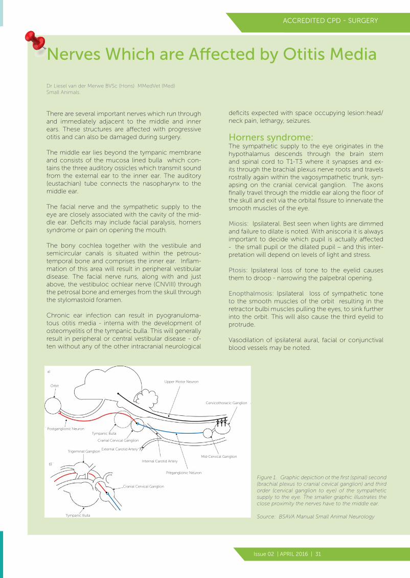
Horners syndrome:The sympathetic supply to the eye originates in the hypothalamus descends through the brain stem and spinal cord to T1-T3 where it synapses and ex-its through the brachial plexus nerve roots and travels rostrally again within the vagosympathetic trunk, syn-apsing on the cranial cervical ganglion. The axons finally travel through the middle ear along the floor of the skull and exit via the orbital fissure to innervate the smooth muscles of the eye.
Miosis: Ipsilateral. Best seen when lights are dimmed and failure to dilate is noted. With aniscoria it is always important to decide which pupil is actually affected - the small pupil or the dilated pupil – and this inter-pretation will depend on levels of light and stress.
Ptosis: Ipsilateral loss of tone to the eyelid causes them to droop - narrowing the palpebral opening.
Enopthalmosis: Ipsilateral loss of sympathetic tone to the smooth muscles of the orbit resulting in the retractor bulbi muscles pulling the eyes, to sink further into the orbit. This will also cause the third eyelid to protrude.
Vasodilation of ipsilateral aural, facial or conjunctival blood vessels may be noted.
Nerves Which are Affected by Otitis Media
Figure 1. Graphic depiction ot the first (spinal) second (brachial plexus to cranial cevical ganglion) and third order (cervical ganglion to eye) of the sympathetic supply to the eye. The smaller graphic illustrates the close proximity the nerves have to the middle ear.
Source: BSAVA Manual Small Animal Neurology
ACCREDITED CPD - SURGERY
Dr Liesel van der Merwe BVSc (Hons) MMedVet (Med) Small Animals.
Postganglionic Neuron
Upper Motor NeuronOrbit
Tympanic Bulla
Cervicothoracic Ganglion
Mid-Cervical Ganglion
Preganglionic Neuron
Internal Carotid Artery
External Carotid Artery
Cranial Cervical Ganglion
Trigeminal Ganglion
Cranial Cervical Ganglion
a)
b)
Tympanic Bulla
April 2016 Vet360 for Madaleen Review.indd 31 2016/03/24 2:24 PM

vet360Issue 02 | APRIL 2016 | 32
© 2015 IDEXX Laboratories, Inc. All rights reserved. • 1509012-0915-EU All ®/TM marks are owned by IDEXX Laboratories,Inc. or its affiliates in the United States and/or other countries. The IDEXX Privacy Policy is available at idexx.com.
For more information visit: idexxsdma.com
Experience the Benefits of Early Detection
As little as 25 % of kidney function remaining at time of
diagnosis with CREA
Up to 60 % of kidney function remaining at time of diagnosis with SDMA
60%25%
In time, kidney diseasewill develop in:
1 in 10 dogs and 1 in 3 cats
NEW
IDEXX SDMA A revolutionary medical breakthrough in kidney diagnostics
After nearly a decade of research and develop-ment, IDEXX is proud to introduce the first and only veterinary specific SDMA test.
The new IDEXX SDMA (symmetric dimethy-larginine) assay enables the identification of chronic kidney disease (CKD) in cats and
dogs substantially earlier than traditional tests.
Identify kidney disease sooner, intervene earlier and find the best approach for each animal. Available only from IDEXX Reference Laboratories.
Reference1. Hand MS, Thatcher CD, Remillard RL, et al., eds. Small Animal Clinical Nutrition. 5th ed. Topeka, KS: Mark Morris Institute; 2010. 2. Grauer GF. Staging and management of canine chronic kidney disease. dvm360. 2009.
http://veterinarynews.dvm360.com/staging-and-management-canine-chronic-kidney-disease. 3. Hall JA, Yerramilli M, Obare E, et al. Comparison of serum concentrations of symmetric
dimethylarginine and creatinine as kidney function biomarkers in cats with chronic kidney disease. J Vet Intern Med. 2014;28(6):1676-1683.
Vestibular Disease ClassificationPeripheral Central
Paresis Never Possible - brainstem disease may affect ascending proprioceptive and and descending motor tracts
Proprioceptive deficits Never Possible – brainstem disease may affect ascending proprioceptive and and descending motor tracts
Consciousness Alert May be confused, distressed, disoriented
May be depressed, stuporous or comatose
Cranial nerve deficits Facial nerve deficits - runs close to middle ear
Facial nerve deficits - exits brain stem close to vestibulocochlear nerve (CN VIII)Additionally Cranial Nerves V and VI all the way down to XII may be affected (brain stem lesion)
Horners syndrome: Possible Unlikely
Nystagmus Horizontal or rotatory - NEVER VERTICALFast phase away from side of lesion Direction doesn’t change with head position
Horizontal, rotatory or vertical Fast phase in any direction Direction may change with head position
Figure 2. Anatomy specimen of a dogs head, showing dissection of the facial nerve. Note the exit from the skull where the horizontal ear canal inserts into the bulla.
ACCREDITED CPD - SURGERY
IDEXX Learning CenterKnowledge you can put into practice
SnippetsShort, self-paced online demonstrations
Online Courses • Archived Webinars • Tutorials • CE ChallengesLearn anytime, anywhere at your pace
Live Webinars • Distance Education • Coach-WebinarsLive, scheduled learning events online
www.idexxlearningcentre.com
IDEXX VetConnect PLUSMake diagnostic decisions more quickly and confidently
Learn how to review patient test requests and results and use other key features on IDEXX VetConnect PLUS• Make care decisions in real time to quickly address patient
health and client concerns.• Easily compare in-house results to reference laboratory re-
sults and see over 10 years of historical data.• View trending graphs that help you quickly spot abnormali-
ties for individual patients.• View pathology images on either the mobile app or web site.• Stay up to the minute on every case with instant results no-
tifications using the VetConnect PLUS mobile app for iP-hone or Android
www.vetconnectplus.com
Dorsal Buccal Branch
Ventral Buccal Branch
Auriculopalpebral Branch
Facial Nerve
April 2016 Vet360 for Madaleen Review.indd 32 2016/03/24 2:24 PM