manifestations of disease - mhprofessionalresources.com · 12-5 chapter e12 atlas of oral...
TRANSCRIPT

12-1
CHA
PTER e12
Atlas of Oral Manifestations of Disease
CHAPTER e12 Atlas of Oral Manifestations of Disease Samuel C. Durso Janet A. Yellowitz
The health status of the oral cavity is linked to cardiovascular dis-ease, diabetes, and other systemic illnesses. Thus, examining the oral cavity for signs of disease is a key part of the physical exam. This chapter presents numerous outstanding clinical photographs illustrating many of the conditions discussed in Chap. 32, Oral Manifestations of Disease. Conditions affecting the teeth, periodon-tal tissues, and oral mucosa are all represented.
Figure e12-1 Gingival overgrowth secondary to calcium channel blocker use.
Figure e12-2 Oral lichen planus.
Figure e12-3 Erosive lichen planus.
Figure e12-4 Stevens-Johnson syndrome—reaction to nevirapine.
Figure e12-5 Erythematosus candidiasis under a denture (i.e., the patient should be treated for this fungal infection).
Copyright © 2012 The McGraw-Hill Companies, Inc. All rights reserved.

12-2
PART 2
Cardinal Manifestations and Presentation of Diseases
Figure e12-6 Severe periodontitis.
Figure e12-7 Angular cheilitis.
Figure e12-8 Sublingual leukoplakia.
Figure e12-9 A. Epulis (gingival hypertrophy) under denture. B. Epulis fissuratum.
Copyright © 2012 The McGraw-Hill Companies, Inc. All rights reserved.
12-3
CHA
PTER e12
Atlas of Oral Manifestations of Disease
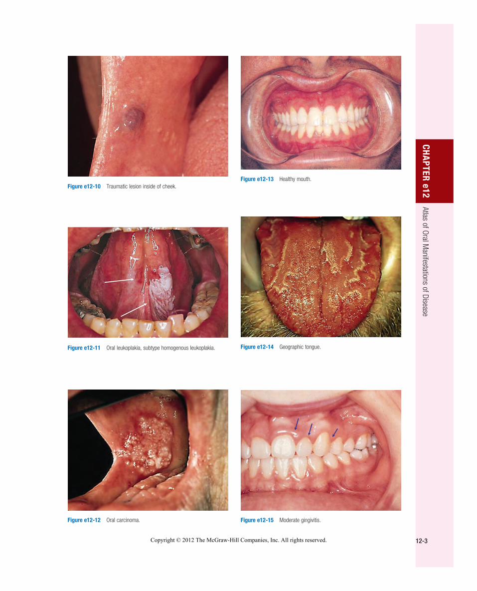
Figure e12-10 Traumatic lesion inside of cheek.
Figure e12-11 Oral leukoplakia, subtype homogenous leukoplakia.
Figure e12-12 Oral carcinoma.
Figure e12-13 Healthy mouth.
Figure e12-14 Geographic tongue.
Figure e12-15 Moderate gingivitis.
Copyright © 2012 The McGraw-Hill Companies, Inc. All rights reserved.

12-4
PART 2
Cardinal Manifestations and Presentation of Diseases
Figure e12-16 Gingival recession.
Figure e12-17 Heavy calculus and gingival inflammation.
Figure e12-18 Severe gingival inflammation and heavy calculus.
Figure e12-19 Root cavity in presence of severe periodontal disease.
Figure e12-20 Ulcer on lateral border of tongue—potential carcinoma.
Figure e12-21 Osteonecrosis.
Copyright © 2012 The McGraw-Hill Companies, Inc. All rights reserved.
12-5
CHA
PTER e12
Atlas of Oral Manifestations of Disease
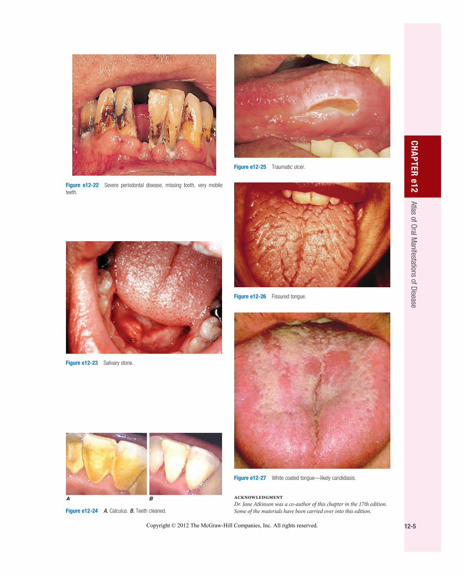
Figure e12-22 Severe periodontal disease, missing tooth, very mobile teeth.
Figure e12-23 Salivary stone.
Figure e12-24 A. Calculus. B. Teeth cleaned.
Figure e12-25 Traumatic ulcer.
Figure e12-26 Fissured tongue.
Figure e12-27 White coated tongue—likely candidiasis.
AcknowledgmentDr. Jane Atkinson was a co-author of this chapter in the 17th edition. Some of the materials have been carried over into this edition.
Copyright © 2012 The McGraw-Hill Companies, Inc. All rights reserved.

Copyright © 2012 The McGraw-Hill Companies, Inc. All rights reserved.