manipulations of pka in chick limb development reveal ... · (lincolnshire), fertilized quail eggs...
TRANSCRIPT

05 (2007) 312–324www.elsevier.com/locate/ydbio
Developmental Biology 3
Manipulations of PKA in chick limb development reveal roles in digitpatterning including a positive role in Sonic Hedgehog signaling
Eva Tiecke a,1, Roisin Turner b, Juan Jose Sanz-Ezquerro a,2, Anne Warner b, Cheryll Tickle a,⁎
a Cell and Developmental Biology, School of Life Sciences, University of Dundee, Dow Street, Dundee, DD1 5EH, UKb Anatomy and Developmental Biology, University College London, Gower Street, London, WC1E 6BT, UK
Received for publication 27 September 2006; revised 9 February 2007; accepted 13 February 2007Available online 21 February 2007
Abstract
Sonic Hedgehog (Shh) signaling by the polarizing region, at the posterior of the vertebrate limb bud, is pivotal in determining digit number andidentity. Shh establishes a gradient of the bifunctional transcriptional effector, Gli3, with high levels of full-length activator (Gli3A) in theposterior bud, where digits form, and high levels of shorter repressor (Gli3R) in the anterior. Repressor formation depends on protein kinase A(PKA), but in Drosophila, PKA also plays a role in activator function. Increasing PKA levels in chick limb development using Forskolin had noeffect on posterior polarizing activity but weak polarizing activity, based on ligand-independent Shh signaling, was induced in anterior limb budcells resulting in extra digits. Manipulating PKA activity levels directly with a retrovirus expressing activated PKA induced extra digits similar tothose induced by Forskolin treatment suggesting that PKA may have a previously unrecognized positive role in Shh signaling in vertebrate limbs.Expressing dominant negative PKA also induced extra, sometimes multiple digits, from anterior limb bud demonstrating the negative role in Shhsignaling. PKA levels in the limb bud are high posteriorly and low anteriorly, suggesting that PKA activity may influence the outcome of Shhsignaling in normal development.© 2007 Elsevier Inc. All rights reserved.
Keywords: Chick; Limb; Sonic hedgehog; Gli3; PKA
Introduction
A key question in embryonic development is how differentparts of the body arise in the proper places. A good model toaddress this question is the digit pattern in vertebrate limbs,which is controlled by signaling of the polarizing region. Theextracellular signaling molecule, Sonic Hedgehog (Shh), is nowknown to be pivotal in this process (Riddle et al., 1993) andconsiderable progress has been made in dissecting out theHedgehog (Hh) signal transduction pathway in vertebrates(reviewed by Cohen, 2003; Hooper and Scott, 2005). Mutationsin a growing number of genes that encode components of thepathway, whose functions, in many cases, are not yet well-
⁎ Corresponding author.E-mail address: [email protected] (C. Tickle).
1 Current address: Developmental Genetics DKBW Centre for Biomedicine,Mattenstrasse 28, Ch-4058 Basel, Switzerland.2 Current address: Departmento de Immunologia y Oncologia, Centro
Nacional de Biotechnologia, Campus de Cantoblanco–UAM, 28049 Madrid,Spain.
0012-1606/$ - see front matter © 2007 Elsevier Inc. All rights reserved.doi:10.1016/j.ydbio.2007.02.017
understood, lead to changes in digit number and pattern, forexample the ta3 gene (Davey et al., 2006) and genes that encodeintraflagellar proteins (Huangfu et al., 2003; Liu et al., 2005).Therefore, it is becoming increasingly important to dissect outthe pathway in detail and understand how it is regulated. Here,we examine the role of protein kinase A (PKA), which has beenimplicated in modulating several steps in Hh signal transduc-tion, in patterning the digits of the chick wing.
The polarizing region is a region of mesenchyme cells at theposterior margin of the limb bud and its signaling propertieswere discovered through its ability to induce extra digits(polarizing activity) when grafted to the anterior margin of asecond wing bud (Saunders and Gasseling, 1968). Following apolarizing region graft, an extra set of digits 432 arise from theanterior part of the wing bud and are arranged in mirror-imagesymmetry with the normal set 234. Signaling has been shown tobe dose-dependent with, for example, grafts of small numbersof polarizing region cells inducing only an extra digit 2, andincreasing numbers resulting in an additional digit 3 and finallyan additional digit 4 (Tickle, 1981). Similar effects can be

313E. Tiecke et al. / Developmental Biology 305 (2007) 312–324
produced by varying the length of exposure to a polarizingregion signal (Smith, 1980) and it has been suggested that cellsfirst form an anterior digit and then are promoted over time toform more posterior digits (Eichele et al., 1985).
Shh is one of three vertebrate Hedgehog genes and isexpressed in the polarizing region (Riddle et al., 1993). Shh hasbeen shown to diffuse across the posterior part of the limb bud(Gritli-Linde et al., 2001; Zeng et al., 2001). Application of Shhto the anterior margin of chick limb buds induces digitduplications in a dose- and time-dependent fashion (Yanget al., 1997). Shh binds to the receptor patched (Ptc) onresponding cells, which allows signaling by the transmembraneprotein smoothened (Smo) through a signaling complexcontaining the Gli family of transcriptional effectors, Gli1,Gli2 and Gli3 (reviewed Cohen, 2003; Hooper and Scott, 2005).In the presence of Hh ligand, full-length Gli protein (Gli3Fl)becomes converted through an unknown process into atranscriptional activator (GliA) (Chen et al., 1999), whichtranslocates to the nucleus and activates expression of targetgenes. In the absence of Hh ligand, Gli proteins, in particularGli3, are processed to a shorter form (GliR), which can alsotranslocate to the nucleus but acts as a transcriptional repressor(reviewed Koebernick and Pieler, 2002).
In normal chick limb buds, Gli3A is high in posterior and lowin anterior regions whereas Gli3R is high in anterior and low inposterior regions (Wang et al., 2000). Analysis of Shh−/−,Gli3−/− and Gli3−/−; Shh−/− mouse embryos has shown thatthe main function of Shh in the limb, is to relieve Gli3mediated repression of genes required for digit patterning. Thisrelief of Gli3R leads to formation of a series of patterned digitsfrom the posterior part of the limb bud while the anterior part,where Gli3R persists, does not give rise to digits (Litingtung etal., 2002; te Welscher et al., 2002).
Proper functioning of the Hh pathway is known to involveseveral kinases, for example, GSK3, CK1, PKA and ubiquitinligase, that modify components so that they transduce the Hhsignal (or lack of Hh signal) properly (Hooper and Scott, 2005).In several vertebrate systems, PKA has been shown to be anantagonist of hedgehog signaling (Epstein et al., 1996;Hammerschmidt et al., 1996; Ungar and Moon, 1996) andthis has been ascribed to its role in regulating processing of theGli3 protein to the repressor form (Wang et al., 2000).
Here we investigate the role of PKA in modulating the outputof Shh signaling pathway in chick limb development both byapplying the drug Forskolin and by overexpressing constitu-tively active and dominant negative PKA using the RCAS(Replication Competent Avian Sarcoma) viral system. For-skolin increases adenylate cyclase activity thereby increasingintracellular cyclic AMP and thus PKA activity. Forskolin hasbeen shown in several developing systems including somite,brain, feather bud, tooth and testis (Fan et al., 1995; Hynes et al.,1995; Noveen et al., 1996; Cobourne et al., 2001; Yao andCapel, 2002) to inhibit Shh signaling. We showed that Forskolintreatment had no effect on polarizing region signaling butinstead, surprisingly, induced anterior limb mesenchyme toform digits. We further showed that manipulating levels of PKAdirectly also resulted in the formation of extra digits.
Materials and methods
Embryos
Fertilized White Leghorn chicken eggs were obtained from Henry Stewart(Lincolnshire), fertilized quail eggs from the Roslin Institute (Edinburgh) andstandard pathogen-free (spf) fertilized eggs from Lohmann Tierzucht (Ger-many). All eggs were incubated at 37 °C and staged according to Hamburgerand Hamilton (1951).
Micromass culture
Micromass cultures were prepared from chick or quail limb buds (stages20–21; Vogel and Tickle, 1993; Makarenkova et al., 1997). A 10 μl dropcontaining 4×10⁴ cells/ml or 2×10⁷ cells/ml was placed into the center of eachwell of a 4-well dish (Nunc). After 1 h, 500 μl of culture medium (F12/DMEM,50/50; Gibco BRL with 10% FCS, 1% glutamine and 1% antibiotic/antimycotic) was added and cultures were maintained for 24 h in a 37 °Cincubator gassed with 5% CO2. Forskolin (Sigma) dissolved in DMSO wasadded to cultures at a final concentration of 100 μM.
Grafts of micromass culture and carriers soaked in Forskolin
After 24 h, micromass cultures were scraped off the dish, cut into 8 piecesand grafted under a loop made by lifting away the apical ectodermal ridge at theanterior margin of a stage 20–21 chick limb buds (Fig. 1A). Various carriers,including heparin beads (Sigma), Affigel blue beads (Biorad), nitrocellulosewith 2 different pore sizes (1.2 μm and 8 μm respectively, Millipore) and DEAEpaper (Whatmann; Tickle et al., 1982), were soaked in 50, 100, 250 and 500 μMForskolin for 1–2 h at room temperature and then grafted to the anterior marginhost limb.
Alcian green staining
Embryos were fixed in 5% trichloroacetic acid at day 9 of incubation, stainedfor cartilage using 0.1% Alcian Green in 70% acetic acid, dehydrated in ethanoland cleared in methylsalicylate.
QCPN staining
Wings of chicken embryos with quail cell grafts were fixed at 9 days ofincubation in 4% paraformaldehyde (PFA) in phosphate-buffered saline (PBS),embedded in O.C.T. compound (Miles), frozen and sectioned at 10 μm. Sectionswere stained as previously described (Tanaka et al., 2000) except that thesecondary antibody was FITC anti-mouse (Sigma). Slides mounted in Citifluor(Agar Scientific) were viewed with a fluorescence compound microscope.
RT-PCR
RNA was isolated from tissue using a QiaShredder column and QuiagenRNA Easy kit (Quiagen). cDNA was synthesized using AMV ReverseTranscriptase Kit (Roche) using oligo dT primers to select for mRNA only.The RT-PCR was carried out using the following primers.
Chick Shh: forward 5′-AGAGAGACAGCCAGGGATAGG-3′ and reverse5′-TGCCACTGAGTTTTCTGCTTT-3′,chick Gli3 :forward 5′-GACGGCAGGTATTAGTTCAAGG-3′ and reverse5′-GCGTTCCTTCTCTCTCTGTGAT-3′,chick GAPDH: 5′-AGCTTACTGGAATGGCTTTCC-3′ and 5′-ACCAT-CAAGTCCACAACACGGTTGCTG-3′
In situ hybridization
In situ hybridization was performed as previously described (Wilkinson andNieto, 1993) with probes synthesized from the following templates: Shh(Roelink et al., 1994), Ihh (Vortkamp et al., 1996), dHAND (Fernandez-Teran
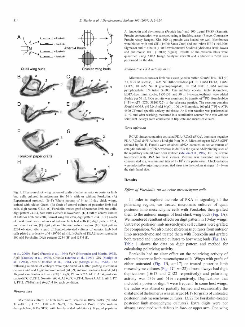
Fig. 1. Effects on chick wing pattern of grafts of either anterior or posterior limbbud cells cultured in micromass for 24 h with or without Forskolin. (A)Experimental protocol. (B–F) Whole mounts of 9- to 10-day chick wings,stained with Alcian Green. (B) Graft of control culture of posterior limb budcells, digit pattern ?3234. (C) Forskolin-treated graft of posterior limb bud cells,digit pattern 24334, note extra element in lower arm. (D) Graft of control cultureof anterior limb bud cells, normal wing skeleton, digit pattern 234. (E, F) Graftsof Forskolin-treated cultures of anterior limb bud cells (E) digit pattern 2234,note absent radius; (F) digit pattern 334, note reduced radius. (G) Digit pattern2234 obtained after a graft of Forskolin-treated cultures of anterior limb budcells plated at a density of 4×10⁴/10 μl. (H, I) Grafts of DEAE paper soaked in100 μM Forskolin. Digit patterns 2234 (H) and (33)4 (I).
314 E. Tiecke et al. / Developmental Biology 305 (2007) 312–324
et al., 2000), Bmp2 (Francis et al., 1994) Fgf4 (Niswander and Martin, 1992),Fgf8 (Crossley et al., 1996), Gremlin (Merino et al., 1999), Gli1 (Marigo etal., 1996a), Hoxa13 (Nelson et al., 1996), Ptc (Marigo et al., 1996b). Thefollowing numbers of embryos were hybridized 24 h after grafting micromasscultures. Shh and Fgf8: anterior control (AC) 9; anterior Forskolin treated (AF)16; posterior Forskolin treated (PF) 5. Fgf4, Ptc and Gli1: AC 2; AF 4; posteriorcontrol (PC) 2; PF 2.Gremlin: AC 4; AF 6; PC 4; PF 4.Hoxa13: AC 2; AF 3; PC1; PF 2. dHAND and Bmp2: 4 for each condition.
Western blot
Micromass cultures or limb buds were isolated in RIPA buffer (50 mMTris–HCl pH 7.5, 150 mM NaCl, 1% Nonidet P-40, 0.5% sodiumdeoxycholate, 0.1% SDS) with freshly added inhibitors (10 μg/ml pepstatin
A, leupeptin and chymostatin (Peptide Inc.) and 100 μg/ml PMSF (Sigma)).Protein concentration was assessed using a Bradford assay (Pierce, CoomassieProtein Assay Reagent Kit). 100 μg protein was loaded per well. Membraneswere blotted with anti-Gli3 (1:500; Santa Cruz) and anti-rabbit HRP (1:10,000;Sigma) or anti-α-tubulin (1:50; Developmental Studies Hybridoma Bank, Iowa)and anti-mouse HRP (1:5000; Sigma). Results of the Western blots werequantified using AIDA Image Analyzer ver3.28 and a Student's T-test wasperformed on the data.
Radioactive PKA activity assay
Micromass cultures or limb buds were lysed in buffer: 50 mM Tris–HCl pH7.4, 0.27 M sucrose, 1 mM Na Ortho-vanadate pH 10, 1 mM EDTA, 1 mMEGTA, 10 mM Na B glycerophosphate, 10 mM NaF, 5 mM sodiumpyrophosphate, 1% triton X-100. One inhibitor cocktail tablet (Complete,EDTA-free, mini, Roche, 1836153) and 50 μl β-mercaptoethanol were addedfreshly per 50 ml. PKA activity was monitored by transfer of 32PO3 from Isoblue[32P]-γ-ATP (ICN, 38101X.2) to the substrate peptide. The reaction contains50 mMMOPS, pH 7.0, 5 mMMgCl2, 100 μMKemptide, 100 μM [32P]-γ-ATP;4500 Ci/mmol specific activity and tissue. An 8-min reaction was performed at37 °C and, after washing, measured in a scintillation counter for 2 min withoutscintillant. Assays were conducted in triplicate and means calculated.
Virus infection
RCAS viruses containing activated PKA (RCAS-aPKA), dominant negativePKA (RCAS-dnPKA; both a kind gift from Dr. A. Münsterberg) or RCAS-eGPF(cloned by Dr. E. Farrell) were obtained. aPKA contains an active mutant ofcatalytic subunit C of PKAwhereas in dnPKA the cyclic AMP binding sites ofthe regulatory subunit have been mutated (Mellon et al., 1989). DF1 cells weretransfected with DNA for these viruses. Medium was harvested and virusconcentrated to give a minimal titer of 1×10⁸ virus particles/ml. Chick embryoswere infected by injecting concentrated virus into the coelom at stages 13–14 onthe right hand side.
Results
Effect of Forskolin on anterior mesenchyme cells
In order to explore the role of PKA in signaling of thepolarizing region, we treated micromass cultures of quailposterior limb mesenchyme cells with Forskolin, then graftedthem to the anterior margin of host chick wing buds (Fig. 1A).We monitored resultant effects on digit pattern in 10-day wings.Untreated posterior limb mesenchyme cell cultures were graftedfor comparison. We also made micromass cultures from anteriorlimb mesenchyme and treated them with Forskolin and graftedboth treated and untreated cultures to host wing buds (Fig. 1A).Table 1 shows the data on digit pattern and method forcalculating polarizing activity.
Forskolin had no clear effect on the polarizing activity ofcultured posterior limb mesenchyme cells. Wings with grafts ofeither untreated (Fig. 1B, n=17) or treated posterior limbmesenchyme cultures (Fig. 1C, n=22) almost always had digitduplications (16/17 and 21/22 respectively) and polarizingactivity was 53% and 61% respectively. Duplications thatincluded a posterior digit 4 were frequent. In some host wings,the radius was absent or partially formed and occasionally thedistal endof thehumeruswasenlarged(4/17 forgraftsofuntreatedposterior limbmesenchyme cultures; 13/22 for Forskolin-treatedposterior limb mesenchyme cultures). Extra digits were notalways associated with defects in fore- or upper arm. One wing

Table 1Effects on host chick wing digit pattern of grafting anterior or posterior quail limb micromass cultures treated for 24 h with or without Forskolin
Treatment n Total number ofdigits formed
Type of extra digits obtained Polarizingactivity
Wings withextra digits
3 4 5 Mean digitnumber
Normal digitpattern 234
Additionalblip
Additional2
Additional ortransformed 3
Additional4
(%) (%)
Posterior control 17 6 8 3 3.8 1 0 2 11 3 53 94Posterior Forskolin 22 6 12 4 3.9 1 0 2 12 7 61.2 95Anterior control 17 17 0 0 3 17 0 0 0 0 0.0 0⁎ 35 35 0 0 3 35 0 0 0 0 0 0Anterior Forskolin 47 36 11 0 3.2 25 4 11 7 0 14.4 47⁎ 36 28 8 0 3.1 28 0 8 0 0 5.6 22
All grafts were from micromasses in which 2×10⁵ cells/10 μl were plated apart from rows indicated by ⁎ in which 4×10⁴ cells/10 μl were plated.Bifurcated digits were counted as 1.Additional blip includes b234 and bb234.Additional 2 include b2234, (22)34, 2234.Additional 3 include 32(34), 3234, 334, 3334, 3b34, ?3234, (b3)234, 3(22)34, 32234, (33)234, b32234.Additional 4 include 24334, 34.334, 4334, 43b34, (43)234, 43(22)34, 43234.
ðdigit 4Þ � 4þ ðdigit 3Þ � 2þ ðdigit 2Þ � 1þ ðblipÞ � 0:5total number of wings� 4
� 100 ¼ polarizing activity
315E. Tiecke et al. / Developmental Biology 305 (2007) 312–324
grafted with a Forskolin-treated posterior limb mesenchymeculture had an extra element in the forearm (Fig. 1C).
Wings with grafts of untreated cultures of anterior limbmesenchyme cultures never had extra digits (Fig. 1D, quail tochick n=17; chick to chick n=35) although some had a radiusdefect. Unexpectedly, 47% of chick wings grafted with culturedquail anterior limb mesenchyme cells treated with Forskolin hadchanges in digit pattern (n=22/47, Figs. 1E, F) while 28% ofwings with grafts of Forskolin-treated chick anterior limbmesenchyme cultures also showed changes (8/28; Fig. 1G). Inboth sets of experiments, the change in digit pattern consisted,nearly always, of an extra anterior digit 2 (Fig. 1E), i.e.,duplication of the most anterior digit; sometimes a digit 2 wasreplaced by a digit 3 (Fig. 1F). Defects in more proximalelements, mainly a missing or reduced radius, were observed insome cases. Chick anterior limb mesenchyme cells cultured atlower cell densities (10 μl of 4×10⁴ cells/ml) also induced extradigits when treated with Forskolin (5/15), while grafts of cellsfrom untreated anterior limb mesenchyme cultures at the samecell densities had no effect on wing patterning (10/10). Thustreatment of cultured quail or chick anterior limb mesenchymecells with Forskolin unexpectedly results in formation of anextra digit 2 or replacement of digit 2 by a digit 3 when thesecells are grafted back into host limbs.
To investigate whether the extra or altered digit arises fromthe Forskolin-treated anterior limb mesenchyme cells or fromhost tissue, we studied the fate of cultured quail anterior limbmesenchyme cells treated with Forskolin grafted to chick limbbuds. Graft quail and host chick cells can be distinguished usingthe QCPN antibody. The extra anterior digit (digit 2; Figs. 2A′,A″) consisted of both quail and chick cells (Fig. 2A‴, n=3).Graft cells were found in host mesenchyme along the anterioredge and at the base of the extra digit (Fig. 2A‴, arrows). Theywere also found more proximally in ectopic feather buds (Fig.2A‴, arrowheads) and cartilage. For comparison, we examineddistribution of quail cells from cultures of posterior limb
mesenchyme treated with Forskolin when grafted to a hostchick wing. In this case, the extra digit, a digit 3 (Fig. 2B′, B″),also contained both graft and host cells. Graft cells were foundin host mesenchyme along the anterior margin of the extra digit,as with grafts of Forskolin-treated anterior limb mesenchymecells, but extended more distally (Fig. 2B‴; arrows). No quailcells were detected in the cartilage of the extra digit but therewere quail cells in more proximal cartilage elements of the wing(data not shown). We conclude that Forskolin-treated anteriorlimb mesenchyme cells entrain host anterior limb mesenchymecells to participate in extra digit formation and behave similarlyto Forskolin-treated posterior cultured cells or polarizing regioncells (CT unpublished).
We then tested whether applying Forskolin directly to theanterior margin of chick limb buds on inert carriers could lead toformation of extra anterior digits or transform identity of theanterior digit. With most of the carriers, no extra digits formed(124/161 cases) but, with Forskolin soaked pieces of DEAEpaper (Whatmann), similar digit duplications were induced inhost wing buds to those produced by grafts of Forskolin-treatedmicromasses of anterior limb mesenchyme cells in 10/37 cases.Either an extra digit 2 (Fig. 1H) was induced or a digit 3 formedin place of a digit 2 (Fig. 1I). The optimal concentration ofForskolin was 100 μM (Table 2), the concentration used to treatmicromasses. Both methods gave roughly the same polarizingactivity. Thus either treating anterior limb mesenchyme cultureswith Forskolin or treating anterior limb buds directly withForskolin leads to additional digit formation or promotes a digit2 to a digit 3.
Gene expression in anterior limb mesenchyme after Forskolintreatment
The digit pattern change produced by grafts of Forskolin-treated anterior limb mesenchyme cultures could be explainedby the induction of Shh expression in the cells. However, we

Fig. 2. QCPN staining to study distribution of quail graft and chick host cells in extra digits and RT-PCR to examine Shh expression in cultured cells. (A′) Host chickwing grafted with Forskolin-treated culture of quail anterior limb bud cells; digit pattern 2234. (A″) High power of panel A′ showing extra digit 2. (A‴) Frozen sectionthrough extra digit showing labeled quail cells along anterior edge and in ectopic feather buds. (B′) Host chick wing grafted with Forskolin-treated quail posterior limbbud cells; digit pattern 3234. (B″) High power of panel B′ showing extra digit 3. (B‴) Frozen section through extra digit 3 showing labeled quail cells along anterioredge (C) RT-PCR for Shh in micromass cultures at 1 and 24 h and in stage 20 limb bud. (C′) RT-PCR for GAPDH in same samples as control. AC=untreated anteriormesenchyme culture, AF=Forskolin-treated anterior mesenchyme culture, PC=untreated posterior mesenchyme culture, PF=Forskolin-treated posteriormesenchyme culture, A=anterior limb bud, P=posterior limb bud.
316 E. Tiecke et al. / Developmental Biology 305 (2007) 312–324
could detect no Shh expression by RT-PCR in Forskolin-treatedcultures of anterior limb mesenchyme cells after either 1 or 24 hof culture (Fig. 2C) as was the case with untreated cultures ofanterior limb mesenchyme cells. Treated and untreated culturesof posterior limb mesenchyme cells expressed Shh at 1 h asexpected but showed no detectable expression of Shh at 24 hconsistent with previous reports (Kimura and Ide, 1998).GAPDH was used as a positive control (Fig. 2C′).
Even though no Shh expression could be detected in theForskolin-treated micromasses of anterior limb mesenchymecells, Shh expression might be switched on when the anteriorlimb mesenchyme cell micromasses are placed under the apicalridge in a host wing bud. Therefore, we examined Shhexpression 24 h after grafting treated and untreated cultures ofboth anterior and posterior limb mesenchyme into host limbbuds by in situ hybridization. However, Shh expression couldnot be detected in any of these grafts (Figs. 3A–C). There havebeen reports of redundancy between the different vertebrate Hhs(Yang et al., 1998; Pathi et al., 2001), and Ihh is known to beexpressed during chondrogenesis (Vortkamp et al., 1996;
Table 2Effects on host chick wing digit pattern of grafting DEAE soaked in various concen
Concentration ofForskolin (μM)
n Normal digitpattern 234
Type of extra digits obtained
Additional blip Additio
0 8 850 6 4 1 1100 8 3 2 2150 7 5 1 1250 8 7 1
Additional 2 include (22)34, 2234.
Koyama et al., 1996). We therefore examined Ihh expressionin grafted cultures but no expression could be found after 24 h(data not shown). We conclude that the effect of Forskolin onanterior limb mesenchyme cells is not dependent on inductionof Shh or Ihh expression.
One hypothesis that would explain our observations is thatForskolin activates the Shh signaling pathway in anterior limbmesenchyme in the absence of ligand. This hypothesis wastested by examining, in host limb buds grafted with Forskolin-treated anterior limb mesenchyme cells, expression patterns ofgenes known to be read outs of Shh signaling and associatedwith digit formation. Gene expression was compared with thatin limb buds grafted with untreated anterior limb mesenchymecells and with untreated and treated posterior limb mesenchymecells. We examined expression of genes, Ptc and Gli1, knownto be activated by Shh and also expression of genes which morelikely depend on repression by Gli3R, Hoxa13, Gremlin,dHAND and Bmp2 (Litingtung et al., 2002; te Welscher et al.,2002). We also examined the effects on the apical ectodermalridge and expression of Fgf genes, including Fgf4, which
trations of Forskolin
Polarizingactivity (%)
Embryos withextra digits (%)
nal 2 Additional/Transformed 3 [(33)4]
0 06.3 34
1 15.6 624.7 293.1 13

Fig. 3. Expression of Shh, Fgf8, Fgf4, Ptc and Gli1 in chick limb buds 24 h after grafts of either anterior or posterior limb bud cell cultures treated for 24 h with orwithout Forskolin. Graft outlined. (A–C) Ventral view; Shh and Fgf8. (A′–C′) Edge on view of anterior margin, dorsal side up. Grafts of anterior control (A, A′),anterior Forskolin (B, B′), posterior Forskolin (C, C′) cultures. Note the extent and morphology of Fgf8 expression in ridge, expression stronger in panels B′ and C′,weaker in panel A′. (D–G). Frontal view; Fgf4. Grafts of anterior control (D, D′), anterior Forskolin (E, E′), posterior control (F, F′), posterior Forskolin (G, G′)cultures. (H–K) Ventral view; Ptc. Grafts of anterior control (H), anterior Forskolin (I), posterior control (J) posterior Forskolin (K) cultures. (L–O) Dorsal view; Gli1.Grafts of anterior control (L), anterior Forskolin (M), posterior control (N) posterior Forskolin (O) cultures. Note the light ectopic Gli1 expression around graft ofanterior Forskolin-treated cultures (with arrows); stronger expression around grafts of posterior control and posterior Forskolin-treated cultures (with arrows).
317E. Tiecke et al. / Developmental Biology 305 (2007) 312–324
contributes to a positive feed-back loop that co-ordinatessignaling between polarizing region and ridge (Zuniga et al.,1999).
Forskolin-treated grafts had a clear effect on the apical ridge(Figs. 3A′–C′). After 24 h, the ridge had grown over all thegrafts, but in limb buds with grafts of Forskolin-treated culturesof anterior limb mesenchyme cells Fgf8 was expressed morewidely anteriorly (Fig. 3B′) than in limb buds grafted withuntreated cultures. Furthermore, the morphology of the Fgf8expressing anterior apical ridge appeared more similar to theposterior ridge of normal limb buds and to anterior ridge Fgf8expression following grafts of posterior limb mesenchyme cells
(compare Figs. 3B′ and C′). The Fgf4 expression domain wasalso shifted anteriorly and was wider in limb buds with grafts ofanterior limb mesenchyme cells treated with Forskolin (Fig. 3Ecompare with contralateral limb E′). A shift in Fgf4 expressionanteriorly was also seen in limb buds with grafts of both treatedand untreated cultures of posterior limb mesenchyme cells, butthe widening of the Fgf4 domain seemed more marked afterForskolin treatment (compare Figs. 3F and G; F′ and G′ arecontralateral limbs).
Ptc, a direct target of Hh signaling, was expressed at highlevels in grafts of Forskolin-treated anterior limb mesenchymecultures and in both treated and untreated posterior limb

318 E. Tiecke et al. / Developmental Biology 305 (2007) 312–324
mesenchyme cultures but not in untreated anterior limbmesenchyme cultures (Figs. 3H–K). Ptc expression did notappear to be induced in host tissue adjacent to the graftssuggesting that this consequence of Forskolin treatment is cellautonomous. By contrast, another target gene, Gli1, was notexpressed within any of the grafts (Figs. 3L–O) but a faint rimof expression was seen in host tissue around grafts of Forskolin-treated anterior limb mesenchyme cultures and more stronglyaround grafts of treated and untreated posterior limb mesench-yme cell cultures (with arrows in Figs. 3M–O).
Expression of genes that appear to be normally repressed inanterior regions of the limb bud by Gli3R, Hoxa13, Gremlin,dHand and Bmp2 was also modified by grafts of Forskolintreatment anterior limb mesenchyme (Fig. 4). Hoxa13 wasdetected in grafts of Forskolin-treated cultures of anterior limbmesenchyme cells, as well as in grafts of both treated anduntreated cultures of posterior limb mesenchyme cells, but notin grafts of untreated cultures of anterior limb mesenchymecells (Figs. 4A–D). Hoxa13 expression was not induced in
Fig. 4. Expression of Hoxa13, Gremlin, dHAND and Bmp2 in chick limb buds 24 hwith or without Forskolin. Graft outlined. (A–D) Ventral view;Hoxa13. Grafts of ante(D). (E–H) Dorsal view; Gremlin. Grafts of anterior control (E), anterior Forskoliextended and Gremlin is also expressed in graft (compare F–H with E). (I–L) Dorscontrol (K), posterior Forskolin (L) cultures. Ectopic expression around graft in paneanterior Forskolin (N), posterior control (O), posterior Forskolin (P) cultures. Ectop
host limb buds. Expression of Gremlin, which is involved inthe Shh-Fgf feedback loop, was also similarly detected ingrafts of Forskolin-treated cultures of anterior limb mesench-yme cells, treated and untreated cultures of posterior limbmesenchyme cells but not in grafts of untreated cultures ofanterior limb mesenchyme cells (Figs. 4E–H). However,expression was patchy in the graft and not seen in everygrafted limb of Forskolin-treated anterior mesenchyme (n=3/4). Marked changes were also seen in Gremlin expression inthe host limb buds and expression extended towards grafts ofForskolin-treated cultures of anterior limb mesenchyme cellsand both treated and untreated cultures of posterior limbmesenchyme cells (Figs. 4 F, G, H). Gremlin expression inhost limb buds grafted with untreated anterior mesenchymecells was normal. Expression of dHAND was upregulated inlimbs with grafts of Forskolin-treated cultures of anterior limbmesenchyme cells (n=1/4), treated (n=4/4) and untreated(n=3/4) cultures of posterior limb mesenchyme cells but notin limbs with grafts of untreated cultures of anterior limb
after grafts of either anterior or posterior limb bud cells cultures treated for 24 hrior control (A), anterior Forskolin (B), posterior control (C), posterior Forskolinn (F), posterior control (G), posterior Forskolin (H) cultures. Host expressional view; dHAND. Grafts of anterior control (I), anterior Forskolin (J), posteriorls J–L compared to I. (M–P) Dorsal view; Bmp2. Grafts of anterior control (M),ic expression around the graft in panels N–P compared to M.

319E. Tiecke et al. / Developmental Biology 305 (2007) 312–324
mesenchyme cells (n=4/4; Figs. 4I–L). Bmp2 expression wasextended and detected around the grafts of Forskolin-treatedcultures of anterior limb mesenchyme cells (n=1/4), treated(n=4/4) and untreated (n=3/4) cultures of posterior limbmesenchyme cells and not in limbs with grafts of untreatedcultures of anterior limb mesenchyme cells (Figs. 4M–P).Altogether, these data show that downstream target genes ofShh signaling can be activated in graft and host tissue in theabsence of the ligand. We also attempted to perform whole-mount in situ hybridization analysis for these genes on limbbuds grafted with DEAE paper soaked in Forskolin, butunfortunately, the detection solution reacted with the DEAEpaper making the results impossible to interpret.
Fig. 5. PKA activity in cultures after Forskolin treatment and effects of altering PKAassay in micromasses after 24 h cultured with or without Forskolin. Forskolin treatmeGFP expression in entire right wing, anterior right leg but not left wing or leg. (C, D)(11)234. Note increased cartilage, humerus, radius (C); femur (D). (E, F) RCAS-dnP(F) Leg, duplicated digit 1, digit pattern 11234.
Effects of manipulating PKA activity
Forskolin elevates cAMP and should lead to an increase inPKA activity. Therefore, the effects on PKA activity of treatingmicromasses of limb mesenchyme cells with Forskolin weremeasured using incorporation of 32P in a known PKA substrate.As predicted, Forskolin treatment led to increased PKA activity(Fig. 5A). The basal PKA level was slightly higher in cultures ofposterior limb mesenchyme cells than in cultures of anteriorlimb mesenchyme cells. After 24-h Forskolin treatment,cultures of both anterior and posterior limb mesenchymecultures showed an increase in PKA activity compared tocontrols, confirming the predicted cAMP-driven rise in PKA
activity by retroviral infection. (A) PKA activity determined using a radioactiveent increases PKA activity more markedly in anterior cells. (B) RCAS-eGFP,
RCAS-aPKA, (C) Wing, digit pattern 2(33)4. (D) Leg, duplicated digit 1, patternKA, (E) Wing, 3 extra digits anteriorly probably all 2s, digit pattern (22)(22)34.

320 E. Tiecke et al. / Developmental Biology 305 (2007) 312–324
activity. The increase was more marked in anterior cultures.Furthermore, we found that this increase in PKA activity wasmaintained in cultures at 48 h (data not shown). We do notknow, however, how long high level PKA activity would bemaintained in the cells once grafted into the limb bud.
cAMP has many other functions in the cell so to test whetherthe extra digits, that form following Forskolin treatment ofanterior cultured limb bud cells, are a direct consequence ofaltering PKA levels, we manipulated PKA activity using RCASviruses. To increase PKA activity, we used a virus containing anactive mutant of catalytic subunit C of PKA (aPKA), and todecrease PKA activity, we used a virus containing theregulatory subunit in which cAMP binding sites have beenmutated (dnPKA; Mellon et al., 1989). These constructs havepreviously been used in other organisms to manipulate PKAlevels (Hammerschmidt et al., 1996; Ungar and Moon, 1996).We infected the presumptive chick wing bud region with theseviruses by injecting concentrated virus into the coelom of stage13–14 embryos adjacent to the region that will give rise to theright wing. Concentrated virus containing eGFP (RCAS-eGFP)was injected as a control. In the first series of experiments, weused normal White Leghorn chick embryos (wl); in the secondseries, pathogen-free chick embryos (spf). Fig. 5 and Table 3show the results.
Injections of control RCAS-eGFP into the presumptive limbregion (wl, n=17 and spf, n=18) did not change digit pattern(Fig. 5B). In 18/18 pathogen-free embryos, fluorescent EGFPwas detected and the whole wing was infected. Not all WhiteLeghorn embryos were infected in every run. For example, inone run (n=11) we could detect no fluorescence in fiveembryos, while limbs of 3 embryos appeared to be fully infectedand 3 partly infected. In some embryos (both pathogen-free andWhite Leghorn), expression was also observed in the leg,mainly in digits 1 and 2.
When RCAS-aPKA was injected into the presumptive limbregion, 4.5% of White Leghorn embryos and 18.8% ofpathogen-free embryos (wl, n=2/44 and spf, n=3/16) had achange in digit pattern (Figs. 5C and D). The altered digitpattern in the wing consisted of either an extra digit 2 in threecases, in one case in the wing, digit 3 was bifurcated resulting ina digit pattern 2(33)4. In one case in the leg, digit 2 wasduplicated resulting in a digit pattern 12234. In limbs of some
Table 3Effects on digit pattern of modifying PKA activity by infecting limb buds with RCA
Virus Type of eggsused
n Type of extra digits obtained
Normal digitpattern 234
Additionalblip
Additional 2(or in leg ad
RCAS-eGFP Normal 17 17spf 18 18
RCAS-aPKA Normal 44 42 2spf 16 13 1
RCAS-dnPKA Normal 39 30 2 5spf 19 11 1 8
Additional 2 include (22)34, 2234, (22)(22)34.Additional 1 includes (11)234, 11234, 111234.a In both these cases an additional digit was formed, i.e., 2334 and 12234.
RCAS-aPKA-injected embryos, chondrogenesis was affectedand the humerus (wl; n=4/44, spf; n=11/16) or femur (wl;n=0/44, spf; n=5/16) was thicker and shorter (Fig. 5D) thanthose in the uninfected contralateral limbs. On some occasions,tibia and fibula were fused and the radius was missing.
When RCAS-dnPKAwas injected into the presumptive limbregion, 23% of White Leghorn embryos and 42% of pathogen-free embryos (wl, n=9/39 and spf, n=8/19) had extra digitsanteriorly. Sometimes limbs had more than one extra digit atthe anterior margin of the limb bud (Fig. 5E); in these cases, allthe extra digits looked similar but could be fused andoccasionally truncated (these were scored as digit 2 forcalculating polarizing activity). Following injections of bothtypes of viruses, digit duplication was sometimes observed inthe leg (Figs. 5D, F) but not the wing and in one case (forRCAS-dnPKA) both wing and leg had extra digits. Forcalculating polarizing activity, the duplications obtained in theleg were added to the ones obtained for the wings and the sumof all the wing patterns and leg duplications were divided bythe total number of embryos.
Effects of Forskolin on Gli3 protein and levels of PKA innormal limb buds
Digit formation in the anterior region of the limb isnormally suppressed by Gli3R therefore we monitored theeffects of 24 h of Forskolin treatment on the processing of Gli3protein in micromass cultures of chick limb cells by Westernblot analysis and compared this to the distribution of the Gli3protein (Gli3Fl and Gli3R as the antibody cannot distinguishbetween Gli3A and Gli3Fl) in limb tissue divided into anterior(A), middle (M) and posterior (P) regions (see Fig. 1A; n=4for each type of culture/tissue). Gli3Fl was present at similarlevels throughout the limb bud whereas Gli3R was higher inthe anterior part of the limb bud than in the posterior part (Fig.6A). Forskolin treatment of limb mesenchyme culturesappeared to increase protein levels of both Gli3Fl and Gli3Rin cultures of both anterior and posterior cells (Figs. 6B and C).Measurements of the intensity of bands in triplicate blotsnormalized with respect to alpha tubulin showed that the ratioof Gli3Fl/Gli3R in normal limb buds was 1.4 in the posteriorhalf and 0.25 in the anterior half. When cells were cultured for
S viruses
Polarizingactivity (%)
Embryos withextra digits (%)
ditional 1)Additional 3 or transformed 3(or in leg 2)
0.00 0.000.00 0.001.10 4.54
2 a 7.81 18.752 9.30 23.070 11.18 42.11

Fig. 6. Gli3 protein levels in cell cultures and limb buds and PKA activity innormal limb buds. (A) Western blot for Gli3 in anterior, middle and posteriorthirds of limb buds. (B) Western blot for Gli3 in untreated and treated cultures ofanterior (AC, AF) or posterior (PC, PF) limb mesenchyme cultures and in limbbuds cut into anterior, middle and posterior parts. (C) Ratio of full-length Gli3(Gli3Fl) versus Gli3R in treated and untreated cultures of anterior or posteriorlimb mesenchyme and in limb buds cut into anterior and posterior parts. Increasebetween AC and AF is significant (p<0.007) but not between PC and PF(p>0.174). Error bars represent the standard deviation from the mean value ofthe three experiments. (D) RT-PCR for Gli3 and GAPDH in untreated andtreated cultures of anterior (AC, AF) or posterior (PC, PF) limb mesenchyme.(E) PKA activity in chick anterior, middle and posterior limb buds at stages 19,20 and 21 of development. PKA activity generally low anteriorly, highposteriorly. Error bars as in panel C.
321E. Tiecke et al. / Developmental Biology 305 (2007) 312–324
24 h this ratio was maintained, while Forskolin treatment leadsto an increase in Gli3Fl/Gli3R. This increase was significant inanterior treated cultures (p<0.007) but not in cultures ofposterior treated cultures (p<0.174). The increase in the ratioof Gli3Fl/Gli3R in both anterior and posterior cultures ismostly due to an increase in Gli3Fl, although Gli3R alsoincreases but to a lesser extent (Fig. 6C). This increase in totalGli3 protein (Fl and R) was not due to a change in transcriptionof Gli3 as semi-quantitative RT-PCR did not show any changein the level of Gli3 transcripts in anterior control versusanterior Forskolin-treated cultures and in posterior controlversus posterior Forskolin-treated cultures (n=3; Fig. 6D).
Finally we determined endogenous PKA activity in differentregions of normal chick limb buds across the anterior–posterior
axis (Fig. 6E). PKA activity was graded across the antero-posterior axis of stage 19 limb buds being highest in theposterior region, lowest in the anterior region and intermediatein the middle. At stages 20 and 21, PKA activity was alsohighest at the posterior, as at stage 19, but the differencebetween activity in posterior and anterior regions was much lessand activity was lowest in the middle (Fig. 6E).
Discussion
Our main finding is that anterior wing bud cells treated withForskolin form an extra digit 2 or digit 2 is replaced by a digit 3(i.e., the most anterior digit of the wing is posteriorized). Thisappears to occur via activation of the Shh signaling pathway inthe absence of ligand. Forskolin treatment leads to changes inthe ratio between full-length Gli3 and Gli3R and genesdownstream of Shh signaling are expressed. We show thatthis effect of Forskolin can be mimicked, at least in part, byincreasing PKA activity directly and that the digit patternsresulting from these manipulations are similar to those obtainedafter Forskolin treatment. Decreasing PKA activity also inducedchanges in digit pattern with one or sometimes more than oneextra digit 2 forming at the anterior of the wing (digit 1 in theleg). PKA activity appears to be graded across the antero-posterior axis of early chick limb buds suggesting that locallevels of PKA might play a role in normal digit patterning.Altogether, our results suggest that PKA has a positive role inShh signaling in the chick limb bud in addition to acting as anegative regulator.
The changes in digit pattern and gene expression induced byForskolin indicate that the Shh pathway has been activated inanterior mesenchyme and resemble changes in digit patternobtained with low doses or short exposure to Shh (Yang et al.,1997). In many systems Forskolin has been reported toantagonize Shh signaling. However, there is a precedent forForskolin activation of the vertebrate Shh pathway in left/right(L/R) patterning of the body (Kawakami and Nakanishi, 2001;Rodriguez-Esteban et al., 2001). Ectopic application ofForskolin to early chick embryos induced expression of Nodaland Pitx2, downstream targets of Shh signaling which specify L/R asymmetry, in the absence of ligand. Furthermore, Forskolindid not appear to antagonize endogenous Shh signaling in theearly chick embryo, similar to our findings in the limb.
We have shown that the positive effect of Forskolin onanterior mesenchyme can be mimicked by elevating PKA levelsdirectly. When we infected chick limbs with a virus containingan activated PKA, additional digits formed or the existinganterior digit became posteriorized, albeit in a small number ofcases. In these limbs, occasionally, the cartilage of the rest of thelimb was affected (Fig. 6C and D). An increase in chondrogen-esis in response to Forskolin has previously has been describedby Solursh et al. (1981) in micromass culture. This stimulationof Hh signaling by activated PKA contrasts with a previousfinding in the CNS of zebrafish where overexpression ofactivated PKA suppressed cell types normally associated withHh signaling (Hammerschmidt et al., 1996; Epstein et al., 1996;Ungar and Moon, 1996).

322 E. Tiecke et al. / Developmental Biology 305 (2007) 312–324
In our experiments, in which we grafted Forskolin-treatedcells into limb buds, there was also evidence for a non-autonomous effect. It has been known for many years thatForskolin and cAMP increase gap junctional communicationbetween cells. In addition, there is recent direct evidence thatcAMP diffusing into neighboring cells via gap junctions canmediate PKA phosphorylation in these cells (Ponsioen et al.,2006). Thus an increase in gap junctional communicationcoupled with intercellular diffusion of cAMP might explain theeffects of grafts of Forskolin-treated cells on host cells.Interestingly, we have previously shown that gap junctionalcommunication plays a role in digit patterning in the chick limb(Allen et al., 1990).
It is not clear in our system whether other effects of Forskolintreatment, which are not directly mediated by PKA, alsocontribute to inducing the digit changes. For example, there isevidence that Forskolin can activate Akt/PKB (Sable et al.,1997; Filippa et al., 1999), which affects GSK3, a component ofthe Wnt signaling pathway (Frame and Cohen, 2001). Inaddition the effect of Forskolin on PKA could also be affectingother signaling pathways as demonstrated by Chen et al. (2005).
We have shown that infecting chick limbs with a viruscontaining dnPKA also induced additional digits. With thistreatment, however, unlike infection with activated PKA ortreatment with Forskolin, multiple extra digits were sometimesproduced. In a preliminary analysis of gene expression inlimbs of embryos infected with dnPKA, a small patch ofectopic Hoxd13 expression was detected in one case,consistent with what would be expected if Gli3R wasdecreased. In other wings, Alx4 expression was extended(n=1), Grm expression was reduced (n=1) while expressionof Ptc, Gli1 and Bmp2 appeared unaltered (n=12 for eachgene). Thus, although this analysis in part supports the ideathat the extra digits that develop ectopically after infecting withdnPKA are due to de-repression of Gli3R, this remains to besubstantiated.
In Drosophila, PKA is known to exert both negative andpositive effects on Hh signaling. Phosphorylation of Ci, theDrosophila homologue of Gli, by PKA is necessary for theproduction of Ci-75, which functions as a repressor (Jiang andStruhl, 1995; Chen et al., 1998; Price and Kalderon, 1999). Thepositive effect of PKA on Hh signaling is not so extensivelystudied but has been reported to be mediated through effects onSmo (Jia et al., 2004; Zhang et al., 2004; Apionishev et al.,2005), Ci itself (Wang and Holmgren, 2000) and activation ofother, as yet unknown, factors (Zhou et al., 2006). As vertebrateSmo lacks critical PKA phosphorylation sites (reviewedHuangfu and Anderson, 2006), it seems unlikely that theactivation of Shh signaling we observe after PKA treatment ismediated by effects on Smo. Furthermore, with respect to theimportance of PKA phosphorylation of Ci, experiments ininsects using different driver lines have given contradictoryresults (Chen et al., 1999; Price and Kalderon, 1999; Wang etal., 1999; Wang and Holmgren, 2000). Thus even in insects theexact targets of PKA that are required for activation of Hhsignaling and that might operate in vertebrates still remain to befully elucidated.
Other possible mechanisms by which PKA could beinvolved in Gli3 function in vertebrates are suggested by theeffects of PKA on the functioning of other transcription factors,such as PBX1, where phosphorylation by PKA is necessary fornuclear import and functioning of this protein (Kilstrup-Nielsenet al., 2003) and dHAND, where PKA phosphorylation hasbeen suggested to modulate dimerization properties with otherfactors (Firulli et al., 2003, 2005). dHAND was one of the Shhtargets that we found to be ectopically expressed in host limbswith Forskolin-treated anterior mesenchyme grafts. However, itseems unlikely that PKA acts via dHAND in inducing thepattern changes that we see since hyperphosphorylation ofdHAND results in limb truncation and not additional digits(Firulli et al., 2003).
We found that Forskolin treatment of cultures of anteriorand posterior limb mesenchyme increased the levels of bothfull-length Gli3 and Gli3R and that the ratio between full-length Gli3 and Gli3R increased by the same amount inanterior and posterior mesenchyme. However, the ratio of full-length Gli3/Gli3R in Forskolin-treated anterior cells is stilllower that that found in untreated posterior mesenchyme or theposterior region of normal limb buds. This could explain whythe duplications obtained are similar to those resulting fromlow level Shh treatment. Our data and data from others (forexample Davey et al., 2006) suggest that it is not only thelevels of Gli3 protein that are important but the ratio betweenfull-length and repressor forms of the protein. The fact that wedo not see a significant difference in digit pattern after grafts oftreated and untreated of the posterior mesenchyme suggeststhat the system is saturated. In normal limbs, there is a gradientof PKA activity, with high activity posteriorly and low activityanteriorly. The high activity in the posterior part of the limbbud is surprising since Gli3R levels are lower in this region ofthe limb. One possibility is that high PKA may be part of afeedback mechanism to restrain the extent of polarizingactivity in the posterior region of the limb bud.
Acknowledgments
We thank Andrew Bain for the skeletal staining, Dr. AndreaMünsterberg for the PKA viruses, Dr. Elizabeth Farrell for theRCAS-EGFP and Dr. Dave Becker for the comments on themanuscript. This work was supported by the MRC and TheRoyal Society. Eva Tiecke was funded by a BoehringerIngelheim Fonds studentship and the MRC.
References
Allen, F., Tickle, C., Warner, A., 1990. The role of gap junctions in patterning ofthe chick limb bud. Development 108, 623–634.
Apionishev, S., Katanayeva, N.M., Marks, S.A., Kalderon, D., Tomlinson, A.,2005. Drosophila smoothened phosphorylation sites essential for Hedgehogsignal transduction. Nat. Cell Biol. 7, 86–92.
Chen, Y., Gallaher, N., Goodman, R.H., Smolik, S.M., 1998. Protein kinase Adirectly regulates the activity and proteolysis of cubitus interruptus. Proc.Natl. Acad. Sci. U. S. A. 95, 2349–2354.
Chen, C.H., von Kessler, D.P., Park, W., Wang, B., Ma, Y., Beachy, P.A., 1999.Nuclear trafficking of cubitus interruptus in the transcriptional regulation ofHedgehog target gene expression. Cell 98, 305–316.

323E. Tiecke et al. / Developmental Biology 305 (2007) 312–324
Chen, A.E., Ginty, D.D., Fan, C., 2005. Protein kinase A signalling via CREBcontrol myogenesis induced by Wnt proteins. Nature 433, 317–322.
Cobourne, M.T., Hardcastle, Z., Sharpe, P.T., 2001. Sonic hedgehog regulatesepithelial proliferation and cell survival in the developing tooth germ.J. Dent. Res. 80, 1974–1979.
Cohen Jr., M.M., 2003. The hedgehog signaling network. Am. J. Med. Genet.123A, 5–28.
Crossley, P.H., Minowada, G., MacArthur, C.A., Martin, G.R., 1996. Roles forFGF8 in the induction, initiation and maintenance of chick limb development.Cell 84, 127–136.
Davey, M.G., Paton, I.R., Yin, Y., Schmidt, M., Bangs, F.K., Morrice, D.R.,Smith, T.G., Buxton, P., Stamataki, D., Tanaka, M., Munsterberg, A.E.,Briscoe, J., Tickle, C., Burt, D.W., 2006. The chicken talpid3 gene encodesa novel protein essential for Hedgehog signaling. Genes Dev. 20,1365–1377.
Eichele, G., Tickle, C., Alberts, B.M., 1985. Studies on the mechanism ofretinoid-induced pattern duplications in the early chick limb bud: temporaland spatial aspects. J. Cell Biol. 101, 1913–1920.
Epstein, D.J., Marti, E., Scott, M.P., McMahon, A.P., 1996. AntagonizingcAMP-dependent protein kinase A in the dorsal CNS activates a conservedSonic hedgehog signaling pathway. Development 122, 2885–2894.
Fan, C.M., Porter, J.A., Chiang, C., Chang, D.T., Beachy, P.A., Tessier-Lavigne,M., 1995. Long-range sclerotome induction by sonic hedgehog: direct roleof the amino-terminal cleavage product and modulation by the cyclic AMPsignaling pathway. Cell 81, 457–465.
Fernandez-Teran, M., Piedra, M.E., Kathiriya, I.S., Srivastava, D., Rodriguez-Rey, J.C., Ros, M.A., 2000. Role of dHAND in the anterior–posteriorpolarization of the limb bud: implications for the Sonic hedgehog pathway.Development 127, 2133–2142.
Filippa, N., Sable, C.L., Filloux, C., Hemmings, B., Van Obberghen, E., 1999.Mechanism of protein kinase B activation by cyclic AMP-dependent proteinkinase. Mol. Cell. Biol. 19, 4989–5000.
Firulli, B.A., Howard, M.J., McDaid, J.R., McIlreavey, L., Dionne, K.M.,Centonze, V.E., Cserjesi, P., Virshup,D.M., Firulli, A.B., 2003. PKA,PKCandthe protein phosphatase 2A influence HAND factor function: a mechanism fortissue-specific transcriptional regulation. Mol. Cell 12, 1225–1237.
Firulli, B.A., Krawchuk, D., Centonze, V.E., Vargesson, N., Virshup, D.M.,Conway, S.J., Cserjesi, P., Laufer, E., Firulli, A.B., 2005. Altered Twist1 andHand2 dimerization is associated with Saethre–Chotzen syndrome and limbabnormalities. Nat. Genet. 37, 373–381.
Frame, S., Cohen, P., 2001. GSK3 takes centre stage more than 20 years after itsdiscovery. Biochem. J. 359, 1–16.
Francis, P.H., Richardson, M.K., Brickell, P.M., Tickle, C., 1994. Bonemorphogenetic proteins and a signalling pathway that controls patterning inthe developing chick limb. Development 120, 209–218.
Gritli-Linde, A., Lewis, P., McMahon, A.P., Linde, A., 2001. The whereaboutsof a morphogen: direct evidence for short- and graded long-range activity ofhedgehog signaling peptides. Dev. Biol. 236, 364–386.
Hamburger, V., Hamilton, H.L., 1951. A series of normal stages in thedevelopment of the chick embryo. J. Morphol. 88, 49–92.
Hammerschmidt, M., Bitgood, M.J., McMahon, A.P., 1996. Protein kinase A isa common negative regulator of Hedgehog signaling in the vertebrateembryo. Genes Dev. 10, 647–658.
Hooper, J.E., Scott, M.P., 2005. Communicating with Hedgehogs. Nat. Rev.,Mol. Cell Biol. 6, 306–317.
Huangfu, D., Anderson, K.V., 2006. Signaling from Smo to Ci/Gli: conservationand divergence of Hedgehog pathways from Drosophila to vertebrates.Development 133, 3–14.
Huangfu, D., Liu, A., Rakeman, A.S., Murcia, N.S., Niswander, L., Anderson,K.V., 2003. Hedgehog signalling in the mouse requires intraflagellar transportproteins. Nature 426, 83–87.
Hynes, M., Porter, J.A., Chiang, C., Chang, D., Tessier-Lavigne, M., Beachy,P.A., Rosenthal, A., 1995. Induction of midbrain dopaminergic neurons bySonic hedgehog. Neuron 15, 35–44.
Jia, J., Tong, C., Wang, B., Luo, L., Jiang, J., 2004. Hedgehog signalling activityof Smoothened requires phosphorylation by protein kinase A and caseinkinase I. Nature 432, 1045–1050.
Jiang, J., Struhl, G., 1995. Protein kinase A and hedgehog signaling in Droso-phila limb development. Cell 80, 563–572.
Kawakami, M., Nakanishi, N., 2001. The role of an endogenous PKAinhibitor, PKIalpha, in organizing left-right axis formation. Development128, 2509–2515.
Kilstrup-Nielsen, C., Alessio, M., Zappavigna, V., 2003. PBX1 nuclear export isregulated independently of PBX-MEINOX interaction by PKA phosphor-ylation of the PBC-B domain. EMBO J. 22, 89–99.
Kimura, J., Ide, H., 1998. Shh, Bmp-2 and Hoxd-13 gene expression in chicklimb bud cells in culture. Dev. Growth Differ. 40, 457–464.
Koebernick, K., Pieler, T., 2002. Gli-type zinc finger proteins as bipotentialtransducers of Hedgehog signaling. Differentiation 70, 69–76.
Koyama, E., Leatherman, J.L., Noji, S., Pacifici, M., 1996. Early chick limbcartilaginous elements possess polarizing activity and express hedgehog-related morphogenetic factors. Dev. Dyn. 207, 344–354.
Litingtung, Y., Dahn, R.D., Li, Y., Fallon, J.F., Chiang, C., 2002. Shh and Gli3are dispensable for limb skeleton formation but regulate digit number andidentity. Nature 418, 979–983.
Liu, A., Wang, B., Niswander, L.A., 2005. Mouse intraflagellar transportproteins regulate both the activator and repressor functions of Glitranscription factors. Development 132, 3103–3111.
Makarenkova, H., Becker, D.L., Tickle, C., Warner, A.E., 1997. Fibroblastgrowth factor 4 directs gap junction expression in the mesenchyme of thevertebrate limb Bud. J. Cell Biol. 138, 1125–1137.
Marigo, V., Johnson, R.L., Vortkamp, A., Tabin, C.J., 1996a. Sonic hedgehogdifferentially regulates expression of GLI and GLI3 during limb develop-ment. Dev. Biol. 180, 273–283.
Marigo, V., Scott, M.P., Johnson, R.L., Goodrich, L.V., Tabin, C.J., 1996b.Conservation in hedgehog signaling: induction of a chicken patchedhomolog by Sonic hedgehog in the developing limb. Development 122,1225–1233.
Mellon, P.L., Clegg, C.H., Correll, L.A., McKnight, G.S., 1989. Regulation oftranscription by cyclic AMP-dependent protein kinase. Proc. Natl. Acad.Sci. U. S. A. 86, 4887–4891.
Merino, R., Rodriguez-Leon, J., Macias, D., Ganan, Y., Economides, A.N.,Hurle, J.M., 1999. The BMP antagonist Gremlin regulates outgrowth,chondrogenesis and programmed cell death in the developing limb.Development 126, 5515–5522.
Nelson, C.E., Morgan, B.A., Burke, A.C., Laufer, E., DiMambro, E.,Murtaugh, L.C., Gonzales, E., Tessarollo, L., Parada, L.F., Tabin, C.,1996. Analysis of Hox gene expression in the chick limb bud.Development 122, 1449–1466.
Niswander, L., Martin, G.R., 1992. Fgf-4 expression during gastrulation,myogenesis, limb and tooth development in the mouse. Development 114,755–768.
Noveen, A., Jiang, T.X., Chuong, C.M., 1996. cAMP, an activator of proteinkinase A, suppresses the expression of sonic hedgehog. Biochem. Biophys.Res. Commun. 219, 180–185.
Pathi, S., Pagan-Westphal, S., Baker, D.P., Garber, E.A., Rayhorn, P., Bumcrot,D., Tabin, C.J., Blake Pepinsky, R., Williams, K.P., 2001. Comparativebiological responses to human Sonic, Indian and Desert hedgehog. Mech.Dev. 106, 107–117.
Ponsioen, B., van Zeijl, L., Moolenaar, W.H., Jalink, K., 2006. Directmeasurement of cyclic AMP diffusion and signaling through connexin43gap junctional channels. Exp. Cell Res. 313, 415–423.
Price, M.A., Kalderon, D., 1999. Proteolysis of cubitus interruptus in Droso-phila requires phosphorylation by protein kinase A. Development 126,4331–4339.
Riddle, R.D., Johnson, R.L., Laufer, E., Tabin, C., 1993. Sonic hedgehogmediates the polarizing activity of the ZPA. Cell 75, 1401–1416.
Rodriguez-Esteban, C., Capdevila, J., Kawakami, Y., Izpisua Belmonte, J.C.,2001. Wnt signaling and PKA control Nodal expression and left–rightdetermination in the chick embryo. Development 128, 3189–3195.
Roelink, H., Augsburger, A., Heemskerk, J., Korzh, V., Norlin, S., Ruiz i Altaba,A., Tanabe, Y., Placzek, M., Edlund, T., Jessell, T.M., et al., 1994. Floorplate and motor neuron induction by vhh-1, a vertebrate homolog ofhedgehog expressed by the notochord. Cell 76, 761–775.

324 E. Tiecke et al. / Developmental Biology 305 (2007) 312–324
Sable, C.L., Filippa, N., Hemmings, B., Van Obberghen, E., 1997. cAMPstimulates protein kinase B in a Wortmannin-insensitive manner. FEBS Lett.409, 253–257.
Saunders Jr., J.W., Gasseling, M.T., 1968. Ectodermal–Mesodermal Interactionsin the Origin of Limb Symmetry. Williams and Wilkins, Baltimore.
Smith, J.C., 1980. The time required for positional signalling in the chick wingbud. J. Embryol. Exp. Morphol. 60, 321–328.
Solursh, M., Reiter, R.S., Ahrens, P.B., Vertel, B.M., 1981. Stage- and position-related changes in chondrogenic response of chick embryonic wingmesenchyme to treatment with dibutyryl cyclic AMP. Dev. Biol. 83, 9–19.
Tanaka, M., Cohn, M.J., Ashby, P., Davey, M., Martin, P., Tickle, C., 2000.Distribution of polarizing activity and potential for limb formation in mouseand chick embryos and possible relationships to polydactyly. Development127, 4011–4021.
te Welscher, P., Zuniga, A., Kuijper, S., Drenth, T., Goedemans, H.J., Meijlink,F., Zeller, R., 2002. Progression of vertebrate limb development throughSHH-mediated counteraction of GLI3. Science 298, 827–830.
Tickle, C., 1981. The number of polarizing region cells required to specifyadditional digits in the developing chick wing. Nature 289, 295–298.
Tickle, C., Alberts, B.,Wolpert, L., Lee, J., 1982. Local application of retinoic acid tothe limb bond mimics the action of the polarizing region. Nature 296, 564–566.
Ungar, A.R., Moon, R.T., 1996. Inhibition of protein kinase A phenocopiesectopic expression of hedgehog in the CNS of wild-type and cyclops mutantembryos. Dev. Biol. 178, 186–191.
Vogel, A., Tickle, C., 1993. FGF-4 maintains polarizing activity of posteriorlimb bud cells in vivo and in vitro. Development 119, 199–206.
Vortkamp, A., Lee, K., Lanske, B., Segre, G.V., Kronenberg, H.M., Tabin, C.J.,1996. Regulation of rate of cartilage differentiation by Indian hedgehog andPTH-related protein. Science 273, 613–622.
Wang, Q.T., Holmgren, R.A., 2000. Nuclear import of cubitus interruptus isregulated by hedgehog via a mechanism distinct from Ci stabilization and Ciactivation. Development 127, 3131–3139.
Wang, G., Wang, B., Jiang, J., 1999. Protein kinase A antagonizes Hedgehogsignaling by regulating both the activator and repressor forms of cubitusinterruptus. Genes Dev. 13, 2828–2837.
Wang, B., Fallon, J.F., Beachy, P.A., 2000. Hedgehog-regulated processing ofGli3 produces an anterior/posterior repressor gradient in the developingvertebrate limb. Cell 100, 423–434.
Wilkinson, D.G., Nieto, M.A., 1993. Detection of messenger RNA by in situhybridization to tissue sections and whole mounts. Methods Enzymol. 225,361–373.
Yang, Y., Drossopoulou, G., Chuang, P.T., Duprez, D., Marti, E., Bumcrot, D.,Vargesson, N., Clarke, J., Niswander, L., McMahon, A., Tickle, C., 1997.Relationship between dose, distance and time in Sonic Hedgehog-mediatedregulation of anteroposterior polarity in the chick limb. Development 124,4393–4404.
Yang, Y., Guillot, P., Boyd, Y., Lyon, M.F., McMahon, A.P., 1998. Evidence thatpreaxial polydactyly in the Doublefoot mutant is due to ectopic IndianHedgehog signaling. Development 125, 3123–3132.
Yao, H.H., Capel, B., 2002. Disruption of testis cords by cyclopamine orForskolin reveals independent cellular pathways in testis organogenesis.Dev. Biol. 246, 356–365.
Zeng, X., Goetz, J.A., Suber, L.M., Scott Jr., W.J., Schreiner, C.M., Robbins,D.J., 2001. A freely diffusible form of Sonic hedgehog mediates long-range signalling. Nature 411, 716–720.
Zhang, C., Williams, E.H., Guo, Y., Lum, L., Beachy, P.A., 2004. Extensivephosphorylation of Smoothened in Hedgehog pathway activation. Proc.Natl. Acad. Sci. U. S. A. 101, 17900–17907.
Zhou, Q., Apionishev, S., Kalderon, D., 2006. The contributions of proteinkinase A and smoothened phosphorylation to hedgehog signal transductionin Drosophila melanogaster. Genetics 173, 2049–2062.
Zuniga, A., Haramis, A.P., McMahon, A.P., Zeller, R., 1999. Signal relay byBMP antagonism controls the SHH/FGF4 feedback loop in vertebrate limbbuds. Nature 401, 598–602.