manoalide, a natural sesterterpenoid that inhibits calcium channels”
TRANSCRIPT

THE JOURNAL OF BIOLOGICAL CHEMISTRY Vol. 262, No. 14, Issue of May 15, pp. 6531-43538.1987 Printed in U.S.A.
Manoalide, a Natural Sesterterpenoid That Inhibits Calcium Channels” (Received for publication, November 18,1986)
Larry A. WheelerSBlI, Georgs Sachsll , Gerald De Vries$, Danon Goodrum$, Elizabeth Woldemussie**, and Shmuel Muallem$$ From the Departments of $Biochemistry and * * P h a r m b g y , Dkeovery Research, Allergan InelHerbert Labs, Irvine, C~~ifornia 92715, the $Department of Pharmacology, University of California Los Angeles, Los Angeles, California 90024, )I Wadsworth Veterans Administratwn Hospital, Los Angela, California 90024, and the $$Research Institute, Cedar Sinai Medical Center, UCLA School of Medicine, Los Angeles, California 90048
Manoalide is a marine natural product that has anti- inflammatory and anti-proliferative activities and is an irreversible inhibitor of phospholipase Az and phos- pholipase C. It is now shown that the compound is a potent inhibitor of Ca2+ mobilization in several cell types. In A431 cells the increase in epidermal growth factor receptor-mediated Ca2+ entry and release from intracellular Ca2+ stores were blocked by manoalide in a time-dependent manner with an IC5o of 0.4 PM. The effect of manoalide on phosphoinositide metabolism, namely the production of inositol monophosphate, did not coincide with its effect on the epidermal growth factor response. In GH3 cells, manoalide blocked the thyrotropin-releasing hormone-dependent release of Ca2+ from intracellular stores without inhibition of the formation of inositol phosphates from phosphatidyli- nositol 4,Ei-bisphosphate. Manoalide also blocked the K+ depolarization-activated Ca” channel in these cells as well as the activation of the channel by Bay K8644 with an ICBo of 1 GM. In addition, manoalide also inhib- ited the Ca2+ influx induced by concanavalin A in mouse spleen cells in a time- and temperature-sensitive manner with an IC,, of 0.07 PM. However, neither forskolin-activated adenylate cyclase in A431 cells nor the distribution of the potential sensitive dye, 3,3‘- djpropylthiodicar~yanide iodide in GHs cells was af- fected by manoalide. Thus, manoalide acts as a Ca2+ channel inhibitor in all cells examined. This action may account for its effects on inflammation and prolifera- tion and may be independent of its effect on phospho- lipases.
Manoalide is a novel marine natural product isolated from the sponge L u ~ a r ~ l ~ v a r ~ b ~ ~ ~ that has been shown (1) to be apotent inhibitor of bee venom (ICso = 0.05 pM) and (2) cobra venom (ICrn = 2 PM) phospholipase A2. Glaser and Jacobs (3) recently have shown that manoalide irreversibly binds to bee venom phospholipase A2 and suggest that it may represent a novel class of anti-inflammatory agent.
The discovery that manoalide is a potent phospholipase A, inhibitor provided an opportunity to use it as a pharmacolog- ical probe of the role and importance of phospholipases in calcium (Ca2+) mobilization. A wide variety of receptors when activated produce their characteristic effects on cell function through mobilization of Ca2+ (for reviews see Refs. 4 and 5).
* The costs of publication of this article were defrayed in part by the payment of page charges. This article must therefore be hereby marked “aduertisement” in accordance with 18 U.S.C. Section 1734 solely to indicate this fact.
1 To whom reprint requests should be addressed Dept. of Biochem- istry, Allergan Inc./Herbert Labs, 2525 Dupont Dr., Irvine, CA 92715.
.~
Calcium can be mobilized from two sources-intracellular and extracellular. A key step in the action of Ca2+-mobilizing agonists that release Ca2+ from intracellular stores is the activation of phospholipase C and the subsequent hydrolysis (6) of phosphatidylinositol 4,5-bisphosphate to inositol 1,4,5- trisphosphate (IPS).l IPB, in turn, acts as a second messenger and binds to an intracellular receptor on the rough endo- plasmic reticulum, releasing Ca2+ from intracellular stores (4, 5). Bennett et ai. (7) have recently reported that manoalide inhibits a phosphatidylinositol-specific phospholipase C pu- rified from guinea pig uterus (ICbo = 1.5 pM). The mechanisms that affect the uptake of Ca2+ from extracellular sources are less defined.
Two types of channels have been identified in the plasma membrane-~ormone-activated channels and voltage-oper- ated channels (8,9). Since phospholipases release arachidonic acid from the cell membrane, arachidonic acid or its metabo- lites have been postulated to play a role in opening the hormone-activated channel (4, 10-12).
We chose representative cells and tissues where the mech- anisms of Ca2+ mobilization have been established to test manoalide’s activity. EGF has been shown to open a hormone- activated plasma Ca2+ channel in A431 cells using quin-2 dye techniques (14). In GHs cells, Ca*+ release from intracellular stores is apparent with TRH stimulation, whereas K+ depo- larization or Bay K8644 opens a voltage-sensitive Ca2+ chan- nel that depends on extracellular Ca2+ (15-17). Both A431 and GH, cells are tumor lines. Therefore, normal mouse spleen cells were used to determine the effects of manoalide in non-transformed cells.
In the study reported here, we show that manoalide is a potent inhibitor of Ca2+ mobilization whether it is released from intracellular stores or through opening of plasma mem- brane voltage-operated or hormone receptor-activated Ca2+ channels. Furthermore, Bay K8644 action was inhibited. Re- sults in both A431 and GH3 cells showed that at a concentra- tion of manoalide that totally blocked Ca2+ mobilization (1.5- 3.0 rM) there was no effect on phosphatidylinositol hydroly- sis, nor on CAMP levels nor on diS-C3-(5) distribution between cells and medium. These results taken together suggest that a major action of manoalide is inhibition of calcium mobili- zation.
The abbreviations used are: IPS, inositol 1,4,5-t~sphosphate [Ca2+]i, cytoplasmic (intracellular) free Ca2+ concentration; [Ca2+l0, extracellular Ca2+ concentration; ConA, concanavalin A; EGF, epi- dermal growth factor; EGTA, [ethylenebis(oxyethylenenitrilo)]~t- raacetic acid; IP, inositol 1-phosphate; TRH, thyrotropin-releasing hormone; HEPES, 4-(2-hydroxyethyl)-l-piperazineethanesulfonic acid; diS-C3-(5), 3,3’-dipropylthiodicarbocyanide iodide, Me2S0, di- methyl sulfoxide.
6531

6532 Manoalide and Calcium Channels
EXPERIMENTAL PROCEDURES Cells-Human A431 (clone 8) epidermoid carcinoma cells were
obtained from Dr. Gordon N. Gill, Dept. of Medicine, University of California San Diego School of Medicine, La Jolla, CA. Cells were plated in 75-mm flasks and grown in monolayer culture in RPMI 1640 medium containing 10% fetal calf serum, 10 mM HEPES buffer, penicillin (100 unita/ml), streptomycin (100 units/ml), 2 mM L- glutamine, and 40 p~ 2 - m e r c a p t ~ t h ~ o l . Cells were used 2-3 days after plating when cultures were 80-90% confluent.
GH3 cells, a clonal strain of rat pituitary tumor cells, were obtained from American Type Tissue Culture collection. Cells were grown either as monolayer cultures or in suspension at 37 "C under 95% air, 5% C02 in Ham's F-10 medium supplemented with donor horse serum (15%), fetal calf serum (2.5%), penicillin (100 units/ml), and strep- tomycin (100 units/ml).
Spleen cells were obtained from BALB/c mice. Single cell suspen- sions were made and red blood cells lysed with a Tris-buffered ammonium chloride solution. Viable cells were counted with fluores- cein diacetate and resuspended at approximately 5 X lo6 cells per ml.
Reagents-Fura-2 and diS-C3-(5) were purchased from Molecular Probes Inc., (Junction City, OR) and were stored as 1 mM stock solutions in dimethyl sulfoxide or methanol, respectively, at -70 "C. EGF (culture grade) was purchased from Collaborative Research (Lexington, MA). Bay K8644 was kindly provided by Dr. Fairhurst (Dept. of Pharmacology, University of California, Irvine). Other substances were obtained from the following sources: TRH from Behring Diagnostics; concanavalin A, pyruvate, digitonin from Sigma; Dowex AG 1-X8 (formate form, 100-200 mesh) anion-exchange resin from Bio-Rad; my0-[2-~H]inositol (16.3 Ci/mmol) from Amersham Cow. Manoalide was purified from the L. uariabilis sponge by the Synthetic Chemistry Department, Discovery Research, Allergan Inc./ Herbert Labs. Manoalide was stored dry at -70°C in polypropylene tubes and solubilized in MeZSO or polyethylene glycol 200 for exper- imental use.
Fura-2 and quin-2 Loading of Cells for Determining [Ca2+li-A431 cells were detached using a 5-10-min trypsin-EDTA treatment, whereas GH3 cells were treated 2-5 min with a 1% pancreatin solu- tion. Cells were immediately washed twice in a 20 mM HEPES buffer (pH 7.4) containing 120 mM NaCl, 6 mM KCl, 1 mM MgSOI, 1 mg/ ml glucose, 1 mg/ml pyruvate, and 1.4 mM calcium (medium A). Approximately 5 X lo6 cells were suspended in medium A and incubated with 4 p~ fura-2-AM for 15 min at 37 "C. After washing the fura-2-loaded cells, the uptake of dye was checked using fluores- cence microscopy and found to be evenly distributed in the cytosol of all cells. Fluorescence was continuously recorded with a Perkin-Elmer LS-5 spectrofluorometer. The excitation wavelength was set at 340 nm and emission wavelength set at 500 nm. The cell suspension was continually stirred, maintained a t 37 "C, and equilibrated for approx- imately 5 min before addition of various agents. [CaZ+], was calculated using the following formula:
All fluorescence values were measured relative to an EGTA-quenched signal determined as follows: F was the relative fluorescence mea- surement of the sample. Fmm was determined by lysing the cells with digitonin (lo0 plfml) in MeSO. After Fmax was determined the pH was adjusted to 8, with NaOH and CaZ+ chelated with 3 mM EGTA to totally quench the fura-2 signal and obtain F-.
When quin-2 was used, cells were incubated with 10 p~ quin-2 at 37 "C for 1 h, washed, and then used The method of calibration was as described by Moolenaar et al. (14).
Uptake of L3iS-C3-{5) by CH3 Cells-A solution of medium A and 1 p~ diS-C3-(5) was continually stirred, maintained at 37 "C, and equilibrated for at least 5 min before addition of GH, cells (1 X 10' cells/ml). Fluorescence was continuously recorded with a Perkin- Elmer LS-5 spectrofluorometer (excitation wavelength = 606 nm; emission wavelength = 664 nm) to follow the uptake and distribution of diS-C,-(5) between cells and media.
Cyclic AMP in A431 Cells"A431 cells were plated at 4 X lo5 cells per well in 24-well cluster dishes. Media from confluent cultures were replaced with Earle's medium containing 1 mM 3-isobutyl-1-meth- ylxanthine and 6 mM ascorbic acid. The cultures were treated with vehicle or manoalide in 0.1% MezSO for 10 min. Vehicle or forskofin in ethanol was then added and the incubation continued for an additional 15 min. The reaction was stopped by the addition of
trichloroacetic acid. The cells were scraped from the culture dishes and transferred, together with the incubation medium, to 12 X 75- mm tubes. The trichloroacetic acid was removed with water-washed ethyl ether, the samples acetylated according to the method of Harper and Brooker (lS), and the cyclic AMP content measured by radioim- munoassay (19).
PHlrnositol Labeling of A431 and GH, Cells, Isolation, and Quan- titation of ~HJInositol Metabolites-Medium M199 was used for labeling cells because it contains a low concentration of cold inositol and enhanced cellular incorporation of [3H]inositol.
A431 cells were plated at 4 X l@ per well (24-well cluster dish) in M199 medium plus 10% fetal bovine serum and incubated overnight with 4 pCi/ml ~~H]inositol (Amersham Corp., 16.3 Ci/mmol). Cells were left attached for EGF stimulation. The reaction was stopped by adding chlo~form/methanol/4 N HCl (100200:2, v/v/v). The assay of water-soluble [3H]inositol phosphates was done as described by Beaven et ol. (13). Suspensions of GH3 cells were plated in 150-mrn Petri dishes at 5 X 10' cell per ml in M199, 15% horse serum, 2.5% fetal calf serum, and f3H]inositol (4 pCi/ml). The cells were incubated another 72 h before the experiment. Isolation and quantitation of inositol phosphates are as described above.
Dose-response Curves for Manoalide-Since this compound shows a time- and temperature-dependent effect, the IC, values stated in the text refer to only the specific conditions of the experiments (5 min preincubation at 37 "C).
RESULTS
Effect of Manoalide on [Ca2+Ji Changes A431 Celk-The addition of EGF induced a rapid, transient
rise in [Ca2+ji in fura-2-loaded cells (Fig. lA). A maximal response was obtained after about 30-60 s and had not re- turned to base line by 16 min. The peak values ranged from 246 to 394 nM ( X = 315 f 20, n = 16), which represented a 2-fold increase of [Ca2+li over basal levels (148 k 5, n = 41). The EGF-dependent rise in [Ca2+li was dose-dependent with a maximal response at 100 ng/ml. In A431 cells loaded with quin-2, no change in [Ca2+li was seen even with maximal levels of EGF in Ca2+-free medium as described previously (14). Quin-2 is a stronger Ca2+ buffer than fura-2, and some inhibitory actions of this dye have been described on Na+/ Ca2' exchange (20). When fura-2-loaded cells in the same Ca2+-free medium were treated with EGF, a transient increase in [Ca2+li was observed. Again, there was an approximate doubling of the [Ca2+Ii level, from 118 to 284 nM (Fig. 1B). The time course of the response was similar, but [Ca2+Ii levels returned to base line after 2 min. When Ca2+ was then added to the medium to raise the concentration to 1.4 mM, [CaZ+ji increased to 284 nm, after which cells slowly reduced Ca2+ toward resting levels (Fig. 1B). An alternative means ofshow- ing intracellular Ca2+ release by an agonist is to block Ca2+ entry. As shown in Fig. lC, 100 p~ La3' was not able to prevent the EGF-induced rise in [Ca"],. However, as in Ca2+- free media, the cells reduced [Ca2+Ii rapidly to resting levels. These data indicated that EGF was able both to mobilize intracellular Ca2+ stores, and to increase CaZ+ entry into A431 cells.
Preincubation of these cells with manoalide blocked these E G F responses. The addition of manoalide in this cell type produced a small transient change in [Ca2+]i that was depend- ent o n [Ca2'], (Fig. 1, I) and E ) . 150 nM manoalide required about 30 min incubation at 37 "C to abolish the EGF effect, whereas 1.5 p~ required only 5 min preincubation. Similar results were obtained in Ca2+-free medium, showing that manoalide did not discriminate between the Ca2+ pathways affected by the stimulus, EGF.
The time dependence of inhibition by manoalide is shown in Fig. 2. Thus, manoalide was preincubated with A431 cells at concentrations of 150 nM, 300 nM, and 1.5 pM. Cells were sampled at different times and the EGF-mediated response

Manoalide and Calcium Channels 6533
da 153 c "-
EGF 2 min 2 Min EGF Caz*
EGF
E 403-
f YLD EGF
FIG. 1. Effect of manoalide on the EGF-induced increase in [Ca2+]i. A431 cells were loaded with fura-2, and [Ca"]; was measured as described under "Experimental Procedures." A, A431 cells (7 X 10' cells/ml) resuspended in medium A containing 1.4 mM CaCL Where indicated, the cells were stimulated with 100 ng/ml EGF. This experiment is representative of 16 others. B, A431 cells added to Ca2+-free medium A and stimulated with 100 ng/ml EGF. Where indicated, 1.4 mM CaC12 were added to the incubation medium (n = 3). C, A431 cells resuspended in medium A containing 1.4 mM CaC12. Where indicated, 100 pM Lac4 and then 100 ng/ml EGF were added to the incubation medium (n = 3). D, A431 cells suspended in medium A containing 1.4 mM CaCl2.0.15 p~ manoalide from a stock solution of 1 mM in Me2S0 was then added. After 30 min of incubation at 37 "C, the cells were stimulated with 100 ng/ml EGF (n = 3). E, experimental procedure as in D, except that the cells were treated with 1.5 WM manoalide for 5 min before stimulation with EGF (n = 5). MLD, manoalide.
I
100 + L % I N H 1 75
A I 1 I I
0 1.0 10.0 100.0
MINUTES FIG. 2. Time course of manoalide inhibition of EGF effect
in A431 cells at three manoalide concentrations. A431 cells were loaded with fura-2, and [Ca2+]; was measured as described under "Experimental Procedures." The cells were then washed and resus- pended at a concentration of 4 X lo6 cells/ml in medium A containing 1.4 mM CaC12. 50 pl of the cells were added to 3 ml of medium A containing 1.4 mM CaC12 and either 0.15 (W), 0.3 (e), or 1.5 p M (A) manoalide. After the indicated incubation times at 37 "C, the cells were stimulated with 100 ng/ml EGF and [Ca2+]; was measured. The EGF-mediated increase in [Ca2+]; in the absence of manoalide (con- trol) was taken as 100% response and was measured before each incubation time with manoalide.
of [Ca2+Ii was monitored. As seen from Fig. 2,100% inhibition of the response was obtained after 5 min with 1.5 pM drug, 15 min at 300 nM, and 30 min at 150 nM. Therefore, the dose- response curve for manoalide was obtained with a standard- ized preincubation time of 5 min at 37 "C. Manoalide had an IC,, of 0.4 p~ for the total response of [Ca2+Ii to EGF in A431 cells. The inhibition of EGF-induced intracellular Ca2+ release by manoalide in Ca2+-free media was identical to the combined response seen in medium A with 1.4 mM CaC1" (IC, = 0.4 PM).
GH3 Celk-This cell line has been used to study two types of Ca2+ responses-the release of Ca2+ from intracellular stores and depolarization-dependent Ca2+ entry induced by elevation of medium K+ (15). This cell model therefore allows assessment of the effect of manoalide on a proven IP3-medi- ated response and on a voltage-operated channel.
The addition of TRH induced a transient rise in [Ca2+Ii from a basal level of 207 f 5 nM ( n = 43) to 511 f 87 nM ( n = 7) that decayed to almost base-line values in about 5 min (Fig. 3A). 0.1 p~ TRH produced a maximal rise in [Ca2+Ii that was independent of medium Caz+ (Fig. 3B). However, in the absence of medium Ca", the effect of TRH was short- lived compared to the effect in the presence of medium Ca". An increase of medium K+ induced an increase in [Caz+Ii (Fig. 3A) that was dependent on the presence of medium Ca2+ (Fig. 3B). 0.5 p M manoalide for 20 min or 3.0 p M for 5 min completely blocked both the hormone and voltage effects on [Ca2+Ii (Fig. 3, C and D).
The Ca2+ agonist Bay K8644, when added to GH3 cells incubated in medium A containing 12 mM K', induced a rise in [Ca2+Ii from 208 to 520 nM that was dependent on the presence of medium Ca" (Fig. 4, A and B). The effect of Bay K8644 was dose-dependent with an EDso of 0.3 ~ L M (Fig. 4 0 ) . This compound is known to affect the voltage-operated Ca2+ channel (15-17). Manoalide inhibited the effect of Bay K8644,

6534 Manoalide and Calcium Channels A
770
455 5 r;l
L?
mm 262
155
1 361
64
1
D
? 268 A 260
148
p 372-
~ 298 -
' 0 1 2 2 4 - -rn 258-
t t t ' I ' 5 Mln I I MLD I I I
MLD TRH KC1 TRH KC1
FIG. 3. Effect of manoalide on TRH and KCl. induced increase in [Ca''], in GHs cells. GH3 cells were detached from culture plates and loaded with fura-2 as described under "Experimental Procedures." A, about lo5 cells/ml suspended in medium A containing 1.4 mM CaClz and incubated at 37 "C with continuous stirring in a fluorometer cuvette. Where indicated, 0.1 p M TRH and then 50 mM KC1 were added. This experiment is one of six other determinations. B, fura-2-loaded GH3 cells washed once (by centrifugation for 5 min at 300 X g) and resuspended in Ca2+-free medium A containing 0.2 mM EGTA. Where indicated, 0.1 p~ TRH and 50 mM KC1 were added to the incubation medium (n = 3). C, 0.5 p M manoalide from a 1 mM stock solution in Me2S0 added to GH3 cells suspended in medium A containing 1.4 mM CaClZ. After 20 min of incubation at 37 "C, 0.1 p M TRH and 50 mM KC1 were added as indicated in the figure (n = 3). D, the experimental protocol was as for C, except that the cells were incubated with 3 uM manoalide for 5 min before stimulation with 0.1 uM TRH and 50 mM KC1 (n = 5 ) . MLD, manoalide.
as shown in Fig. 4C. With 5 min preincubation at 37 "C, the IC50 values for the four responses (TRH, f1.4 mM medium Ca2+, K+ depolarization, and Bay K8644) were similar and were found to be 1.0 p ~ .
Mouse Splenic Lymphocytes-These cells were chosen to provide a model of a non-tumor cell line. In fura-2-loaded cells, the addition of ConA produced a 1.5-fold rise in [Ca2+Ii to a level of 272 f 11 nM ( n = 8) followed by a slow reduction toward base line as illustrated in Fig. 5A. The ConA-mediated increase in [Ca2+]; was largely dependent on [Ca2+l0 (Fig. 5B). Addition of 1.4 mM Ca2+ after ConA increased the [Ca2+Ii to 403 nM (Fig. 5B). The addition of Ca2+ to unstimulated cells in Ca2+-free medium resulted in a 60 nM increase in [Caz+Ii. As shown in Fig. 5C, preincubation of cells with 0.5 pM manoalide for 5 min inhibited the ConA response. The ICso for the compound was found to be 70 nM. As for the tumor cell lines, the effect of manoalide was time-dependent and irreversible.
The effect of the incubation temperature on manoalide inhibition of the ConA response is shown in Fig. 6. In prelim- inary experiments it was found that the manoalide effect was irreversible, although any excess manoalide can be washed from the cells (not shown). Therefore, to test the effect of the incubation temperature on manoalide effect, lymphocytes were incubated with 0.1 p~ manoalide for 5 min at the indicated temperature. Then the cells were washed with man- oalide-free medium, resuspended in 3 ml of incubation me- dium at 37 "C, and stimulated with ConA. The manoalide effect was sensitive to the preincubation temperature; when above 20 "C, the potency of manoalide increased exponen- tially. An irreversible, temperature-dependent effect was also reported for manoalide inhibition of phospholipase A, (2, 3).
Effect of Manoalide on Inositol Phosphate Production A431 Cells-The stimulation of phosphatidylinositol turn-
over in A431 cells by EGF has been shown to depend on
extracellular Ca2+ (21). Due to the newly described ability of EGF to release Ca2+ from intracellular stores (Fig. l ) , we re- examined the Ca2+ requirement for EGF-induced phosphati- dylinositol turnover (Fig. 7). In the absence of Ca2+ in the medium after 15 min there was no elevation of inositol phos- phate production, but at longer time intervals there was a significant increase even in the absence of medium Ca2+. An increase of the effective medium Ca2+ concentration from 50 nM to 5 p~ restored 70% of the EGF-induced increment of IP levels. The IP levels increased linearly for 60 min. The maximal increase of IP was produced at 100 pM medium Ca2+ and was the same as that found at 1.4 mM. Separation of the inositol phosphates showed that the major product was ino- sitol monophosphate, and no increase in IP3 or inositol 1,4- bisphosphate was detected (Table I). The apparent EGF- induced release of Ca" (Fig. 1) suggested that IP3 would be formed in the first 2 min. However, in spite of lengthy incubation of the cells in my~-[~H]inositol, no effect of EGF was found on IP3. As will be seen later, this was not true of TRH stimulation of GH3 cells.
Fig. 8 shows the effect of 1.5 p~ manoalide for 10 min on the EGF-stimulated formation of inositol phosphates over a 60 min observation period. As before, in the absence of EGF there was no increase of IP formation over the basal level. The effect of EGF was statistically significant at 5 min and rose thereafter. Manoalide at 1.5 p~ did not inhibit EGF- stimulated IP production. This experiment provides further evidence that a change in [Ca2+]i is not required for EGF- mediated stimulation of IP production. However, higher man- oalide concentrations (10 p ~ ) produced a 40% inhibition of EGF action on IP formation. When the EGF-dose response of IP formation at 60 min was compared in the absence and presence of manoalide, no effect of 1.5 p~ manoalide was observed at doses of EGF up to 150 ng/ml (Fig. 9). Thus, the

A 6 8 6 7
Manoalide and Calcium Channels
A 377 7
6535
208 i r - I +
BAY K8644
205
5 131
c 365 3054
MLD BAY K8644 198
0 10 I 00 10 00
R A Y K86JJCONCENTKAlION l vh l l
FIG. 4. Effect of Bay Kt3644 and manoalide on [Ca2+Ii in GHs cells. Fura-2-loaded GHs cells (10' cells/ml) were suspended in medium A containing 1.4 mM CaC12 and 12 mM KCl. A, where indicated, 1 pM Bay K8644 from a stock solution of 1 mM in ethanokwater (5050, v/v) was added to the incubation medium. B, cells washed and suspended in Ca2+-free medium A containing 12 mM KC1 and 0.2 mM EGTA as described in the legend to Fig. 3. Where indicated, the cells were stimulated with 1 p~ Bay K8644. C, GH3 cells suspended in incubation medium similar to that in A incubated with 1 p~ manoalide for 5 min before stimulation with 1 p~ Bay K8644 (n = 3). D, to obtain the dose-response curve for Bay K8644- mediated increase in [Ca2+]; in GH3 cells, the protocol of experiment A was used except that the indicated concentrations of Bay K8644 were added and the peak increase in [Ca2+]i was measured. MLD, manoalide.
EDa for EGF was unchanged by manoalide. Apparently, in A431 cells, the action of manoalide on [Ca2+Ii responses could be dissociated from its effect on IP formation and hence from its effect on phospholipase C.
GH3 Cells-The action of TRH on the level of IP3 is shown in Fig. 10. The change in IPS levels was found to be independ- ent of medium Ca2+ and similar to previously described ex- periments (22-24). Preincubation with manoalide at 1 and 3 FM had no effect on the TRH-induced increment of IP3, inositol 1,4-bisphosphate (not shown), and IP (not shown). However, at 10 p ~ , there was significant inhibition of IP, release. Thus, manoalide at high concentrations inhibited IP3 formation, presumably due to its effect on phospholipase C. However, a t concentrations where the [Ca2+Ii increase was blocked, no effect of manoalide on IP3 levels was found. Thus, as for the A431 cells, the effect of the compound on Caz+ signals was dissociated from its effect on inositide metabolism.
Effect of Manoalide on Other Membrane Functions-In A431 cells forskolin stimulated dose-responsive increases in CAMP that were not inhibited by 1 and 10 FM manoalide. The action of the compound did not extend, therefore, to the membrane-bound adenylate cyclase (Table 11). GH3 cells re- spond to depolarization by activation of a voltage-dependent Ca2+ channel. Thus, if manoalide altered cell membrane po- tential, this voltage effect might be blocked. Accordingly, the uptake of the potential sensitive dye, diS-C3-(5) was moni- tored in control and manoalide-treated cells. No difference in the redistribution of the dye was noted (Fig. 11) in GH3 cells
172 -1 e- l +
Con A
Con A &+
M io Coh A FIG. 5. Effect of manoalide on ConA-induced increase in
[Ca2+Ii in mouse spleen lymphocytes. Mouse spleen lymphocytes were isolated and loaded with fura-2 as described under "Experimen- tal Procedures." A, about 7 X lo5 cells/ml resuspended in medium A containing 1.4 mM CaC12 and then stimulated with 25 pg/ml of ConA. The experiment shown is representative of eight others. B, cells washed and resuspended in Caz+-free medium A before stimulation with 25 pg/ml ConA. When [Ca"]i stabilized, 1.4 mM CaCl, was added to the medium. Addition of 1.4 mM CaCl, to cells suspended in Ca2+-free medium and without ConA increased [Ca"]; only by about 60 nM. C, cells in medium A containing 1.4 mM CaC12 incubated with 0.5 p~ manoalide for 5 min before stimulation with 25 pg/ml ConA. MLD, manoalide.
even in the presence of Ca2+ inhibitory concentrations of manoalide.
DISCUSSION
The data from these studies show that manoalide can block a variety of Ca2+ responses, namely hormone-operated plasma membrane Ca2+ channels, voltage-operated plasma membrane Ca2+ channels, and intracellular Ca2+ release. Both tumor cell lines and normal cells were chosen, to ensure that the actions described had general applicability. In addition, the relation- ship between the Ca2+ changes and alterations in phosphoi- nositide metabolism was investigated as a function of the production of water-soluble inositol phosphates.
A431 cells were selected initially because of the previous finding (14) that EGF raised intracellular Ca2+ due to Ca2+ entry. In these studies quin-2, which has a greater buffering capacity than fura-2, was used but this dye or its metabolites
6536 Manoalide and Calcium Channels
ioo i %
I 75" N H I E 50 I T I
--
O 25" N
0 , I I 1 1 i o 15 20 25 a0 35 40
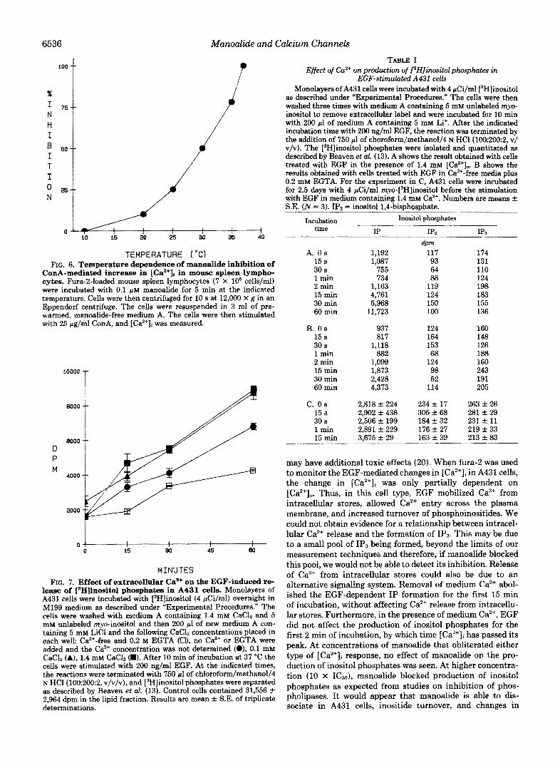
TEMPERATURE ("Cf FIG. 6. Temperature dependence of manoalide inhibition of
ConA-mediated increase in [Ca2'Ii in mouse spleen lympho- cytes. Fura-2-loaded mouse spleen lymphocytes (7 X lo6 cells/ml) were incubated with 0.1 p~ manoalide for 5 min at the indicated temperature. Cells were then centrifuged for 10 s at 12,000 X 8 in an Eppendorf centrifuge. The cells were resuspended in 3 ml of pre- warmed, manoalide-free medium A. The cells were then stimulated with 25 pg,fml ConA, and [Ca2+Ji was measured.
10000
9000
D P M
8000
4000
2000
0
MINUTES FIG. 7. Effect of extracellular CaB+ on the EGF-induced re-
lease of [SH]inositol phosphates in A431 cells. Monolayers of A431 cells were incubated with [3~]inositol (4 pCi/ml) overnight in M199 medium as described under "Experimental Procedures." The cells were washed with medium A containing 1.4 mM CaClz and 5 mM unlabeled myo-inositol and then 200 pl of new medium A con- taining 5 mM LiCl and the following CaC& concentrations placed in each well: Ca'+-free and 0.2 M EGTA (El), no Ca2+ or EGTA were added and the Ca2+ concentration was not determined (O), 0.1 mM CaClz (A), 1.4 mM CaClz (M). After 10 min of incubation at 37 "C the cells were stimulated with 200 ngfml EGF. At the indicated times, the reactions were terminated with 750 pl of chloroform/methanol/4 N HCl(lOO2002, v/v/v), and [3H]inositol phosphates were separated as described by Beaven et al. (13). Control cells contained 31,556 f 2,964 dpm in the lipid fraction. Results are mean Itt. S.E. of triplicate determinations.
TABLE I Effect of Go2+ on production of rHJinosit01 phosphutes in
EGF-stimulated A431 cells Monolayers of A431 cells were incubated with 4 pCi/ml [3H]inositol
as described under "Experimental Procedures." The cells were then washed three times with medium A containing 5 mM unlabeled myo- inositol to remove extracellular label and were incubated for 10 min with 200 pl of medium A containing 5 mM Li+. After the indicated incubation time with 200 ng/ml EGF, the reaction was terminated by the addition of 750 pl of chorofo~/methanol/4 N HC1(100:2002, v/ v/v). The [3H]inositol phosphates were isolated and quantitated as described by Beaven et al. (13). A shows the result obtained with cells treated with EGF in the presence of 1.4 mM [Ca2+j0. B shows the results obtained with cells treated with EGF in Ca2+-free media plus 0.2 mM EGTA. For the experiment in C, A431 cells were incubated for 2.5 days with 4 pCi/ml myo-['Hlinositol before the stimulation with EGF in medium containing 1.4 mM Ca2+. Numbers are means f S.E. ( N = 3). IP2 = inositol 1,4-bisphosphate.
Incubation Inositol phosphates time IP
" IP2 IPa
dPm -.
A. Os 1,192 117 174 15 s 1,087 93 131 30 s 755 64 110 1 min 734 88 124 2 min 1,163 119 198 15 min 4,761 124 183 30 min 6,968 150 155 60 min 11,723 100 136
B. Os 937 124 160 15 s 817 164 148 30 s 1,118 153 126 1 min 882 68 188 2 min 1,099 124 160 15 min 1,873 98 243 30 min 2,428 52 191 60 min 4,373 114 205
c. o s 2,818 rt 224 234 +- 17 263 k 26 15 s 2,902 f 438 305 f 68 281 Itt. 29 30 s 2,506 It 199 184 f 32 231 f 11 1 min 2,891 f 229 176 f 27 219 f 33 15 min 3,675 rt 29 163 f 39 213 f 83
may have additional toxic effects (20). When fura-2 was used to monitor the EGF-mediated changes in [Ca2+Ii in A431 cells, the change in [Ca2+Ii was only partially dependent on [Caz+]o. Thus, in this cell type, EGF mobilized Ca2+ from intracellular stores, allowed Caz+ entry across the plasma membrane, and increased turnover of phosphoinositides. We could not obtain evidence for a relationship between intracel- lular Ca2+ release and the formation of IPS. This may be due to a small pool of IPS being formed, beyond the limits of our measurement techniques and therefore, if manoalide blocked this pool, we would not be able to detect its inhibition. Release of Ca2+ from intracellular stores could also be due to an alternative signaling system. Removal of medium Ca2+ abol- ished the EGF-dependent IP formation for the first 15 min of incubation, without affecting ea2+ release from intracellu- lar stores. Furthermore, in the presence of medium Ca", EGF did not affect the production of inositol phosphates for the first 2 min of incubation, by which time [Ca2+Ii has passed its peak, At concentrations of manoalide that obliterated either type of [Ca2+Ii response, no effect of manoalide on the pro- duction of inositol phosphates was seen. At higher concentra- tion (10 X ICm), manoalide blocked production of inositol phosphates as expected from studies on inhibition of phos- pholipases, It would appear that manoalide is able to dis- sociate in A431 cells, inositide turnover, and changes in

Manoalide and Calcium Channels 6537
aOooo T
20000
P D M 15000/ /d 10OOO
5000 3 0 4 0 10 20 a0 4 0 5 0 80
MINUTES FIG. 8. Effect of manoalide on EGF-stimulated [‘HIIP pro-
duction in A431 cells. Cells were labeled with [3H]inositol as described under “Experimental Procedures.” The cells were prepared as described in the legend to Fig. 8. Manoalide at 1.5 pM (H) or vehicle (0) was then added and incubated at 37 “C for 10 min. The cells were then stimulated with 200 ng/ml EGF. Addition of mano-
triangle. At the indicated times, the reaction was terminated, and alide (1.5 p ~ ) with no EGF stimulation is represented by the solid
total water-soluble [3H]inositol phosphates determined. Control cells contained 96,269 f 3,835 dpm of 3H label in the lipid fraction. The data shown are mean f S.E. of triplicate determinations.
40000
a0000
D p 20000
M
10000
0 O
I I I I I
50 100 150 200
EGF (NG/ML) FIG. 9. Dose-dependent curve for EGF stimulation of IP
formation in control and manoalide-treated A431 cells. La- beling of cells with [3H]inositol and the experimental protocol was the same as in the legend to Fig. 8. The cells in medium A containing 1.4 mM CaC12 and 5 mM LiCl were incubated with 1.5 p~ manoalide (H) or vehicle (0) for 10 min at 37 “C before stimulation with the indicated concentration of EGF. After 60 min of incubation at 37 “C, the reaction was terminated, and total water-soluble [3H]inositol phosphates quantitated. The results shown are the mean f S.E. of triplicate determinations.
[Ca2+Ii. This would be anticipated if manoalide was acting as an inhibitor of Ca2+ channels.
GH3 cells possess two systems of interest in this connection. TRH mobilized intracellular Ca2+ stores by means of hydrol-
4500 i D
M .oo/ Jr 1500 4
0 0 1 2 a 4 5
MINUTES FIG. 10. Effect of manoalide on TRH-mediated IPS produc-
tion in GHs cells. Suspensions of GH3 cells were labeled with 4 pCi/ ml [3H]inositol as described under “Experimental Procedures.” The cells were then washed twice in medium A containing 5 mM myo- inositol and 1.4 mM CaCIZ, and then resuspended in medium A containing 1.4 mM CaClZ and 5 mM LiCl and incubated for 10 min at 37 “C. Cells were incubated for 5 min at 37 “C with either vehicle (O), 1 pM (H), 3 phi (A), or 10 pM (0) manoalide, before stimulation with 0.1 p~ TRH. At the indicated times, samples were removed, the reaction was terminated, and IP3 was separated on Dowex columns as described (13). The results shown are the mean f S.E. of three determinations.
TABLE I1 Effect of manoalide on forskolin-stimulated cyclic AMP production
in A431 cells A431 cells were incubated with 0,1, or 10 p~ manoalide for 10 min
at 37 “C. Then the indicated concentrations of forskolin were added and the incubation at 37 “C allowed to proceed for a further 15 min. At the end of the incubation with forskolin, the reaction was termi- nated by the addition of 0.5 ml of 30% trichloroacetic acid. The supernatant was collected for measurements of CAMP as described under “Experimental Procedures.” Values for control and 1 p~ man- oalide represent the mean f S.E. of three experiments in which the effect of each drug concentration was determined in triplicate. Values for 10 p~ manolide represent the mean k S.E. of triplicate determi- nations in one experiment.
Additions Cyclic AMP
Control Manoalide, 1 NM Manoalide, 10 p~
nmol/mg protein Vehicle 0.08 f 0.04 0.04 f 0.003 0.10 k 0.05 Forskolin
1 pM 0.20 f 0.07 0.22 f 0.07 ND”
100 LLM 6.94 f 1.90 7.55 k 2.00 8.49 k 0.36 10 pM 1.23 f 0.33 1.34 f 0.34 ND
a ND, not determined.
ysis of phosphatidylinositol 1,4,5-bisphosphate and release of IPS as well as increasing Ca2+ entry. TRH more than doubled [Ca2+Ii, and the initial increase was independent of [Ca2+l0. Manoalide inhibition of the TRH-dependent [Ca2+Ii signal had an apparent of 1 WM; a 10-fold greater manoalide concentration was required to inhibit partially the TRH in- duced increase of IP3 and other inositol phosphates (data not shown). In contrast t o A431 cells: we were able to show that, in GH3 cells, manoalide’s effect on [Ca2+Ii is dissociated from an effect of the drug on IPS production. Manoalide also blocked the K’ depolarization-activated Ca2+ channel as well

6538 Manoalide and :alcium Channels
A 1 derive more from its Ca" effects rather than from its effects on lipid metabolism.
Acknowkdgements-We thank S. Docheff and M. A. Pitale for their secretarial assistance and typing the manuscript. We also thank Drs. M. Garst and E. Tallman for isolating, purifying, and supplying the manoalide used in these studies and Dr. S. Dietrich for doing the log p calculation for manoalide.
FIG. 11. Effect of manoalide on diS-Cs-(5f distribution in GHa cells. GH3 cells were treated with vehicle (A), 1 p~ (B), 3 p~ (C), or 10 p~ (D) manoalide for 5 min at 37 "C before addition to medium A containing 1.4 mM CaC& and 1 p~ diS-C3-(5). The fluo- rescence of the dye was measured as described under ''Experimental Procedures."
as the activation of the channel by Bay K8644. Evidently, [Ca2+], changes with this protocol are due to Ca2+ entry. Manoalide readily blocked these responses with an ICso of 1.0 I.IM with similar characteristics as shown for the other Ca2+ pathways, in terms of ICW and time dependence. Thus, in these two cultured cell lines, manoalide was able to block hormone-operated plasma membrane Ca2+ pathways, path- ways of intracellular Ca2+ release, and voltage-operated plasma membrane pathways. The action of manoalide appears to inhibit IP3-linked agonist-induced release of intracellular Ca2+ distal to the generation of inositol phosphates.
Manoalide was found not to inhibit forskolin-stimulated adenylate cyclase in A431 cells or the uptake of the membrane potential sensitive dye diS-C3-(5). These two membrane-as- sociated activities were quantitated to determine whether the hydrophobicity (calculated log p = 4.5; method described in Ref. 25) of manoalide might produce nonspecific effects on cell membranes. DiS-C3-(5) is a lipid-permeable cation that accumulates in membrane-enclosed spaces as a function of a negative interior potential, and thus serves as a monitor of plasma membrane potential as well as mitochondrial poten- tial. Because manoalide did not alter the distribution of the dye, we conclude that manoalide affects Ca2+ mobilization across plasma and endoplasmic reticular membranes without interference with processes that determine potential differ- ences.
In the case of both bee venom (26,27) and cobra venom (2) phospholipase Az, manoalide has been suggested to bind ir- reversibly to a lysine group at or near the active site. The time and temperature dependence of the action of manoalide on Ca2+ mobilization, as well as the difficulty of reversal by washing, suggests that derivatization of cellular sites other than phospholipase C or phospholipase A2 might be respon- sible for the effects observed in these experiments. The activ- ity of manoalide allows some dissection of the Ca2+ signals from phosphoinositide metabolism and thus provides a probe for studying Ca2+ signaling in a variety of normal and trans- formed cell types and cellular compa~ments. The anti-inflam- matory and anti-proliferative activities of manoalide may
Note Added in Proof-EGF-induced IP3 turnover has been recently reported by Pike and Eakes (Pike, L. J., and Eakes, A. T. (1987) J. Bwl. Chem. 262, 1644-1651) which is consistent with the release of Ca2+ from intracellular stores reported in this study.
REFERENCES 1. Jacobs, R. C. Culver, P., Langdon, R., O'Brien, T., and White,
2. Lombardo, D., and Dennis, E. (1985) J. BioL Chem. 260, 7234-
3. Glaser, K., and Jacobs, R. (1986) Biochem. PhurmacoL 35,449-
4. Chandra Sekar, M., and Hokin, L. E. (1986) J. Membr. Biol. 89,
5. Berridge, M., and Imine, R. (1984) Nature 312, 315-321 6. Berridge, M. (1983) Bioehem. J. 212,849-858 7. Bennett, C., Mong, S., Wu, H.-L., and Crooke, S. (1986) Phar-
8. Bolton, T. B. (1979) Physiol. Reu. 59,607-718 9. Rasmussen, H., and Barrett, P. (1984) PhyswZ. Rev. 64,938-976 10. Volpi, M., Naccache, P., and Sha'afi, R. (1980) Bioe~im. Bwphys.
11. Rubin, R. (1982) Fed. Proc. 41,2181-2187 12. Putney, J., Weiss, S., Van De Walle, C., and Haddas, R. (1980)
13. Beaven, M., Moore, J., Smith, G., Hesketh, T., and Metcalfe, J.
14. Moolenaar, W., Aerts, R., Tertoolen, L., and de Laat, S. (1986)
15. Drummond, A. (1985) Nature 315,752-755 16. Nowycky, M., Fox, A., and Tsien, R. (1985) Proc. Natl. Acad. Sci.
17. Schramm, M,, Thomas, G., Toware, T., and Franckowiak, G.
18. Harper, J. F., and Brooker, G. J. (1975) J. Cyclic Nucleotide Res.
19. Steiner, A. L., Parker, C. W., and Kipnis, D. M. (1972) J. Bwl.
20. Allen, T. J., and Baker, P. F. (1985) Nature 315, 755-756 21. Sawyer, S., and Cohen, S. (1981) Biochemistry 20,6280-6286 22. MacPhee, C., and Drummond, A. (1984) Mol. Pharmacol. 25,
23. Drummond, A., Bushfield, M., and MacPhee, C. (1984) M O L
24. Strawb, R., and Gershengorn, M. (1986) J. Biol. Chem. 261,
25. Hansch, C., and Leo, A. J. (1979) S ~ s t i ~ ~ n ~ constants for Correlation Analysis in Chemistry and Biology, John Wiley & Sons, New York
W. (1985) T e t r a ~ d r o n Lett. 41,981-984
7240
453
193-210
macologist 28, 538 (abstr.)
Res. Commun. 92,1231-1237
Nature 284,345-347
(1984) J. Bwl. Chem. 259, 7137-7142
J. Bwl. Chem. 261,279-284
u. s. A. 82,2178-2182
(1983) Nature 303,535-537
1,207-218
Chem. 247,110&1113
193-200
Pharmacol. 25, 201-208
2712-2717
26. Glaser, K., and Jacobs, R. (1986) Biochem. Pharmol . , in press 27. Bennett, C., Mong, S., and Crooke, S. (1986) Bkhem. Pharma-
cot., in press