manual of operations - children's hospital
TRANSCRIPT

Version Date 04/11/14 PBTC NIC Manual of Operations Title, Table of Contents, Legends
Page 1 of 5
Pediatric Brain Tumor Consortium
Neuroimaging Center
Manual of Operations
Children's Hospital Boston

Version Date 04/11/14 PBTC NIC Manual of Operations Title, Table of Contents, Legends
Page 2 of 5
Pediatric Brain Tumor Consortium
Neuroimaging Center
Manual of Operations
Created and Edited by
Tina Young Poussaint, MD Children's Hospital, Boston
Robert Mulkern, PhD Children's Hospital, Boston
Fred Fahey, DSc Children's Hospital, Boston
Voula Osganian, MD, ScD Children's Hospital, Boston
This grant is supported by funds from the National Cancer Institute (NIH-NCI U01 CA 81456-12)
First produced: 6/1/03. Revised: 1/22/04, 4/23/04, 5/11/04, 5/27/04, 7/6/04, 8/10/04, 11/03/04, 12/21/04, 02/01/05, 05/05/05, 10/19/05, 02/01/06, 05/17/06, 9/26/06, 02/19/07, 01/01/08,
09/03/08, 10/29/08, 11/10/08, 12/02/08, 04/14/09, 07/07/09, 08/19/09, 11/19/09 01/15/10, 02/02/10, 05/10/10, 06/16/10, 06/22/10, 07/27/10, 06/28/11, 09/07/11, 10/11/11, 03/14/12, 04/13/12, 05/04/12, 05/15/12, 10/30/12, 12/18/12, 03/26/13, 05/15/13, 08/15/13,
01/24/14, 4/11/14

Version Date 04/11/14 PBTC NIC Manual of Operations Title, Table of Contents, Legends
Page 3 of 5
TABLE OF CONTENTS I. Overview and Goals .............................................................. Section I II. Study Organization ................................................................ Section II III. Neuroimaging Studies ............................................................ Section III IV. Procedures for Transfer of Imaging Studies ........................... Section IV V. Guidelines for Reading and Interpreting Imaging Studies ...... Section V VI. Data Management Procedures ............................................... Section VI VII. PBTC NIC Data Management System (DMS) ........................ Section VII VIII. Quality Control Approaches .................................................... Section VIII IX. Appendices ............................................................................. Section IX
Appendix A: PBTC NIC MR Protocols of Brain and Spine Appendix B: PBTC NIC MRI Quality Assurance Procedures Appendix C: PBTC NIC PET Facility Procedures Appendix D: PBTC NIC Case Report Forms

Version Date 04/11/14 PBTC NIC Manual of Operations Title, Table of Contents, Legends
Page 4 of 5
FIGURE, TABLE AND FORM LEGENDS FIGURE LEGEND
Figure 1 PBTC Organization Figure 2 PBTC Neuroimaging Center
Organization TABLE LEGEND
Table 1 PBTC Neuroimaging Committee Table 2 PBTC MR Equipment List Table 3 PBTC PET Protocol Equipment and
Investigators Section A, B, C, D Table 4 PBTC Institutions and Principal
Investigators Table 5 PBTC Site CRA list Table 6 PBTC MR Technologists Contact List Table 7 PBTC Protocols:
Neuroimaging Objectives Table 8 PBTC Protocols:
Imaging Components FORMS LEGEND [Legacy and current forms in use]
Form 100a MRI Brain Data Form 100b MRI Brain Data Form 2 MRI Quality Assurance Form 3 Retired: No longer in use Form 4 PET Brain Data Form 5 PET Quality Assurance Form 6 MRI Spine Data Form 7 Retired: No longer in use Form 8 Retired: No longer in use Form 9a MRI Brain Leptomeningeal Data Form 10 Retired: No longer in use Form 11 CT Hemorrhage Data Form 12 Retired: No longer in use Form 13 Retired: No longer in use Form 14 PET Support Data Form 15 PET Phantom Acquisition Form Form 16 Merged: Now part of Form 1a Form 17 Fusion Volumetrics Form 18 MRI Brain Permeability Form 19 Site Visit Mandatory Reporting Form Form 20 68Ge ACR Phantom Acquisition Form Form 21 Spinal Tumor Volume Form 22 Comments Form Form 23 RECIST Measurements Form 24 MacDonald Criteria

Version Date 04/11/14 PBTC NIC Manual of Operations Title, Table of Contents, Legends
Page 5 of 5
FORMS LEGEND [Legacy and current forms in use] Continued Form 25 Diffusion Tensor Imaging Form 26 MR Contrast Survey Form 27 ADC Histogram Analysis Form 28 MRI Brain Permeability
Section IPBTC NIC Manual of OperationsOverview and Goals

Version Date 01/01/2008 PBTC NIC Manual of Operations Section I – Overview and Goals
Page 1 of 1
I. Overview and Goals The Pediatric Brain Tumor Consortium (PBTC) is a multidisciplinary cooperative research organization devoted to the study of correlative tumor biology and new therapies for primary CNS tumors of childhood. Its mission is to contribute rapidly and effectively to the understanding and cure of these tumors through the conduct of multi-center, multidisciplinary, innovative studies with designs and analyses based on uniformly high quality statistical science. While the primary mission of the PBTC is to identify through laboratory and clinical science superior treatment strategies for children with brain cancers, the PBTC investigators recognize their profound responsibility to meet the special needs of the children and families as they face this enormous challenge. Members are committed to working within their institutions and communities to improve support services and follow up care for these patients and their families. The goals of the PBTC Neuroimaging Center (NIC) are:
To provide leadership in the diagnostic imaging component of all clinical PBTC protocols including the development, implementation and / or coordination of all current and future research protocols for diagnostic imaging studies. These include but are not limited to MRI, MR diffusion, MR perfusion, MR spectroscopy, CT and PET;
To direct quality assurance activities for the neuroimaging studies including the oversight of compliance with imaging protocols and assessments of diagnostic image quality, data integrity and storage/retrieval;
To provide centralized review and interpretation of all diagnostic neuroimaging studies and analyze CT, MRI, MRS, PET and other images to generate summary data that will be provided to the Operations and Biostatistics Center (OBC) for statistical analysis;
To develop a correlative imaging research plan related to the novel therapeutic interventions of the PBTC;
To ensure PBTC’s ability to incorporate imaging endpoints in its overall research program;
To oversee cross-platform translations for comparative analyses of MRI, MRS, and PET studies.

Section IIPBTC NIC Manual of OperationsOrganization

Version Date: 08/15/2012 PBTC NIC Manual of Operations Section II – Organization
Page 1 of 15
II. Organization
The overall organization of the Pediatric Brain Tumor Consortium (PBTC) is outlined in Figure 1. The consortium includes 10 participating Institutions and a special member (Neurooncology Branch NCI/NINDS), for the recruitment of patients into PBTC protocols, an Operation and Biostatistics Center, a Neuroimaging Center, and Central Laboratories. The Steering Committee is the primary decision-making body for the study with scientific oversight provided by an external Scientific Advisory Board. The work of the Consortium is accomplished through several ad-hoc and standing committees. Specifically, the Neuroimaging Committee (NC) is composed of one neuroradiologist from each participating institution and the PI of the NIC, Dr. Tina Young Poussaint. In addition there are external and internal liaisons on the committee. Under the leadership of Dr. Poussaint, the NC is responsible by consensus for the development of clear and standard procedures for the performance of all PBTC MR Neuroimaging studies across the sites and for all MR neuroimaging quality control procedures. In addition, the NC reviews imaging study data quality reports on a regular basis, provides recommendations regarding possible neuroimaging research questions that may be appropriate to be considered in relevant treatment protocols; and assists in the evaluation of research neuroimaging protocol feasibility, methods, and scientific merit. A PET investigator committee consisting of one PET investigator per site, provides input regarding PET QA, PET analyses, protocols, radiopharmaceuticals and PET research projects.
Figure 1 presents the PBTC organizational structure. Table 1 presents the MR Contacts.
Figure 2 presents the organizational structure of the NIC. Figure 3 presents the organizational structure of the Clinical Research Center at Children’s Hospital Boston. Table 2 lists the MR Equipment information for each site. Table 3 lists the PET Equipment and Investigators from each site. Table 4 lists the PIs at each respective site. Table 5 lists the CRAs at each site. Table 6 lists the contact MR Technologists at each site.

Version Date 08/15/2013
PBTC NIC Manual of Operations Section II – Organization
Page 2 of 15
Figure 1. Pediatric Brain Tumor Consortium Organization

Version Date 08/15/2013
PBTC NIC Manual of Operations Section II – Organization
Page 3 of 15
Table 1. MR Neuroimaging Contacts
Name Institution Email Address Position
Poussaint, Tina Young Boston Children’s/DFCI [email protected] Chair
Vezina, Gilbert Children’s National [email protected]
Zimmerman, Robert CHOP* [email protected]
Lascola, Christopher Duke [email protected]
Panigrahy, Ashok Children’s Pittsburgh [email protected]
Patay, Zoltan St. Jude [email protected]
Shaw, Dennis Seattle Children’s* [email protected]
Jones, Jeremy Texas Children’s [email protected]
Cha, Soonmee UCSF* [email protected]
Evers, Robert NIH [email protected]
Burrowes, Delilah Lurie Children’s [email protected]
Jones, Blaise Cincinnati Children’s [email protected]
Nelson, Marvin CHLA [email protected]
Haque, Sofia MSKCC [email protected]
Yeom, Kristen Stanford [email protected]
Mulkern, Robert Boston Children’s* [email protected]
*Temporary Member Institutions

Version Date 08/15/2013
PBTC NIC Manual of Operations Section II – Organization
Page 4 of 15
Figure 2. PBTC Neuroimaging Center Organization

Version Date 08/15/2013
PBTC NIC Manual of Operations Section II – Organization
Page 5 of 15
Figure 3. Children’s Hospital Boston Clinical Research Center Organization

Version Date 08/15/2013
PBTC NIC Manual of Operations Section II – Organization
Page 6 of 15
Table 2. MR Equipment List (2013)
Site Code
Site # of
Magnets MRI Manufacturer Field Strength Software Version
11 Children’s National
Washington, DC Dr. Gilbert Vezina
2 GE Discovery 450 GE Optima 450W
1.5T 1.5T
20.1 21.0 M3
1 GE Discovery 750 (1) 3.0T 20.1
12 Children’s Hospital of Philadelphia
Philadelphia, PA Dr. Robert Zimmerman
5 Siemen Verio (2) Siemens Avanto (2) Siemen Symphony (1)
3T 1.5 T 1.5 T
B17 & B17 B17 & B17 B17
2 Siemen Trio (2) 3.T B17
13 Duke University Medical Center
Durham, NC Dr. Christopher Lascola
6 GE Signa 1.5 (4) Siemens Avanto (2)
1.5T 1.5T
15xM4 VB15
2 Siemens Trio 3.0 (1) 3.0T VB 15 VB 15
1 Siemens Skyra 3.0T
Siemens Aera 1.5T
14 Children’s Hospital Boston
Boston, MA Dr. Robert Mulkern
1 GE 1.5 T 15.0
4 Siemens 3 T VB17
2 Siemens 1.5 T VB17
15 Children’s Hospital Pittsburgh
Pittsburgh, PA Dr. Ashok Panigrahy
2 GE 1.5T (2) 1.5T 16.0
1 GE 3.0T (1) 3.0T 16.0
1 Siemens Skyra (1) 3.0T Not available.
16 St. Jude Children’s Research Hospital
Memphis, TN Dr. Zoltan Patay
2 Siemens Avanto (2) 1.5T VB15
1 Siemens Trio (1) 3.0T VB15
1 Siemens Skyra (1) 3.0T VB17
17 Children’s Hospital Regional Medical Center
Seattle, WA Dr. Dennis Shaw
1 Siemens 1.5 Avanto (2) [Siemens Trio - not used for PBTC]
1.5T [3T]
VB17 VB17
18 Texas Children’s Hospital at Baylor
Houston, TX Dr. Jeremy Jones
4
Philips 1.5T Achieva Philips 1.5T Achieva Philips Achieva Philips Achieva
1.5T 1.5T 1.5T 1.5T
Manufacturer supplied software
1 Philips 3.0T Achieva 3.0T
19 UCSF Children’s Hospital 4 GE 1.5T (4)
1.5T 12.0, 14.0(2), 15.0

Version Date 08/15/2013
PBTC NIC Manual of Operations Section II – Organization
Page 7 of 15
San Francisco, CA Dr. Soonmee Cha
3 GE Excite 3T (3) 3.0T 14.0, 750(2)
Site Code
Site # of
Magnets MRI Manufacturer Field Strength Software Version
20 National Institutes of Health
Bethesda, MD Dr. Nicholas Patronas
2 Siemens 3.0T VB17
3 Philips (1) 1.5T 2.6.3 ~ 3.1
21 Lurie Children’s Hospital
Chicago, IL Dr. Delilah Burrowes
2 Siemens Aera (2) 3.0T Not available.
1 Siemens Skyra (1) 3T Not available.
1 GE Signa HDxt (1) 1.5T Not available.
1 GE Signa Excite (1) 1.5T Not available.
22 Cincinnati Children’s Hospital Medical Center
Cincinnati, OH Dr. Blaise Jones
6
GE (3) GE (1) GE (1) GE (1)
1.5T
16HDxT 15HDx 16HDxT/3.54ONI 15HDx/3.53ONI
2 Philips (2) 3T R3.2.1
1 Siemens 1.5T VB17
1 Bruker 7T Paravision 5.1
23 Children’s Hospital Los Angeles
Los Angeles, CA Dr. Marvin Nelson
1 GE 1.5T Signa – 9.1 M4
1 Philips 1.5T Achieva 3.0
2 Philips 3.0T Achieva 3.0
24 Memorial Sloan-Kettering Cancer Center
New York, NY Dr. Sofia Haque
1 GE Signa 3T V15.0
2 GE Signa 1.5T v12.0
1 GE 450W 1.5T V12.0
1 GE 750 3T V15.0
25 Lucile Packard Children’s Hospital at Stanford
Palo Alto, CA Dr. Kristen Yeom
2 GE MR750 (1) GE MR450W (1)
3T 1.5T
HD 16.0_v01 DV22_Z02

Version Date 08/15/2013
PBTC NIC Manual of Operations Section II – Organization
Page 8 of 15
Table 3. PET Equipment List (2013) SECTION A: Equipment
Site Code
Institution Equipment Vendor/Model
Contact Name
Personnel Contact Telephone
Contact Email
11 Children’s National Medical Center Washington, DC
GE Discovery 690 PET/MDCT Pranav Vyas, MD Thomas Fearon, PhD
PET Physician PET Physicist
202-476-5630 202-476-5075
[email protected] [email protected]
13 Duke University Medical Center Durham, NC
GE Discovery STE PET/CT GE Discovery 690 PET/CT
James, Olga, MD Turkington, Timothy PhD Hawk, Thomas, BS
PET Physician PET Physicist Computer Specialist
919-684-7647 919-684-7706 919-684-7714
[email protected] [email protected] [email protected]
14 Boston Children’s Hospital Boston, MA
Siemens mCT 40 PET/CT Treves, Ted, MD Fahey, Fred, DSc Cao, Xinhua, PhD
PET Physician PET Physicist Computer Specialist
617-355-7935 617-355-2809 617-355-5563
[email protected] [email protected] [email protected]
15 Children’s Hospital Pittsburgh of UPMC Pittsburgh, PA
GE Discovery DVCT PET/CT Joyce, Judith, MD N/A Czachowski, Michael, MBA
PET Physician PET Physicist Nuclear Tech Supervisor
412-647-0104 412-692-9518
[email protected] [email protected]
16 St Jude Children’s Research Hospital Memphis, TN
GE Discovery 690 Shulkin, Barry, MD, MBA Brady, Samuel, PhD Foster, Nancy
PET Physician PET Physicist Computer Specialist
901-595-3347 [email protected] [email protected] [email protected]
18 Texas Children’s Hospital at Baylor Houston, TX
Philips 3T PET/MRI TF Philips Gemini TF Select 16 Slice PET/CT
Seghers, Victor, MD, PhD McMullen, Tara Walters, Veronica
PET Physician PET Technologist Admin. Asst.
832-826-8525 832-826-8578 832-826-8525
[email protected] [email protected] [email protected]
20 National Institutes of Health Bethesda, MD
GE Advance (2) PET/CT HRRT Siemens MCT PET/CT
Herscovitch, Peter, MD Barker, Craig, PhD Fraser, Charles
PET Physician PET Physicist Computer Specialist
301-451-4248 301-451-3558 301-451-3591
[email protected] [email protected] [email protected]
21 Lurie Children’s Hospital of Chicago Chicago, IL
GE PET/CT Discovery 690 12123PT6
Shore, Richard, MD Delilah Burrowes, MD Christina Sammet, PhD
PET Physician PET Physician PET Physicist
312-227-3504 312-227-4388 312-227-3393
[email protected] [email protected] [email protected]
22 Cincinnati Children’s Hospital Medical Center Cincinnati, OH
Philips Ingenuity TF Michael J. Gelfand, MD Susan E. Sharp, MD (backup) Lisa Lemen, PhD Jay Moskovitz (information tech) Leonid Rozhkov (analytic methods)
PET Physician PET Physician PET Physicist Computer Specialist Computer Specialist
513-636-7650 513-636-6662 513-558-2197 513-636-5981 513-803-0545
[email protected] [email protected] [email protected] [email protected] [email protected]
23 Children’s Hospital Los Angeles Los Angeles, CA
Philips/Gemini GXL16 PET/CT Hollie A. Jackson, MD PET Physician 323-361-4570 [email protected]
24 Memorial Sloan-Kettering Cancer Center New York, NY
General Electric PET/CT Neeta Pandit-Taskar, MD Sadek Nehmeh Ross Schmidtlein Brad Beattie John Humm Hovanes Kalaigian
PET Physician PET Physicist PET Physicist PET Physicist PET Physicist Computer Specialist
212-639-7372 212-639-2127 212-639-8082 646-888-2579 212-639-7367 212-639-7382
[email protected] [email protected] [email protected] [email protected] [email protected] [email protected]
25 Lucile Packard Children’s Hospital at Stanford Palo Alto, CA
GE D690 PET/CT GE D600 PET/CT
Andrew Quon, MD Craig Levin, PhD
PET Physician PET Physicist
650-725-4711 650-736-7211

Version Date 08/15/2013
PBTC NIC Manual of Operations Section II – Organization
Page 9 of 15
Table 3. PET Equipment List (2013) SECTION B Radiopharmaceuticals SECTION C Image Analysis Equipment SECTION D Research Interests
Site Code
Institution Radiopharmaceuticals Used with Brain imaging
Image Analysis Equipment
Brain Imaging research at site unrelated to the PBTC
11 Children’s National Washington, DC
FDG GE AW workstation Projects in fetal MRI, fMRI
13 Duke University Medical Center Durham, NC
F-FDG (18), F-FLT (18), O2 (15), C (15) o2, h2 (15) O
GE AW and Xeleris, Unix WS
Radiolabeled mab therapy for brain tumors – PET f/u, Metabolic effects of radiation therapy on cognitive function (FDG, H2(15) O), dementia, plaque imaging compounds for dementia , FLT
14 Boston Children’s Hospital Boston, MA
FGD, FLT Hermes Medical Solution Workstation
Animal Micro PET, Epilepsy-spect
Dana Farber Cancer Institute Boston, MA
FDG, FLT (pending) ECAT Software (CTI), Xeleris/AW (GE), Hermes, Siemens Leonardo/True D
Adult brain tumors. Central PET core lab for global multi-center trial of lapatinib in brain metastases in women with breast cancer
15 Children’s Hospital Pittsburgh of UPMC Pittsburgh, PA
FDG GE Discovery DVCT Possible FLT in near future
16 St Jude Children’s Research Hospital Memphis, TN
11 C-MET-PET 18 F-FDG
Hermes Medical Solution Workstation
C-11 methionine uptake for CNS tumor survey Relation of histology to FDG uptake
18 Texas Children’s Hospital at Baylor Houston, TX
FDG
MIM vista software Philips Portal
No participation in Brain imaging research prior to PBTC
20 National Institutes of Health Bethesda, MD
FDG, H20, fdopa, fcway, flumazenil, nnc, leucine, dasb, fallypride, raclopride, methylreboxitine, DTBZ, FP-TZTP, arachidonic acid, docosahexaenoic acid
PACS Stroke, epilepsy, Alzheimer’s, Parkinson’s, schizophrenia, depression, obsessive compulsive disorder, tumor, alcohol abuse, movement disorders, Fragile X
21 Lurie Children’s Hospital of Chicago Chicago, IL
FDG GE PET/CT Discovery 690 12123PT6 AW
N/A
22 Cincinnati Children’s Hospital Medical Center Cincinnati, OH
F-18-FDG (2,3), Tc-99m-ECD (2,3), Tc-99m-HMPAO (2,3), F-18-fallypride (1,3,4), F-18-FDOPA (1,2,3), F-18-choline (1,3,5), C-11-methionine (1,2,6) *
Analyze 10, SPM, Brain Lab Epileptogenic focus localization, Dopamine transporter density (1)
* (1)=Available in co-operation with Kettering Medical Center, Dayton, OH. (2)=Used for clinical studies. (3)=Available locally. (4)=Used for human research studies only. (5)=Only used for non-human experiments to date. (6)=Will require patient transportation to off campus site.
23 Children’s Hospital Los Angeles Los Angeles, CA
F18-FDG N/A N/A
24 Memorial Sloan-Kettering Cancer Center New York, NY
FDG, FLT, and FACB GE PET VCAR Hermes Medical Systems
F-choline in brain (PI at site is Kathryn Beall)
25 Lucile Packard Children’s Hospital at Stanford Palo Alto, CA
FDG, FLT, Amyvid GE Advantage Windows Amyvid (AV-45)

Version 08/15/2013 PBTC NIC Manual of Operations Section II – Organization
Page 10 of 15
Table 4. PBTC Institutions and Principal Investigators
CHILDREN’S NATIONAL MEDICAL CENTER
CHILDREN’S HOSPITAL OF PHILADELPHIA
Roger Packer, M.D. Children's National Medical Center
Department of Neurology 111 Michigan Avenue, NW Washington, DC 20010 TEL: 202/884-2120 FAX: 202/884-5226 E-MAIL: [email protected] SITE 11
Peter Phillips, M.D. The Children's Hospital of Philadelphia
Division of Neuro-Oncology 4
th Floor Wood Building
3400 Civic Center Boulevard Philadelphia, Pennsylvania 19104 TEL: 215/590-2800 FAX: 215/590-3709 E-MAIL: [email protected] SITE 12
BRAIN TUMOR CENTER AT DUKE DANA-FARBER CANCER INSTITUTE
Sri Gururangan, M.D. The Brain Tumor Center at Duke
Duke University Medical Center Box 3624 Durham, North Carolina 27710 TEL: 919/668-6288 FAX: 919/681-1697 E-MAIL: [email protected] SITE 13
Mark W. Kieran, M.D., Ph.D. Dana-Farber Cancer Institute
Department of Pediatrics 44 Binney Street Boston, Massachusetts 02115 TEL: 617/632-4907 FAX: 617/632-4248 E-MAIL: [email protected] SITE 14
ST. JUDE CHILDREN’S RESEARCH HOSPITAL
James M. Boyett, Ph.D. Executive Director, PBTC Operations & Biostatistics Center
St. Jude Children’s Research Hospital Department of Biostatistics Memphis, TN 38105 TEL: 901/595-4986 FAX: 901/595-4184 EMAIL: [email protected] Site 16
Larry Kun, M.D. St. Jude Children's Research Hospital
Department of Radiation Oncology 262 Danny Thomas Place Memphis, Tennessee 38105-3678 TEL: 901/595-3604 FAX: 901/595-3113 E-MAIL: [email protected] SITE 16
CHILDREN’S HOSPITAL OF PITTSBURGH CHILDREN’S HOSPITAL & REGIONAL MEDICAL CENTER – SEATTLE
Ian Pollack, M.D. Children's Hospital of Pittsburgh
Department of Neurological Surgery 3705 Fifth Avenue Pittsburgh, Pennsylvania 15213 TEL: 412/692-5090 FAX: 412/692-5921 E-MAIL: [email protected] SITE 15
J. Russell Geyer, M.D. Seattle Children’s Hospital
Department of Pediatrics 4800 Sand Point Way NE B6553 Seattle, Washington 98105 TEL: 206/987-6664 FAX: 206/987-3946 E-MAIL:[email protected] SITE 17
TEXAS CHILDREN’S CANCER CENTER / BAYLOR
UNIVERSITY OF CALIFORNIA, SAN FRANCISCO
Murali Chintagumpala, M.D. & Patricia Baxter, M.D. Texas Children’s Cancer Center
6621 Fannin Street, MC 3-3320 Houston, Texas 77030 TEL: 800/CANCER9 FAX: 832/825-4299 E-MAIL: [email protected]; [email protected] SITE 18
Michael Prados, M.D. University of California, San Francisco
Department of Neurological Surgery 400 Parnassus, Room A808 San Francisco, California 94143-0372 TEL: 415/353-2966 FAX: 415/353-2167 E-MAIL: [email protected] SITE 19

Version 08/15/2013 PBTC NIC Manual of Operations Section II – Organization
Page 11 of 15
Table 4. PBTC Institutions and Principal Investigators (continued)
NATIONAL INSTITUTES OF HEALTH CHILDREN’S MEMORIAL HOSPITAL
Kathy Warren, M.D. NIH/NCI
Pediatric Oncology Branch Bldg 10, CRC, Rm 1-5750 Bethesda, MD 20892-1104 TEL: 301/435-4683 FAX: 301/451-7052 E-MAIL: [email protected] SITE 20
Stewart Goldman, M.D. Children's Memorial Hospital
2300 Children's Plaza Chicago, IL 60614-3394 TEL: 773/ 880-4562 FAX: 773/880-3223 E-MAIL: [email protected] SITE 21
CINCINNATI CHILDREN’S HOSPITAL MEDICAL CENTER
CHILDREN’S HOSPITAL LOS ANGELES
Maryam Fouladi, M.D. CCHMC
Medical Director, Neuro-Oncology Program 3333 Burnet Avenue Cincinnati, OH 45229-3062 TEL: 513/636-4266 FAX: 513/636-3549 E-MAIL: [email protected] SITE 22
Girish Dhall, M.D. CHLA
Neural Tumors Program 4650 Sunset Boulevard, MS #54 Los Angeles, CA 90027-6016 TEL: 323/361-4629 FAX: 323/361-8165 E-MAIL: [email protected] SITE 23
MEMORIAL SLOAN-KETTERING CANCER CENTER
LUCILE PACKARD CHILDREN’S HOSPITAL AT STANFORD
Ira Dunkel, M.D. MSKCC
Pediatric Oncology Branch 1275 York Avenue New York, NY 10065 TEL: 212/639-2153 FAX: 212/717-3239 E-MAIL: [email protected] SITE 24
Paul Graham Fisher, M.D. Stanford
Neurology and Pediatrics 750 Welch Road, Suite 317 Palo Alto, CA 94304-1510 TEL: 650/721-5889 FAX: 650/723-7299 E-MAIL: [email protected] SITE 25

Version 08/15/2013 PBTC NIC Manual of Operations Section II – Organization
Page 12 of 15
Table 5. PBTC Clinical Research Associates/Nurses
LURIE CHILDREN’S HOSPITAL OF CHICAGO
Kelly Verel Lurie Children’s (773) 880-8147 [email protected]
Jillian Tabares Lurie Children’s (312) 227-4810 [email protected]
Carrie Kempler Lurie Children’s (773) 883-6186 [email protected]
CHILDREN’S NATIONAL MEDICAL CENTER
Cynthia Arevalo Children’s National (202) 476-5115 [email protected]
Erin Watson Children's National (202) 476-6493 [email protected]
Chad Stephens Children’s National (202) 476-4481 [email protected]
Meenal Pathak Children’s National (202) 476-4481 [email protected]
CHILDREN’S HOSPITAL OF PITTSBURGH
Sharon Dibridge Children’s Pittsburgh (412) 692-7070 [email protected]
Angela Krol Children’s Pittsburgh (412) 692-8047 [email protected]
Judith Cawley Children’s Pittsburgh (412) 692-5059 [email protected]
CHILDREN’S HOSPITAL OF PHILADELPHIA
Ratnakar Patti CHOP (267) 426-5503 [email protected]
DANA FARBER CANCER INSTITUTE
Lianne Hawes Dana Farber (617) 632-6740 [email protected]
BRAIN TUMOR CENTER AT DUKE
Jennifer King Duke (919) 668-5364 [email protected]
Waynette Freeman Duke (919) 684-3440 [email protected]
Jeanne Krauser Duke (919) 681-2546 [email protected]
Denise Jaggers Duke (919) 613-6803 [email protected]
NATIONAL INSTITUTES OF HEALTH
Linda Ellison NIH (301) 496-8009 [email protected]
Natasha Brunson NIH (301) 451-7747 [email protected]
Sharon Mavroukakis NIH (301) 231-4876 [email protected]
Candace Menke NIH (301) 451-5007 [email protected]
CHILDREN’S HOSPITAL & REGIONAL MEDICAL CENTER – SEATTLE
Kim Starr Children’s Seattle (206) 987-7274 [email protected]
Jonathan Liaw-Gray Children’s Seattle (206) 884-5184 [email protected]
Cory Hoeppner Children’s Seattle (206) 987-3176 [email protected]
Celeste Oglesby Children’s Seattle (206) 987-1457 [email protected]

Version 08/15/2013 PBTC NIC Manual of Operations Section II – Organization
Page 13 of 15
Table 5: PBTC Clinical Research Associates/Nurses (continued)
ST. JUDE’S CHILDREN’S RESEARCH HOSPITAL
Samantha Buhler St. Jude (901) 595-5124 [email protected]
Annemarie McClellan St. Jude (901) 595-2734 [email protected]
Kate Marshall St. Jude (901) 595-4922 [email protected]
Molly Clarke St. Jude (901) 595-3567 [email protected]
Letitia Williams St. Jude (901)595-5597 [email protected]
TEXAS CHILDREN’S CANCER CENTER / BAYLOR
Susan Burlingame Texas (832) 824-1532 [email protected]
Manju George Texas (832) 824-1531 [email protected]
Bridget Medina Texas (832) 824-6858 [email protected]
Suzanne Hegemier-Wheeler Texas (832) 824-4217 [email protected]
Renee Klenke Texas (832) 824-4570 [email protected]
Susan Milligan Texas (832) 824-2138 [email protected]
Megan Tucker-Hall Texas (832) 824-4306 [email protected]
UNIVERSITY OF CALIFORNIA, SAN FRANCISCO
Sharon Lee-Kresge UCSF (415) 514-3658 [email protected]
Linda Li UCSF (415) 476-7910 [email protected]
Annie Le UCSF (415) 353-9387 [email protected]
CINCINNATI CHILDREN’S HOSPITAL MEDICAL CENTER
Lori Backus CCHMC (Cincinnati) (513) 636-2047 [email protected]
Courtney Blank CCHMC (513) 803-3255 [email protected]
Renee Doughman CCHMC (513) 803-0471 [email protected]
CHILDREN’S HOSPITAL LOS ANGELES
Jennifer Harrington CHLA (323) 361-7319 [email protected]
Corazon (Cora) Cruz CHLA (323) 361-4126 [email protected]
Kelley Haley CHLA (323) 361-2480 [email protected]
MEMORIAL SLOAN-KETTERING CANCER CENTER
Marilyn Winchester MSKCC (646) 888-5723 [email protected]
Judy Persaud MSKCC (646) 888-5722 [email protected]
LUCILE PACKARD CHILDREN’S HOSPITAL AT STANFORD
Carissa Bailey Stanford (650) 725-4708 [email protected]
Christina Huang Stanford (650) 723-0574 [email protected]

Version 08/15/2013 PBTC NIC Manual of Operations Section II – Organization
Page 14 of 15
Table 6. MR Technologists
CHILDREN’S MEMORIAL HOSPITAL – CHICAGO
Marci Messina 773-880-3404 [email protected]
Kimberly Ritzke (QA contact) 773-880-3404 [email protected]
CHILDREN’S HOSPITAL BOSTON
Diane Biagiotti 617-355-5154 [email protected]
CHILDREN’S HOSPITAL OF PHILADELPHIA
Glenn Ferrick 267-426-3199 [email protected]
Christine Harris 215-590-4116 [email protected]
BRAIN TUMOR CENTER AT DUKE
Joe Hinton 919-684-7254 [email protected]
CHILDREN’S HOSPITAL OF PITTSBURGH
Dennis Willaman 412-692-3038 [email protected]
ST. JUDE’S CHILDREN’S RESEARCH HOSPITAL
Mary Jo Freeman 901-595-2013 (office) 901-595-3578 # 0933 (pager) [email protected]
Carolyn Phillips (QA contact) 901-595-8728 [email protected]
CHILDREN’S HOSPITAL & REGIONAL MEDICAL CENTER – SEATTLE
Kara Mallon 206-987-2716 [email protected]
TEXAS CHILDREN’S CANCER CENTER / BAYLOR
Dionne Dowdy 832-824-6330 [email protected]
UNIVERSITY OF CALIFORNIA, SAN FRANCISCO
Ben Mow 415-353-1250 [email protected]
CHILDREN’S NATIONAL MEDICAL CENTER
Anne Holder 202-884-2969 [email protected]
NATIONAL INSTITUTES OF HEALTH
Bonita Damaska 301-402-6278 [email protected]

Version 08/15/2013 PBTC NIC Manual of Operations Section II – Organization
Page 15 of 15
Table 6. MR Technologists (continued)
MEMORIAL SLOAN-KETTERING CANCER CENTER
Thomas Mair 212-639-6342 [email protected]
CINCINNATI CHILDREN’S HOSPITAL MEDICAL CENTER
Julie Young 513-636-5805 [email protected]
LUCILE PACKARD CHILDREN’S HOSPITAL AT STANFORD
Allan White 650-497-8376 [email protected]
CHILDREN’S HOSPITAL LOS ANGELES
Arthur Kohatsu 323-361-2411 [email protected]

Section IIIPBTC NIC Manual of OperationsNeuroimaging Studies

Version Date 01/24/14 PBTC NIC Manual of Operations
Section III – Neuroimaging Studies Page 1 of 13
III. Neuroimaging Studies This section outlines and provides an overview of the different types of neuroimaging studies that are used in various PBTC protocols. The NIC assists in therapeutic protocol development by providing standardized language describing diagnostic qualitative and quantitative neuroimaging procedures and their implementation in participating institutions. Protocols and implementation are developed by consensus and in collaboration with the Neuroimaging and PET Investigator Committees. The detailed procedures for standard sequences, contrast doses and infusion times, image data storage, image data transfer, tumor measurement and response criteria, and volumetric analysis can be found in each specific imaging study protocol in the Appendix. A brief description of each study follows. A. MRI: Standard Sequences MRI is the primary neuroimaging modality used for monitoring the effects of the PBTC therapeutic protocols. MRI is sufficiently mature to allow for a standardization of imaging protocols between sites despite disparities between MRI manufacturers. The NIC staff provides each site with a minimum set of MRI sequences, which must be performed for inclusion into the database including T1, T2, FLAIR, gradient sequences and susceptibility weighted sequences. Standardization of all pulse sequence parameters including slice plane selection, slice thickness, in-plane matrices, spatial resolution, repetition times, echo times, and echo train lengths is mandated among the sites.
Data analysis of standard MR images includes tumor location, tumor signal characteristics, and presence of hemorrhage, particularly in antiangiogenesis protocols. Tumor volume is obtained from FLAIR or T2 sequences and T1 gadolinium sequences (enhancing and nonenhancing: cyst/necrosis) using the Vitrea workstation (Vital Images, Plymouth, MN) (See Form 1a). For leptomeningeal disease, tumor location, presence of disease (whether linear or nodular (single or multiple)) is recorded. (See form 6 and 9).
For hemorrhage assessment, presence of hemorrhage is recorded and an estimate of the percentage of hemorrhage in the tumor is determined and categorized as 0-25%, 26-50%, 51-75% and greater than 75%. The signal characteristics of the hemorrhage are determined on the following sequences: T1, T2, FLAIR, and diffusion, perfusion, gradient echo and suscepitiblity weighted series (See Form 1a).
B. CT Parameters will be established for CT data acquisition for those PBTC protocols requiring CT imaging. Standardization of all parameters will be done. CT scans are done as needed for hemorrhage assessment in the acute setting (See Form 11). C. MR Spectroscopy
MRS offers a noninvasive in vivo approach to biochemical analysis. MRS provides additional quantitative information regarding cellular metabolites since signal intensity is linearly related to steady-state metabolic concentration. MRS may detect cellular biochemical changes prior to the detection of morphological changes by MRI or other imaging modalities. Some of the more important applications of proton MRS in the assessment of CNS neoplasia at the current time are to evaluate cellular response to

Version Date 01/24/14 PBTC NIC Manual of Operations
Section III – Neuroimaging Studies Page 2 of 13
therapy, to direct tissue biopsies, to plan surgical resections, to define disease extent and to differentiate tumor progression from treatment effects such as necrosis. Single voxel MRS PRESS sequences has been required at all sites which can be substituted by multivoxel technique at sites with this capability. Please note all sites should do a screen save of the MRS spectrum with the ratios displayed on the image. In addition MRS raw data should be saved and transferred to the OBC (See MRS raw data transfer-Section 4C).
Calculation of MRS ratios includes NAA/tCr, choline/tCr, lipid/tCr. Other metabolites of interest include citrate, myoinositol, and taurine.
D. MR Diffusion Diffusion imaging is standardized using single-shot echoplanar spin echo based diffusion sequences. MR diffusion has been useful in the characterization of tissue, tumor cellularity, tumor grading, tumor response to treatment and distinction of tissue types. Diffusion tensor imaging (DTI) provides visualization of fiber bundle direction and integrity which has applications including presurgical planning or coregistration of tractography data with radiosurgical planning and functional imaging MR data as well as assessing treatment-induced white matter changes.
Standardization of sequence parameters includes slice thicknesses, axial locations, spatial resolution, TE and b-factor values. For standard MR diffusion, a region of interest is placed over the solid part of the tumor divided by the MR diffusion region of interest in the frontal white matter and the ratio value recorded. Coordination with region-of-interest (ROI) from the CBV map is done as necessary. For DTI, fractional anisotropy (FA), apparent diffusion coefficient (ADC) and RGB-orientation color maps are created using software created for this purpose using IDL (ITT Visual Information Solutions, Boulder, CO). TrackVis is used to visualize and analyze fiber track data from diffusion MR imaging. E. MR Perfusion
MR perfusion imaging is currently being used to evaluate cerebral perfusion dynamics by analyzing hemodynamic parameters including relative cerebral blood volume, relative cerebral blood flow, and transit time, all as complementary to conventional MR imaging. The MRI perfusion data is analyzed using the method of Ostergaard et al developed at MGH, including maps of rCBV, rCBF, CBV corrected for permeability changes, and a T2-based permeability-surface area product measurement.
An MR perfusion region of interest is placed in the solid part of the tumor with highest CBV and divided by MR perfusion region of interest from the frontal white matter and the ratio value recorded. Parameters for echoplanar gradient echo mode single-shot perfusion imaging are standardized. MR permeability imaging is done particularly in antiangiogenesis protocols with 3D axial T1 dynamic contrast enhanced sequences which with kinetic modeling yield permeability (Kps) and CBV measurements. A modified Tofts model is used (2-compartment, bi-directional flux) to estimate both fractional blood volume (fBV) and microvascular permeability (Kps). DCE permeability MRI metrics also include the volume transfer constant between plasma and extravascular extracellular space (Ktrans), fractional blood-plasma volume (Vp), and the volume of the extravascular extracellular space per unit volume tissue (Ve).

Version Date 01/24/14 PBTC NIC Manual of Operations
Section III – Neuroimaging Studies Page 3 of 13
F. Positron Emission Tomography (PET)
PET imaging is used for evaluation of drug distribution and metabolism, to assess metabolic activity in brain tumors, to assess tumor progression, radiation necrosis and treatment-associated changes in tumor metabolism. Standard PET procedures are available for those sites having PET capabilities. As a high degree of anatomic localization is required, the PET images are co-registered with MRI data on a workstation used for registration and analysis (Hermes Medical Solutions, Stockholm, Sweden) and converted to Interfile 3.3 image format (See Form 4). Specific PET information is recorded by the site at submission of the PET study (See Form 14 PET support form). G. PBTC Imaging Protocol Tables Tables 7 and 8 below outline each PBTC study protocol and the neuroimaging studies that must be completed for that protocol. Study patients are scheduled for their procedure by the site P.I. and the requisition clearly marked to indicate that this is a PBTC study patient along with the PBTC Study Protocol Number in which the patient is enrolled. It should be noted that if more than one neuroimaging study is required and all sequences cannot be completed due to unforeseen circumstances, the priority of MR measurements are as follows: 1) standard MR sequences 2) MR perfusion, 3) MR diffusion, 4) and MR spectroscopy.

Version Date 04-11-14
PBTC Protocols Page 4 of 13
Table 7: PBTC Protocols [001 – 009] Protocol Title Strata Status Neuroimaging Objective/Test
PBTC-001
A Pilot Study of Systemic and Intrathecal Chemotherapy followed by Conformal Radiation for Infants with Embryonal Intracranial Central Nervous System Tumors
None Study completed
No correlative objective. Estimate PFS, pattern of failure. Central review of MRI scan at end of study
PBTC-002 A Phase I Study of SU5416 in Pediatric Patients With Recurrent or Progressive Poor Prognosis Brain Tumors
Stratum 1: Patients not on enzyme-inducing anticonvulsant drugs Stratum 2: Patients receiving enzyme-inducing anticonvulsant drugs
Study completed
To identify the signal characteristics and biologic correlates of tumors after SU5416 treatment.
PBTC-003 A Phase I Trial of Escalating Oral Doses of SCH 66336 in Pediatric Patients with Refractory or Recurrent Brain Tumors
None Study completed
No correlative objective. MRI for response only. No central review.
PBTC-004 A Phase I Study of Intrathecal SpartajectTM-Busulfan in Children with Neoplastic Meningitis
None Study completed
No correlative objective. MRI for response only with central review
PBTC-005 A Phase I Trial of Temozolomide and O6-Benzylguanine in Pediatric Patients with Recurrent Brain Tumors
Stratum 1: patients previously not treated with RT or only focal RT Stratum 2: patients with prior craniospinal irradiation or myeloblative therapy.
Study completed
No correlative objective. MRI for response only with central review
PBTC-006 A Phase I/II Trial of STI571 in Children with Newly Diagnosed Poor Prognosis Brainstem Gliomas and Recurrent Intracranial Malignant Gliomas
Stratum 1: newly diagnosed localized brainstem tumors Stratum 2A: recurrent intracranial malignant gliomas - not using EIACD Stratum 2B: recurrent intracranial malignant gliomas - using EIACD
Study completed
To develop exploratory data concerning surrogate endpoints of therapeutic activity, using physiological neuroimaging studies and correlative biological studies
PBTC-007
A Phase I/II Trial of ZD1839 (Iressa™) and Radiation in Pediatric Patients Newly Diagnosed with Brain Stem Tumors or Incompletely Resected Supratentorial Malignant Gliomas with Phase II Limited to Brain Stem Tumors
Stratum 1: Newly diagnosed intrinsic brain stem glioma or incompletely resected supratentorial malignant gliomas not receiving enzyme-inducing anti-convulsant drugs Stratum 2: Incompletely resected supratentorial malignant gliomas receiving enzyme-inducing anti-convulsant drugs
Study completed
To compare hemodynamic MR parameters to metabolic FDG-PET scanning and correlate both with clinical response or progression in this population
PBTC-009 A Phase I Trial of GLIADEL® and O6-Benzylguanine in Pediatric Patients with Recurrent Malignant Gliomas
None Study completed
MRI / MRS / MR perfusion & diffusion for assessment of response and toxicity
*If neutropenia is the dose limiting toxicity, additional patients will be accrued allowing the use of G-CSF to establish whether higher doses of temozolomide can be administered with this form of hematological support (Stratum 1b &/or 2b)

Version Date 04-11-14
PBTC Protocols Page 5 of 13
Table 7: PBTC Protocols [010 – 016]
Protocol Title Strata Status Neuroimaging Objective/Test
PBTC-010
A Phase II Study of Oxaliplatin in Children with Medulloblastoma, Supratentorial Primitive Neuroectodermal Tumors and Atypical Teratoid Rhabdoid Tumors after Failure of Initial Therapy
Stratum IA: medulloblastoma patients with measurable disease Stratum IB: medulloblastoma patients with only positive CSF cytology or with linear leptomeningeal disease; Stratum II: will include patients with supratentorial primitive neuroectodermal tumor (S-PNET) including pineoblastomas, and ependymoblastomas; Stratum III: patients with atypical teratoid rhabdoid tumors (ATRT).
Study completed
No correlative objective. MRI for response only with central review.
PBTC-011 A Phase I/II Trial of Intracerebral IL13-PE38QQR Infusions in Pediatric Patients with Recurrent Malignant Glioma
No strata in Phase I Study completed
No correlative objective. MRI for response only with central review.
PBTC-012 A Phase I Study of Cilengitide (EMD 121974) in Children with Refractory Brain Tumors
No strata Study completed
MRI, MRS, MR perfusion, PET for tumor blood flow, metabolic activity and volume.
PBTC-013
A Phase I/II Study of a Recombinant Chimeric Protein Composed of Transforming Growth Factor (TGF)- and a Mutated Pseudomonas Exotoxin Termed PE38 (TP-38) in Pediatric Patients with Recurrent or Progressive Supratentorial High Grade Gliomas
No strata Study completed
No correlative objective. MRI for response only with central review.
PBTC-014 A Phase I/II Trial of R115777 and Radiation in Pediatric Patients Newly Diagnosed Non-disseminated Intrinsic Diffuse Brainstem Gliomas
No strata Study completed
To characterize radiographic changes using MRI, MRS, perfusion and diffusion imaging and PET scans.
PBTC-015
A Phase II Trial of 06-Benzylguanine and Temozolomide in Pediatric Patients with Recurrent or Progressive High-Grad Gliomas and Recurrent or Progressive Brainstem Tumors
Stratification Patients will be stratified according to tumor type: Stratum A Recurrent or progressive high grade gliomas Stratum B Recurrent or progressive brain stem tumors
Study completed
To evaluate changes in MR spectroscopic patterns, MR diffusion and MR perfusion in children with refractory or recurrent high-grade gliomas or brainstem gliomas who are treated with the combination of O6-BG and TMZ.
PBTC-016
A Phase I, Molecular Biology and Phase II Study of Lapatinib (GW572016) in Pediatric Patients with Recurrent or Refractory Medulloblastoma, Malignant Glioma or Ependymoma
Phase I: Stratum 1: those who are not receiving steroids; Stratum 2: those who are receiving steroids;
Study completed
To characterize radiographic changes using MRI, MRS, perfusion and diffusion imaging and PET scans.

Version Date 04-11-14
PBTC Protocols Page 6 of 13
Table 7: PBTC Protocols [017 – 021]
Protocol Title Strata Status Neuroimaging Objective/Test
PBTC-017 A Phase I Study of CLORETAZINE™ (VNP40101M) in Children with Recurrent, Progressive or Refractory Primary Brain Tumors
Stratum 1: no prior XRT or focal XRT only and/or < 2 prior myelosupressive chemotherapy regimens; Stratum 2: prior craniospinal XRT, high-dose chemotherapy, and/or > 2 prior myelosupressive chemotherapy regimens
Study completed
No correlative component. Central review for response based on the first MRI scan obtained after 2 courses of chemotherapy.
PBTC-018 A Phase I Trial of CC-5013 in Pediatric Patients with Recurrent or Refractory Primary CNS Tumors
No strata Study completed
1. To evaluate changes in circulating endothelial cells, circulating endothelial cell precursors, and angiogenic modulators and correlate these changes with changes in MR perfusion and clinical outcome. 2. To evaluate changes in MR spectroscopy, MR perfusion and diffusion during treatment.
PBTC-019 A Phase I Pharmacokinetic Optimal Dosing Study of Intrathecal Topotecan for Children with Neoplastic Meningitis
No strata Study completed
MRI central review for treatment effects and response at study completion.
PBTC-020 A Phase I Clinical Trial of AZD2171 in Children with Recurrent or Progressive Central Nervous System (CNS) Tumors
Stratum 1: Those who are not receiving enzyme inducing anticonvulsant drugs (EIACD) Stratum 2: Stratum 1: Those who are receiving enzyme inducing anticonvulsant drugs (EIACD)
Study completed
1. To explore correlations in changes in CECs, CEPs and angiogenic modulators with changes in MR perfusion. 2. To obtain preliminary evidence of biologic activity of AZD2171 by evaluating alterations in tissue perfusion, tumor blood flow and metabolic activity using MR perfusion and diffusion imaging, MRS as well as PET analysis and correlating these findings with changes in tumor size by standard MRI. 3. To continue the PBTC investigation of imaging assessments of antiangiogenesis effects by combining data from this trial with other PBTC trials of similar agents.
PBTC-021
A Phase I Trial of Capecitabine Rapidly Disintegrating Tablets and Concomitant Radiation Therapy in Children with Newly Diagnosed Brainstem Gliomas and High Grade Gliomas
No strata Study completed
To characterize radiographic changes in non-disseminated, newly diagnosed intrinsic brainstem gliomas and high-grade gliomas treated with radiation and capecitabine using MRI, MRS, perfusion and diffusion imaging and PET scans

Version Date 04-11-14
PBTC Protocols Page 7 of 13
Table 7: PBTC Protocols [022 – 025] Protocol Title Strata Status Neuroimaging Objective/Test
PBTC-022
Phase II study of Bevacizumab plus Irinotecan (CamptosarTM) in Children with Recurrent, Progressive, or Refractory Malignant Gliomas, Diffuse/Intrinsic Brain Stem Gliomas, Medulloblastomas, Ependymomas and Low Grade Gliomas
Stratum A: Recurrent, progressive or refractory high-grade gliomas Stratum B: Recurrent, progressive or refractory Intrinsic brain stem tumors Stratum C: Recurrent or progressive Medulloblastomas Stratum D: Recurrent or progressive Ependymomas Stratum E: Recurrent low grade gliomas
Study completed
1. To document changes in MR perfusion and diffusion scans obtained within 24-48 hours following the 2nd dose of Bevacizumab as compared to baseline and correlate with response. 2. To correlate functional changes in tumor with responses to treatment with Bevacizumab + irinotecan using MR perfusion/diffusion imaging, and Fluoro-deoxyglucose (FDG) positron emission tomography (PET). 3. To estimate vascular endothelial growth factor receptor-2 (VEGF-R2) expression in peripheral blood mononuclear cells (PBMC) prior to treatment and its down-regulation following two doses of single-agent Bevacizumab and correlate this finding with permeability changes in the tumor on MR perfusion imaging obtained 24-48 hours following the 2nd dose Bevacizumab
PBTC-023 Phase I and Pharmacokinetic Study of Enzastaurin (LY317615) in Children and Adolescents with Refractory Primary CNS Tumors
No strata Study completed
To explore changes in correlative magnetic resonance imaging in children receiving enzastaurin. Specifically to evaluate changes in MR perfusion and diffusion scans obtained within 15 ± 2 days after initiation of enzastaurin therapy as compared to baseline and to correlate these changes with clinical outcome, as applicable. 1. Results of imaging studies will be combined across similar PBTC protocols to increase the power for detecting correlations among scans and with outcome.
PBTC-024
A Phase I Study of MK-0752 in Pediatric Patients with Recurrent or Refractory CNS Malignancies
No strata Study completed
To explore changes in correlative magnetic resonance imaging in children receiving MK-0752. Volumetric MR imaging findings may be combined across similar PBTC protocols to increase the power for detecting correlations among scans and associations with outcome.
PBTC-025
A Phase I Pharmacokinetic and Safety Study in Children with Recurrent or Refractory Medulloblastoma to Identify a Pharmacokinetic Based Dose for GDC-0449
No strata Study completed
Central review of MR knee, standard MR brain and spine.

Version Date 04-11-14
PBTC Protocols Page 8 of 13
Table 7: PBTC Protocols [025B – 033]
Protocol Title Strata Status Neuroimaging Objective/Test
PBTC-025B
A Phase I Pharmacokinetic and Safety Study in Children with Recurrent or Refractory Medulloblastoma to Identify a Pharmacokinetic Based Dose for GDC-0449
No strata Open to accrual
Central review of neuro-imaging studies by neuro-radiologists will be conducted to confirm response only in patients reported by the treating site to have experienced an objective response. MRI of the Brain and spine with and without contrast (brain) and post contrast (spine) will be performed.
PBTC-026 A Feasibility Study of SAHA combined with Isotretinoin and Chemotherapy in Infants with Embryonal Tumors of the Central Nervous System
No strata Open to accrual
To estimate the preliminary response rate of this approach in patients with measurable residual disease (primary site and/or metastatic sites. Central review of standard MR brain and spine.
PBTC-027 A Phase I Study of ABT-888, an Oral Inhibitor of Poly(ADP-ribose) Polymerase and Temozolomide in Children with Recurrent/Refractory CNS Tumors
No strata Study completed
No correlative objective. MR for response only.
PBTC-029 Phase I and Pharmacokinetic Study of AZD6244 for Recurrent or Refractory Pediatric Low Grade Glioma
No strata Open to accrual
Central review of MR imaging studies. To assess diffusion imaging contributions to tumor behavior (type, grade) and response to therapy.
PBTC-030
A Phase II Trial of Capecitabine Rapidly Disintegrating Tablets and Concomitant Radiation Therapy in Children with Newly Diagnosed Brainstem Gliomas
No strata Study completed
To describe and explore changes in diffusion tensor imaging variables in brainstem gliomas in response to therapy and prior to progression.
PBTC-031 Phase I and Pharmacokinetic Trial of PTC299 in Pediatric Patients with Refractory or Recurrent CNS Tumors
No strata Open to accrual
To obtain preliminary evidence of biologic activity of PTC299 by using MR diffusion to assess tumor cellularity.
PBTC-032 A Phase II Clinical Trial Evaluating the Efficacy and Safety of GDC-0449 in Children with Recurrent or Refractory Medulloblastoma
Strata A: Patients without evidence of activation of Hedgehog signaling pathway Strata B: Patients with evidence of activation of Hedgehog signaling pathway
Open to accrual
MR of the knee: To assess for side effects of this drug on growth cartilage MR of brain/spine: Central review of MR scans of brain and spine will be performed to confirm sustained responses and other clinical events as may be needed.
PBTC-033
A Plae I/II Study of ABT-888, an Oral Poly (ADP-ribose) Polymerase Inhibitor, and Concurrent Radiation Therapy, Followed by ABT-888 and Temozolomide, in Children with Newly Diagnosed Diffuse Pontine Gliomas (DIPG)
No strata Open
To explore the quantitative MR measures of relative cerebral blood volume (rCBV), vascular permeability (Ktrans, vp, and ve values), and apparent diffusion coefficient (ADC) within the first six months of initiating protocol treatment to correlate with disease outcome and determine whether such metrics differentiate patients with pseudoprogression from those with true early progressive disease.

Version Date 04-11-14
PBTC Protocols Page 9 of 13
Table 7: PBTC Protocols [036] Protocol Title Strata Status Neuroimaging Objective/Test
PBTC-036
A Molecular Biology and Phase II Study of Imetelstat (GRN163L) in Children with Recurrent High-Grade Glioma, Ependymoma, Medulloblastoma/Primitive Neuroectodermal Tumor and Diffuse Intrinsic Pontine Glioma
No strata Open To assess changes in tumor size, enhancement, and diffusion characteristics.
PBTC-041
A Phase I Trial of p28 (NSC745104), a Non-HDM2 mediated peptide inhibitor of p53 ubiquitination in pediatric patients with recurrent or progressive CNS tumors
No strata Open No correlative objective.
MR for response only.

Version Date 04-11-14
PBTC Protocols Page 10 of 13
Table 8: PBTC Protocols [001 – 013]
Protocol Trial MR
Brain MR
DiffusionMR
Perfusion
MR Spectro-scopy
Cister-nogram
MR Spine
Bone Scan
PET CT
PBTC-001
A Pilot Study of Systemic and Intrathecal Chemotherapy followed by Conformal Radiation for infants with Embryonal Intracranial Central Nervous System Tumors
+ + + +
PBTC-002 A Phase I Study of SU5416 in Pediatric Patients With Recurrent or Progressive Poor Prognosis Brain Tumors
+ + + + +
PBTC-003 A Phase I Trial of Escalating Oral Doses of SCH 66336 in Pediatric Patients with Refractory or Recurrent Brain Tumors
+ +
PBTC-004 A Phase I Study of Intrathecal SpartajectTM-Busulfan in Children with Neoplastic Meningitis
+ +
PBTC-005 A Phase I Trial of Temozolomide and O6-Benzylguanine in Pediatric Patients with Recurrent Brain Tumors
+ +
PBTC-006 A Phase I/II Trial of STI571 in Children with Newly Diagnosed Poor Prognosis Brainstem Gliomas and Recurrent Intracranial Malignant Gliomas
+ + + + + +
PBTC-007
A Phase I/II Trial of ZD1839 (IressaTM) and Radiation in Pediatric Patients Newly Diagnosed with Brain Stem Tumors or Incompletely Resected Supratentorial Malignant Gliomas with Phase II limited to Brain Stem Tumors
+ + + +
PBTC-009 A Phase I Trial of GLIADEL® and O6-Benzylguanine in Pediatric Patients with Recurrent Malignant Gliomas
+ + + +
PBTC-010
A Phase II Study of Oxaliplatin in Children with Recurrent or Refractory Medulloblastoma, Supratentorial Primitive Neuroectodermal Tumors and Atypical Teratoid Rhabdoid Tumors
+ +
PBTC-011 A Phase I/II Trial of Intracerebral IL13-PE38QQR Infusions in Pediatric Patients with Recurrent Malignant Glioma
+ +
PBTC-012 A Phase I Study of Cilengitide (EMD 121974) in Children with Refractory Brain Tumors
+ + + + +
PBTC-013
A Phase I/II Study of a Recombinant Chimeric Protein Composed of Transforming Growth Factor (TGF)-a and a Mutated Form of the Pseudomonas Exotoxin Termed PE38 (TP-38) in Pediatric Patients with Recurrent or Progressive Supratentorial High Grade Gliomas
+ +

Version Date 04-11-14
PBTC Protocols Page 11 of 13
Table 8: PBTC Protocols [014-022]
Protocol Trial MR
Brain MR
DiffusionMR
Perfusion MR
SpectroscopyCister-nogram
MR Spine
Bone Scan
PET CT
PBTC-014 A Phase I/II Trial of R115777 and Radiation in Pediatric Patients Newly Diagnosed Non-disseminated Intrinsic Diffuse Brainstem Gliomas
+ + + + +
PBTC-015
A Phase II Trial of 06-Benzylguanine and Temozolomide in Pediatric Patients with Recurrent or Progressive High-Grad Gliomas and Recurrent / Progressive Brainstem Tumors
+ + + +
PBTC-016
A Phase I, Molecular Biology and Phase II Study of Lapatinib (GW572016) in Pediatric Patients with Recurrent or Refractory Medulloblastoma, Malignant Glioma or Ependymoma
+ + + + +
PBTC-017 A Phase I Study of CLORETAZINE™ (VNP40101M) in Children with Recurrent, Progressive or Refractory Primary Brain Tumors
+ +
PBTC-018 A Phase I Trial of CC-5013 in Pediatric Patients with Recurrent or Refractory Primary CNS Tumors
+ + + + +
PBTC-019 A Phase I Pharmacokinetic Optimal Dosing Study ofIntrathecal Topotecan for Children with Neoplastic Meningitis
+ +
PBTC-020 A Phase 1 Clinical Trial of AZD2171 in children with Recurrent or Progressive Central Nervous System (CNS) Tumors
+ +
T1 permeability followed by
T2*
+ +
PBTC-021
A Phase I Trial of Capecitabine Rapidly Disintegrating Tablets and Concomitant Radiation Therapy in Children with Newly Diagnosed Brainstem Gliomas and High Grade Gliomas
+ + + + +
PBTC-022
Phase II study of Bevacizumab plus Irinotecan (CamptosarTM) in Children with Recurrent, Progressive, or Refractory Malignant Gliomas, Diffuse/Intrinsic Brain Stem Gliomas, Medulloblastomas, Ependymomas and Low Grade Gliomas
+ +
T1 permeability followed by
T2*
+ +

Version Date 04-11-14
PBTC Protocols Page 12 of 13
Table 8: PBTC Protocols [023-033]
Protocol Trial MR
Brain MR
DiffusionMR
Perfusion MR
SpectroscopyCister-nogram
MR Spine
Bone Scan
PET CT
PBTC-023 A Phase I and Pharmacokinetic Study of Enzastaurin (LY317615) in Children and Adolescents with Refractory Primary CNS Tumors
+ +
T1 permeability followed by
T2*
+
PBTC-024 A Phase I Study of MK-0752 in Pediatric Patients with Recurrent or Refractory CNS Malignancies
+ +
PBTC-025
A Phase I Pharmacokinetic and Safety Study in Children with Recurrent or Refractory Medulloblastoma to Identify a Pharmacokinetic Based Dose for GDC-0449 **
+ +
PBTC-025B
A Phase I Pharmacokinetic and Safety Study in Children with Recurrent or Refractory Medulloblastoma to Identify a Pharmacokinetic Based Dose for GDC-0449
+ +
PBTC-026 A Feasibility Study of SAHA combined with Isotretinoin and Chemotherapy in Infants with Embryonal Tumors of the Central Nervous System
+ +
PBTC-027 A Phase I Study of ABT-888, an Oral Inhibitor of Poly(ADP-ribose) Polymerase and Temozolomide in Children with Recurrent/Refractory CNS Tumors
+ +
PBTC-029 A Phase I and Pharmacokinetic Study of AZD6244 for Recurrent or Refractory Pediatric Low Grade Glioma
+ + +
PBTC-030
A Phase II Trial of Capecitabine Rapidly Disintegrating Tablets and Concomitant Radiation Therapy in Children with Newly Diagnosed Brainstem Gliomas *
+ +
PBTC-031 A Phase I and Pharmacokinetic Trial of PTC299 in Pediatric Patients with Refractory or Recurrent CNS Tumors
+ + +
PBTC-032 A Phase II Clinical Trial Evaluating the Efficacy and Safety of GDC-0449 in Children with Recurrent or Refractory Medulloblastoma
+ +
PBTC-033
A Phase I/II Study of ABT-888, an Oral Poly (ADP-ribose) Polymerase Inhibitor, and Concurrent Radiation Therapy, Followed by ABT-888 and Temozolomide, in Children with Newly Diagnosed Diffuse Pontine Gliomas (DIPG)
+ +
T1 permeability followed by
T2*
+

Version Date 04-11-14
PBTC Protocols Page 13 of 13
Table 8: PBTC Protocols [036]
Protocol Trial MR
Brain MR
DiffusionMR
Perfusion MR
SpectroscopyCister-nogram
MR Spine
Bone Scan
PET CT
PBTC-036
A Molecular Biology and Phase II Study of Imetelstat (GRN163L) in Children with Recurrent
High-Grade Glioma, Ependymoma, Medulloblastoma/Primitive Neuroectodermal Tumor
and Diffuse Intrinsic Pontine Glioma
+ + +
PBTC-041
A Phase I Trial of p28 (NSC745104), a Non-HDM2 mediated peptide inhibitor of p53 ubiquitination in
pediatric patients with recurrent or progressive CNS tumors
+ + +
PBTC-29 B
A Phase 1 and Phase II Study of AZD6244 for Recurrent or Refractory Pediatric Low Grade Glioma
+ + +
PBTC- 39
A Phase II study of Peginterferon alfa-2b (PEGIntron) for pediatric patients with unresectable or recurrent craniopharyngioma
+
* DTI ** MR of knee required

Section IVPBTC NIC Manual of OperationsProcedures for Transfer of Imaging Studies

IV. Procedures for Transfer of Imaging Studies A. MR and PET Image Transfer
The PBTC neuroimaging studies are sent across a secure tunnel from the participating sites to the OBC at St. Jude's Hospital and then to the Neuroimaging Center at Children’s Hospital Boston. Software in the Operations Office is used to replace the patient identifiers with PBTC codes. It is important that the patient identifiers are stripped and replaced with PBTC codes at the central Operations office, as it not only maintains patient confidentiality before the studies are sent to the Neuroimaging Center of the PBTC; it also provides us with the accountability to match correct patient data with the imaging studies. Throughout the transmission process from the PBTC sites to the Operations Office of PBTC and then onwards to the Neuroimaging Center of PBTC, the OBC is employing a Virtual Private Network (VPN) solution. The data are highly encrypted and encapsulated in this secure tunnel. This VPN solution has been used to transmit the PBTC database that has all patient information from the PBTC sites to the Operations Office. The OBC staff is fully aware of the HIPAA regulations and has made sure that the Operations Office conforms to the HIPAA regulations in their work for the Consortium. MR and PET studies are downloaded from the OBC after email notification to the NIC. This is followed by a compliance assessment, followed by the completion of a web-based form to indicate that download is completed, and a sign-off procedure on each designated series of the exam for presence and analyzability. The studies are sorted into the appropriate protocol folder on the NIC PC workstation and backed up to the PBTC NIC server.
To NIC and To PBTC Sites:Web-based reporting on Imaging Studies sent to OBC from sites and signed off by NIC
PBTC Imaging Server
at OBC
Upload Imaging Studies via secure VPN using the Internet
Email on successful receipt of Imaging Studies
NIC retrieves Imaging Studies via secure VPN and eFilm
using the Internet
Email from OBC of images to be retrieved by NIC and email
from NIC on successful retrieval of Imaging Studies
Neuro-Imaging Center (NIC)
at Children’s Boston
Send Imaging Studies (MRI / MRS) via eFilm over the Intranet
PBTC site’s Diagnostic Imaging Scanner
PBTC LaptopAt Site
PBTC site’s PETScanner
Send PET studies via eFilm over the Intranet
PBTC NeuroPBTC Neuro--Imaging Transport /Archive SystemImaging Transport /Archive System
Image Analysis Database (at OBC)
NIC enters processed-image data for statistical analyses
B. PBTC NIC Operations Overview for MR & PET Image Manipulation The process of downloading, compliance assessment, sign-off and sorting is described as follows in stepwise outline form.
Version Date 05/04/2012 PBTC NIC Manual of Operations Section IV – Procedures for Transfer
of Imaging Studies
Page 1 of 22
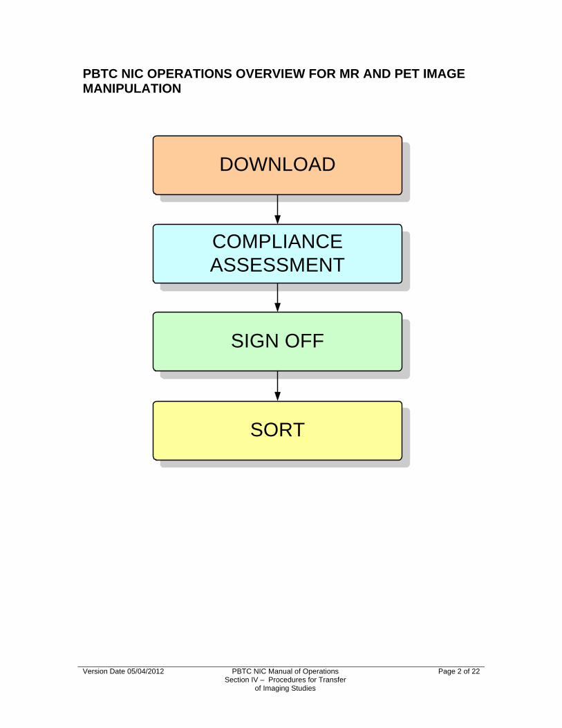
PBTC NIC OPERATIONS OVERVIEW FOR MR AND PET IMAGE MANIPULATION
DOWNLOAD
SIGN OFF
COMPLIANCEASSESSMENT
SORT
Version Date 05/04/2012 PBTC NIC Manual of Operations Section IV – Procedures for Transfer
of Imaging Studies
Page 2 of 22

I. DOWNLOAD
Version Date 05/04/2012 PBTC NIC Manual of Operations Section IV – Procedures for Transfer
of Imaging Studies
Page 3 of 22
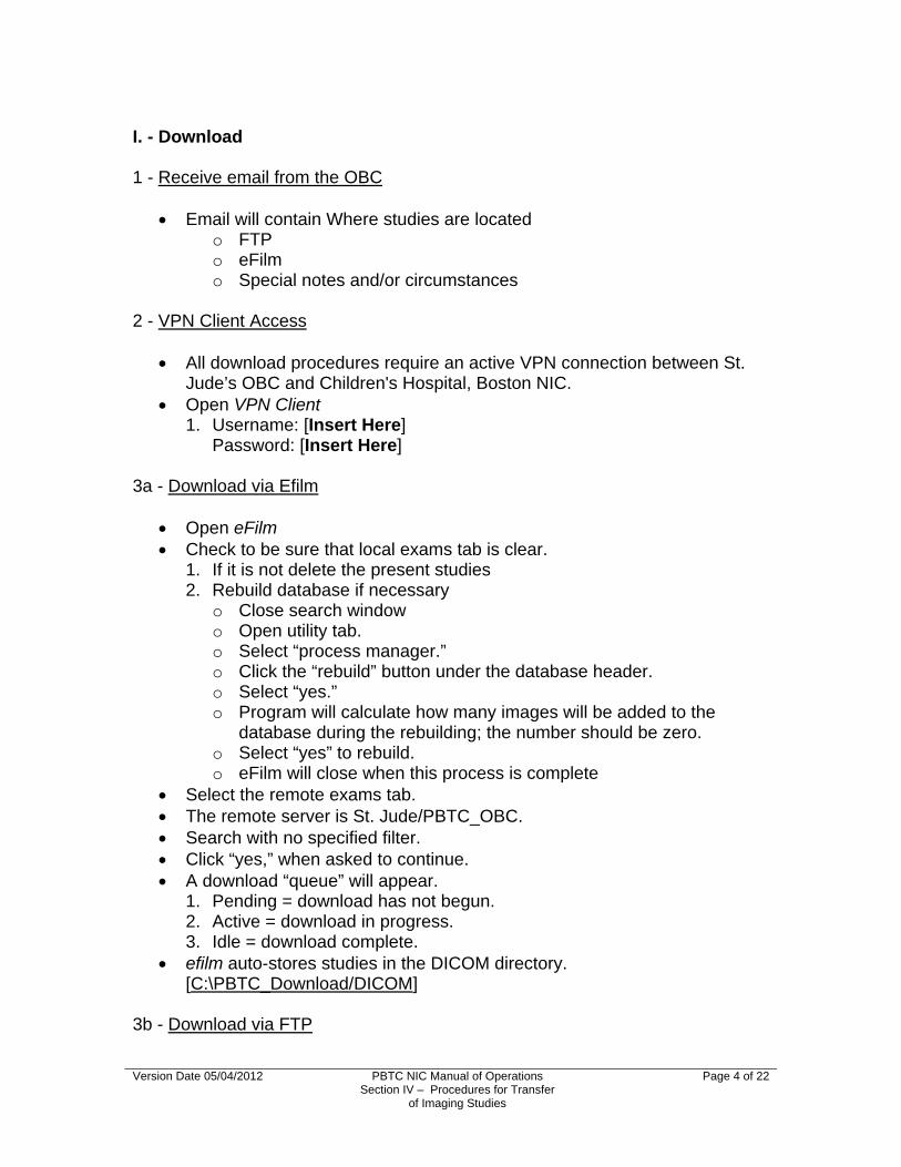
I. - Download 1 - Receive email from the OBC
Email will contain Where studies are located o FTP o eFilm o Special notes and/or circumstances
2 - VPN Client Access
All download procedures require an active VPN connection between St. Jude’s OBC and Children's Hospital, Boston NIC.
Open VPN Client 1. Username: [Insert Here]
Password: [Insert Here] 3a - Download via Efilm
Open eFilm Check to be sure that local exams tab is clear.
1. If it is not delete the present studies 2. Rebuild database if necessary
o Close search window o Open utility tab. o Select “process manager.” o Click the “rebuild” button under the database header. o Select “yes.” o Program will calculate how many images will be added to the
database during the rebuilding; the number should be zero. o Select “yes” to rebuild. o eFilm will close when this process is complete
Select the remote exams tab. The remote server is St. Jude/PBTC_OBC. Search with no specified filter. Click “yes,” when asked to continue. A download “queue” will appear.
1. Pending = download has not begun. 2. Active = download in progress. 3. Idle = download complete.
efilm auto-stores studies in the DICOM directory. [C:\PBTC_Download/DICOM]
3b - Download via FTP
Version Date 05/04/2012 PBTC NIC Manual of Operations Section IV – Procedures for Transfer
of Imaging Studies
Page 4 of 22

Open Filezilla Client Connect to “remote browser”
1. Remote server IP = [Insert Here] 2. Log-in set to [Insert Here]
The OBC will supply the folder name of the download in an email. Copy files from OBC Server to NIC Workstation folder “DICOM_FTP”
[C:\PBTC_Download\DicomFTP] NOTE: Compliance assessment of these files will be delivered via DICOM
Works. 4a - Compliance Assessment Preparation - MR
Open Excel File open; [C:\Shared\PBTC Download-Sign-off.xls]
1. File should open to “download raw MR” tab, if not switch to that tab. Open “Internet Explorer.” Open “PBTC Web Report”
1. http://10.1.1.118/PBTC_WebReport/I8M1A3G9E7D1U6K2E9/default.html Log-in
1. OBC has provided personal usernames and passwords. Mouse-click the “click here” link attributed to “To sign-off downloaded
MRI/MRD/MRP/MRS/CT studies on OBC server, please click here.” A new browser window will open. Highlight the MR spreadsheet that appears and copy.
1. Important: Be sure to omit the header rows when copying MR data. Header rows are provided in the Excel file.
Return to Excel Paste MR data onto the “download raw MR” tab.
1. Be sure that data is pasted in the first free cell of the first column. [A4] 2. The drop-down menus the data may take a few minutes to appear.
Once loaded, copy the MR data From “Download Raw MR” tab. Paste in the “download format MR” tab. Format MR page for printing
1. “Find all” for “institution.” 2. Replace all “institution” with “inst.” 3. Decrease the width of “confirmed by.” [J] to 3.00 4. Reformat cells under column header name, “scan date.” [C] Data column should be formatted to “dd/mm/yy”
5. Remove column with header name, “Phase ID.” [E] 6. Remove column with header name, “Images downloaded &
components confirmed during previous upload.” [G] 7. Remove column with header name, “comments.” [I] 8. Change fill color for all columns with the header name “analyzable” to
“light yellow” [J, L, O, R, T, V, X, Z, AC, AE, AG, AI, AK].
Version Date 05/04/2012 PBTC NIC Manual of Operations Section IV – Procedures for Transfer
of Imaging Studies
Page 5 of 22

Version Date 05/04/2012 PBTC NIC Manual of Operations Section IV – Procedures for Transfer
of Imaging Studies
Page 6 of 22
9. Change fill color for columns with the header names of “Diffusion.” “T2* Perf”, “T1 Perm Perf”, “Spec Sing”, “DTI” to “light grey” [ S, U, W, and Y respectively].
10. View “page break preview.” Entire spreadsheet should fit in portrait on legal-sized paper. Be sure page break does not interrupt length or width of data
columns or rows. 11. View “print preview.” Be sure that records do not cut off or interfere with the footer.
12. “Save as” date of OBC email. 4b - Compliance Assessment Preparation - PET
Return to Internet Explorer and the web report. Mouse-click the “click here” link attributed to “To sign-off downloaded PET
studies on OBC server, please click here.” A new browser tab will open. Highlight the entire spreadsheet including headers, and copy. Return to Excel Paste PET Data in the “Download Raw PET” tab.
1. Be sure that data is pasted in the first free cell of the first column. [A1] 2. The drop-down menus the data may take a few minutes to appear.
Once loaded, copy the PET Data from the “Download Raw PET” tab. Paste in the “Downloaded Format PET” tab. Save
5 – Print
Sign-off worksheets require the use of legal-sized printer paper (11” x 14”) and can easily be printed using \\arctic\wol03mfp01

II. COMPLIANCE ASSESSMENT
Version Date 05/04/2012 PBTC NIC Manual of Operations Section IV – Procedures for Transfer
of Imaging Studies
Page 7 of 22

II. - Compliance Assessment 1 - Meetings
There are three types of scans in each download: 1. Head and spine MR imaging 2. Diffusion, Perfusion and Spectroscopic Imaging 3. PET imaging
Each type of scan requires the review of a different specialist. 1. Tina Young Poussaint, MD Call/email and schedule an appointment to meet.
2. Sridhar Vajapeyam Ph.D. Call/email and schedule an appointment to meet.
3. Frederick Fahey D.Sc. Copy “Download Format PET” spreadsheet and paste it into an
email. 1) If PET studies were downloaded via eFilm, he will view images
via a pre-existing sharing function in eFilm.
2 - The Compliance Assessment Worksheet
Filling out the component portion of the sign-off sheet— 1. Components received by the NIC should match those sent by the institution. If this is the case, check the box. If this is NOT the case, cross out the institution and confirmation.
Write in the corrected value. 2. If all components are present— Write “all” in the column with the header name, “Images
downloaded & components confirmed.” Write “no” in the column with the header name, “Re-upload
needed?” 3. If not all components are present— Write “some” in the column with the header name, “Images
downloaded & components confirmed.” The study may need re-uploading, depending on the component
missing. Check “Tables 7 and 8: PBTC Protocols”, to see which components are mandatory for which protocol.
4. If none of the components are present— Write “none” in the column with the header name, “Images
downloaded & components confirmed”. Write “yes” in the column with the header name “Re-upload
needed?” 5. If Components are present but not listed, hand-write a comment stating such— Space is limited so abbreviations and short-hand are encouraged.
Version Date 05/04/2012 PBTC NIC Manual of Operations Section IV – Procedures for Transfer
of Imaging Studies
Page 8 of 22

Version Date 05/04/2012 PBTC NIC Manual of Operations Section IV – Procedures for Transfer
of Imaging Studies
Page 9 of 22
Filling out the analyzability portion of the sign-off sheet: 1) If a component is analyzable, check the box. 2) If a component is not analyzable, write “no”
Scans that are not analyzable do not require re-upload. Comments
1. Comments should be made on the PBTC Assessment Sheet and transcribed during the sign-off process.

III. SIGN-OFF
Version Date 05/04/2012 PBTC NIC Manual of Operations Section IV – Procedures for Transfer
of Imaging Studies
Page 10 of 22

III. Sign-off
1 - Completed Compliance Assessment Forms
All three scan types must be reviewed by the appropriate specialist prior to sign-off. MR Diffusion/Perfusion/Spectroscopy PET
The completed Compliance Assessment Worksheet should make note of the following: 1. The download received is compliant with the study list provided by the
OBC. 2. The studies are compliant with their respective protocols. 3. Studies that are present are analyzable. 4. Missing or incomplete studies that require a re-upload request. 5. Itemized comment list of any incidental cases that may have been
downloaded but not tracked by the sites. 2 - VPN Client Access
All sign-off procedures require an active VPN connection. Open VPN Client
1. Username: [Insert Here] Password: [Insert Here]
3a Sign-off Procedure for MR/Diffusion/Perfusion/Spectroscopy studies
Open Internet Explorer Open favorite site: “PBTC Web Report”
2. [http://10.1.1.118/PBTC_WebReport/I8M1A3G9E7D1U6K2E9/default.html] Log-in
1. OBC has provided personal usernames and passwords. Mouse-click the “click here” link attributed to “To sign-off downloaded
MRI/MRD/MRP/MRS/CT studies on OBC server, please click here” on the OBC web report.
A new browser window will open. Transcribe the data collected on the Compliance Assessment Worksheet
during the Compliance Assessment Meetings into the component portion of the sign-off web report.
If all of the components uploaded by the institution are confirmed with institution's information, please select "yes, all" in the column with the header name, “Images downloaded & components confirmed.” 1. By selecting “yes, all” you are automatically populating the “yes” to
every “component” column offered on the web report.
Version Date 05/04/2012 PBTC NIC Manual of Operations Section IV – Procedures for Transfer
of Imaging Studies
Page 11 of 22

2. Be aware that by selecting “yes, all” you are self-populating the “Re-upload needed?” column with a “no” response.
3. When a “yes, all” selection does not populate the “analyzable” column, it will require manual population.
If some of the components uploaded by the institution are confirmed with institution's information, please select "yes, some" in the column with the header name, “Images downloaded & components confirmed.” 1. By selecting “yes, some” you need to manually populate each “yes/no”
drop-down menu for each component column on the web report. 2. Be aware that by selecting “yes, some,” the “Re-upload needed?”
column will not self-populate and thus, will need to be manually populated. The study may need re-uploading depending on the component
and the protocol. Check “Tables 7 and 8: PBTC Protocols” to see which components are mandatory for which protocols.
3. When a “yes, some” selection does not populate the “analyzable?” column, it will require manual population.
If none of the components uploaded by the institution are confirmed with institution's information, please select "no, none" in the column with the header name, “Images downloaded & components confirmed.” 1. By selecting “no, none,” you are automatically populating the “no” to
every component column offered on the web report. 2. Be aware that by selecting “no, none,” you are self-populating the
column with the header name “Re-upload needed?” with a “yes” response.
3. A “no, none” selection will self-populate the “analyzable?” columns to “no.”
If components are present, but not listed by the OBC, hand-write a comment in the “comments” column stating such. 1. Space is limited so abbreviations and short-hand are encouraged.
Click the “sign-off neuro-imaging studies downloaded from OBC Server” button at the very bottom of the MRI/Diffusion/Perfusion sign-off page. 1. Missing values will result in an error message.
A new browser window will open with a summary of the just submitted data.
3b - Update Download/Sign-off Excel for MR/Diffusion/Perfusion/Spectroscopy
Open Excel File open; [C:\Shared\PBTC Download-Sign-off.xls]
1. File should open to the “Download Raw MR” tab. 2. Select “Sign-off MR” tab.
Return to “Internet Explorer” and the web report submission summary page.
Highlight the most recent data, omitting the header rows. Right mouse-click to copy.
Version Date 05/04/2012 PBTC NIC Manual of Operations Section IV – Procedures for Transfer
of Imaging Studies
Page 12 of 22

Return to Excel file [PBTC Download-Sign-off.xls] Paste the new data into the left uppermost free cell. [A4] Save.
4a - Sign-off Procedure for PET Studies
Open Internet Explorer. Open favorite site: “PBTC Web Report.”
1. http://10.1.1.118/PBTC_WebReport/I8M1A3G9E7D1U6K2E9/default.hml Log-in
2. OBC has provided personal usernames and passwords. Mouse-click the “click here” link attributed to “To sign-off downloaded PET
studies on OBC server, please click here.” If the study was received from the OBC by the NIC, select “yes” in the
column named “Images downloaded & confirmed.” 1. Manual population of “Re-upload needed?” & “PET scan analyzable?”
required. If the study was not received from the OBC by the NIC, select “no” in the
column named “Images downloaded & confirmed.” 1. Self populates “yes” for “Re-upload needed?” and “no” for “PET scan
analyzable?” Comments added in designated column as needed. Click the “Sign-off Neuro-imaging studies downloaded from OBC Server”
button at the very bottom of the PET sign-off page. 1. Missing values will result in an error message. 2. A new browser window will open with a summary of the just submitted
data. 4b - Update Download/Sign-off Excel PET
Return to Excel. File open; [C:\Shared\PBTC Download-Sign-off.xls]
1. File should open to the “download MR Raw” tab. 2. Select “Signoff PET” Tab.
Return to Internet Explorer and the web report submission summary page. Highlight the most recent data, including the header rows. Right mouse-click to copy. Return to Excel file [PBTC Download-Sign-off.xls] Paste the new data into the left uppermost free cell. [A1] Save.
Version Date 05/04/2012 PBTC NIC Manual of Operations Section IV – Procedures for Transfer
of Imaging Studies
Page 13 of 22

5 - Update Protocol Spreadsheets - MR
Open Internet Explorer. Open favorite site: “PBTC Web Report.”
1. [http://10.1.1.118/PBTC_WebReport/I8M1A3G9E7D1U6K2E9/default.html] Log in to the PBTC Web Report.
1. OBC has provided personal usernames and passwords. Locate the “drop-down” menu and “view” button associated with “To view
MRI/MRD/MRP/MRS studies sent to OBC and downloaded to NIC, in Excel format, select protocol.” 1. Each drop-down menu option will open an Excel file listing of all
studies obtained for that protocol by accession number.
Go to F:\ Drive of the Workstation [10.6.60.222\C$ ] 3. Username: [Insert Here]
Password: [Insert Here] Select folder named, “PBTC Protocol Spreadsheets” Contained within each protocol-specific folder is a spreadsheet for that
protocol: [{protocol number}_PBTC-MR-image-Studies-list.xls] 1. Be aware that some protocols have more than one run, or phases. 2. An additional phase will be represented by a lower case Roman
numeral directly following the {protocol number} in the file name. Ex: 014ii = Protocol 14, Phase 2
3. A phase breakdown list can be found on the F:\ Drive of the Workstation [\NIC Download Resources\Phase Information]
Updating the spreadsheets 1. Compliance Assessment Worksheet 2. Select the first protocol/accession number listed on the Compliance
Assessment Worksheet. 3. F:\Drive 4. Open the Excel spreadsheet for that protocol. 5. Select the dated tab, if the spreadsheet does not automatically open to
that tab. 6. OBC WebReport 7. Select that protocol on the drop-down menu of the OBC Web Report. 8. View. 9. A new browser window will open as well as a “file download – security
warning.” 10. Open. This will open the OBC version of the spreadsheet in the new
browser window. 11. Using the Compliance Assessment Worksheet as a guide, select the
entire row corresponding to the protocol/accession number/date combination for the study in that download.
12. Copy. 13. Return to C:\Drive.
Version Date 05/04/2012 PBTC NIC Manual of Operations Section IV – Procedures for Transfer
of Imaging Studies
Page 14 of 22

Version Date 05/04/2012 PBTC NIC Manual of Operations Section IV – Procedures for Transfer
of Imaging Studies
Page 15 of 22
14. Paste the new row in the first open row of the dated tab. 15. Continue copy/paste until each newly downloaded entry for that
protocol is completed. 16. Rename the tab on the spreadsheet to reflect the day the update took
place. 17. Continue this process for each protocol, until the entire download has
been reflected on the spreadsheets.
Protocols 020, 022, 023, 030, & 030 1. These protocols, in particular, require two types of perfusion diffusion
tensor imaging.(DTI) T2* gradient echo perfusion T1 permeability DTI
2. If it is the case that the values on the sign off page do not reflect the studies received by the NIC for T2* perfusion, T1 permeability or diffusion imaging then short hand notation is used to accurately record the studies received on the Compliance Assessment Worksheet. and this information is then hand keyed into the Protocol Specific spreadsheets.
3. Analyzability data for the perfusion studies will have been recorded on the Compliance Assessment Worksheet.
4. Open Excel [{020/022/023/030/033}_PBTC-MR-image-Studies-list.xls]. 5. Select the DTI/Perf tab. 6. Transcribe the analyzability of T2*, T1 permeability, and DTI in their
respective hand key (orange) columns. Analyzability is recorded with the following short-hand.
i. 0 = not present/not analyzable ii. 1 = present/not analyzable iii. 2 = present/analyzable

IV. SORT
Version Date 05/04/2012 PBTC NIC Manual of Operations Section IV – Procedures for Transfer
of Imaging Studies
Page 16 of 22

IV. - Sort
1a - Raw File Backup - eFilm
Before any actual sorting takes place a copy of the entire download is backed up on an external hard drive.
Open the two local browsers: 1. C:\PBTC_Download\DICOM 2. E:\PBTC Download Backup Create a file with the download date given by the OBC in the
notification email. o Copy the entire “C:\PBTC_Download\DICOM” into “E:\PBTC
Download Backup\[DoD].” 1. Where DoD = date of download
1b - Raw File Backup – FTP
Open the two local browsers: 1. C:\PBTC_Download\DicomFTP 2. E:\PBTC Download Backup Create a file with the download date supplied by the OBC in the
notification email. Copy the entire “C:\PBTC_Download\DicomFTP” into “E:\PBTC
Download Backup\DoD.” 1. Where DoD = date of download
2 - Format via DICOM Works
Files sent from the OBC have a numerical dicom file header name. In an
effort to avoid confusion during subsequent analysis, the NIC formats each file header name to be alpha-numeric. The NIC uses DICOM Works for this procedure.
Open DICOM Works. 1. DICOM Works should be the only application running. 2. DICOM Works will open to the last directory it had access to. The size
of that directory will dictate how long it will take DICOM Works to load. Be patient.
Escape (Esc) out of the credit overlay. The bottom-left icon should read, “This list is current.”
1. If it does not, “double- click” as instructed. Right mouse-click on the shaded check box on the upper left-hand side of
the data list. Select “other.”
1. This will allow for the individual selection of studies. Select the first study listed.
Version Date 05/04/2012 PBTC NIC Manual of Operations Section IV – Procedures for Transfer
of Imaging Studies
Page 17 of 22

On the right side of the screen an option for “update thumbnails” will appear.
Select “update thumbnails.” Select studies to be exported.
1. The export process will vary with the number and type of studies selected.
2. Select only one type of study at a time—either MR or PET. 3. Select no more than four studies of any one type at a time.
3a - Export – MR
Select studies to be exported. Click “export” button from the top tool bar. The “export main wizard” menu will appear. Make sure the following settings are maintained:
1. Source: “checked items.” 2. File Format: DICOM (native format) 3. Destination: a local directory
Click “next.” Click “next.” “Choose a destination directory”
[C:\PBTC_Download\DicomMrToBeSorted]. Click “save.” Repeat until all MR cases have been exported.
3b - Export – PET
Select studies to be exported. Click “export” button from top toolbar. Make sure the following settings are maintained:
1. Source: “checked items.” 2. File Format: DICOM (native format). 3. Destination: a local directory.
Click “next.” Click “next.” Choose a destination directory
[C:\PBTC_Download\DicomPetToBeSorted]. Click “save.” Repeat until all PET cases have been exported.
4a - Text File - MR
Open a Command Prompt window.
Version Date 05/04/2012 PBTC NIC Manual of Operations Section IV – Procedures for Transfer
of Imaging Studies
Page 18 of 22

The opening directory will be [C:\Documents and Settings\[log in profile name].
Enter the command [cd ..\..] 1. Where “cd” = “change directory” and the “..\..” = the number of levels
retraced to bring one to the main C:\ directory. 2. This will bring you to the main C:\ directory.
Enter the [\PBTC_Download\DicomMrToBeSorted] Enter dir > [file name]mr.txt.
1. The “dir” command creates a text file list of the contents of that directory.
2. The file name should be the “date of the download” [dd-mm-yy]. 3. The file name is followed by the file type [MR]. 4. The file extension is [.txt].
Click “enter.” 4b - Text File – PET
Open a Command Prompt window. The opening directory will be [C:\Documents and Settings\[log in profile
name]. “Enter” the command [cd ..\..]
1. Where “cd” = “change directory” and the “..\..” = the number of levels retraced to bring one to the main C:\ directory.
2. This will bring you to the main C:\ directory. Enter the [\PBTC_Download\DicomPetToBeSorted] Enter [dir >{file name}pet.txt.
1. The “dir” command creates a text file list of the contents of that directory.
2. The file name should be the “date of the download” [dd-mm-yy]. 3. The file name is followed by the file type [PET]. 4. The file extension is [.txt].
“Enter.”
5 - Move Text Files
. Open a local browser.
1. NIC Workstation [C:\NIC Download Resources\TxtByDate] New folder Name folder with date of download.
1. [dd-mm-yy] Open an additional local browser.
1. Workstation [C:\PBTC_Download\DicomMrToBeSorted]. Select “DicomMrToBeSorted.” Move [filename]mr.txt file to NIC Workstation.
Version Date 05/04/2012 PBTC NIC Manual of Operations Section IV – Procedures for Transfer
of Imaging Studies
Page 19 of 22

Version Date 05/04/2012 PBTC NIC Manual of Operations Section IV – Procedures for Transfer
of Imaging Studies
Page 20 of 22
Go up one level. Select “DicomPetToBeSorted.” Move [filename]pet.txt file to NIC Workstation.
6a - Move to the NIC Server – MR
Open the two local browsers: 1. Workstation [PBTC_Download\DicomMrToBeSorted] 2. Server [Poussaint on RC-FS(RC-FS.tch.harvard.edu)] Folders are Protocol Specific
Move files from the workstation into respective protocol specific folder on the server.
Repeat until all files in workstation have been moved to the server. 6b - Move to the NIC Server – PET
Open the two remote browsers: 1. Workstation [PBTC_Download\DicomPetToBeSorted] 2. Server [Poussaint on RC-FS(RC-FS.tch.harvard.edu)]] Folders are protocol-specific
Move files from the workstation into respective protocol specific folder on the server.
Repeat until all files in workstation have been moved to the server.

C. MRS Raw Data Transfer Contact Person: Sridhar Vajapeyam, PhD 617-355-7870; Email: [email protected] Please note all sites should do a screen save of the MRS spectrum with the ratios displayed on the image. 1.) GE Scanners The spectroscopy raw data files are stored in the directory /usr/g/mrraw. Open a C shell and go to the directory /usr/g/mrraw by typing the command
“cd /usr/g/mrraw.” List the files in the directory by typing “ls –l.” You will see several files with names like Pxxxxx.7. You will also see the date and time the files were created. Pick the right files based on the date and time the MRS study was conducted
and copy them to a separate directory and then transfer them to OBC of the PBTC.
2.) Siemens Scanners The Syngo Database will list the spectroscopy raw data files associated with
a study. Export these files to a new folder and then transfer them to OBC of the PBTC. Since Sonata System has no means of writing any data other than patient
data (in DICOM format) into a CD ROM, you would have to first export the raw data to a temporary directory. You will then ftp this file to a PC or a Unix workstation for further processing. The data is in Siemsn 'rdx' format following export.
Exporting data:
a. Drag raw data into the "spectroscopy task card" just as you would when processing spectroscopy data.
b. Click on "options" on the menubar and then choose "export raw data". c. You will be asked for a directory and a file name to export the data. d. Normally you would use the "C:\temp" Directory. Data now is exported.
ftp data:
a. First log into Windows NT system by holding 'CTRL" and "Esc" keys > together. This will allow you to get access to the Microsoft Windows > environment.
b. Click on "start" button on the lower left corner of the screen, click on "advanced user" if you have not logged into the NT systems previously. This will give you a prompt window for your password input. Factory default password is "meduser1" unless it has been previously altered.
Version Date 05/04/2012 PBTC NIC Manual of Operations Section IV – Procedures for Transfer
of Imaging Studies
Page 21 of 22

Version Date 05/04/2012 PBTC NIC Manual of Operations Section IV – Procedures for Transfer
of Imaging Studies
Page 22 of 22
c. After you log into the NT systems, hit "CTRL" and "Esc" simultaneously again. Go to "start" then "programs" and the "command prompt.” This will open up a DOS window with a prompt "C:\". Change to "temp" directory (or other directory you created when you export your raw data) by typing "cd temp.”
d. You can now ftp your data out to other PC or workstations. Do not forget to set to "binary" mode when you ftp or your data will be garbled.
e. Example: "ftp 123.45.678.99" (connect to your ftp server PC or unix system) (enter user name/password) "bin" (change from ASCII mode to binary) "put file name" (upload data) "quit."
3.) Phillips Scanners Raw spectroscopy data and the corresponding header information are
stored as .SDAT and .SPAR files associated with the patient identifier. The localizer image data and header are stored as .REC and .PAR
respectively. Transfer all these files for each spectroscopy study into a separate folder and
then transfer to OBC of the PBTC. Transfer to OBC of the PBTC MRS data will require an ftp connection between the site's scanner workstation and the PBTC laptop. Once the files reach the PBTC laptop at the site, then the CRA at the site can ftp it over the secure channel to OBC of the PBTC.

Section VPBTC NIC Manual of OperationsGuidelines for Reading and Interpreting Imaging Studies

Version Date 05/02/2012 PBTC NIC Manual of Operations Section V – Guidelines for Reading
and Interpreting imaging studies
Page 1 of 1
V. Guidelines for Reading and Interpreting Imaging Studies
The NIC is responsible for image analysis of all PBTC research neuroimaging studies. Each study is reviewed by 1-2 Neuroradiologists from the NIC and the final interpretation of results reached by consensus.
Before image analysis, imaging studies are evaluated for readability and compliance with the protocol and the protocol compliance form is completed by the data volumetric analyst and data analysis engineer in consultation with the neuroradiologist.
The standard FLAIR, T2, and gadolinium sequences are sent to the Vitrea workstation (Vital Images, Plymouth, MN) and volumetric analysis is done. The data volumetric analyst consults with one of two neuroradiologists to determine the tumor site and regions of interest to be measured. Automated segmentation software defines the tumor regions on FLAIR or T2 images and nonenhancing and enhancing tumor regions on gadolinium T1 axial images. The perfusion, diffusion and MRS components of each study are transferred from the PC workstation to a dedicated workstation for image analysis (see data form MR). After reviewing the post-analysis data, the information is backed up on the NIC workstation, research computing server and a dedicated backup server system through CHB Research Computing.

Section VIPBTC NIC Manual of OperationsData Management Procedures

Version Date 08/15/2013 PBTC NIC Manual of Operations Section VI – Data Management
Procedures
Page 1 of 1
VI. Data Management Procedures
Data is collected on hard copy forms by NIC data analysts, NIC nuclear medicine physicist/physician and neuroradiologists at the time of image analysis. The NIC Research Coordinator, Gloria Faboyede, ensures that all necessary data are collected in a timely and efficient fashion. The study coordinator reviews all hard copy data forms and completes data entry at the CRC. The data forms are entered directly through the PBTC web-based data management system and managed by the PBTC Operations and Biostatistics Center. All other data such as quality assurance data are entered into an SPSS database at the CRC. For these data, the CRC generates data entry status reports and generates frequency distributions of study variables to provide timely and relevant feedback to the NIC. A password protected study directory is established on the program’s server for all database files and analysis files. Datasets are cleaned, verified, archived with documentation and then used to create documented analysis files. Access to programs and datasets is denied by password to non-study staff. All analytic and database files are backed up daily. No patient identifiers are present in the database or on forms. Each study participant has a unique PBTC assigned study numeric study identifier that includes a site, protocol, and subject identifier.
The Department of Radiology at the NIC has its own Information and
Technology (IT) dedicated staff under the leadership of John Speziale (Director of Radiology IT) who oversee the networking and security of the imaging data received. A dedicated file system on the ftp server is used to store and disseminate data. Imaging data is transferred via a secure tunnel from St. Jude's to our dedicated server. Imaging studies are backed up onto the NIC server, research computing share drive and backed daily by the Research Computing at Children’s Hospital Boston under the supervision of Ryan Callahan.

Section VIIPBTC NIC Manual of OperationsPBTC NIC Data Management System
VII. PBTC NIC Data Management System The Clinical Research Information Technology Core will be used as a central location for data processing and management. Children's Hospital Boston, with collaboration from a consortium of institutional partners, has developed a software toolset and workflow methodology for electronic collection and management of research and clinical trial data. REDCap (Research Electronic Data Capture) data collection projects rely on a thorough study-specific data dictionary defined in an iterative self-documenting process by all members of the research team with planning assistance from the Clinical Research Center. The iterative development and testing process results in a well-planned data collection strategy for individual studies. REDCap servers are housed in a local data center at Children's and all web-based information transmission is encrypted. REDCap has been disseminated for use locally at other institutions and currently supports > 230 academic/non-profit consortium partners on six continents and 24,000 research end-users (www.project-redcap.org)
The PBTC NIC Clinical Data Management System “Users Manual” diagrammatic
flow chart, and protocol matrix follows.
Version Date 05/02/2012 PBTC NIC Manual of Operations Section VII –
PBTC NIC Data Management System
Page 1 of 8

Clinical Data Management System
Users Manual
Project: Pediatric Brain Tumor Consortium
Neuroimaging Center
PI: Tina Young Poussaint, MD
Scientific Research Information System Informatics Team, Clinical Research Program
Children's Hospital, Boston
Version Date 05/02/2012 PBTC NIC Manual of Operations Section VII –
PBTC NIC Data Management System
Page 2 of 8

Data Management System (User Manual) Data Management System (User Manual).........................................................3 Introduction.........................................................................................................4 System Overview................................................................................................4 Requirements......................................................................................................4 Application Security............................................................................................4 Login Screen.......................................................................................................5 Home Screen......................................................................................................6 Data Entry Screen Selection...............................................................................6 Patient Accession Selection/Entry.......................................................................7 Form and Event Selection....................................................................................7 Data Entry............................................................................................................8 Validation Error Messages...................................................................................8
Version Date 04.13.2012 PBTC NIC Manual of Operations Section VII –
PBTC NIC Data Management System
Page 3 of 8

Introduction A Data Management System (DMS) is a software program of varying size and sophistication by which data collected on case report forms are stored, tracked and prepared for analysis. The data management system for this study is Web accessible and supports the following features.
Basic Security (user authentication and authorization)
Data Capture
o Real-Time Validations
o Outlier Identification
o Read-Only Access
Protocol Tracking
o Custom Data Management Reports
Audit Trails
This data management system is programmed to adhere to the operations of the study protocol (See pages 20 & 21). Clinical staff may use the DMS to track the progress and state of each case report form for each study participant. Standard data management reports will be added to the system to facilitate preparation of data for analysis. Many of the features outlined above are described in detail in this manual. Further questions regarding DMS functionality should be directed to the clinical research specialist liaison of the Clinical Research Center office.
System Overview
Requirements
As stated previously, the DMS is a Web-based system accessible from inside the Children's Hospital, Boston intranet only. The Web site requires users to employ Internet Explorer version 8 or higher. The data management system is accessible through the following URL. http://redcap-qi.tch.harvard.edu/redcap_edc/
Application Security
CRC provides user access to key personnel for initial website access. Once access is granted, users will use their Children’s ID and password to log in. User access to the database is determined by individual role on project.
Version Date 04.13.2012 PBTC NIC Manual of Operations Section VII –
PBTC NIC Data Management System
Page 4 of 8

Login Screen
All users are directed to the Login Screen at start-up (see below).
If a user has difficulty logging in, they may select the Clinical Research Center link and request help.
Version Date 05/02/2012 PBTC NIC Manual of Operations Section VII –
PBTC NIC Data Management System
Page 5 of 8

Home (Portal) Screen
Upon logging in, the user must select the My Projects tab. From there, the user will click on the link labeled Pediatric Brain Tumor Consortium Neuroimaging Center.
Next, the user will select the Data Entry link on the left side of the page.
Version Date 05/02/2012 PBTC NIC Manual of Operations Section VII –
PBTC NIC Data Management System
Page 6 of 8

On the next screen, the user must select the arm (or PBTC Protocol), enter the Patient Accession #, and then press the Enter or Tab key.
Next, the user must select the appropriate Data Collection Instrument (or Form) and Event Number for data entry. Red dots designate forms that have not been entered yet. Once a form has been entered and saved, the red dot will change to a green dot.
Version Date 05/02/2012 PBTC NIC Manual of Operations Section VII –
PBTC NIC Data Management System
Page 7 of 8

On the next screen, the user will be able to enter the data for the selected Data Collection Instrument (or Form) and Event Number. Once the form is entered, the user can specify whether the form is Complete, Incomplete, or Unverified and then click the Save Record button.
If an entered value is outside the specified range, a warning message will appear. The user will have the opportunity to accept or re-enter the out-of-range value.
Version Date 05/02/2012 PBTC NIC Manual of Operations Section VII –
PBTC NIC Data Management System
Page 8 of 8

Section VIIIPBTC NIC Manual of OperationsQuality Control Approaches

VIII. Quality Control Approaches A. Training and Certification Requirements of the Neuroradiologist
The neuroradiologists at each site must be a Board certified radiologist with expertise in neuroimaging and/or CAQ accreditation. B. Manuals of Operation
The NIC Manual of Operations documents all procedures or standard clinical protocols and research protocols, which address diagnostic standard protocols; intravenous contrast doses and infusion times when appropriate; image data storage; image data transfer; tumor volumetric analysis. Manuals are provided to each site for implementation of all imaging protocols and updated by the NIC throughout the study as appropriate. C. Coordination and Communication The NIC coordinates regular communication and meetings among the participating neuroradiologists of the Neuroimaging Committee and PET investigators of the PET Investigator Committee. This includes at least quarterly conference calls and biannual meetings as well as regular e-mail exchange for routine correspondence and questions. Dedicated consensus conferences and/or neuroradiology symposia for PET and MR investigators are held on a biannual basis at Children’s Hospital Boston or PBTC meetings. D. Site Visits
The NIC will conduct site visits as needed to each site to review
procedures and compliance with all PBTC protocols. During the visit, participating sites will be expected to provide materials and answer questions directed by the NIC. An evaluation checklist will be completed at these site visits for inclusion in a Site Visit Report to the Principal Investigator and PBTC Steering Committees (see Site Visit Monitoring Report attached).
E. Reader Reliability
The reproducibility of the central image interpretation is evaluated by selecting a random 20% subsample of exams from each protocol for repeat interpretation at the NIC. All NIC neuroradiologists are blinded to the exams. Inter-rater and Intra-rater reliability is assessed by examining the percent agreement with the first reading (see Form 1b). F. Data Quality
Version Date 05/02/2012 PBTC NIC Manual of Operations Section VIII – Quality Control
Approaches
Page 1 of 7

Regular meetings of the NIC staff occur with each download where images from the participating institutions are reviewed for routine compliance with the standardized imaging protocols set by the NIC. These data are entered into a web-based from at the OBC. NIC staff assess the diagnostic image quality and data integrity prior to submitting the CT, MR and PET data for analysis. Dr. Mulkern reviews the current QA protocols at each of the participating institutions including MR and MRS phantom controls in use. Compliance is insured with the QA protocols throughout the course of the study. In addition, the NIC, with the OBC, generates summary statistics and a report on the image quality and compliance with PBTC protocols by site and overall. These are distributed to the NC and Steering Committee for review on a biannual basis. An overview of the quality control procedures for each imaging study are as follows.
MRI Quality Control: Each site should have an American College of Radiology (ACR) phantom, which can be purchased (J.M. Specialty Parts, 11689 Sorrento Valley Rd, San Diego, CA 92121, 858-794-7200). This phantom comes equipped with a guide to performing specific tests of geometric accuracy, high contrast spatial resolution, slice thickness accuracy, slice position accuracy, image intensity uniformity, percent signal ghosting, and low contrast detectability. Each scanner used for the trial, scans the ACR phantom quarterly. These QC data sets are sent to the NIC on a regular basis where they are evaluated according to the recommendations of the ACR accreditation manual. Site scanner equipment includes Siemens, Phillips and GE scanners. A video for the MR QC process is posted on the website of the NIC: www.childrenshospital.org/research/pbtcnic Statistical data from the QA program is generated in the form of reports on an ongoing basis. CT Quality Control: A Quality Control Program similar to the program established by the ACR for the Accreditation of CT Scanners shall be established. This consists of minimum Qualifications for physicians, technologists, and medical physicist involved in the CT program. Equipment standards for the CT scanners must also be met. For protocols that involve CT scanning, the ACR phantom for CT will be used. This phantom allows the following performance parameters of a CT scanner, its display workstation, or hard copy film to be periodically monitored In addition to meeting initial minimum Qualifications, e.g. certification by either American Board of Radiology or American Registry of Radiologic Technologists, personnel must meet continuing education requirements. The physicians also must interpret and report a minimum of 100 CT examinations annually.
A Quality Control (QC) program must be established and implemented under the supervision of a qualified medical physicist. Initial performance testing by the physicist is required upon installation and must be repeated at least annually. These tests shall include, but not be limited to the following:
Version Date 05/02/2012 PBTC NIC Manual of Operations Section VIII – Quality Control
Approaches
Page 2 of 7

Annual Tests Performed by Qualified Physicists 1. Alignment light accuracy 2. Alignment of table to gantry 3. Table/Gantry tilt 4. Slice localization from scanned projection radiograph (localized image) 5. Table incrementation accuracy 6. Slice thickness accuracy 7. Image Quality
a. High contrast resolution b. Low contrast resolution c. Image uniformity d. Noise e. Artifact evaluation
8. CT number accuracy and linearity 9. Display devices
a. Video display b. Hard-copy display
10. Dosimetry a. CTDI b. Patient radiation dose for representative exams
11. Safety evaluation a. Visual Inspection b. Audible/visual signals c. Posting requirements d. Scattered radiation measurements
Continuous Quality Control A continuous QC program must be established for all CT units with the assistance of a qualified medical physicist. The qualified physicist will determine the frequency of each test and who should perform it based on the facility and CT usage. An on-site radiological technologist shall be identified to be responsible for conducting routine quality control. This continuous QC program shall include, but not be limited to, the following: 1. Alignment light accuracy 2. Slice thickness accuracy 3. Image Quality
a. High contrast resolution b. Low contrast resolution c. Image uniformity d. Noise e. Artifact evaluation
4. CT number accuracy 5. Display devices
Version Date 05/02/2012 PBTC NIC Manual of Operations Section VIII – Quality Control
Approaches
Page 3 of 7

a. Video display b. Hard-copy display
All QC testing must be carried out in accordance with written procedures and methods. Preventive maintenance must be scheduled, performed, and documented by a qualified service engineer or a regular basis. The results of the QC program must be monitored annually by the qualified medical physicist. If the results of a QC test fall outside the control limits, correct action should be taken. A qualified medical physicist should be available to assist in prescribing corrective actions for unresolved problems. All deficiencies must be documented and service records maintained by the CT facility. PET Quality Control: The quality control program for PET assures proper operation of all of the detector blocks and the accuracy of quantitation. Daily “blank” scans should be acquired to evaluate the operation of the detector blocks. Scanners provided by certain vendors require periodic updating of photomultiplier tubes to ensure proper operation. For General Electric scanners, an “update gains” check of the photomultiplier tubes should be performed weekly. For Siemens scanners, a “bucket setup” should be performed quarterly. Philips scanners do not require such a check. Modern PET scanners consist of thousands of small detectors and it is imperative for proper operation that their response be “normalized.” Such normalization of the scanner should be performed quarterly according to the manufacturers’ specifications. After normalization, a calibration must be performed for the scanner to be able to report the in vivo radiopharmaceutical distribution in Bq/mL or μCi/mL. Therefore, this calibration should be performed quarterly. PET scanners are complicated devices that require regular, preventive maintenance. Such preventive maintenance should be performed quarterly.
All members of the PBTC provide quarterly reports with regards to compliance to the PET quality control program described above. These reports require completing Form 5 (PET Quality Assurance Data Form) and sending it to the Neuroimaging Center. Each site completes a one-time analysis of a PET phantom (Form 15). Statistical data from the PET QA program is generated in the form of reports on an ongoing basis. The PBTC NIC was involved in a project with Paul Kinahan, PhD, of the University of Washington and Laurence Clarke, PhD of the NIH, that entails the imaging of a 20 cm cylindrical phantom with “hot” (i.e., higher activity concentration than the background) and “cold” (i.e., lower activity concentration than the background) features. This collaborative project, jointly overseen by the Society of Nuclear Medicine (SNM) and the American Association of Physicists in Medicine (AAPM), centered on the design of a phantom made with an equilibrium mixture of 68Ge/68Ga and equipped with 4 hot features of varying size (8-25 cm) and 3 cold features (all 25 cm of different materials) incorporated into a lid that can be used with the phantom imaged in 2005.
Following an initial evaluation of the phantom and corresponding, standardized imaging protocol and reporting form, the phantom was deployed to
Version Date 05/02/2012 PBTC NIC Manual of Operations Section VIII – Quality Control
Approaches
Page 4 of 7

the respective PET imaging programs of the 10 PBTC NIC sites. We asked each site to 1) image the phantom as if it were a PBTC subject; 2) report which features were visible; and 3) record the SUV values for the background as well as the 4 hot features. The data obtained from this QC procedure enabled the NIC to better understand how quantitation differed among the PBTC sites and how limits of accuracy may varied depending on which clinical application was being tested. This study was published by the Journal of Medical Physics (Fahey FH, Kinahan PE, Doot RK, Kocak M, Thurston H, Poussaint TY. Variability in PET Quantitation Within a Multicenter Consortium. Med. Phys. 2010 Jul:37(7):3660-3666).
*Image courtesy of University of Washington – Seattle
Version Date 05/02/2012 PBTC NIC Manual of Operations Section VIII – Quality Control
Approaches
Page 5 of 7

*Image courtesy of University of Washington – Seattle PET Data Analysis:
All PET imaging data acquired via a PBTC protocol is forwarded to the OBC and subsequently sent to the NIC for analysis by download or FTP.
All PET imaging data, along with their corresponding MR data, are uploaded to a multi-modality processing and review station (Hermes Medical Systems). Whether the imaging data is present and readable is determined and noted on Form 4. If the data are not readable, a reason is given on Form 4. If readable, the PET data are registered to the MR and are subjectively reviewed in conjunction with the MR. A representative image transverse image plane through the tumor is selected for review. Uptake in the tumor (as defined by the MR) is subjectively judged on a 5-point scale (FDG less than white matter, FDG uptake similar to white matter, FDG uptake more than white matter but less than gray matter, FDG uptake similar to gray matter, FDG uptake greater than gray matter) The uniformity of the uptake is judged to be 0-25%, 26-50%, 51-75% or 76-100% of the tumor as defined by MR. It is also determined whether the FDG uptake correlates with regions of MRI contrast enhancement. This subjective analysis is followed by quantitative, region of interest (ROI) analyses. On the
Version Date 05/02/2012 PBTC NIC Manual of Operations Section VIII – Quality Control
Approaches
Page 6 of 7

Version Date 05/02/2012 PBTC NIC Manual of Operations Section VIII – Quality Control
Approaches
Page 7 of 7
representative transverse plane selected above, 4 ROIs are drawn: one around the tumor, one in comparable gray matter, one in comparable white matter and one about the entire brain. If the tumor is not in a transverse slice that contains comparable gray and white matter, a second transverse plane is selected for the comparison ROIs. The mean and maximum pixel values within each of the ROIs are recorded. Ratios of tumor/gray matter, tumor/white matter and tumor/whole brain using both mean and maximum pixel counts are determined.
3D Image Analysis
In selected cases 3D image analysis has been done. 3D Image Analysis Tumor volumes of interest (VOIs) are defined on both FLAIR and T1 post-contrast (PC) MR images. The areas defined are the areas of hyperintense signal on the FLAIR images and enhancing tumor on the T1 post-contrast MR images. After volumes of interest are manually drawn on the MR images, they are confirmed by a nuclear medicine physicist and pediatric neuroradiologist and transferred to the co-registered PET images. Four 3D FDG uptake metrics have been determined for each PET VOI. The mean pixel value within the VOI is determined and this value is normalized by the mean value within the comparison VOI of either normal gray or white matter yielding the Tumor Gray Matter Ratio (Mean) and Tumor White Matter Ratio (Mean) values, respectively. The Tumor Gray Matter Ratio (Maximum) and Tumor White Matter ratio (Maximum) is defined similarly except the maximum pixel value within the VOI is used instead of the mean. For the Tumor Gray matter Ratio (Total) and the Tumor White Matter Ratio (Total), all of the pixel values within the VOI are added yielding a cumulative metric, and this value is normalized by a similar value for the gray or matter comparison VOI, respectively. The inhomogeneity index is defined as the ratio of the standard deviation of the pixel values within the VOI normalized by the mean.

Section IXPBTC NIC Manual of OperationsAppendices, Protocols and Forms

Version Date 01/24/14
PBTC NIC Manual of Operations Section IX – Neuroimaging Studies
Protocols and Forms
Page 1 of 11
IX. APPENDICES OF NEUROIMAGING STUDIES PROTOCOLS AND FORMS APPENDIX A: PBTC NIC MR PROTOCOLS OF BRAIN AND SPINE A1. PBTC MR Protocol Brain 0) Localizer of Choice Note: Efforts should be made to have same slice locations and thicknesses for sequences 2, 3 and 5. 1) Axial T1-weighted spin echo (SE) whole head, TR/TE = (500-700)/minimum
full, receiver bandwidth (RB) = 16 kHz, FOV = 18-24 cm, slice thickness/gap = 4/0 (mm), NEX = 2, no phase wrap option, matrix = 256 x 192 (frequency x phase), frequency direction = A/P
1*) For hemorrhage: Axial 2D gradient echo sequence whole head: TR/TE/FA =
(500-700)/25/90, 256x192 (frequency x phase), FOV = 18-24 cm, slice thickness/gap = 5/0, NEX = 2, frequency directions = A/P
2) Axial T2-weighted fast spin echo (FSE), TR/ETE = (4000-6000)/80-100, ETL
= 10-16, RB = 16 kHz, FOV = 18-24 cm, slice thickness/gap = 4/0 interleaved, NEX = 2, matrix = 256 x 192, flow compensation option, frequency direction A/P
2a) 3T optional replacement) Axial 3D Fast Spin Echo sequence with variable flip
angle (GE XETA sequence, Siemens SPACE sequence). ETL = 65, parallel imaging acceleration = 2, TR = 3 s, Effective TE = 80 ms, 200 slices, 1 mm thickness, 6 minute scan times
3) Axial FLAIR, TR/TI/ETE = 10,000/2200/162, ETL = 16, RB = 32 kHz, FOV = 18-24 cm, slice thickness/gap = 4/0 mm, NEX = 1, flow comp, frequency direction A/P
4a) Perfusion –T2* Gradient Echo Technique Perfusion scan axial echo planar imaging, gradient echo mode, single shot,
(reduced coverage 10 slices only), receiver bandwidth = 64 kHz, matrix = 128 x 128, TR/TE = 1500/45-60), slice thickness/gap = 6/3 for supratentorial mass, 3/0 for brainstem mass, FOV = 18-24 cm, NEX = 1, frequency direction R/L, 45 to 60 phases, 10 phases prior to bolus injection of GdDTPA, frequency direction R/L, flip angle=90. For child <10 kg, injection rate 1-2cc/sec; >10kg to <35kg, injection rate =2cc/sec; >35kg, injection rate =3 - 4 cc/sec, 22G IV minimum.
4b) MR permeability consists of dynamic T1-weighted 3D SPGR (FLASH, FFE),
minimum resolution 128x128x16, 24cm FOV, TR min, TE min, flip = 30 degrees, which is repeated 40 times (using multiphase/dynamics option).

Version Date 01/24/14
PBTC NIC Manual of Operations Section IX – Neuroimaging Studies
Protocols and Forms
Page 2 of 11
The dose of Gadolinium DTPA (GdDTPA) is 0.1mmol/kg body weight and injected at a rate of 2ml/sec approximately 10 seconds after scanning starts (to collect 2-3 baseline image sets). Kinetic modeling of the dynamic signal changes yields estimates of regional fractional blood volume and microvascular permeability (Ktrans), a sensitive indicator of blood brain disruption and a putative correlate of angiogenesis.
5) Axial T1-weighted spin echo (SE) post contrast whole brain, TR/TE = 500-
700/minimum full, receiver bandwidth = 16 kHz, FOV = 18-24 cm, slice thickness/gap = 4/0 (interleaved acquisitions), NEX = 2, frequency direction = A/P, matrix = 256 x 192
6) PROBE-P, PRESS single voxel spectroscopy, TR/TE = 1500/144, FOV = 24
cm, voxel size = 15 x 1 x 1 cm3 (approximately) positioned in solid tumor using post-GD T1, FLAIR and T2 images, total scans = 128
7) Diffusion imaging, single-shot echo planar spin echo, TR/TE = 2000/80,
matrix = 128 x 128, b-factor = 5/1000 s/mm2, one baseline plus 6 diffusion sensitization directions for diffusion tensor and trace imaging, receiver
bandwidth = 64 kHz, frequency direction = R/L, slice thickness/gap = 5 or 6/0 whole brain, decrease thickness for brainstem tumors.
8a) Diffusion Tensor Imaging Protocol: GE DTI protocol for 1.5T machines: DWI-EPI with ASSET(no fat sat)(with pure calibration) Image options: EPI, DIFF ASSET-2.00 Ph, #shots1 matrix size 96x96x50 (50 is the number of slices) TE: min NEX: 1 Number of baselines: 6 Phase FV: 1(full) Shim Auto Scan direction: Axial IS Bandwidth: 62.5 Spatial resolution: 2.5 mm by 2.5 mm by 2.5 mm TR: < 10 seconds – Can increase TR up to 12-13 seconds. FOV: 24 Freq: R/L Phase correction b-value: 1000 directions: 30 Select ASSET-2.00ph and PURE user CVs(control variables): CV6-CV0.00
Other diffusion options: recon all images

Version Date 01/24/14
PBTC NIC Manual of Operations Section IX – Neuroimaging Studies
Protocols and Forms
Page 3 of 11
8b) Diffusion Tensor Imaging Protocol: Siemens DTI protocol for 1.5T machines: DWI-EPI Mode = MDDW Number of B values: 2 b=0 and b=1000 directions: 30 Number of baselines: (problematic on SIEMENS) matrix size 96x96x50 Spatial resolution: 2.5 mm by 2.5 mm by 2.5 mm Slice thickness: 2.5 mm Base resolution: 96x96 Phase resolution: 100% Number of averages: 1 FOV: 240mm Phase FOV: 100% Shim Auto Scan direction: Axial Bandwidth: 1302 Hz/Px TR: < 10 seconds TE: min (as low as allowed) Freq: R/L Phase correction Fat sat: off GRAPPA = 2 Diffusion recon options: Tensor checkbox should also be checked (but make
sure your PACS system can manage these tensor recon files, otherwise just send them to PBTC if your PACS cannot handle them)
A2. Intra-Tumoral Hemorrhage (ITH) 1.) Neuroimaging Definition of Intratumoral Hemorrhage (ITH):
The PBTC neuroimaging committee has developed the following definition for ITH: Acute or subacute hemorrhage based on T1, T2 and gradient echo images that are greater than punctate in extent. Evidence of chronic blood products alone or punctate hemorrhage will not be considered an ITH for purposes of the stopping rule. (Note: any evidence of ITH, including old blood or punctate hemorrhage, prior to beginning treatment would make the patient ineligible). 2.) Definition of Symptomatic ITH: A symptomatic ITH is defined as: neurologically significant events temporally related to an MRI demonstrating ITH, in which either:
i. Patients with grade 2 or less neurologic dysfunction at baseline have an increase to grade 3 or 4 dysfunction, OR
ii. Patients with grade 3 dysfunction have an increase to grade 4. 3.) ITH and Stopping Rule

Version Date 01/24/14
PBTC NIC Manual of Operations Section IX – Neuroimaging Studies
Protocols and Forms
Page 4 of 11
The following ITH occurrences will apply to the stopping rule unless the attribution is definitely not related to the drug (e.g., definite disease progression with associated ITH may be attributed to disease rather than to the investigational agent).
i. Patients with symptomatic ITH ii. Asymptomatic hemorrhage will not require that the patient be removed
from study, or count during the stopping rule unless hemorrhage is progressive. MRI must be repeated 2 weeks following scan demonstrating asymptomatic hemorrhage. If there is evidence of new hemorrhage, the patient will be removed from study and the event will count towards the stopping rule.
A3. PBTC NIC MR Protocol Spine For leptomeningeal seeding: 1) Sagittal T1-weighted spin echo, TR/TE = 400/minimum full, FOV = 24 cm can
increase to 48 cm with 0.75 partial FOV option, thickness/gap = 3/0 mm,
spatial sat A/P, NEX = 2, frequency direction = S/I, receiver bandwidth = 16 kHz, matrix = 192 x 256, can increase to 384 x 512
2) PRN Axial T1-weighted spin-echo, TR/TE = 600/minimum full, FOV = 16 cm,
thickness/gap = 4/1, NEX = 2, frequency direction R/L, matrix = 256 x 224,
receiver bandwidth = 16 kHz If concern about spinal cord damage/injury: 1) Axial T2-weighted FSE, TR/ETE/ETL = 4000/85/12, FOV = 16 cm with 0.75
partial FOV option, thickness/gap = 4/1 mm, frequency direction R/L, NEX =
2, matrix = 256 x 256, receiver bandwidth = 16 kHz, flow comp option, FSE-XL if available
2) Sagittal T2-weighted FSE, TR/ETE/ETL = 4000/85/12, FOV = 24 cm with 0.75
partial FOV option, thickness/gap = 3/0 mm, spatial sat S/I, NEX = 2, matrix = 256 x 256, flow comp option, FSE-XL if available
A4. Data Form Completion and Entry All images are read by the NIC neuroradiologists. The NIC neuroradiologist completes the PBTC MRI compliance and data form for the brain and spine, and the forms are provided to Audrey Gill at the Clinical Research Center for data entry through the PBTC web-based data management system. These data are managed centrally for statistical analysis by the PBTC OBC.
Version Date 01/24/14
PBTC NIC Manual of Operations Section IX – Neuroimaging Studies
Protocols and Forms
Page 5 of 11
A5. Questions For any questions regarding the MR Protocol of the Brain or Spine, sites should contact Robert Mulkern, Ph.D., 617-355-3737, email: [email protected]. APPENDIX B: PBTC NIC MRI QUALITY ASSURANCE PROCEDURES A complete report of the current state of MRI QC with quantitative listings of the results from each site is compiled and a running update of the overall QC report is handled by the Clinical Research Center at Children’s Hospital Boston. Dr. Mulkern receives the completed forms from each site as they perform quarterly (at minimum) tests. He archives the hard copy, performs a close inspection of each report to identify errors or outliers in the form; then the new data is entered by the CRC into the QC report. Data and statistics associated with each of the seven tests for each site are maintained. Regular meetings (monthly or more) between Dr. Mulkern and the CRC personnel have been established to maintain an active QC MRI program. Sites will be notified when action level values exceeding 15% of the mean for any measurement are noted by the NIC. Each site should follow its own institutional procedures in assessing and/or repairing the scanner in question. Sites should also have a spectroscopy phantom as supplied by their manufacturer. In the case of General Electric sites, the phantom is referred to as the Braino phantom as it contains concentrations of the major metabolites similar to that found in the human brain. At least quarterly, this phantom should be scanned and the values for the three resonances NAA, Cho and Cr associated ratios should be recorded in the PBTC QA form The primary quality control testing should be done quarterly using the American College of Radiology (ACR) MRI test phantom which is also used for the accreditation process of MRI systems. Each site should purchase the phantom which comes with manuals on how to scan and analyze the subsequent data. For ACR phantom purchase, please contact: J.M. Specialty Parts 11689-Q Sorrento Valley Rd. San Diego, CA 92121 858 794-7200 B1. Typical Report: The ACR phantom was imaged according to the guidelines of the ACR pamphlet “Site scanning instructions for use of the MR phantom” and subsequent measurements were made from sagittal and axial images acquired with T1-weighted and T2-weighted spin echo sequences.

Version Date 01/24/14
PBTC NIC Manual of Operations Section IX – Neuroimaging Studies
Protocols and Forms
Page 6 of 11
B2. Test Procedure Seven specific tests are performed from a scanning session that should last less than 20 minutes. A description of each exam and analysis as performed here at Children’s Hospital, Boston is provided below. Note that all 7 tests provided results which were within the acceptable limits of image quality specified by the ACR which will also be used as the PBTC criteria for acceptable scanning. Test 1: Geometric Accuracy Linear measurements of the dimensions of the phantom along several directions are made and compared with known dimensions of the phantom. Measured
values must be within 2 mm of known dimensions. Results: a) end-to-end-sagittal view, localizer slice, measured value = 147 mm, actual
value = 148 mm. b) top-to-bottom axial view, slice 1, measured value = 189 mm, actual value =
190 mm c) left-to-right axial view, slice 1, measured value = 189 mm, actual value = 190
mm d) top-to-bottom axial view, slice 7, measured value = 190 mm, actual value =
190 mm e) left-to-right axial view, slice 7, measured value = 190 mm, actual value = 190
mm f) diagonal 1, axial view, slice 7, measured value = 190 mm, actual value = 190
mm g) diagonal 2, axial view, slice 7, measured value = 189 mm, actual value =
190mm Test 2: High Contrast Spatial Resolution Three pairs of 4 x 4 arrays of 1.1 mm, 1.0 mm and 0.9 mm holes are imaged and counted with imaging sequences having a 1 mm spatial resolution specification. The number of distinguishable holes in each array are identified. Array 1 (1.1 mm holes) – all holes identified Array 2 (1.0 mm holes) – all holes identified Array 3 (0.9 mm holes) – only ¼ holes identified Test 3: Slice Thickness Accuracy The slice thickness insert of the phantom consists of two crossed ramps which yield estimates of the actual vs. proscribed slice thickness of 5 mm. Length measurements of the top and bottom ramp signal intensities were 63 mm and 41

Version Date 01/24/14
PBTC NIC Manual of Operations Section IX – Neuroimaging Studies
Protocols and Forms
Page 7 of 11
mm respectively from which the actual slice thickness is calculated using the formula Slice thickness = 0.2 x (63 x 41/(63 + 41)) = 4.97 mm
Which is well within the specified range of 5.0 0.7 mm required for acceptance. Test 4: Slice Position Accuracy Slices 1 and 11 are aligned with the vertices of crossed 45º wedges so that when perfectly aligned, two black bars will appear next to each other in each of the images of slice 1 and 11. For perfect slice position, the bars will have equal length. If the difference between the bar lengths is less than 5 mm, then the center of the slices are within 2.5 mm of the prescribed locations and within tolerance. The bar length differences in slices 1 and 11 were approximately 3 mm, well within tolerance (less than 5 mm) for this measurement. Test 5: Image Intensity Uniformity A uniform region of the phantom (axial slice 7, 19,849 mm2 area) was selected and two regions-of-interest (ROI’s), 100 mm2 each, with the highest and lowest signal intensities were identified. Signal intensities from these two ROI’s were used to calculate the percent integral uniformity (PUI) through the equation PUI = 100 x (1 – (ROIhigh – ROIlow)/(ROIhigh + ROIlow)) The ROIhigh and ROIlow were measured as 1374 and 1227, respectively, yielding a PUI value of 94 %. This is above the minimum PUI value of 87.5 % required for acceptance. Test 6: Percent Signal Ghosting The large ROI identified for image intensity uniformity (Test 5) is used to estimate signal intensity within the ACR phantom (ROIlarge). Four additional measurements are made in the air outside of the phantom to estimate noise values along both the frequency and phase encode dimensions. Ghost signal occurs only along the phase encoding direction so that the ghost signal is calculated as Ghosting ratio = (left + right – top – bottom)/(2 x ROIlarge) Measurements were 1321, 11.1, 10.9, 17.8 and 16.8 for the large, top, bottom, left and right ROI’s respectively where the left and right were the phase encode noise measurements containing the ghost signals. The ghosting ratio was thus calculated as 0.005 which is well below the 0.025 limit specified for acceptance.

Version Date 01/24/14
PBTC NIC Manual of Operations Section IX – Neuroimaging Studies
Protocols and Forms
Page 8 of 11
Test 7: Low Contrast Object Detectability Slices 8 – 11 of the ACR phantom contain 10 spokes each containing 3 low contrast objects per spoke with contrast decreasing from 5.1 %, 3.6 %, 2.5 % and 1.4 % for slices 11 – 8, respectively. One counts the number of spokes for which all three objects can be seen in each slice and sums the number of spokes so-counted through all four slices (a spoke is not counted if any one of the three objects are not visualized). From the T1 spin echo scans, 36 spokes were counted and for the T2-weighted spin-echo sequence 37 spokes were counted. This far exceeds the 9 spokes required per scan for acceptance. B3. Data Form Completion and Entry Sites should complete the PBTC MRI QUALITY ASSURANCE DATA FORM - FORM #2 each time the phantom is scanned and fax the form to Audrey Gill, Research Coordinator, at Children’s Hospital Boston (fax #617-730-0828). B4. Reporting and Follow-up Sites will be notified when action level values exceeding 15 % of the mean for any measurement are noted by the NIC. Each site should follow its own institutional procedures in assessing and/or repairing the scanner in question. B5. Questions For any questions regarding the implementation of the quality assurance procedures, sites should contact: Robert Mulkern, Ph.D. at 617-355-3737, email: [email protected]

Version Date 01/24/14
PBTC NIC Manual of Operations Section IX – Neuroimaging Studies
Protocols and Forms
Page 9 of 11
APPENDIX C: PBTC NIC PET FACILITY PROCEDURES GE ADVANCE C1. NEURO: FDG Brain Tumor Protocol Patient Prep: Adult- NPO for 4 hours prior to F-18 FDG injection (water encouraged). Infant- NPO for 2 hours prior to F-18 FDG injection (water encouraged). I.V. or hep lock is necessary along with a possible sedation order. Radiopharmaceutical: F-18 FDG Dose: Determined by patient weight with minimum and maximum values. Adult- 5 - 15 mCi Pediatric- 0.5-10.0 mCi (0.143 mCi/kg) Acquisition mode: 3D Acquisition time: 6 mins Attenuation correction: Performed using either a calculated method, a measured method
with 3 min transmission scan with segmentation applied or CT if the study is acquired on a PET/CT scanner.
C2. Method 1) Patient is interviewed for pertinent medical history and discussion of PET procedure. 2) Injection of F-18 FDg is made with the patient in supine position. After the injection the
room lights are dimmed. The patient is instructed to stay awake but quiet. Approximately 80% of the glucose localization occurs in 20 minutes, so movement and/or sedation may proceed 20 minutes after injection.
3) Patient is asked to empty bladder after injection. 4) Imaging begins at 40-60 minutes post F-18 FDG injection. 5) Patient is positioned in PET head holder. 6) Patient is asked to empty bladder post scanning; hydration and frequent bladder emptying
are encouraged. 7) Any variation in the above technique should be brought to the attention of the PET
attending physician. 8) Any possibility of pregnancy and/or breastfeeding must be ruled out prior to
injection/inhalation of radioactive material.

Version Date 01/24/14
PBTC NIC Manual of Operations Section IX – Neuroimaging Studies
Protocols and Forms
Page 10 of 11
C3. Data Form Completion and Entry All images are read by the NIC PET nuclear medicine physician and physicist in conjunction with the NIC neuroradiologist. The NIC PET physician completes the PBTC PET compliance and data form and the form is provided to Rajna Filip-Dhima at the Clinical Research Center for data entry into the PBTC web-based data management system. These data are managed centrally by the PBTC Biostatistics and Operations Center. C4. Questions For any questions regarding the PET procedures, sites should contact Frederic Fahey at (617) 355-2809 or e-mail at [email protected]. Data for calculation of SUV, i.e. injected activity, time of injection, time of scan, patient weight and patient height as well as additional parameters (see PET support form 14) are recorded for each PET exam and uploaded with the study to the OBC.

Version Date 01/24/14
PBTC NIC Manual of Operations Section IX – Neuroimaging Studies
Protocols and Forms
Page 11 of 11
APPENDIX D: PBTC NIC CASE REPORT FORMS
Form 100a MRI Brain Data
Form 100b MRI Brain Data
Form 2 MRI Quality Assurance
Form 3 Retired: No longer in use
Form 4 PET Brain Data
Form 5 PET Quality Assurance
Form 6 MRI Spine Data
Form 7 Retired: No longer in use
Form 8 Retired: No longer in use
Form 9a MRI Brain Leptomeningeal Data
Form 10 Retired: No longer in use
Form 11 CT Hemorrhage Data
Form 12 Retired: No longer in use
Form 13 Retired: No longer in use
Form 14 PET Support Data
Form 15 PET Phantom Acquisition Form
Form 16 Merged: Now part of Form 1a
Form 17 Fusion Volumetrics
Form 18 MRI Brain Permeability
Form 19 Site Visit Mandatory Reporting Form
Form 20 68Ge ACR Phantom Acquisition Form
Form 21 Spinal Tumor Volume
Form 22 Comments Form
Form 23 RECIST Measurements
Form 24 Macdonald Criteria
Form 25 Diffusion Tensor Imaging
Form 26 MR Contrast Survey
Form 27 ADC Histogram Analysis
Form 28 MRI Brain Permeability