mapping of serotonin-like immunoreactivity … · whole mount preparations of the ventral nerve...
TRANSCRIPT

0270.6474/83/0303-0585$02.00/O The Journal of Neuroscience Copyright 0 Society for Neuroscience Vol. 3, No. 3, pp. 585-602 Printed in U.S.A. March 1983
MAPPING OF SEROTONIN-LIKE IMMUNOREACTIVITY IN THE LOBSTER NERVOUS SYSTEM’
BARBARA S. BELTZ” AND EDWARD A. KRAVITZ
Neurobiology Department, Harvard Medical School, Boston, Massachusetts 02115
Received July 29, 1982; Revised October 27, 1982; Accepted October 29, 1982
Abstract
Serotonin exerts a wide range of physiological actions on many different lobster tissues. To begin the examination of the role of serotonin in lobsters at a cellular level, we have used immunohisto- chemical methods to search for presumptive serotonergic neurons, their central and peripheral projections, and their terminal fields of arborization. Whole mount preparations of the ventral nerve cord and various peripheral nerve structures have been used for these studies. With these tissues, more than 100 cell bodies have been found that show serotonin-like immunoreactivity. Although a few of the cell bodies are located peripherally (near the pericardial organs, a well known crustacean neurohemal organ), the vast majority are located in central ganglia. Every ganglion in the ventral nerve cord contains at least one immunoreactive cell body. The projections of many of the neurons have been traced, and we have constructed a map of the system of serotonin-immunoreactive cell bodies, fibers, and nerve endings. In addition, a dense plexus of nerve endings showing serotonin-like immunoreactivity surrounds each of the thoracic second roots in the vicinity of groups of peripheral neurosecretory neurons. These peripheral nerve plexuses originate from central neurons of the ventral nerve cord. In some cases we have been able to trace processes from particular central cell bodies directly to the peripheral nerve root plexuses; in other cases we have traced ganglionic neuropil regions to these peripheral endings.
In lobsters the monoamines serotonin and octopamine and a peptide resembling proctolin are found both in peripheral nervous tissues and within central ganglia of the ventral nerve cord. In the periphery the three sub- stances share similar patterns of distribution throughout an extensive neurosecretory network (Sullivan et al., 1977; Livingstone et al., 1981; Schwarz et al., 1981). Plex- uses of nerve endings containing these substances are found at two locations along thoracic second nerve roots of the ventral nerve cord. All three substances can be released from these regions directly into the hemolymph, where they are likely to circulate as neurohormones able to influence the physiological state of many different lobster tissues (Maynard and Welsh, 1959; Cooke, 1966; Sullivan and Barker, 1975; Sullivan et al., 1977; Sullivan,
I We are grateful to Harvey Karten for his time and assistance in introducing us to immunohistochemical techniques. We thank Cheryl Tracy and Linda Kobierski for technical assistance, and Delores Cox and Joe Gagliardi for their help in preparing this manuscript. We also acknowledge the valuable help of our colleagues, both within and outside this department, who critiqued the manuscript. This work was supported by National Institutes of Health Grants NS-07848 and NS-
02253. B. S. B. was supported by a National Institutes of Health postdoctoral fellowship.
’ To whom correspondence should be addressed.
1979; Kravitz et al., 1980). Some tissues are affected by all three substances, with differences in the details of their action. For example, on neuromuscular prepara- tions, serotonin increases transmitter release from both excitatory and inhibitory nerve terminals and enhances the contractility of muscle fibers (Florey and Florey, 1954; Grundfest and Reuben, 1961; Dudel, 1965; Glusman and Kravitz, 1982), whereas proctolin and octopamine have the same general effect as serotonin on muscle fibers but have no effect on transmitter release (Battelle and Kravitz, 1978; Kravitz et al., 1980; Schwarz et al., 1980). In other cases one or the other of the substances has a selective action on a peripheral tissue. Thus octop- amine alone enhances clotting of the hemolymph (Bat- telle and Kravitz, 1978), whereas serotonin and proctolin are far more effective than octopamine in increasing the strength and frequency of contraction of the heart (Cooke, 1966; Battelle and Kravitz, 1978; Florey and Rathmayer, 1978; Miller and Sullivan, 1981).
Within the central ganglia, in general, less information is available on the roles of these substances. Recent experiments have demonstrated, however, that serotonin and octopamine, when injected into freely moving lob- sters, induce sustained and opposite postures. Serotonin induces a flexed and octopamine an extended posture:
585

586 Beltz and Kravitz Vol. 3, No. 3, Mar. 1983
the postures result from selective activation of opposing motor programs, as determined by recording the activity of excitatory and inhibitory neurons innervating the pos- tural flexor and extensor muscles (Livingstone et al., 1980).
In surveying the actions of these three substances on different lobster tissues, we observe a patchwork of phys- iological effects: sometimes the substances function in concert; sometimes they act antagonistically; occasion- ally only one acts on a tissue. This diversity makes it difficult to understand the role of these substances in the normal physiology of lobsters. Much of the difficulty stems from the fact that we have not thus far been able to identify and study individual nerve cells that contain and utilize one or the other of these materials as a neurotransmitter, a neurohormone, or both. (See Bishop and O’Shea (1982) for a very nice recent paper mapping sites showing proctolin-like immunoreactivity in the cockroach central nervous system.)
In the studies presented in this paper we begin to deal with this difficulty. We have used immunohistochemical procedures to locate serotonin-immunoreactive cells, pro- cesses, and endings in the lobster nervous system. These studies have revealed a complex system of more than 100 neurons located primarily in central ganglia. Many of these cells and their processes are organized in charac- teristic patterns in the ganglia. In addition, the peripheral plexuses of serotonergic nerve endings found along the thoracic second nerve roots have been partially traced back to their origins in certain central neurons.
Materials and Methods
All tissues were processed for immunohistochemistry using an indirect immunofluorescence technique (Hok- felt et al., 1975), adapted by us for whole mounts or sectioned lobster tissue.
Whole mounts. Immature lobsters (Homarus ameri- canus) 7 to 10 cm in length (25 to 50 gm weight) and approximately 1 year old were used in all these experi- ments because their small central ganglia are readily penetrated by antibody preparations. Central ganglia, with attached thoracic second roots, were dissected in cold lobster saline of the following composition: 462 mM NaCl; 16 InM KCl; 26 mM CaC12; 8 mM MgC12; 11 mM glucose; 10 mM Tris; 10 mM maleic acid; and sufficient NaOH to adjust the pH to 7.4 (Otsuka et al., 1967; Evans et al., 1976). Dissected tissues were fixed for 12 to 36 hr in 4% paraformaldehyde in 0.1 M phosphate buffer (pH 7.4). In some experiments 1.37% lysine monohydrochlo- ride and 0.214% sodium metaperiodate were added to the fixative (McLean and Nakane, 1974). These fixatives yielded identical results. After fixation, ganglia were rinsed in several changes of 0.1 M phosphate buffer (pH 7.4) containing 0.3% Triton X-100 and 0.1% sodium azide for 6 to 8 hr. Tissues were then incubated at 4°C in a 1:200 dilution of antiserotonin antibody (the primary antibody) for 15 to 20 hr. The antiserotonin antibodies were obtained from Immunonuclear Corp. and were gen- erated in rabbits against a formaldehyde cross-linked serotonin-bovine serum albumin (BSA) conjugate (Stein- busch et al., 1978). Following primary antibody treat-
ment, the tissues were rinsed again in phosphate/Triton X-lOO/azide for 6 to 8 hr, then incubated with the sec- ondary antibody, which was goat antirabbit IgG labeled with fluorescein isothiocyanate (FITC). This antibody preparation was an affinity-isolated IgG produced by Boehringer Mannheim Biochemicals. Tissues were in- cubated in IgG-conjugated FITC at 4°C for 15 to 20 hr. Excess secondary antibody was removed by multiple rinses in 0.1 M phosphate buffer (4 to 6 hr total). Samples were then rinsed once in 4 mM sodium carbonate buffer (pH 9.5), mounted in 80% glycerol in 20 mM carbonate buffer, and viewed with a Zeiss ICM 405 photoinverto- scope using epifluorescent excitation. Exciter-barrier fil- ter and reflector combination cubes were used, containing a blue excitation at 440 to 490 nm and a selective barrier filter at 520 to 560 nm.
Pericardial organs and thoracic second roots were dis- sected from adult lobsters and processed by the same immunohistochemical technique. With these tissues, in- cubation and rinse times were shortened to 1 to 2 hr because the tissues are thin and readily penetrated by the solutions used.
Sectioned material. Tissues to be sectioned were fixed in the 4% paraformaldehyde or the paraformaldehyde/ lysine/periodate (PLP) fixative used for whole mount preparations. After 12 to 36 hr of fixation, tissues were infiltrated with 20% sucrose in 0.1 M phosphate buffer for 2 to 12 hr. Samples were then mounted and frozen with dry ice in preparation for cryostat sectioning. Sections (10 to 25 pm) were cut, mounted on gelatin-coated slides, and processed by the same procedure used for whole mounts, except that incubations were at room tempera- ture and incubation times were shortened to 1 to 1.5 hr.
Specificity controls. Absorption controls were con- ducted by pre-incubating the antiserotonin antibody, at the working dilution, with the following antigens: (I) serotonin creatinine sulfate (1 mg/ml); (2) formaldehyde cross-linked serotonin-BSA in which the BSA concen- tration was 300 pg/ml, with a BSA:serotonin ratio of approximately 1O:l (w/w); (3) BSA (500 pg/m!); (4) octopamine (1 mg/ml). The serotonin - BSA conjugate was supplied by Immunonuclear Corp. and the concen- trations were determined from their analysis of the com- pound. The antigen/antiserotonin antibody mixture was incubated at 4°C for 16 to 24 hr with occasional gentle agitation, centrifuged at 100,000 X g for 20 min, and the supernatant fluid (pre-absorbed serum) was collected. In some experiments, pairs of thoracic second roots were dissected from adult lobsters, fixed with PLP for 3 to 24 hr, and rinsed with phosphate/Triton X-lOO/azide. Sec- ond roots from one side of the animal were incubated with a 1:200 dilution of antiserotonin antibody, and the contralateral second roots were incubated in the pre- absorbed serum. In other experiments, dissected central ganglia were incubated in pre-absorbed serum and com- pared with ganglia, dissected from another lobster, which were treated with a 1:200 dilution of antiserotonin anti- body.
Immunonuclear Corp. has also performed cross-reac- tivity tests on the serotonin antisera. They found no inhibition of staining, using raphe magnus and hypothal- amus as test tissues, when the sera were pre-absorbed

The Journal of Neuroscience Serotonin-like Immunoreactivity in Lobsters 587
with 1 mg/ml of norepinephrine, epinephrine, or dopa- mine.
5,7-Dihydroxytryptamine depletion experiments. The neurotoxin 5,7-dihydroxytryptamine, which selectivity destroys serotonergic endings in the lobster (Livingstone et al., 1981), was also used to test the specificity of the immunofluorescence. Immature lobsters (25 to 50 gm) were injected in the ventral hemolymph sinus with 4 mg of 5,7-dihydroxytryptamine (Sigma) dissolved in 0.5 ml of saline containing 0.5 mg/ml of L-ascorbate. Animals were injected two to three times in 5 days and were left for 2 to 4 weeks in artificial sea water tanks (Instant Ocean) before their ventral nerve cords were removed and processed for immunohistochemistry as above (see “Whole mounts”).
Results
Immunohistochemistry for serotonin reproducibly stains an extensive system of cell bodies, axons, and neuropil in the central and peripheral nervous system of the lobster. In the first part of “Results” we describe this system. At the end of “Results” we present evidence concerning the specificity and sensitivity of the method.
The peripheral neurosecretory system
Previous studies have shown that the highest amounts of serotonin in the lobster nervous system are at two peripheral locations along second nerve roots of thoracic ganglia: close to a bifurcation in the proximal regions of roots in association with groups of neurosecretory neu- rons, and at the distal ends of roots in the pericardial organs (PCOs). In proximal regions of roots, autoradi- ographic studies with [3H]tryptophan and r3H]sero-
tonin demonstrated widespread labeling of nerve endings in the superficial connective tissue surrounding the roots and of three small diameter nerve fibers within each root. No labeling was seen, however, of the cell bodies of the peripheral neurosecretory neurons (Livingstone et al., 1981). In the present studies, identical patterns of labeling are obtained using immunohistochemical techniques. Antiserotonin antibodies stain a dense plexus of varicos- ities and fine nerve processes that entirely surrounds each root in this region (Figs. 1 and 5), as well as three axons within each root (Fig. 1). No staining of the neu- rosecretory cell bodies is seen. When PCOs are processed for immunohistochemistry, we observe a dense plexus of nerve endings identical to that seen in the proximal regions of the second roots, but in addition an occasional 30 to loo-pm structure, probably a cell body, is stained (Fig. 2). Such cell bodies are found only rarely in the PC0 preparations (three PCOs out of 22 processed had one to three immunoreactive presumptive cell bodies). Several axons in the PCOs show serotonin-like immu- noreactivity, but we do not know where these processes originate or terminate.
The central nervous system
There are approximately 100 neuronal cell bodies in central ganglia of the lobster that show serotonin im- munoreactivity. Every ganglion in the ventral nerve cord contains at least two immunoreactive cell bodies. Among other ganglia, the brain contains approximately 34 pre- sumptive serotonergic neurons, and each of the paired circumesophageal ganglia contains a single immunoreac- tive neuron. The stomatogastric, esophageal, and cardiac ganglia contain no immunoreactive neurons (E. Marder
Figure 1. Whole mount immunohistochemical preparation of a thoracic second root. This photograph shows immunoreactivity in three fibers (circles) in the root and in a dense superficial plexus of varicosities (arrowheads) and fine fibers surrounding the root. Calibration bar = 50 p.m.

Beltz and Kravitz Vol. 3, No. 3, Mar. 1983
Figure 2. Photograph of part of a pericardial organ showing three immunoreactive presumptive cell bodies (arrowheads) and a superficial plexus (asterisk) like that seen surrounding the thoracic second roots (see Fig. 1). Calibration bar = 100 pm.
and B. S. Beltz, unpublished results). Figure 3 summa- rizes the locations of the presumptive serotonergic neu- rons. Many of the cells contribute processes to three bilaterally symmetrical fiber systems (described below) that run rostrocaudally through most of the length of the nerve cord. Because these whole mounts were prepared from 25 to 50-gm lobsters, the sizes quoted for cell body and fiber diameters apply only to these small animals.
The rostrocaudal immunoreactive fiber bundles (mid- line, central, and lateral). There are three pairs of im- munoreactive fiber bundles that run longitudinally in the ventral nerve cord (Figs. 3 and 4). One pair, the midline fiber bundles (MFBs), are the most medial and, with their complement of large axons, are the most prominent of the three fiber bundles. They run the entire length of the ventral nerve cord from the subesophageal ganglion to the 6th abdominal ganglion. At the level of the thoracic ganglia, each MFB contains one to two large axons (5 to 10 pm) plus two to four finer fibers; at the level of the abdominal ganglia, the fibers tend to be fewer and smaller. Although the numbers of fibers in these bundles are relatively constant as they course through thoracic ganglia, the actual population of fibers changes. Some fibers (one or two, depending on the ganglionic segment) leave, others branch off fibers in the MFB and travel to the periphery to form part of the plexus of immunoreac- tive varicosities that surrounds thoracic second roots (Fig. 9), and new processes join the bundle within each ganglion. In the abdominal ganglia new processes again join the bundle within each ganglion, but no fibers are seen branching off the MFBs. While we have not defined the cellular origins of all fibers in these bundles, two of the largest fibers in each thoracic MFB come from pairs
of large cell bodies in the 5th thoracic and 1st abdominal ganglia (Figs. 3c and 4).
The central fiber bundles (CFBs) are found only in the thoracic ganglia and connectives and contain several (three to five) very small diameter fibers (Figs. 3, b and c, and 4). Many (possibly all) of the fibers in these bundles come from anteriorly and posteriorly directed branches from large cell bodies in the 1st through 4th thoracic ganglia. There appear to be no peripheral pro- jections from the CFBs.
The lateral fiber bundles (LFBs), which appear discon- tinuous within the ganglia, extend from the subesopha- geal ganglion to the 6th abdominal ganglion (Figs. 3 and 4). Fibers from the LFBs join the MFBs in all of the thoracic and abdominal ganglia (see Fig. 4, b and c; fibers can be easily traced from the 1st abdominal cells and LFBs to the 5th thoracic MFBs). While it has been difficult to trace the cellular origins of many fibers in the LFBs, pairs of immunoreactive cells in the 5th thoracic and 1st abdominal ganglia do send large processes ante- riorly in these bundles (Figs. 3c and 4). The LFBs also are connected with the neuropil of the subesophageal, thoracic, and abdominal ganglia (Fig. 3).
The brain (supraesophageal ganglion). The brain con- tains approximately 34 cell bodies showing serotonin-like immunoreactivity. Most of these neurons are located anteriorly near the origin of the optic stalks in two clusters of small (20 to 30 pm in diameter) cell bodies (Fig. 3a). Each cluster contains approximately 15 cell bodies that send processes posteriorly. Since these are very fine processes, they are difficult to trace over long distances. There are also two pairs of larger cell bodies (100 pm and 150 to 175 pm) lateral and posterior to the clusters of cells; their processes have not been visualized.
Circumesophageal ganglia (commissural ganglia). The circumesophageal ganglia (CEG) are paired ganglia located on the connective between the subesophageal ganglion and the brain (Fig. 3a). These ganglia contain the densest immunoreactive neuropil of any ganglion studied (Figs. 3a and 8). Each ganglion also contains a single immunoreactive cell body (60 ym) which sends a process anteriorly toward the brain (out of the plane of focus in Fig. 8). A single large immunoreactive fiber is found in one of the circumesophageal nerve roots, the superior esophageal nerve (SON, Fig. 3a) which joins the stomatogastric nerve. This fiber can be traced through the esophageal ganglia and into the stomatogastric gan- glion (E. Marder and B. S. Beltz, unpublished results). The cell body of origin of this fiber has not been identi- fied.
The ventral nerve cord
Subesophageal ganglion. Twenty-six small (25 to 40 pm diameter) immunoreactive cell bodies have been la- beled in the subesophageal ganglion (SEG) (Fig. 3a). These cells generally appear as bilaterally paired cells or as midline clusters of three to six cells. Processes from individual cell bodies are traceable for only short dis- tances because they are very fine. The immunoreactive neuropil of the subesophageal ganglion forms a dense ring of tissue around the circumference of the ganglion.
Thoracic ganglia. Each of the five thoracic ganglia

The Journal of Neuroscience Serotonin-like Immunoreactivity in Lobsters 589
contains a pair of cell bodies containing serotonin-like immunoreactivity and a sparse neuropil region located laterally in each hemiganglion (Figs. 3, b and c, and 4, a and b). In most ganglia a fine process from the LFBs in the anterior ipsilateral connective can be seen ramifying to form these neuropil regions. The cell bodies from which these LFB fibers originate have not been identi- fied. Each lateral neuropil region sends a single process out of the ipsilateral thoracic second root (Fig. 9) that forms part of the plexus of endings that surrounds the root. The other axons (one or two, depending upon the segment) that contribute to each second root plexus originate at the ipsilateral MFBs (see “The Rostrocaudal Immunoreactive Fiber Bundles” and Fig. 9). The immu- noreactive cell bodies of the thoracic ganglia are large and increase in diameter as one moves from more ante- rior to more posterior ganglia; cell bodies in the 1st thoracic ganglion are 50 to 60 pm in diameter, whereas the 5th thoracic cell bodies are 130 to 160 pm in diameter.
The patterns of organization of the immunoreactive neurons are similar in thoracic ganglia 1 to 4, but quite different in ganglion 5. In the four anterior thoracic ganglia, the cell bodies are located posteriorly and lat- erally (Figs. 3, b and c, and 4a). Each cell body sends a process anteriorly toward the midline where the pro- cesses cross each other (Figs. 3, b and c, 4a, and 6). The fibers continue to travel laterally and anteriorly until they join the contralateral CFB. Each cell sends one projection anteriorly in this bundle and a second, finer diameter process posteriorly in the same bundle. In con- trast, in the 5th thoracic ganglion, cell bodies are located medially, and each sends a single, large process anteriorly and laterally rather than medially (Figs. 3c and 4b). These processes join the ipsilateral LFB and travel an- teriorly in the connective as part of this bundle. When they enter the 4th thoracic ganglion, they leave the LFBs, move medially, and join the ipsilateral MFBs. They then travel as part of the MFBs further anteriorly, into the 3rd thoracic ganglion. It is difficult to distinguish the fibers of these cells from other fibers of the MFBs over long distances, and, therefore, we have not been able to determine whether fibers from 5th thoracic cell bodies are also part of the MFBs in the 1st and 2nd thoracic ganglia. As mentioned above, processes from the MFBs exit the ipsilateral thoracic second roots of the same segment. Thus the processes of the 5th thoracic cell bodies, which are part of the MFB in the 4th and 3rd thoracic ganglia, may send branches that contribute to the nerve plexuses of the 4th and 3rd (and possibly more anterior) thoracic second roots. Also, unlike the neurons of the more anterior thoracic ganglia, the 5th thoracic cell bodies form a dense arborization of fine fibers close to the cell bodies (Fig. 7). The absence of varicosities along these fibers suggests that they may be a “dendritic,” or input, region of these neurons.
The abdominal ganglia. Four different patterns of immunoreactive cell bodies (described below) are seen within the six abdominal ganglia. Posterior to the 1st abdominal ganglion, which contains three large (100 to 125 pm) cell bodies, most of the immunoreactive neurons are small (40 to 60 Earn) with fine diameter processes (see Fig. 3d). All of the intraganglionic processes that are traceable from these cell bodies travel in an anterior
direction. The MFBs and LFBs are present throughout the abdominal system, but the CFBs are not seen pos- terior to the 5th thoracic ganglion. There is a region on the MFBs in the center of each ganglion that has a fuzzy appearance (Fig. 10). On close inspection, this region appears to be composed of very fine unbeaded processes; this may be a synaptic input region to the MFBs. There is also a sparse neuropil region located laterally in each hemiganglion that is connected both anteriorly and pos- teriorly with fibers in the LFBs.
First abdominal ganglion. There are three large im- munoreactive cell bodies in the 1st abdominal ganglion (Figs. 3c and 4~). These include a pair of large (100 pm diameter) cells positioned anterolaterally in the ganglion, each of which sends a process anteriorly in the ipsilateral LFB. Branches from these processes project out the ipsilateral second and third roots of the 5th thoracic ganglion (Fig. 3~). When the primary projections from these cells reach the 5th thoracic ganglion, they travel medially and join the ipsilateral MFB. The same axonal projection pattern is followed by processes of the pair of immunoreactive neurons in the 5th thoracic ganglion. When 1st abdominal fibers within the MFBs reach the 4th thoracic ganglion, they send a branch out the ipsilat- era1 second root. The same 1st abdominal fibers continue to travel anteriorly, but we do not know if they continue to send branches out the second roots of more anterior ganglia. The third unpaired cell (125 pm diameter) is found in a medial position in the ganglion and sends a single, small diameter process anteriorly. Because this process is fine, we have not been able to follow it after it leaves the ganglion.
Second to fourth abdominal ganglia. Each of these ganglia contains two pairs of small (40 to 60 pm) immu- noreactive neurons. One pair is located anteromedially and the other pair in a more lateral and posterior position (Fig. 3d). In the 2nd abdominal ganglion, the more an- terior paired cells send fine diameter processes into the anterior connectives that join the ipsilateral LFBs (Fig. 3d). The origin of the other fibers in the LFBs at this ganglionic level is not known. We have had difficulty tracing the fibers of the other immunoreactive paired cells in these ganglia, but we have been able to observe that they project anteriorly.
Fifth abdominal ganglion. The 5th abdominal gan- glion, like the lst, contains three immunoreactive cell bodies: two paired neurons and a large, unpaired medial neuron (120 pm diameter) (Fig. 3d). The paired cells are much smaller (50 pm diameter) than those of the 1st abdominal ganglion. Processes from the paired cells travel into the anterior connective, where they join the ipsilateral LFBs (Fig. 3d). The medial unpaired neuron sends a single process anteriorly that divides within the ganglion; each branch then travels anteriorly in the con- nective (Fig. 3d). We do not know if these fibers join the major immunoreactive fiber bundles.
Sixth abdominal ganglion. With some variation by one or two cells in different preparations, there are ap- proximately seven immunoreactive neurons in the 6th abdominal ganglion (Fig. 3d). Five cells (approximately 40 to 50 pm in diameter) are found in an anteromedial position in the ganglion. Two larger (60 to 70 pm diame- ter) unpaired immunoreactive cells are located on the

Beltz and Kravitz Vol. 3, No. 3, Mar. 1983
ANTERIOR
POSTERIOR
3b ANTERIOR
POSTERIOR Figure 3. Schematic diagrams of the immunoreactive ,cell bodies, fibers, and neuropil of the ventral nerve cord. This is a
composite drawing of whole mount preparations of 10 ventral nerve cords. In general, major details are similar in the different preparations. When differences are seen they are in the clarity of the preparations and, therefore, in the ability to trace fine processes. a to d represent continuous lengths of ventral nerve cord. Cell bodies are drawn as large, solid, round or elongate circles. Heavy black lines represent immunoreactive fibers that have been traced to their cell bodies of origin. Fine lines indicate immunoreactive fibers that have not been connected with cell bodies. Each of the fine lines of the LFBs, CFBs, and MFBs represents several fibers. Dashed lines (b and c) indicate fibers that have not been directly visualized in these immunohistochem- ical preparations, but which we believe exist because the patterns of staining are similar from ganglion to ganglion. Stippled regions represent fine processes and varicosities of neuropil and plexus regions. In some areas the drawing has been simplified for the sake of clarity. The text should, therefore, be used in conjunction with these drawings to provide additional details. a, The brain, circumesophageal ganglia (CEG) and subesophageal ganglion (SEG). Two pairs of roots of the SEG are surrounded by plexuses of fine processes and varicosities that are analagous to the thoracic second roots. SON, supraesophageal nerve. b, The 1st (Tl ), 2nd (TZ), and 3rd (T3) thoracic ganglia. Each second root receives a process from the ipsilateral MFB and a process from the ipsilateral neuropil region of the same ganglion (see also Fig. 9). c, The 4th (T4) and 5th (T5) thoracic ganglia and 1st abdominal ganglion (Al). This is the same region of the nerve cord that appears in Fig. 4, a to c. Note that in Al there is a stippled region on the MFB. In the actual whole mount preparations, this region shows fine, unbeaded processes; this may be an input region to the MFB. d, Abdominal ganglia 2 to 6 (A2 to A6). Each ganglion contains lateral neuropil regions and (stippled) central regions representing fine, unbeaded processes on the MFB.

The Joul xal of Neuroscience Serotonin-like Immune
3c ANTERIOR
.eactivity in Lobsters
3d ANTERIOR
II Ikd-MFB
POSTERIOR POSTERIOR
Figure 3 continued

b ~LFB
*CFI~
O-MFB
8

The Journal of Neuroscience Serotonin-like Immunoreactivity in Lobsters 593
ganglionic midline: one in the center, the other at the posterior edge of this ganglion (Fig. 3d). In this ganglion, unlike the other abdominal ganglia, immunoreactive fi- bers can be traced from the LFBs out two posterior medial nerve roots running to peripheral tissues.
Other ganglia. The stomatogastric, esophageal, and cardiac ganglia have been examined in Homarus ameri- canus, Panulirus interruptus, and Cancer borealis (E. Marder and B. S. Beltz, unpublished results). In Homa- rus, preliminary results show no ceil bodies staining, but immunoreactive fibers of unknown origin are present in each of these ganglia; in addition, the stomatogastric ganglion contains a dense, stained neuropil.
Sensitivity of the whole mount technique
Control studies. The experimental results with whole mount preparations are highly reproducible. However, since the ability of antibodies to penetrate large pieces of tissue is a concern, even in the presence of a detergent (Triton X-100), representative ganglia from immature lobsters were sectioned in a cryostat prior to immunohis- tochemical processing. Twenty-micron sections were cut and processed as outlined under “Materials and Methods.” Sections were examined, and we recorded the numbers of immunoreactive cell bodies, their sizes, po- sitions, and projections. The sectioned and reconstructed
Figure 9. Part of a 2nd thoracic ganglion, showing fibers projecting out of the thoracic second root. One fiber comes from the lateral neuropil and another from the MFB. An arrowhead points to one of the paired 2nd thoracic cell bodies. Calibration bar = 100 pm.
Figure 4. Photographs of the 4th thoracic (a), 5th thoracic (b), and 1st abdominal (c) ganglia following whole mount immunohistochemical processing. Photographs of the connectives linking the ganglia together have been removed for this illustration. Fiber bundles are, however, continuous from one ganglion to the next in actual preparations. These photographs can be compared with Fig. 3c, the schematic diagram of the same region. Calibration bar = 240 pm. a, The 4th thoracic ganglion contains a pair of lateral cell bodies, each of which sends a process anteromedially (for enlargement, see Fig. 6). Each of these processes eventually joins the contralateral central fiber bundle. The arrowhead indicates a lateral neuropil region. The asterish is over one of the second roots (see Fig. 5). 6, The 5th thoracic ganglion. The paired cells are located medially in this ganglion. Each has a region of fine, unbeaded fibers branching off the main cell process (for enlargement of this area in a different preparation, see Fig. 7). c, The 1st abdominal ganglion. Processes of the paired cells project anterolaterally, as do those of the 5th thoracic paired cells (b). Lateral neuropil regions, present in each of the abdominal ganglia, are out of the focus plane of this photograph. The arrowhead points out the third, unpaired cell.
Figure 5. Part of the plexus of varicosities and fine fibers found surrounding each thoracic second root. Calibration bar = 30 w.
Figure 6. Enlargement of cell bodies and processes of the 4th thoracic ganglion shown in Figure 4a. The fibers of these cells project anteromedially and decussate in the midline of the ganglion (arrowhead). Calibration bar = 150 pm.
Figure 7. The 5th thoracic cell bodies and fine unbeaded processes branching off the main axon. This photograph is from a different preparation than that shown in Figure 4b. Calibration bar = 75 pm.
Figure 8. The circumesophageal ganglion. A single cell body is shown at the arrowhead. This ganglion also shows a dense neuropil region and darkly staining fibers in both the posterior and anterior connectives. Calibration bar = 150 pm.

594 Beltz and Kravitz Vol. 3, No. 3, Mar. 1983
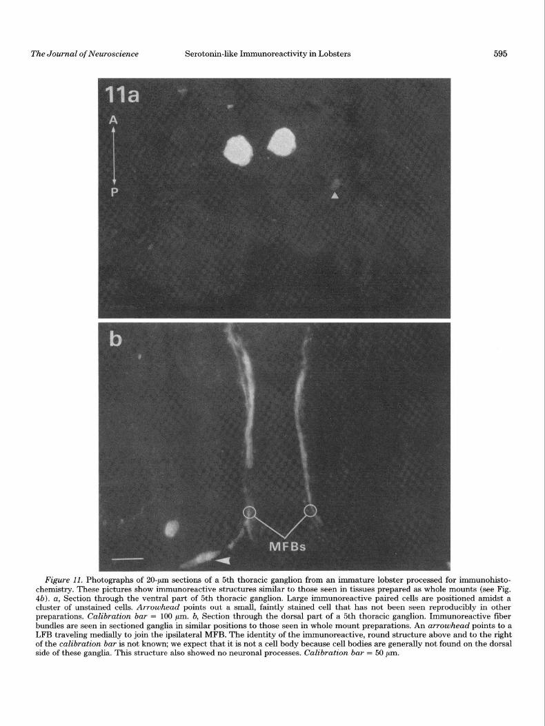
ganglia (subesophageal, 2nd to 5th thoracic, and 1st abdominal) were then compared with whole mount prep- arations (Fig. 11). In all cases, sectioned material yielded results similar to those of the whole mounts. This sug- gests that processing the tissues as whole mounts was giving us a realistic picture of the organization of these ganglia. Occasionally faintly stained cells were seen in sections that had not been seen in whole mount prepa- rations (Fig. lla); however, these cells were few in num- ber and were not reliably seen in repeated experiments.
In several experiments, ganglia were incubated in tryp- tophan (lop7 M), serotonin (IO-* M), or colchicine (7 mM) for 3 hr prior to fixation in an attempt to increase serotonin levels in the ganglia (Livingstone et al., 1980) and thereby reveal other immunoreactive cells, pro- cesses, or nerve endings. Following these incubations, no additional immunoreactive cells were identified, and the pattern of organization of neuropil regions was un- changed. The neurosecretory cell bodies in the thoracic second roots also remained unstained.
Controls for age of animals
In most of the experiments in our laboratory we use adult (5- to 6-year-old, 0.5 kg) lobsters. In all of the experiments in these studies, however, we used younger (approximately l-year-old) and smaller (25 to 50 gm) lobsters. In order to assure ourselves that the patterns of neuronal labeling were similar, at least in some ganglia, in immature and adult animals, we fixed, sectioned, and examined 5th thoracic and 1st abdominal ganglia of adult animals. The numbers and positions of immunoreactive cell bodies were the same in young and adult animals
(Fig. 12a), and the fiber bundles found in the immature lobsters were also present in adults (Fig. 12b). The main difference between ganglia from the two groups of ani- mals was in the density and size of the thoracic neuropil. In ganglia from young animals, thoracic neuropil regions were present but sparse (Figs. 4, a and b and 9). In an adult 5th thoracic ganglion, neuropil regions were large in diameter (800 pm) and the region stained was densely filled with fine processes and varicosities (Fig. 12~). This increase in size and density of the neuropil is much greater than one might expect by simply comparing overall size increases of the whole ganglia between im- mature and adult animals.
Specificity of the immunofluorescence
Control studies. In the studies reported in this paper, identical results were obtained with two lots of serotonin antisera raised in different rabbits. In addition to the specificity tests performed by Immunonuclear Corp. (see “Materials and Methods”), we examined the specificity of the staining by pre-absorption of antisera with sero- tonin, a serotonin-BSA conjugate, BSA, and octopamine (see “Materials and Methods”). For these experiments pairs of second roots (proximal bifurcation regions) were used: a thoracic second root from one side of the animal was treated with the normal 1:200 dilution of antiserum and served as the control for the experimental contralat- era1 root which was treated with pre-absorbed serum. A control incubation is shown in Figure 13a. No fluores- cence is seen in second roots after pre-absorption of serum with the serotonin-BSA conjugate (Fig. 136),
Figure 10. MFBs of the 5th abdominal ganglion. A fuzzy, unbeaded region can be seen that is composed of very fine fibers. A process in the LFB on each side travels anteriorly, and then medially, to join the MFBs. Arrowheads point to anterior parts of the lateral neuropil regions. Further details of this region are out of the focus plane of this photograph. Cell bodies in this ganglion are not shown in this picture. Calibration bar = 100 pm.
The Journal of Neuroscience Serotonin-like Immunoreactivity in Lobsters 595
Figure 11. Photographs of ZO-pm sections of a 5th thoracic ganglion from an immature lobster processed for immunohisto- chemistry. These pictures show immunoreactive structures similar to those seen in tissues prepared as whole mounts (see Fig. 4b). a, Section through the ventral part of 5th thoracic ganglion. Large immunoreactive paired cells are positioned amidst a cluster of unstained cells. Arrowhead points out a small, faintly stained cell that has not been seen reproducibly in other preparations. Calibration bar = 100 pm. b, Section through the dorsal part of a 5th thoracic ganglion. Immunoreactive fiber bundles are seen in sectioned ganglia in similar positions to those seen in whole mount preparations. An arrowhead points to a LFB traveling medially to join the ipsilateral MFB. The identity of the immunoreactive, round structure above and to the right of the calibration bar is not known; we expect that it is not a cell body because cell bodies are generally not found on the dorsal side of these ganglia. This structure also showed no neuronal processes. Calibration bar = 50 pm.

Figure 12. Photographs of 20-pm sections through an adult 5th thoracic ganglion processed for immunohistochemistry. a, A ventral section, showing the paired cells amidst unstained cells. The asterisks are on the lateral neuropil regions, one of which is shown more completely in c. Compare this photograph to Figures 4b (5th thoracic whole mount, immature lobster) and lla (sectioned 5th thoracic, immature lobster). Calibration bar = 200 pm. b, Section through the dorsal part of the ganglion, showing a fiber from the lateral fiber bundle (arrowhead) projecting medially to join the ipsilateral MFB. Compare to Figures 4b and llb. Calibration bar = 100 pm. c, One of the two lateral neuropil regions found on either side of the paired cells (see a). Compare to the relatively sparse lateral neuropil found in ganglia of immature lobsters (see Figs. 4, a and b, and 9). Calibration bar = 200 pm.
596

The Journal of Neuroscience Serotonin-like Immunoreactivity in Lobsters
Figure 13. Absorption controls using thoracic second roots. Calibration bars = 40 p. a, This second root was incubated in the working dilution of antiserotonin antibody. The immunoreactive plexus of fine fibers and varicosities is characteristic of thoracic second roots. b, Photograph of a second root that was incubated in serum pre-absorbed with a serotonin-BSA conjugate. No immunoreactivity is seen in the plexus surrounding the root. c, Picture of a second root that was incubated in serum pre-absorbed with serotonin. Slight immunoreactivity is seen in the second root plexus. d, Picture of a second root that was incubated in serum pre-absorbed with BSA. The immunoreactivity in the plexus appears normal (compare with a).

598 Beltz and Kravitz Vol. 3, No. 3, Mar. 1983
whereas a nearly complete block of staining is seen when serum is pre-absorbed with serotonin alone (Fig. 13~). The slight fluorescence that remains in the latter case may be due to antibodies with a particularly high affinity for the serotonin-BSA conjugate and a lower affinity for free serotonin. Such antibodies might have a far greater preference for serotonin fixed to tissue than for the free serotonin used in the pre-absorption and might, there- fore, bind to the tissue antigen preferentially. BSA alone (Fig. 13d) and octopamine (not shown) did not interfere with the immunofluorescence.
In a second set of experiments, we examined the spec- ificity of staining in central ganglia with similar results. Pre-absorption with a serotonin-BSA conjugate com- pletely blocked the fluorescence, whereas BSA alone had no effect.
5,7-Dihydroxytryptamine (5,7-DHT) injections. Liv- ingstone et al. (1981) demonstrated that 2 to 3 weeks after adult lobsters were injected with 5,7-DHT, the endogenous serotonin content of thoracic second roots was reduced by 90% and the capacity of this region to synthesize serotonin from tryptophan by 85%. 5,7-DHT treatment did not change the octopamine content of roots (which is about 10 times the serotonin content) or its rate of synthesis from precursor compounds (tyrosine or tyramine) (Livingstone et al., 1981). Therefore, using this treatment we can distinguish between serotonin immunoreactivity and any possible cross-reactivity of the antibody with octopamine. We injected immature lob- sters with 5,7-DHT (see “Materials and Methods”) and 2 to 4 weeks after injection, thoracic second roots were examined by immunohistochemical techniques. Two types of results were seen. Either the nerve plexuses surrounding the roots were completely depleted of fluo- rescence (Fig. 14a) or a few punctate spots remained that were not at all comparable to the profuse strings of endings seen in control tissues (Fig. 14b). Occasionally both kinds of results were seen in paired roots. The punctate pattern may represent a remnant of the periph- eral plexus in the process of degeneration.
In central ganglia of adult animals, 5,7-DHT treatment was less effective in reducing the serotonin content (Liv- ingstone et al., 1981). For example, the endogenous ser- otonin content was reduced 44% in thoracic ganglia, 52% in the subesophageal ganglion, and only 14% in the brain. The immunohistochemical results also indicate a partial depletion of serotonin in the central nervous system. The cell bodies and central fiber bundles show apparently normal staining properties, whereas neuropil regions show little or no residual fluorescence (compare Fig. 14~ to Fig. 4a).
Discussion
A major goal of these studies was to determine whether immunohistochemical procedures could be used to de- velop a reasonably complete morphological picture of the serotonin-containing system of neurons in lobsters. Such studies would, we felt, complement our prior biochemical and anatomical studies and allow us to begin a physio- logical analysis of this system at a cellular level.
Validity of the observedpattern of staining. The com- pleteness and accuracy of the morphological picture pre- sented in this article depend on: (1) the specificity for
serotonin of the antibody preparations; (2) the ability of the antibodies to penetrate tissues and bind to tissue sites containing serotonin in whole mount preparations; and (3) there being sufficient levels of serotonin in cells to be revealed by the staining procedure.
We tested the first point, the specificity of the anti- bodies for serotonin, in several ways: (1) by inhibiting the immunohistochemical staining by pre-absorbing an- tisera with serotonin-conjugated antigen and with free serotonin (Fig. 13); (2) by failing to block staining by pre- absorption of antisera with other amines found in the lobster nervous system; and (3) by dramatically reducing staining with procedures that reduce serotonin levels in lobsters (Fig. 14). The last test, in which animals were injected with 5,7-dihydroxytryptamine, is a particularly important control. 5,7-DHT injection does not destroy octopamine-containing nerve endings and does not re- duce the levels of octopamine in the lobster nervous system (Livingstone et al., 1981), but it does abolish the staining of nerve endings by the immunohistochemical procedure. This result, and the failure to inhibit staining when sera are pre-absorbed with octopamine, indicate that the antibody preparations are not staining octop- amine-containing structures. We have not, however, ruled out the possibility that indole derivatives other than serotonin might be found in certain of the stained neurons. Serotonin is metabolized in lobsters to P-alanyl and sulfate conjugates and a ,l?-alanyl, sulfate double conjugate (Kennedy, 1977, 1978). We do not know if these or related compounds react with our antibody preparations. For the larger cells, it is possible to isolate individual cell bodies and analyze them directly for ser- otonin. Such studies will be a necessary part of the evidence that the stained cells actually contain serotonin.
We attempted to deal with the second point, the pen- etration of antibodies into tissue whole mounts, both by using immature lobsters where the ventral nerve cord is small, and by comparing the results of whole mount experiments with those where ganglia from adult and immature animals were sectioned prior to immunohis- tochemical processing. We noted no significant differ- ences in the patterns of cell body staining or in the major features of ganglionic organization between sectioned and intact tissues.
In considering the final point, whether sufficient con- centrations of serotonin were present in cells to be de- tectable by these methods, we attempted to raise sero- tonin levels by incubating tissues with tryptophan, sero- tonin (Livingstone et al., 1981), and colchicine prior to fixation. Here again we observed no differences between control and pre-incubated tissues.
These three types of control experiments and the ob- servation that the pattern of staining of cells and their processes is highly reproducible, support the validity of the morphological picture of serotonin-like immunoreac- tivity that we report in this paper.
Serotonin staining in crustaceans by histofluores- cence methods: comparisons with the immunohisto- chemical results. Serotonin-containing structures have been identified previously in a variety of crustaceans using the Falck-Hillarp histofluorescence technique (Falck et al., 1962) (crayfish: Astacus astacus, Elofsson et al., 1966; crab: Carcinus maenas, Libinia emarginata,

The Journal of Neuroscience Serotonin-like Immunoreactivity in Lobsters 599
Figure 14. Thoracic second roots and a 4th thoracic ganglion from lobsters treated with 5,7-dihydroxytryptamine. a and b, In these photographs, the plexus that normally surrounds each thoracic second root contains little ( b ) or no (a) immunoreactivity. Calibration bar = 20 pm. c, Arrowhead at left points to a faint remnant of neuropil in this 4th thoracic ganglion. The lateral neuropil on the right side is completely absent (compare to Fig. 4a). The asterisk is over one second root that is faintly immunoreactive. Arrows point to two vacuole-like structures that appear in some ganglia following 5,7-dihydroxytryptamine treatment. These are probably not cell bodies because they show no neuronal processes. Calibration bar = 300 pm.
Libinia dubia, Cancer irroratus, Cancer borealis, Car- disoma guanhumi, Cooke and Goldstone, 1970; Gold- stone and Cooke, 1971; lobster: Panulirus interruptus, Homarus vulgaris, and Homarus americanus, Kushner and Maynard, 1977; Osborne and Dando, 1970). While these earlier studies have provided valuable information on the existence of presumptive serotonin-containing neurons in crustacean nervous tissue, our studies show over an order of magnitude higher numbers of likely serotonergic neurons. Species differences might partially account for this discrepancy. What is more likely, how- ever, is that with crustacean tissues, particularly marine crustaceans, immunohistochemical methods yield more reliable results. Other authors have pointed out some major problems in using histofluorescence techniques for serotonin: for example, the yellow fluorescence charac-
teristic of serotonin fades rapidly, making thorough ex- amination of tissues difficult (Fuxe and Jonsson, 1967); the often subtle yellow fluorescence can be obscured by the presence of a much more stable green fluorescence characteristic of catecholamines (Fuxe and Jonsson, 1967; Kushner and Maynard, 1977; Parent, 1981); and with marine invertebrates, freeze drying of tissues often yields very poor tissue preservation (for discussion, see Goldstone and Cooke, 1971). Such difficulties are not encountered with the immunohistochemical methods: tissues are adequately preserved by fixation, at least for light microscopy; the antibodies seem to be sensitive and specific for serotonin; and the fluorescence is relatively stable for weeks if tissues are stored at 4°C. Thus far we have not been able to show homology with the Homarus immunoreactive cells and the serotonergic neurons iden-
600 Beltz and Kravitz Vol. 3, No. 3, Mar. 1983
tified previously by the histofluorescence techniques. This is because the organization of the ganglia of the species involved is often different (e.g., fused ganglia) or because the published figures do not provide enough landmarks for comparison.
Cell bodies and their projections in the ventral nerve cord. The majority of the approximately 100 cell bodies that show serotonin immunoreactivity in the ventral nerve cord are small and their axonal processes are too fine to trace visually. We have, however, been able to follow the major projections of the larger cell bodies in the thoracic ganglia and of the pair of large cells in the 1st abdominal ganglion. Several features of these projec- tions raise interesting physiological possibilities. For ex- ample, while stained cells may have small arborizations of dendrites or axons in the same ganglion as their soma, the principal projections of the large cells are intergan- glionic. No major branches of these neurons project into the roots of the same ganglion in which the cell body appears. Instead cells send axons to join one of the three fiber bundles that traverse much of the length of the ventral nerve cord (Figs. 3 and 4). Projections from serotonin-immunoreactive cell bodies may travel through two or more ganglia before reaching their final termina- tions, they may have multiple targets along their route, and within target areas they may arborize extensively (as in the thoracic second root nerve plexuses). A similar structural organization is seen in serotonergic systems of molluscs and vertebrates. The processes of serotonergic cells in these systems are extremely widespread and highly branched (Steinbusch and Nieuwenhuys, 1979; Van der Kooy and Kuypers, 1979; Kupfermann and Weiss, 1981; Parent, 1981). Consistent with extensive arborizations of these cells are the very large cell body sizes seen in neurons of the 4th and 5th thoracic and the 1st abdominal ganglia. These neurons are among the largest found in lobsters. The large cell body size also is typical of many invertebrate serotonergic neurons. For example, the serotonin-containing Retzius cells of the leech (50 to 80 pm diameter) and metacerebral cells of molluscs (150 to 300 pm diameter) are among the largest neurons in the nervous systems of those animals (Rude et al., 1969; Cottrell and Osborne, 1970; Senseman and Gelperin, 1974; Kupfermann and Weiss, 1981). It is also of interest that in the lst, 5th, and 6th abdominal ganglia we have observed unpaired cells showing serotonin-like immunoreactivity. With few exceptions, most lobster neurons that have been mapped in central ganglia are paired (Otsuka et al., 1967; but see Muramoto, 1977). Unpaired octopamine-containing cells have been found in certain insect central nervous systems (Evans and O’Shea, 1977; Hoyle, 1974, 1975, 1978). These cells are known to serve a neuromodulatory role in adult animals (O’Shea and Evans, 1979). The functional activities of the unpaired cells in lobsters remain to be explored.
Origins of the thoracic second root plexus of seroto- nergic endings. Our earlier studies showed that serotonin was found, synthesized, and released at two locations along lobster thoracic second roots. In agreement with these results we now observe immunohistochemical staining of a dense plexus of neuronal fibers and varicos- ities at these sites. At the ultrastructural level the pre-
vious studies identified four morphologically distinguish- able types of nerve endings in these regions, one of which was shown to be associated with serotonin by electron- microscopic autoradiography (Livingstone et al., 1981). A confirmation of this result by immunohistochemistry at the electron microscopic level remains to be done.
A particular benefit of the present studies was that we could trace some of the axons that form the second root plexuses to their origins at central neurons or central neuropil regions. A clear connection was seen between the paired cells of the 1st abdominal ganglion and pro- cesses that project out the second roots of the 4th, and second and third roots of the 5th thoracic ganglia. In the second roots of more anterior thoracic ganglia, nerve processes that originate at the MFBs form the nerve plexuses (see Fig. 9), but we have not traced these pro- cesses to their cell bodies of origin. One of these processes, however, originates from a very large axon in the MFB. We expect that this is the axon of one of the large paired cell bodies of the 5th thoracic or 1st abdominal ganglia. Since at present we have no evidence that the immuno- reactive cells of thoracic ganglia 1 to 4 send projections either into the MFBs or out the thoracic second roots, it may be that all the second roots receive inputs originating from the 5th thoracic and 1st abdominal cell bodies. For this reason, we expect these two pairs of central neurons to be very important in our understanding of the func- tioning of the thoracic second root system. Clear connec- tions have also been seen between central neuropil re- gions and processes entering the thoracic second roots (see Fig. 9). These neuropil regions are connected with LFB fibers in each ganglion, but we have not been able to trace these fibers back to their cells of origin.
Earlier studies from this laboratory revealed that the firing of cell bodies in the thoracic second roots is in- hibited by serotonin (Konishi and Kravitz, 1978). Since the serotonin-immunoreactive varicosities forming sec- ond root plexuses are in close proximity to these cells, it is possible that the central neurons from which these plexuses originate control the firing of the root cells via local release of serotonin. If this is so, the peripheral serotonergic nerve plexuses might serve both as the source of serotonin that functions as a circulating neu- rohormone through direct release of the amine into the hemolymph and also govern the firing, and thereby the release, of the neurosecretory materials (as yet uniden- tified) associated with the peripheral neurosecretory neu- rons.
Physiological actions of serotonin on peripheral and central tissues in lobsters. Our earlier studies and those of other investigators have identified effects of this amine on various peripheral tissues: neuromuscular prepara- tions (Weiss et al., 1978; Glusman and Kravitz, 1982); heart (Cooke, 1966; Cooke and Hartline, 1975); scaphag- nothite (Berlind, 1977). These tissues are very sensitive to serotonin: for example, in some neuromuscular prep- arations, concentrations as low as 5 X lo-” M increase transmitter release from excitatory and inhibitory nerve endings and enhance contractions in muscle fibers (Glus- man and Kravitz, 1982). Since neuromuscular prepara- tions and many other responsive peripheral tissues con- tain no measurable serotonin, the amine may act on

The Journal of Neuroscience Serotonin-like Immunoreactivity in Lobsters 601
many of these tissues in vivo as a neurohormone carried in the hemolymph. Measurements of serotonin levels in lobster hemolymph support this possibility: the amine circulates at close to its physiologically effective concen- tration (lo-’ M range). The circulating serotonin probably comes from the dense plexuses of nerve endings found along the thoracic second nerve roots (see above), and at least some of these release sites originate from identified central cell bodies. We now have an opportunity to study the properties of these serotonin-releasing cells directly using intracellular techniques.
In addition to its actions on peripheral tissues, more recent studies have shown that serotonin affects lobster central ganglia as well (Livingstone et al., 1980; D. Glanz- man and F. Krasne, personal communication). Injecting serotonin into freely moving animals causes a character- istic flexed posture (Livingstone et al., 1980). Studies with isolated central ganglia suggest that the posture is produced by a serotonin-triggered motor program con- sisting of coordinated firing of the excitatory and inhibi- tory neurons innervating postural skeletal muscles (Liv- ingstone et al., 1980). In contrast to the peripheral effects, however, the central actions require concentrations of serotonin orders of magnitude higher than those found circulating in the hemolymph. Such concentrations (lo-” to lop6 M) would not be reached in the hemolymph of lobsters even if animals liberated their entire store of serotonin. Therefore, circulating serotonin is not likely to be responsible for the amine-induced postural changes. We do not know why such high concentrations of sero- tonin are necessary to produce central actions. One pos- sibility is that sensitive sites in central ganglia are not easily reached by serotonin due to permeability barriers; another possibility is that central serotonin receptors respond only to high concentrations of the amine and ignore low concentrations. If serotonin was released from central synaptic sites where the local concentration was high, both of these problems would be circumvented. The presence of immunoreactive neuropil regions in cen- tral ganglia (shown in Figs. 4, 8, 9, and 12) suggests that synaptic sites capable of releasing serotonin might exist in the ventral nerve cord, in addition to the peripheral hormonal release already documented (Sullivan et al., 1977; Livingstone et al., 1981). Electron microscopic ex- amination of the immunoreactive processes within these neuropil regions and direct release experiments, however, will be needed before we can say with any surety that central serotonergic synapses actually exist in the lobster ventral nerve cord.
The morphological studies described in this paper are steps toward unraveling the function of serotonin in lobsters. We are impressed by the extensive and elaborate distribution of this amine, as well as by the diversity of its physiological effects. The immunological methods have identified many neurons throughout the lobster nervous system that are likely to contain serotonin and have set the stage for physiological experiments dealing with the activation and consequences of activation of these cells. Ultimately, through a combined physiologi- cal, biochemical, and anatomical approach, we hope to learn what factors cause serotonin release, both centrally and peripherally, and what consequences the release of this amine has on the behavior of the animal.
References
Battelle, B. A., and E. A. Kravitz (1978) Targets of octopamine action in the lobster: Cyclic nucleotide changes and physio- logical effects in haemolymph, heart, and exoskeletal muscle. J. Pharmacol. Exp. Ther. 205: 438-448.
Berlind, A. (1977) Neurohumoral and reflex control of scaphog- nathite beating in the crab Car&us maenus. J. Comp. Physiol. 116: 77-90.
Bishop, C. A., and M. O’Shea (1982) Neuropeptide proctolin (H-Arg-Tyr-Leu-Pro-Thr-OH): Immunocytochemical map- ping of neurons in the central nervous system of the cock- roach. J. Comp. Neurol. 207: 223-238.
Cooke, I. M. (1966) The sites of action of pericardial organ extract and 5-hydroxytryptamine in the decapod crustacean heart. Am. Zool. 6: 107-121.
Cooke, I. M., and M. W. Goldstone (1970) Fluorescence locali- zation of monoamines in crab neurosecretory structures. J. Exp. Biol. 53: 651-668.
Cooke, I. M., and D. K. Hartline (1975) Neurohormonal alter- ation of integrative properties of the cardiac ganglion of the lobster Homarus americanus. J. Exp. Biol. 63: 33-52.
Cottrell, G. A., and N. N. Osborne (1970) Subcellular localiza- tion of serotonin in an identified serotonin-containing neu- rone. Nature 225: 470-472.
Dudel, J. (1965) Facilatatory effects of 5-hydroxytryptamine on the crayfish neuromuscular junction. Naunyn-Schmiedebergs Arch. Exp. Pathol. Pharmacol. 249: 515-528.
Elofsson, R., T. Kauri, S.-O. Nielsen, and J. 0. Stromberg (1966) Localization of monoaminergic neurons in the central nervous system of Astacus astacus LinnQ (Crustacea). Z. Zellforsch. 74: 464-473.
Evans, P. D., E. A. Kravitz, B. R. Talamo, and B. G. Wallace (1976) The association of octopamine with specific neurons along lobster nerve trunks. J. Physiol. (Lond.) 262: 51-70.
Evans, P. D., and M. O’Shea (1977) The identification of an octopaminergic neurone and the modulation of a myogenic rhythm in the locust. J. Exp. Biol. 73: 235-260.
Falck, B., N.-A. Hillarp, G. Thieme, and A. Torp (1962) Fluo- rescence of catecholamines and related compounds con- densed with formaldehyde. J. Histochem. Cytochem. 10: 348- 354.
Florey, F., and E. Florey (1954) Uber die mogliche Bedeutung von Enteramin (5-oxytryptamine) als nervoser Akterrssub- stanz bei cephalopoden und dekapoden crustacean. Z. Natur- forsch. 96: 58-68.
Florey, E., and M. Rathmayer (1978) The effects of octopamine and other amines on the heart and on neuromuscular trans- mission in decapod crustaceans: Further evidence for a role as a neurohormone. Comp. Biochem. Physiol. 61C: 229-237.
Fuxe, K., and G. Jonsson (1967) A modification of the histo- chemical fluorescence method for the improved localization of 5-hydroxytryptamine. Histochemie 11: 161-166.
Glusman, S., and E. A. Kravitz (1982) The action of serotonin on excitatory nerve terminals in lobster nerve-muscle prepa- rations. J. Physiol. (Lond.) 325: 223-241.
Goldstone, M. W., and I. M. Cooke (1971) Histochemical local- ization of monoamines in the crab central nervous system. Z. Zellforsch. 116: 7-19.
Grundfest, H., and J. P. Reuben (1961) Neuromuscular synaptic activity in lobster. In Nervous Inhibition, E. Florey, ed., pp. 92-104, Pergamon Press, New York.
Hekfelt, T., K. Fuxe, and M. Goldstein (1975) Applications of immunohistochemistry to studies on monoamine cell systems with special reference to nervous tissues. Ann. N. Y. Acad. Sci. 254: 407-437.
Hoyle, G. (1974) A function for neurons (DUM) neurosecretory on skeletal muscle of insects. J. Exp. Zool. 189: 401-406.
Hoyle, G. (1975) Evidence that insect dorsal unpaired median

602 Beltz and Kravitz Vol. 3, No. 3, Mar. 1983
(DUM) neurons are octopaminergic. J. Exp. Zool. 193: 425- 431.
Hoyle, G. (1978) The dorsal, unpaired, median neurons of the locust metathoracic ganglion. J. Neurobiol. 9: 43-57.
Kennedy, M. B. (1977) Amine metabolism: A different pathway in lobsters. Sot. Neurosci. Abstr. 3: 792.
Kennedy, M. B. (1978) Products of biogenic amine metabolism in the lobster: Sulfate conjugates. J. Neurochem. 30: 315-320.
Konishi, S., and E. A. Kravitz (1978) The physiological prop- erties of amine-containing neurones in the lobster nervous system. J. Physiol (Lond.) 279: 215-229.
Kravitz, E. A., S. Glusman, R. M. Harris-Warrick, M. S. Liv- ingstone, T. Schwarz, and M. F. Goy (1980) Amines and a peptide as neurohormones in lobsters: Action on neuromus- cular preparations and preliminary behavioral studies. J. Exp. Biol. 89: 159-175.
Kupfermann, I., and K. R. Weiss (1981) The role of serotonin in arousal of feeding behavior of Aplysia. In Serotonin Neu- rotransmission and Behavior, B. Jacob and A. Gelperin, eds., pp. 255-287, MIT Press, Cambridge, MA.
Kushner, P. D., and E. A. Maynard (1977) Localization of monoamine fluorescence in the stomatogastric nervous sys- tem of lobsters. Brain Res. 129: 13-28.
Livingstone, M. S., R. M. Harris-Warrick, and E. A. Kravitz (1980) Serotonin and octopamine produce opposite postures in lobsters. Science (N. Y.) 208: 76-79.
Livingstone, M. S., S. F. Schaeffer, and E. A. Kravitz (1981) Biochemistry and ultrastructure of serotonergic nerve end- ings in the lobster: Serotonin and octopamine are contained in different nerve endings. J. Neurobiol. 12: 27-54.
Maynard, D. M., and J. H. Welsh (1959) Neurohormones of the pericardial organs of brachyuran Crustacea. J. Physiol. (Lond.) 149: 215-227.
McLean, I. W., and P. K. Nakane (1974) Periodate-lysine-para- formaldehyde fixative. A new fixative for immunelectron microscopy. J. Histochem. Cytochem. 22: 1077.
Miller, M. W., and R. E. Sullivan (1981) Some effects of proc- tolin on the cardiac ganglion of the Maine lobster, Homarus americanus (Mime Edwards). J. Neurobiol. 12: 629-639.
Muramoto, A. (1977) Neural control of rhythmic anal contrac- tion in the crayfish. Comp. Biochem. Physiol. 56A: 551-557.
Osborne, N. N., and M. R. Dando (1970) Monoamines in the stomatogastric ganglion of the lobster Homarus uulgaris. Comp. Biochem. Physiol. 32: 327-331.
O’Shea, M., and P. D. Evans (1979) Potentiation of neuromus- cular transmission by an octopaminergic neurone in the lo-
cust. J. Exp. Biol. 79: 169-190. Otsuka, M., E. A. Kravitz, and D. D. Potter (1967) Physiological
and chemical architecture of a lobster ganglion with partic- ular reference to gamma-aminobutyrate and glutamate. J. Neurophysiol. 30: 725-752.
Parent, A. (1981) The anatomy of serotonin-containing neurons across phylogeny In Serotonin Neurotransmission and Be- hauior, B. Jacobs and A. Gelperin, eds., pp. 3-34, MIT Press, Cambridge, MA.
Rude, S., R. E. Coggeshall, and L. S. Van Orden, III (1969) Chemical and ultrastructural identification of 5-hydroxytryp- tamine in an identified neuron. J. Cell Biol. 41: 832-854.
Schwarz, T. L., R. M. Harris-Warrick, S. Glusman, and E. A. Kravitz (1980) A peptide action in a lobster neuromuscular preparation. J. Neurobiol. 11: 623-628.
Schwarz, T. L., G. Lee, and E. A. Kravitz (1981) Proctolin-like immunoreactivity in the nervous system of the lobster Hom- arus americanus. Sot. Neurosci. Abstr. 7: 253.
Senseman, D., and A. Gelperin (1974) Comparative aspects of the morphology and physiology of a single identifiable neuron in Helix aspersa, Limar maximus, and Agriolimax califor- nica. Malacolog. Rev. 7: 51-52.
Steinbusch, H. W. M., and R. Nieuwenhuys (1979) Serotonergic neuron systems in the brain of the lamprey, Lampetra flu- uiatilis (abstr.) Anat. Rec. 193: 693.
Steinbusch, H. W. M., A. A. J. Verhofstad, and H. W. T. Joosten (1978) Localization of serotonin in the central nervous system by immunohistochemistry: Description of a specific and sen- sitive technique and some applications. Neuroscience 3: 811- 819.
Sullivan, R. E. (1979) A proctolin-like peptide in crab pericar- dial organs. J. Exp. Zool. 210: 543-552.
Sullivan R. E., and D. L. Barker (1975) Octopamine increases cyclic AMP content of crustacean ganglia and cardiac muscle. Sot. Neurosci. Abstr. 1: 394.
Sullivan, R. E., B. J. Friend, and D. L. Barker (1977) Structure and function of spiny lobster ligamental nerve plexuses: Evi- dence for synthesis, storage, and secretion of biogenic amines. J. Neurobiol. 8: 581-605.
Van der Kooy, D., and H. G. J. M. Kuypers (1979) Fluorescent retrograde double labelling: Axonal branching in the ascend- ing raphe and nigral projections. Science 204: 873-875.
Weiss, K. R., J. L. Cohen, and I. Kupfermann (1978) Modula- tory control of buccal musculature by a serotonergic neuron (metacerebral cell) in Aplysia. J. Neurophysiol. 41: 181-203.