mapping the binding domain of immunoglobulin light chains for tamm-horsfall protein
TRANSCRIPT
Mapping the Binding Domain of ImmunoglobulinLight Chains for Tamm-Horsfall Protein
Wei-Zhong Ying and Paul W. SandersFrom the Department of Medicine and Department of Physiology
and Biophysics, Division of Nephrology, Nephrology Research
and Training Center, Comprehensive Cancer Center, and Cell
Adhesion and Matrix Research Center, University of Alabama at
Birmingham, Birmingham, Alabama; and the Department of
Veterans Affairs Medical Center, Birmingham, Alabama
Cast nephropathy, or myeloma kidney, is a potentiallyreversible cause of chronic renal failure. In this condi-tion, filtered light chains bind to a common site onTamm-Horsfall protein (THP), which is produced bycells of the thick ascending limb of the loop of Henle.Subsequent aggregation of these proteins producescasts that obstruct tubule fluid flow and results in renalfailure. In the present study, we used the yeast two-hybrid system to determine the site of interaction oflight chains with THP. The third complementarity-de-termining region (CDR3) of both � and � light chainsinteracted with THP. These findings were confirmed ina series of competition studies using a synthetic peptidethat corresponded to the CDR3 region and purified THPand light chains. Variations in the CDR3 sequence of thelight chain affected binding. Thus, the current studiesincrease our understanding of the process of cast for-mation and provide an opportunity to develop strate-gies that may inhibit this interaction and prevent theclinical manifestations of myeloma kidney. (Am JPathol 2001, 158:1859–1866)
More than 150 years ago, an astute clinician, Dr. WilliamMacintyre, found an abnormal substance in the urine ofone of his patients. He sent the material to Dr. HenryBence Jones, who reported the newly described proteinand the association with multiple myeloma.1 Bence Jonesproteins were subsequently identified as immunoglobulinlight chains.2 Light chains are typically filtered from theblood by the kidney and metabolized similarly to otherlow molecular weight proteins.3 However, these proteinscan be nephrotoxic.4 During the process of absorptionand catabolism, light chains have been shown to causeproximal tubular epithelial cell injury.4,5 When the reab-sorptive capacity of the proximal tubular cells is satu-rated, light chains are presented to the distal nephron,where they form casts that obstruct flow of tubular fluid.The resultant renal failure is known clinically as castnephropathy, or myeloma kidney.6
Cast nephropathy represents the most common causeof renal failure in multiple myeloma.7 To initiate cast for-mation, light chains bind to a specific peptide domain onTamm-Horsfall protein (THP),8–12 which is synthesizedexclusively by cells of the thick ascending limb of theloop of Henle.13,14 Co-aggregation of light chains withTHP produces the intraluminal casts that are the promi-nent feature of myeloma kidney.8 The electrolyte compo-sition of the tubule fluid as well as tubule fluid flow ratesand amount of THP8–12 modulate binding. The structureof the light chain plays an important role in associationwith THP10 and may also promote homotypic aggrega-tion.15 Although myeloma kidney is potentially reversible,prevention of cast formation is the key to controlling theproblem. Understanding the protein interactions involvedin cast formation represents the initial advance in devel-opment of potential treatment strategies designed to pre-vent myeloma kidney. The current study determined thedomain on the light chain involved in binding THP.
Materials and Methods
Yeast Two-Hybrid Studies
The yeast two-hybrid system (Matchmaker LexA Two-Hybrid System; Clontech Lab. Inc., Palo Alto, CA) wasused initially to detect binding interactions between THPand immunoglobulin light chains. This approach was sim-ilar to the original description of the yeast two-hybridassay,16 but was a LexA-based interaction trap system.17
The host strain in these experiments was Saccharomycescerevisiae EGY48[p8op-lacZ]. The bait consisted of twofragments of human THP that were obtained by polymer-ase chain reaction using cDNA that was provided byGenentech, Inc. (South San Francisco, CA). Descriptionof the cloning and characterization of THP has beenpublished.18 All primers used in this study were obtainedcommercially (Operon Tech. Inc., Alameda, CA). Be-cause there is a single binding domain (amino acid res-idues 225 to 233) for light chains on THP,10,12 the present
Supported by The Office of Research and Development, Medical Re-search Service, Department of Veterans Affairs, and National Institutes ofHealth grant (R01 DK46199).
Accepted for publication January 23, 2001.
Address reprint requests to Paul W. Sanders, M.D., Division of Nephrol-ogy/Department of Medicine, 642 Lyons-Harrison Research Building, 701South 19th St., University of Alabama at Birmingham, Birmingham, AL35294-0007. E-mail: [email protected].
American Journal of Pathology, Vol. 158, No. 5, May 2001
Copyright © American Society for Investigative Pathology
1859
study used two fragments of THP that contained this do-main. A 787-bp fragment (encoding amino acid residues148 to 410, termed THP787) was polymerase chain reac-tion-amplified using 5�-CCGGAATTCCAATGTGGTGGGC-AGCTACTT-G-3� as the forward primer and 5�-ACGCTC-GAGCTCCACGGAGCTGGGGTCTGTGC-3� as the reverseprimer. Underlined sequences in the upstream and down-stream primers contained an EcoRI and XhoI site, respec-tively. A 248-bp fragment (encoding amino acid residues188 to 270, termed THP248) was created using 5�-CCG-GAATTCCGCATGGCCGAGACCTGCGTGC as the forwardprimer and 5�-ACGCTCGAGCTCCACGGAGCTGGGGTC-TGTGC3� as the reverse primer. THP787 and THP248 wereeach ligated into pBluescript II SK(�) vector (Stratagene, LaJolla, CA) using EcoRI and XhoI and then cloned. Sequencesof both products were confirmed using a kit (T7 SequenaseDNA sequencing kit; Amersham Life Science, Inc., Cleveland,
OH). THP787 and THP248 were then cloned into the pLexAexpression vector using EcoRI and XhoI.
Representative cDNAs encoding immunoglobulin �and � light chains were generous gifts from Dr. S. LouisBridges, Jr., at the University of Alabama at Birmingham,Birmingham, AL. A total of four unique � and seven � lightchains were used. Descriptions of these molecules havebeen published.19–21 Each cDNA was used as a tem-plate in polymerase chain reactions to generate frag-ments that were initially cloned into pBluescript II SK(�)for sequencing and then inserted in-frame into pB42ADexpression vector, using EcoRI and XhoI. The expressedfusion proteins consisted of the light chain of interest andthe LexA transcriptional activation domain. Primers andlight chains used in these reactions were shown in Tables1 and 2. A series of truncation and deletion mutants of the� light chain, SSH23, and the � light chain, ITPBLL1, were
Table 1. Primers Used to Generate the � Light Chain Constructs
Light chain construct, � Primer name Forward and reverse primers
ITPBL5 LB62 5�-CCGGAATTCGAAATTGTGTTGACGCAGTCTCCA-3�H170 5�-ACGCTCGAGAACTGCTCATCAGATGGCGGGAAG-3�
ITPBL11 LB62 5�-CCGGAATTCGAAATTGTGTTGACGCAGTCTCCA-3�H170 5�-ACGCTCGAGAACTGCTCATCAGATGGCGGGAAG-3�
BC Syn9 LB62 5�-CCGGAATTCGAAATTGTGTTGACGCAGTCTCCA-3�H170 5�-ACGCTCGAGAACTGCTCATCAGATGGCGGGAAG-3�
SSH23 LB62 5�-CCGGAATTCGAAATTGTGTTGACGCAGTCTCCA-3�LSK19 5�-ACGCTCGAGAACTGCTCATCAGATGGCGGGAAG-3�
SSH23a LB62 5�-CCGGAATTCGAAATTGTGTTGACGCAGTCTCCA-3�SSH23a 5�-ACGCTCGAGTGGAGATTGGCCTGGCCT-3�
SSH23b SSH23b 5�-CCGGAATTCAGGCGCCTAATTTATAAG-3�H170 5�-ACGCTCGAGAACTGCTCATCAGATGGCGGGAAG-3�
SSH23c SSH23c 5�-CCGGAATTCTATTACTGCATGCAA-3�H170 5�-ACGCTCGAGAACTGCTCATCAGATGGCGGGAAG-3�
SSH23� LB62 5�-CCGGAATTCGAAATTGTGTTGACGCAGTCTCCA-3�LSK19 5�-ACGCTCGAGAACTGCTCATCAGATGGCGGGAAG-3�
Underlined sequences contained EcoRI and Xho I restriction sites on the forward and reverse primers, respectively.
Table 2. Primers Used to Generate the � Light Chain Constructs
Light chain construct, � Primer name Forward and reverse primers
ITPBLL1 LB75 5�-CCGGAATTCTCCGAATTCTCCTCTCTCACTGCACAG-3�LB69 5�-ACGCTCGAGGGGAATTCGCTCCCGGGTAGAAGTCTCT-3�
ITPBLL1a LB75 5�-CCGGAATTCTCCGAATTCTCCTCTCTCACTGCACAG-3�PBLL1a 5�-ACGCTCGAGTCGCTCAGGGATCTCTGA-3�
ITPBLL1b PBLL1b 5�-CCGGAATTCAGCGACCGGCCCTCAGAG-3�LB69 5�-ACGCTCGAGGGGAATTCGCTCCCGGGTAGAAGTCTCT-3�
ITPBLL1c PBLL1b 5�-CCGGAATTCAGCGACCGGCCCTCAGAG-3�PBLL1c 5�-ACGCTCGAGTAGGACGGTGACCTT-3�
ITPBLL1d LB75 5�-CCGGAATTCTCCGAATTCTCCTCTCTCACTGCACAG-3�PBLL1d 5�-ACGCTCGAGATAGACGACCAGCAC-3�
LKPBLLL2 LB75 5�-CCGGAATTCTCCGAATTCTCCTCTCTCACTGCACAG-3�LB69 5�-ACGCTCGAGGGGAATTCGCTCCCGGGTAGAAGTCTCT-3�
ITPBLL11 LB76 5�-CCGGAATTCTCCGAATTCTCTGCACAGTCTCTGAGGCC-3�LB69 5�-ACGCTCGAGGGGAATTCGCTCCCGGGTAGAAGTCTCT-3�
ITPBLL22 LB77 5�-CCGGAATTCCCCTGAATTCCTCGGCGTCCTTGCTTACTGCA-3�LB69 5�-ACGCTCGAGGGGAATTCGCTCCCGGGTAGAAGTCTCT-3�
ITPBLL68 LB75 5�-CCGGAATTCTCCGAATTCTCCTCTCTCACTGCACAG-3�LB69 5�-ACGCTCGAGGGGAATTCGCTCCCGGGTAGAAGTCTCT-3�
ITPBLL7 LB75 5�-CCGGAATTCTCCGAATTCTCCTCTCTCACTGCACAG-3�LB69 5�-ACGCTCGAGGGGAATTCGCTCCCGGGTAGAAGTCTCT-3�
ITPBLL75 LB75 5�-CCGGAATTCTCCGAATTCTCCTCTCTCACTGCACAG-3�LB69 5�-ACGCTCGAGGGGAATTCGCTCCCGGGTAGAAGTCTCT-3�
Underlined sequences contained Eco RI and Xho I restriction sites on the forward and reverse primers, respectively.
1860 Ying and SandersAJP May 2001, Vol. 158, No. 5
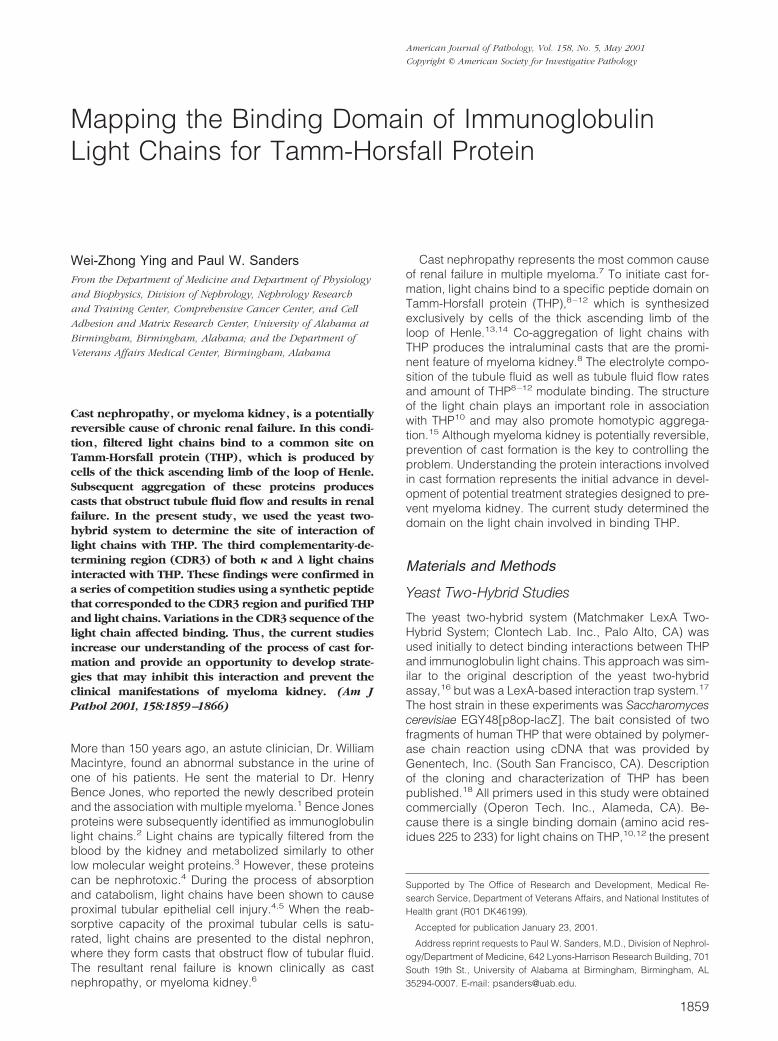
also created (Figure 1). Restricting the 506-bp fragmentwith BsiZI that cut the insert in two unique sites, andsubsequently purifying and intramolecularly re-ligatingthe product created a truncation mutant of SSH23. Togenerate the SSH23pep construct, two 5�-phosphory-lated synthetic oligonucleotides, 5�-CCGGAATTCATG-CAAGGTACACACTGGCCTCCGCTCACTCTCGAGCGT-3 �and 5�-ACGCTCGAGAGTGAGCGGAGGCCAGTGTGT-ACCTTGCATGAATTCCGG-3�, were annealed and li-gated into pBluescript II SK(�) for sequencing and theninto pLexA, using EcoRI and XhoI. This sequence en-coded a 10-amino acid peptide (MQGTHWPPLT) thatcorresponded to the amino acid residues from 94 to 103on SSH23. ITPBLL1pep, which encoded a 10 amino acidpeptide (QVWDSTSDHY) that corresponded to aminoacid residues 88 to 97 of ITPBLL1, was prepared insimilar manner using 5�-phosphorylated 5�-CCGGAAT-TCCAGGTATGGGATAGTACTAGTGATCATTATCTCGA-GCGT-3� and 5�-ACGCTCGAGATAATGATCACTAGTAC-TATCCCATACCTGGAATTCCGG-3�. Both SSH23pepand ITPBLL1pep corresponded to the complementarity-determining region (CDR)3 of light chains. Automated
DNA sequencing confirmed proper construction of allplasmids and authenticity of the sequences.
EGY48[p8op-lacZ] yeast were co-transformed withpLexA-THP787 (or pLexA-THP248) and pB42AD-lightchain construct, using the lithium acetate method (Alkali-Cation yeast transformation kit; BIO101 Inc., La Jolla,CA). EGY48[p8op-lacZ] yeast were also co-transformedwith plasmids (pLexA-Pos alone and pLexA-53/pB42AD-T) that served as positive control experimentsand with plasmids (pLexA-Lam/pB42AD-T) that servedas a negative control. The pLexA-Pos plasmid encoded afusion of the DNA-binding domain (LexA) with the GAL4-activating domain, allowing activation of genes undercontrol of LexA operators (LEU2 and lacZ). pLexA-53 andpB42AD-T provided another positive control experimentsby encoding proteins known to interact (murine p53 andSV40 large T-antigen, respectively). pLexA-Lam en-coded a fusion of the DNA-binding domain with humanlamin C that does not interact with SV40 large T antigenand thus served as a negative control. Double transfor-mants were selected in a standard manner using medium
Figure 1. To determine the THP-binding domain on light chains, a series of mutants of � (SSH23) and � (ITPBLL1) light chains was examined. The four frameworkregions (FR) and three CDRs of the variable subunit of the light chains are shown. The lengths (in bp) of the constructs and interaction of the fusion proteins withTHP are displayed.
CDR3 Domain Binds THP 1861AJP May 2001, Vol. 158, No. 5
that lacked histidine and tryptophan. The transformantswere incubated at 30°C for 4 days. Interactions betweenthe two hybrid proteins were then tested by growth ingalactose-containing, leucine-deficient medium and by�-galactosidase plate and liquid culture assays. In the invivo plate assay, 200 to 400 cfu of transformed yeast weredispersed on to 100-mm agar plates containing 5-bromo-4-chloro-3-indolyl-�-D-galactopyranoside (X-Gal, 80 mg/L), 1� BU salts (26 mmol/L Na2HPO4, 25 mmol/LNa2HPO4, pH 7), and either 2% galactose or 2% dex-trose. The plates were incubated at 30°C for 3 to 6 daysto generate the blue color. The interactions were furtherquantified by liquid culture assay of �-galactosidase ac-tivity using o-nitrophenyl �-D-galactopyranoside (Amer-sham Life Science, Inc., Cleveland, OH) as the substrate.Using other proteins, relative affinities detected with thisreaction have been shown to correlate with interactionsdetected using other biochemical methods.22 Five separatetransformants were each examined in triplicate. Reactionswere performed at 30°C in solutions containing 60 mmol/LNa2HPO4, 40 mmol/L NaH2PO4, 10 mmol/L KCl, 1 mmol/LMgSO4, and 50 mmol/L �-mercaptoethanol. Timing of thereaction began as o-nitrophenyl �-D-galactopyranosidewasadded and the reaction continued until a yellow color wasobserved. The reaction mixture was then centrifuged at14,000 rpm for 10 minutes to pellet cell debris. Absorbanceof the supernatant at 420 and 600 nm was determined.Units of �-galactosidase activity were calculated as follows:units � [1,000*OD400]/[(elapsed time)*(0.1*concentrationfactor)*OD600].To verify expression of the fusion proteins, selected sam-ples were examined using Western blotting. Yeast cellswere pelleted, then resuspended in cracking buffer, 8mol/L urea, 5% sodium dodecyl sulfate, 40 mmol/L Tris-HCl, 0.1 mmol/L ethylenediaminetetraacetic acid, 0.08%�-mercaptoethanol and a combination of protease inhib-itors (Complete; Boehringer Mannheim, Mannheim, Ger-many) was included. The yeast were disrupted by vigor-ous shaking for 3 minutes at 5,000 rpm (MiniBeadbeater;Biospec Products Co., Bartlesville, OK) after addition of425 to 600 �mol/L glass beads (Sigma Chemical Co., St.Louis, MO). Cell debris was removed by centrifugation at14,000 rpm for 5 minutes at 4°C. Western blot detectionof THP and light chains then proceeded in standardmanner.23,24 Briefly, the supernatant fractions wereboiled briefly, then separated using 8 or 12% polyacryl-amide gels and transferred onto nitrocellulose mem-branes. Membranes were then probed using rabbit anti-human THP antiserum (Biomedical Technologies, Inc.,Stoughton, MA) or rabbit anti-human Ig/L-chain typelambda and type kappa (Behringwerke AG, Marburg,Germany).
Purification of Human THP and ImmunoglobulinLight Chains
Human THP and light chains were obtained from urine instandard manner.8–12 THP was purified from urine of ahealthy adult male by precipitation in 0.64 mol/L NaCl,followed by dialysis and lyophilization. Purified human
THP was biotinylated as described previously,25 usingsulfo-N-hydroxysuccinimide biotin (ImmunoPure Sulfo-NHS-Biotin; Pierce Chemical Co., Rockford, IL), followedby dialysis against phosphate-buffered saline (PBS) at4°C for 24 hours to remove free biotin. Sodium dodecylsulfate-polyacrylamide gel electrophoresis identified asingle band at �100 kd (data not shown). Six differentlight chains (three � and three �) were also used in thisstudy and were obtained from patients who had lightchain proteinuria and clinical renal failure. The patientswho donated �2 and �5 had biopsy-proven cast ne-phropathy. Patients who donated �5, �3, and �7 hadclinical presentations that were compatible with cast ne-phropathy, but did not undergo kidney biopsy. The pa-tient who donated �6 had Waldenstrom’s macroglobu-linemia and progressive renal failure and was also notbiopsied. These light chains were purified from urine byprecipitation using 70% ammonium sulfate, followed byion-exchange chromatography.4,5,8,9 Purified light chainswere dialyzed and lyophilized. A single band at �22 kdwas identified by sodium dodecyl sulfate-polyacrylamidegel electrophoresis (data not shown). Proteins were keptat �20°C until use.
Peptide Competition Experiments
A synthetic peptide was obtained commercially (Re-search Genetics Inc., Huntsville, AL). MQGTHWPPLTcorresponded to the CDR3 region of SSH23. Peptideswere purified by high-pressure liquid chromatography;the molecular masses were confirmed by mass spec-trometry.
Competition experiments were performed using solid-phase enzyme-linked immunosorbent assay and Westernand dot blotting. To perform enzyme-linked immunosor-bent assay, wells of microtiter plates were coated witheach of the six purified light chains, 0.2 mmol/L in PBS,and incubated overnight at room temperature. Biotinyl-ated human THP, 0.2 �mol/L, was incubated overnight at4°C alone or with the CDR3 peptide. The concentration ofthe peptides ranged from 0 to 4 mmol/L. After washingthe wells with 0.05% Tween 20 in PBS and blocking with1% bovine serum albumin in PBS, the pre-incubated,THP/peptide mixtures were added to the wells and incu-bated at 37°C for 2 hours. After washing, samples wereincubated with avidin conjugated with horseradish per-oxidase (ImmunoPure avidin, horseradish peroxidaseconjugated; Pierce Chemical Co., Rockford, IL), 1:2,000dilution in PBS. After additional washes, wells were de-veloped using Azino-bis(3-ethylbenzthiazoline-6-sulfonicacid) as substrate in citrate-phosphate buffer, pH 4.2.Optical density was determined at 405 nm using a micro-plate reader (VERSAmax; Molecular Devices Corp.,Menlo Pack, CA).
In other competition experiments, 10 �g samples ofeach of the six light chains were separated by sodiumdodecyl sulfate-polyacrylamide gel electrophoresis using12% polyacrylamide gels and then transferred onto anitrocellulose membrane. Twenty �g of yeast extract con-taining the fusion proteins were dot-blotted directly onto a
1862 Ying and SandersAJP May 2001, Vol. 158, No. 5
nitrocellulose membrane. After blocking and washing,the blots were probed with 0.2 �mol/L biotinylated THP or0.2 �mol/L biotinylated THP that had been pre-incubatedwith CDR3 peptide, 4 mmol/L, overnight at 4°C. Afteradditional washes, the membranes were incubated withstreptavidin-conjugated HRP, 1:10,000 dilution in Tris-buffered saline. The membranes were developed usingECL Western blotting system and Hyperfilm (AmershamPharmacia Biotech, Piscataway, NJ).
Statistical Analysis
All data were presented as mean � SE. Significant dif-ferences were determined using one-way analysis of vari-ance with standard post hoc testing (Statview, version5.0; SAS Institute, Inc., Cary, NC). A P value of 0.05assigned statistical significance.
Results
Human THP Interacted with Human LightChains in Vivo
Initial experiments ensured that the hybrid proteins thatconsisted of the pLexA DNA-binding domain and the twofragments of human THP did not, by themselves, trans-activate the reporter genes. Both peptide segments ofTHP contained the known single binding site for lightchains.10,12 The light chain constructs that were testedare shown in Tables 1 and 2. Western analysis of extractsof representative co-transformed yeast confirmed ex-pression of fusion proteins that contained THP and lightchain (Figure 1). When co-expressed with pLexA-
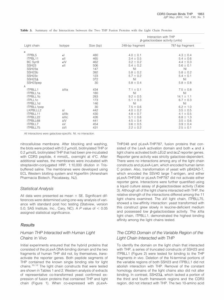
THP248 and pLexA-THP787, fusion proteins that con-sisted of the LexA activation domain and both � and �light chains activated both LEU2 and lacZ reporter genes.Reporter gene activity was strictly galactose-dependent.There were no interactions among any of the light chainconstructs and pLexA-Lam, which encoded human laminC protein. Also, transformation of yeast with pB42AD-T,which encoded the SSV40 large T-antigen, and eitherpLexA-THP248 or pLexA-THP787 did not activate eitherreporter gene. Interactions were further quantified usinga liquid culture assay of �-galactosidase activity (Table3). Although all of the light chains interacted with THP, therelative strength of the interactions differed among the 11light chains examined. The �VI light chain, ITPBLL75,showed a low-affinity interaction: yeast transformed withthis construct grew slowly in leucine-deficient mediumand possessed low �-galactosidase activity. The �IIIalight chain, ITPBLL1, demonstrated the highest bindingaffinity among the light chains tested.
The CDR3 Domain of the Variable Region of theLight Chain Interacted with THP
To identify the domain on the light chain that interactedwith THP, a series of truncated constructs of SSH23 andITPBLL1 (Figure 2) were tested for binding to the THPfragments in vivo. Deletion of the N-terminal portions ofthe variable regions of both SSH23 and ITPBLL1 did notabolish interaction with THP. Removal of the constanthomology domains of the light chains also did not alterbinding. In contrast, SSH23�, which lacked a portion ofthe third and fourth framework regions and the CDR3region, did not interact with THP. The two 10-amino acid
Table 3. Summary of the Interactions between the Two THP Fusion Proteins with the Light Chain Proteins
Light chain Isotype Size (bp)
Interaction with THP�-galactosidase activity (units)
248-bp fragment 787-bp fragment
�ITPBL5 �I 480 4.0 � 0.1 4.3 � 0.4ITPBL11 �II 465 3.4 � 0.5 5.4 � 0.6BC Syn9 �IV 462 3.2 � 0.2 4.4 � 0.3SSH23 �II 506 5.4 � 0.2 5.6 � 0.1SSH23a 147 NI NISSH23b 392 5.8 � 0.2 5.8 � 0.2SSH23c 123 5.7 � 0.2 5.4 � 0.1SSH23� 372 NI NISSH23pep 30 5.8 � 0.4 5.8 � 0.8
�ITPBLL1 �IIIa 434 7.1 � 0.1 7.5 � 0.8ITPBLL1a 180 NI NIITPBLL1b 283 9.2 � 0.5 14.1 � 1.1ITPLL1c 173 5.1 � 0.3 6.7 � 0.3ITPBLL1d 146 NI NIITPBLL1pep 30 7.5 � 0.6 6.2 � 1.0LKPBLLL2 �I 442 4.0 � 0.2 3.5 � 0.5ITPBLL11 �IIIb 430 4.8 � 0.7 4.1 � 0.5ITPBLL22 �IIIc 426 5.1 � 0.6 6.8 � 1.3ITPBLL68 �IV 441 4.5 � 0.4 3.5 � 0.6ITPBLL7 �V 454 3.6 � 0.5 3.9 � 0.4ITPBLL75 �VI 431 2.2 � 0.2 2.5 � 0.1
All interactions were galactose-specific. NI, no interaction.
CDR3 Domain Binds THP 1863AJP May 2001, Vol. 158, No. 5
peptide sequences that represented the CDR3 portionsof both molecules interacted with THP in vivo.
A Synthetic Peptide Corresponding to theCDR3 Domain Inhibited Binding of LightChains to THP
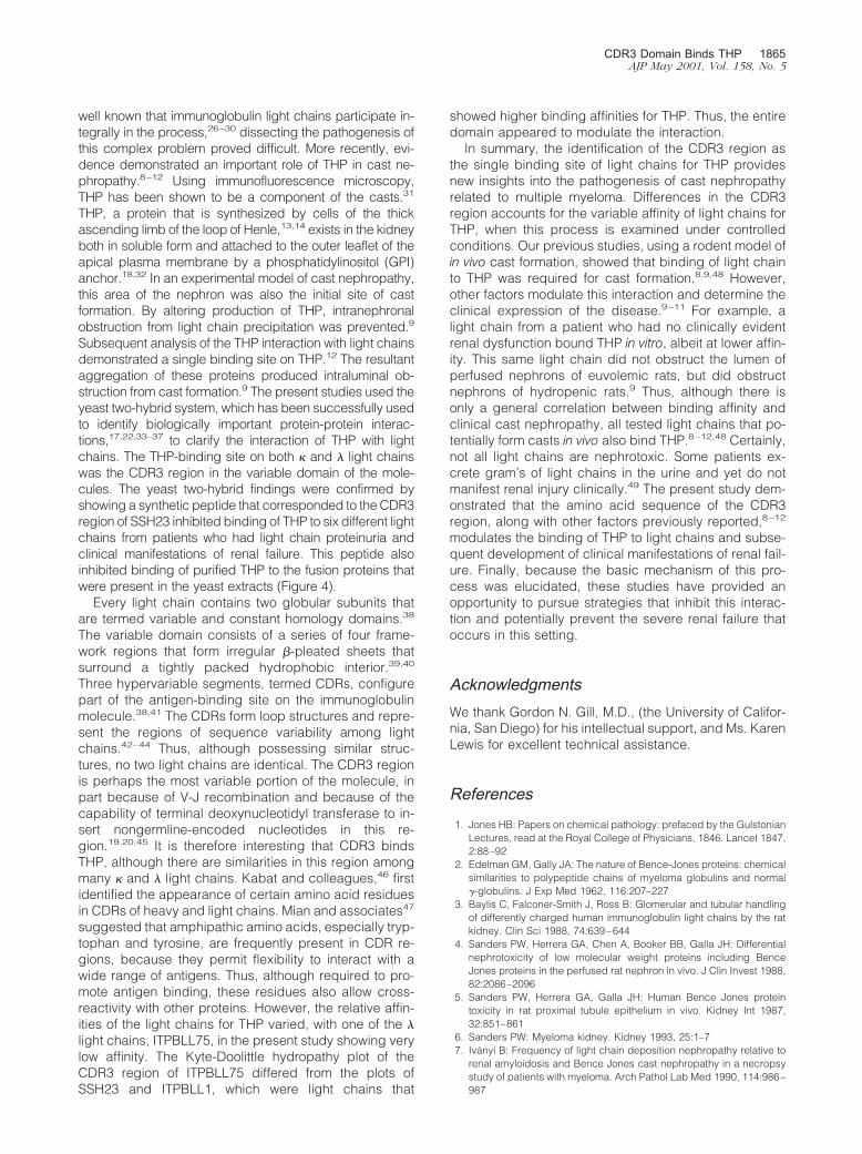
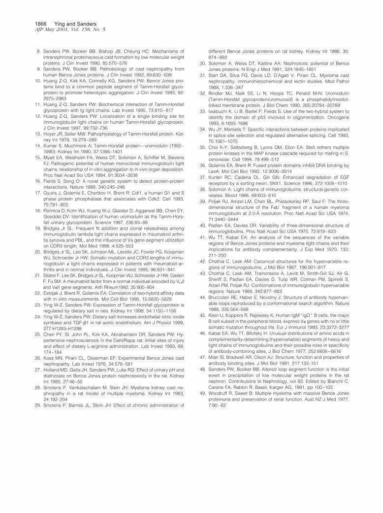
Biotinylated human THP bound to six different lightchains, three � and three �, which had been purified frompatients who had renal failure and light chain proteinuria.Pre-incubation of the THP with a synthetic 10-amino acidpeptide that represented the CDR3 region of SSH23 ef-fectively inhibited interaction of THP with the bound lightchains (Figure 3). Using enzyme-linked immunosorbentassay, this peptide inhibited, in a dose-response manner,binding of THP to these light chains (Figure 3). Extractsfrom yeast that had been transformed with the variouslight chain constructs were dot blotted and then incu-bated with biotinylated THP to show binding (Figure 4).As anticipated from the in vivo studies, THP bound tosamples that contained SSH23, SSH23b, SSH23c, andSSH23pep, but did not interact with samples containingSSH23a and SSH23�. THP also bound to samples thatcontained ITPBLL1, ITPBLL1b, and ITPBLL1c, but notextracts from yeast that expressed ITPBLL1a andITPBLL1d. These findings confirmed the in vivo studiesdescribed above. Pre-incubation of THP with the CDR3peptide resulted in inhibition of binding to these samples.
DiscussionCast nephropathy is a form of progressive renal failure thatoccurs in the setting of multiple myeloma. Although it was
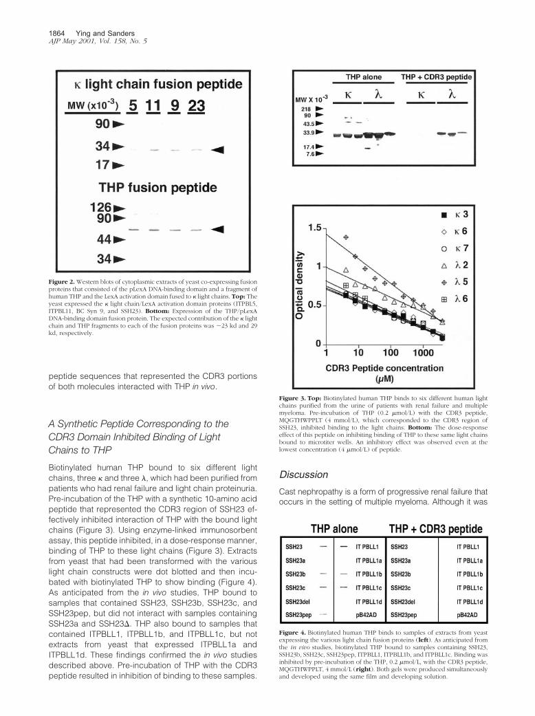
Figure 2. Western blots of cytoplasmic extracts of yeast co-expressing fusionproteins that consisted of the pLexA DNA-binding domain and a fragment ofhuman THP and the LexA activation domain fused to � light chains. Top: Theyeast expressed the � light chain/LexA activation domain proteins (ITPBL5,ITPBL11, BC Syn 9, and SSH23). Bottom: Expression of the THP/pLexADNA-binding domain fusion protein. The expected contribution of the � lightchain and THP fragments to each of the fusion proteins was �23 kd and 29kd, respectively.
Figure 3. Top: Biotinylated human THP binds to six different human lightchains purified from the urine of patients with renal failure and multiplemyeloma. Pre-incubation of THP (0.2 �mol/L) with the CDR3 peptide,MQGTHWPPLT (4 mmol/L), which corresponded to the CDR3 region ofSSH23, inhibited binding to the light chains. Bottom: The dose-responseeffect of this peptide on inhibiting binding of THP to these same light chainsbound to microtiter wells. An inhibitory effect was observed even at thelowest concentration (4 �mol/L) of peptide.
Figure 4. Biotinylated human THP binds to samples of extracts from yeastexpressing the various light chain fusion proteins (left). As anticipated fromthe in vivo studies, biotinylated THP bound to samples containing SSH23,SSH23b, SSH23c, SSH23pep, ITPBLL1, ITPBLL1b, and ITPBLL1c. Binding wasinhibited by pre-incubation of the THP, 0.2 �mol/L, with the CDR3 peptide,MQGTHWPPLT, 4 mmol/L (right). Both gels were produced simultaneouslyand developed using the same film and developing solution.
1864 Ying and SandersAJP May 2001, Vol. 158, No. 5
well known that immunoglobulin light chains participate in-tegrally in the process,26–30 dissecting the pathogenesis ofthis complex problem proved difficult. More recently, evi-dence demonstrated an important role of THP in cast ne-phropathy.8–12 Using immunofluorescence microscopy,THP has been shown to be a component of the casts.31
THP, a protein that is synthesized by cells of the thickascending limb of the loop of Henle,13,14 exists in the kidneyboth in soluble form and attached to the outer leaflet of theapical plasma membrane by a phosphatidylinositol (GPI)anchor.18,32 In an experimental model of cast nephropathy,this area of the nephron was also the initial site of castformation. By altering production of THP, intranephronalobstruction from light chain precipitation was prevented.9
Subsequent analysis of the THP interaction with light chainsdemonstrated a single binding site on THP.12 The resultantaggregation of these proteins produced intraluminal ob-struction from cast formation.9 The present studies used theyeast two-hybrid system, which has been successfully usedto identify biologically important protein-protein interac-tions,17,22,33–37 to clarify the interaction of THP with lightchains. The THP-binding site on both � and � light chainswas the CDR3 region in the variable domain of the mole-cules. The yeast two-hybrid findings were confirmed byshowing a synthetic peptide that corresponded to the CDR3region of SSH23 inhibited binding of THP to six different lightchains from patients who had light chain proteinuria andclinical manifestations of renal failure. This peptide alsoinhibited binding of purified THP to the fusion proteins thatwere present in the yeast extracts (Figure 4).
Every light chain contains two globular subunits thatare termed variable and constant homology domains.38
The variable domain consists of a series of four frame-work regions that form irregular �-pleated sheets thatsurround a tightly packed hydrophobic interior.39,40
Three hypervariable segments, termed CDRs, configurepart of the antigen-binding site on the immunoglobulinmolecule.38,41 The CDRs form loop structures and repre-sent the regions of sequence variability among lightchains.42–44 Thus, although possessing similar struc-tures, no two light chains are identical. The CDR3 regionis perhaps the most variable portion of the molecule, inpart because of V-J recombination and because of thecapability of terminal deoxynucleotidyl transferase to in-sert nongermline-encoded nucleotides in this re-gion.19,20,45 It is therefore interesting that CDR3 bindsTHP, although there are similarities in this region amongmany � and � light chains. Kabat and colleagues,46 firstidentified the appearance of certain amino acid residuesin CDRs of heavy and light chains. Mian and associates47
suggested that amphipathic amino acids, especially tryp-tophan and tyrosine, are frequently present in CDR re-gions, because they permit flexibility to interact with awide range of antigens. Thus, although required to pro-mote antigen binding, these residues also allow cross-reactivity with other proteins. However, the relative affin-ities of the light chains for THP varied, with one of the �light chains, ITPBLL75, in the present study showing verylow affinity. The Kyte-Doolittle hydropathy plot of theCDR3 region of ITPBLL75 differed from the plots ofSSH23 and ITPBLL1, which were light chains that
showed higher binding affinities for THP. Thus, the entiredomain appeared to modulate the interaction.
In summary, the identification of the CDR3 region asthe single binding site of light chains for THP providesnew insights into the pathogenesis of cast nephropathyrelated to multiple myeloma. Differences in the CDR3region accounts for the variable affinity of light chains forTHP, when this process is examined under controlledconditions. Our previous studies, using a rodent model ofin vivo cast formation, showed that binding of light chainto THP was required for cast formation.8,9,48 However,other factors modulate this interaction and determine theclinical expression of the disease.9–11 For example, alight chain from a patient who had no clinically evidentrenal dysfunction bound THP in vitro, albeit at lower affin-ity. This same light chain did not obstruct the lumen ofperfused nephrons of euvolemic rats, but did obstructnephrons of hydropenic rats.9 Thus, although there isonly a general correlation between binding affinity andclinical cast nephropathy, all tested light chains that po-tentially form casts in vivo also bind THP.8–12,48 Certainly,not all light chains are nephrotoxic. Some patients ex-crete gram’s of light chains in the urine and yet do notmanifest renal injury clinically.49 The present study dem-onstrated that the amino acid sequence of the CDR3region, along with other factors previously reported,8–12
modulates the binding of THP to light chains and subse-quent development of clinical manifestations of renal fail-ure. Finally, because the basic mechanism of this pro-cess was elucidated, these studies have provided anopportunity to pursue strategies that inhibit this interac-tion and potentially prevent the severe renal failure thatoccurs in this setting.
AcknowledgmentsWe thank Gordon N. Gill, M.D., (the University of Califor-nia, San Diego) for his intellectual support, and Ms. KarenLewis for excellent technical assistance.
References
1. Jones HB: Papers on chemical pathology: prefaced by the GulstonianLectures, read at the Royal College of Physicians, 1846. Lancet 1847,2:88–92
2. Edelman GM, Gally JA: The nature of Bence-Jones proteins: chemicalsimilarities to polypeptide chains of myeloma globulins and normal�-globulins. J Exp Med 1962, 116:207–227
3. Baylis C, Falconer-Smith J, Ross B: Glomerular and tubular handlingof differently charged human immunoglobulin light chains by the ratkidney. Clin Sci 1988, 74:639–644
4. Sanders PW, Herrera GA, Chen A, Booker BB, Galla JH: Differentialnephrotoxicity of low molecular weight proteins including BenceJones proteins in the perfused rat nephron in vivo. J Clin Invest 1988,82:2086–2096
5. Sanders PW, Herrera GA, Galla JH: Human Bence Jones proteintoxicity in rat proximal tubule epithelium in vivo. Kidney Int 1987,32:851–861
6. Sanders PW: Myeloma kidney. Kidney 1993, 25:1–77. Ivanyi B: Frequency of light chain deposition nephropathy relative to
renal amyloidosis and Bence Jones cast nephropathy in a necropsystudy of patients with myeloma. Arch Pathol Lab Med 1990, 114:986–987
CDR3 Domain Binds THP 1865AJP May 2001, Vol. 158, No. 5
8. Sanders PW, Booker BB, Bishop JB, Cheung HC: Mechanisms ofintranephronal proteinaceous cast formation by low molecular weightproteins. J Clin Invest 1990, 85:570–576
9. Sanders PW, Booker BB: Pathobiology of cast nephropathy fromhuman Bence Jones proteins. J Clin Invest 1992, 89:630–639
10. Huang Z-Q, Kirk KA, Connelly KG, Sanders PW: Bence Jones pro-teins bind to a common peptide segment of Tamm-Horsfall glyco-protein to promote heterotypic aggregation. J Clin Invest 1993, 92:2975–2983
11. Huang Z-Q, Sanders PW: Biochemical interaction of Tamm-Horsfallglycoprotein with Ig light chains. Lab Invest 1995, 73:810–817
12. Huang Z-Q, Sanders PW: Localization of a single binding site forimmunoglobulin light chains on human Tamm-Horsfall glycoprotein.J Clin Invest 1997, 99:732–736
13. Hoyer JR, Seiler MW: Pathophysiology of Tamm-Horsfall protein. Kid-ney Int 1979, 16:279–289
14. Kumar S, Muchmore A: Tamm-Horsfall protein—uromodulin (1950–1990). Kidney Int 1990, 37:1395–1401
15. Myatt EA, Westholm FA, Weiss DT, Solomon A, Schiffer M, StevensFJ: Pathogenic potential of human monoclonal immunoglobulin lightchains: relationship of in vitro aggregation to in vivo organ deposition.Proc Natl Acad Sci USA 1994, 91:3034–3038
16. Fields S, Song O: A novel genetic system to detect protein-proteininteractions. Nature 1989, 340:245–246
17. Gyuris J, Golemis E, Chertkov H, Brent R: Cdi1, a human G1 and Sphase protein phosphatase that associates with Cdk2. Cell 1993,75:791–803
18. Pennica D, Kohr WJ, Kuang W-J, Glaister D, Aggarwal BB, Chen EY,Goeddel DV: Identification of human uromodulin as the Tamm-Hors-fall urinary glycoprotein. Science 1987, 236:83–88
19. Bridges Jr SL: Frequent N addition and clonal relatedness amongimmunoglobulin lambda light chains expressed in rheumatoid arthri-tis synovia and PBL, and the influence of V� gene segment utilizationon CDR3 length. Mol Med 1998, 4:525–553
20. Bridges Jr SL, Lee SK, Johnson ML, Lavelle JC, Fowler PG, KoopmanWJ, Schroeder Jr HW: Somatic mutation and CDR3 lengths of immu-noglobulin � light chains expressed in patients with rheumatoid ar-thritis and in normal individuals. J Clin Invest 1995, 96:831–841
21. Stuber F, Lee SK, Bridges Jr SL, KoopmanWJ, Schroeder Jr HW, GaskinF, Fu SM: A rheumatoid factor from a normal individual encoded by VH2and V�II gene segments. Arth Rheum1992, 35:900–904
22. Estojak J, Brent R, Golemis EA: Correlation of two-hybrid affinity datawith in vitro measurements. Mol Cell Biol 1995, 15:5820–5829
23. Ying W-Z, Sanders PW: Expression of Tamm-Horsfall glycoprotein isregulated by dietary salt in rats. Kidney Int 1998, 54:1150–1156
24. Ying W-Z, Sanders PW: Dietary salt increases endothelial nitric oxidesynthase and TGF-�1 in rat aortic endothelium. Am J Physiol 1999,277:H1293–H1298
25. Chen PY, St. John PL, Kirk KA, Abrahamson DR, Sanders PW: Hy-pertensive nephrosclerosis in the Dahl/Rapp rat: initial sites of injuryand effect of dietary L-arginine administration. Lab Invest 1993, 68:174–184
26. Koss MN, Pirani CL, Osserman EF: Experimental Bence Jones castnephropathy. Lab Invest 1976, 34:579–591
27. Holland MD, Galla JH, Sanders PW, Luke RG: Effect of urinary pH anddiatrizoate on Bence Jones protein nephrotoxicity in the rat. KidneyInt 1985, 27:46–50
28. Smolens P, Venkatachalam M, Stein JH: Myeloma kidney cast ne-phropathy in a rat model of multiple myeloma. Kidney Int 1983,24:192–204
29. Smolens P, Barnes JL, Stein JH: Effect of chronic administration of
different Bence Jones proteins on rat kidney. Kidney Int 1986, 30:874–882
30. Solomon A, Weiss DT, Kattine AA: Nephrotoxic potential of BenceJones proteins. N Engl J Med 1991, 324:1845–1851
31. Start DA, Silva FG, Davis LD, D’Agati V, Pirani CL: Myeloma castnephropathy: immunohistochemical and lectin studies. Mod Pathol1988, 1:336–347
32. Rindler MJ, Naik SS, Li N, Hoops TC, Peraldi M-N: Uromodulin(Tamm-Horsfall glycoprotein/uromucoid) is a phosphatidylinositol-linked membrane protein. J Biol Chem 1990, 265:20784–20789
33. Iwabuchi K, Li B, Bartel P, Fields S: Use of the two-hybrid system toidentify the domain of p53 involved in oligomerization. Oncogene1993, 8:1693–1696
34. Wu JY, Maniatis T: Specific interactions between proteins implicatedin splice site selection and regulated alternative splicing. Cell 1993,75:1061–1070
35. Choi K-Y, Satterberg B, Lyons DM, Elion EA: Ste5 tethers multipleprotein kinases in the MAP kinase cascade required for mating in S.cerevisiae. Cell 1994, 78:499–512
36. Golemis EA, Brent R: Fused protein domains inhibit DNA binding byLexA. Mol Cell Biol 1992, 12:3006–3014
37. Kurten RC, Cadena DL, Gill GN: Enhanced degradation of EGFreceptors by a sorting nexin, SNX1. Science 1996, 272:1008–1010
38. Solomon A: Light chains of immunoglobulins: structural-genetic cor-relates. Blood 1986, 68:603–610
39. Poljak RJ, Amzel LM, Chen BL, Phizackerley RP, Saul F: The three-dimensional structure of the Fab� fragment of a human myelomaimmunoglobulin at 2.0-Å resolution. Proc Natl Acad Sci USA 1974,71:3440–3444
40. Padlan EA, Davies DR: Variability of three-dimensional structure ofimmunoglobulins. Proc Natl Acad Sci USA 1975, 72:819–823
41. Wu TT, Kabat EA: An analysis of the sequences of the variableregions of Bence Jones proteins and myeloma light chains and theirimplications for antibody complementarity. J Exp Med 1970, 132:211–250
42. Chothia C, Lesk AM: Canonical structures for the hypervariable re-gions of immunoglobulins. J Mol Biol 1987, 196:901–917
43. Chothia C, Lesk AM, Tramontano A, Levitt M, Smith-Gill SJ, Air G,Sheriff S, Padlan EA, Davies D, Tulip WR, Colman PM, Spinelli S,Alzari PM, Poljak RJ: Conformations of immunoglobulin hypervariableregions. Nature 1989, 342:877–883
44. Bruccoleri RE, Haber E, Novotny J: Structure of antibody hypervari-able loops reproduced by a conformational search algorithm. Nature1988, 335:564–568
45. Klein U, Kuppers R, Rajewsky K: Human IgMIgD B cells, the majorB cell subset in the peripheral blood, express V� genes with no or littlesomatic mutation throughout life. Eur J Immunol 1993, 23:3272–3277
46. Kabat EA, Wu TT, Bilofsky H: Unusual distributions of amino acids incomplementarity-determining (hypervariable) segments of heavy andlight chains of immunoglobulins and their possible roles in specificityof antibody-combining sites. J Biol Chem 1977, 252:6606–6616
47. Mian IS, Bradwell AR, Olson AJ: Structure, function and properties ofantibody binding sites. J Mol Biol 1991, 217:133–151
48. Sanders PW, Booker BB: Altered loop segment function is the initialevent in precipitation of low molecular weight proteins in the ratnephron. Contributions to Nephrology, vol 83. Edited by Bianchi C,Carone FA, Rabkin R. Basel, Karger AG, 1991, pp 100–103
49. Woodruff R, Sweet B: Multiple myeloma with massive Bence Jonesproteinuria and preservation of renal function. Aust NZ J Med 1977,7:60–62
1866 Ying and SandersAJP May 2001, Vol. 158, No. 5