mapping the human body at cellular resolution -- the nih
TRANSCRIPT

1
Mapping the Human Body at Cellular Resolution -- The NIH Common Fund Human
BioMolecular Atlas Program
Short Title: The Human BioMolecular Atlas Program (HuBMAP) One Sentence Summary: HuBMAP supports technology development, data acquisition, and spatial analyses to generate comprehensive, molecular and cellular 3D tissue maps. HuBMAP Consortium±
Author list can be found in the Appendix.
Contact Author: Michael Snyder, Alway M344, 300 Pasteur Drive, School of Medicine, Stanford University; Tel: (650) 736-8099; Email: [email protected] Word count (2868), Abstract/Preface (100), Figures (3), References (51) ±Author list: *Corresponding author Writing Group Michael P. Snyder1*, Shin Lin2*, Amanda Posgai3*, Mark Atkinson3*, Aviv Regev4,5, Jennifer Rood4, Orit Rozenblatt-Rosen4, Leslie Gaffney4, Anna Hupalowska4, Rahul Satija6,7, Nils Gehlenborg8, Jay Shendure9, Julia Laskin10, Pehr Harbury11, Nicholas A. Nystrom12, Ziv Bar-Joseph13, Kun Zhang14, Katy Börner15, Yiing Lin16, Richard Conroy17, Dena Procaccini17, Ananda L. Roy17, Ajay Pillai18, Marishka Brown19, Zorina S. Galis19
1. Department of Genetics, Stanford School of Medicine, Stanford, CA 94305 2. Department of Medicine, University of Washington, Seattle, WA 98195 3. Department of Pathology, University of Florida Diabetes Institute, Gainesville, FL 32610 4. Klarman Cell Observatory Broad Institute of MIT and Harvard, Cambridge MA 02142 5. Howard Hughes Medical Institute, Koch Institute of Integrative Cancer Research,
Department of Biology, Massachusetts Institute of Technology, Cambridge MA 02140 6. New York Genome Center, New York City, NY 10013 7. New York University, New York NY 10012 8. Department of Biomedical Informatics, Harvard Medical School, Boston, MA 02115 9. Department of Genome Sciences, University of Washington, Seattle, WA 98105 10. Department of Chemistry, Purdue University, West Lafayette, IN 47907 11. Department of Biochemistry, Stanford University School of Medicine, Stanford, CA 94305 12. Pittsburgh Supercomputing Center, Carnegie Mellon University, Pittsburgh, PA 15213 13. Department of Computational Biology, School of Computer Science, Carnegie Mellon
University, Pittsburgh, PA 15213 14. Department of Bioengineering, University of California San Diego, La Jolla, CA 92093 15. Department of Intelligent Systems Engineering, School of Informatics, Computing, and
Engineering, Indiana University, Bloomington, IN 47408 16. Department of Surgery, Washington University School of Medicine, St Louis, MO 63110 17. Division of Program Coordination, Planning, and Strategic Initiatives, National Institutes of
Health, Bethesda, MD 20892

2
18. National Human Genome Research Institute, National Institutes of Health, Bethesda, MD 20892
19. National Heart, Lung, and Blood Institute, National Institutes of Health, Bethesda, MD 20892
HuBMAP Tissue Mapping Centers (TMCs) Caltech-UW TMC Long Cai20*, Jay Shendure9, Cole Trapnell9, Shin Lin2, Dana Jackson9
20. Department of Biology and Biological Engineering, California Institute of Technology, Pasadena CA 91125
Stanford-WashU TMC Michael P. Snyder1*, Garry Nolan20, William James Greenleaf1, Yiing Lin16, Sylvia Plevritis22, Sara Ahadi1,, Hayan Lee1, Stephanie Nevins1, Ed Esplin1, Aaron Horning1, Amir Bahmani1
21. Department of Microbiology, Stanford School of Medicine, Stanford, CA 94305 22. Department of Radiology, Stanford School of Medicine, Stanford, CA 94305
UCSD TMC Kun Zhang14*, Xin Sun14, Sanjay Jain23, James Hagood24, Peter Kharchenko8
23. Department of Medicine, Washington University in St. Louis, St. Louis, MO 63110 24. Department of Pediatrics, University of North Carolina School of Medicine, Chapel Hill,
NC 27599 University of Florida TMC Mark Atkinson3*, Bernd Bodenmiller25, Todd Brusko3, Michael Clare-Salzler3, Harry Nick26, Kevin Otto27, Amanda Posgai3, Clive Wasserfall3, Sergio Maffioletti25
25. Institute of Molecular Life Sciences, University of Zurich, CH-8057 Zurich, Switzerland 26. Department of Neuroscience, University of Florida, Gainesville, FL 32611 27. Department of Biomedical Engineering, University of Florida, Gainesville, FL 32610
Vanderbilt TMC Richard M. Caprioli28*, Jeffrey M. Spraggins28*, Danielle Gutierrez28, Nathan Heath Patterson28, Elizabeth K. Neumann28, Raymond Harris29, Mark deCaestecker29, Raf Van de Plas30, Ken Lau31
28. Mass Spectrometry Research Center, Department of Biochemistry, Vanderbilt University, Nashville, TN 37235
29. Department of Medicine, Vanderbilt University School of Medicine, Nashville, TN 37232 30. Delft Center for Systems and Control, Delft University of Technology, 2628 CD Delft, The
Netherlands. 31. Department of Cell and Developmental Biology, Vanderbilt University, Nashville, TN
37232 Transformative Technology Development Groups (TTDs) California Institute of Technology TTD Long Cai20*, Guo-Cheng Yuan32, Qian Zhu32, Ruben Dries32
3
32. Department of Biostatistics and Computational Biology, Dana-Farber Cancer Institute, Boston. MA 02215
Harvard TTD Peng Yin33, 34*, Sinem K. Saka33, 34, Jocelyn Y. Kishi33, 34, Yu Wang33, 34, Isabel Goldaracena33, 34
33. Wyss Institute for Biologically Inspired Engineering, Harvard University, Boston, MA 02115 34. Department of Systems Biology, Harvard Medical School, Boston, MA 02115
Purdue TTD Julia Laskin10*, DongHye Ye10, 35, Kristin E. Burnum-Johnson36, Paul D. Piehowski36, Charles Ansong36, Ying Zhu36
35. Department of Electrical and Computer Engineering, Opus College of Engineering, Marquette University, Milwaukee, WI 53233
36. Biological Sciences Division, Pacific Northwest National Laboratory, Richland, WA 99352 Stanford TTD Pehr Harbury11*, Tushar Desai37, Jay Mulye11, Peter Chou11, Monica Nagendran37
37. Department of Internal Medicine, Division of Pulmonary & Critical Care, Stanford University School of Medicine, Stanford, CA 94305
HuBMAP Integration, Visualization, and Engagement (HIVE) Collaboratory Carnegie-Mellon - Tools Component Ziv Bar-Joseph13*, Sarah A. Teichmann38*, Benedict Paten39*, Robert F. Murphy13, Jian Ma13, Vladimir Yu. Kiselev38, Carl Kingsford13, Allyson Ricarte13, Matthew Ruffalo13
38. Cellular Genetics Programme, Wellcome Sanger Institute, Hinxton, Cambridgeshire, CB10 1SA, UK
39. Department of Biomolecular Engineering, Jack Baskin School of Engineering, University of California Santa Cruz, Santa Cruz, CA 95064
Harvard Medical School - Tools Component Nils Gehlenborg8*, Peter Kharchenko8, Margaret Vella8, Chuck McCallum8 Indiana University Bloomington - Mapping Component Katy Börner15*, Leonard E. Cross15, Samuel H. Friedman40, Randy Heiland15, Bruce Herr II15, Paul Macklin15, Ellen M. Quardokus15, Lisel Record15, James P. Sluka15, Griffin M. Weber8
40. Opto-Knowledge Systems, Inc., Torrance, CA 90502 University of Pittsburgh - Infrastructure Component Nicholas A. Nystrom12*, Jonathan C. Silverstein41*, Philip D. Blood12, Alexander J. Ropelewski12, William E. Shirey41
41. Department of Biomedical Informatics, University of Pittsburgh, Pittsburgh, PA 15206 University of South Dakota - Collaboration Core Paula Mabee42*, W. Christopher Lenhardt43, Kimberly Robasky43, 44, 45, Stavros Michailidis46

4
42. Department of Biology, University of South Dakota, Vermillion, SD 57069 43. Renaissance Computing Institute, University of North Carolina, Chapel Hill, NC 27517 44. Department of Genetics, University of North Carolina, Chapel Hill, NC 27517 45. School of Information and Library Science, University of North Carolina, Chapel Hill, NC
27517 46. Knowinnovation Inc., Buffalo, NY, 14209
New York Genome Center - Mapping Component Rahul Satija*6, 7, John Marioni38, 47, 48, Aviv Regev4, 5, Andrew Butler6, 7, Tim Stuart6, 7, Eyal Fisher48, Shila Ghazanfar48, Jennifer Rood48, Leslie Gaffney48, Gokcen Ersalan48, Tommaso Biancalani4
47. European Molecular Biology Laboratory, European Bioinformatics Institute (EMBL-EBI),
Wellcome Genome Campus, Hinxton, CB10 1SD, United Kingdom 48. Cancer Research UK Cambridge Institute, University of Cambridge, Li Ka Shing Centre,
Robinson Way, Cambridge, CB2 0RE, United Kingdom NIH HuBMAP Working Group Richard Conroy17, Dena Procaccini17, Ananda Roy17, Ajay Pillai18, Marishka Brown19, Zorina Galis19, Pothur Srinivas19, Aaron Pawlyk49, Salvatore Sechi49, Elizabeth Wilder17, James Anderson17
49. National Institute of National Institute of Diabetes and Digestive and Kidney Diseases, National Institutes of Health, Bethesda, MD 20892
Contact PIs for respective TMCs, TTDs, or HIVE are listed first.
5
Transformative technologies are enabling the construction of three dimensional (3D)
maps of tissues with unprecedented spatial and molecular resolution. Over the next seven
years, the NIH Common Fund Human Biomolecular Atlas Program (HuBMAP) intends to
develop a widely accessible framework for comprehensively mapping the human body at
single-cell resolution by supporting technology development, data acquisition, and
detailed spatial mapping. HuBMAP will integrate its efforts with other funding agencies,
programs, consortia, and the biomedical research community at large towards the shared
vision of a comprehensive, accessible 3D molecular and cellular atlas of the human body,
in health and various disease settings.
6
Introduction
The human body is an incredible machine. Trillions of cells, organized across an array of spatial
scales and a multitude of functional states, contribute to a symphony of physiology. While we
broadly know how cells are organized in most tissues, a comprehensive understanding of the
cellular and molecular states and interactive networks resident in the tissues and organs, from
organizational and functional perspectives, is lacking. The specific 3D organization of different
cell types, together with the effect of cell-cell and cell-matrix interactions in their natural milieu,
have a profound impact on the normal function, natural aging, tissue remodeling, and disease
progression in different tissues and organs. Recently, new technologies have enabled the
molecular characterization of a multitude of cell types1–4 and mapping of their spatial relationships
in complex tissues at unprecedented scale and single cell resolution. These advancements create
an opportunity to build a high-resolution atlas of 3D maps of human tissues and organs.
HuBMAP (see https://commonfund.nih.gov/hubmap) is a NIH-sponsored program with the
goals of developing an open framework and technologies for mapping the human body at cellular
resolution as well as generating foundational maps for several tissues obtained from normal
individuals across a wide range of ages. Whereas GTEx5 is a previous NIH sponsored project
that examined DNA variants and bulk tissue expression patterns across approximately a thousand
individuals, HuBMAP is a distinct project focused on generating molecular maps spatially resolved
at the single cell level but on a more limited number of subjects. To achieve these goals, HuBMAP
has been designed as a cohesive and collaborative organization, having a culture of openness
and sharing using team science-based approaches6. The HuBMAP Consortium
(https://hubmapconsortium.org/) will actively work with other ongoing initiatives including the
Human Cell Atlas7, Human Protein Atlas8, LIfeTime9, and related NIH-funded consortia mapping
specific organs (including brain10, lungs11, kidney12, and genitourinary13 regions) and tissues
(especially pre-cancer and tumors14), as well as other emerging programs.

7
HuBMAP organization and approaches
The HuBMAP consortium (https://hubmapconsortium.org/) is comprised of members with a broad
diversity of expertise (e.g., molecular, cellular, developmental, and computational biologists,
measurement experts, clinicians, pathologists, anatomists, biomedical and software engineers,
and computer and data information scientists) and is organized into three components: 1) Tissue
Mapping Centers (TMCs), 2) HuBMAP Integration, Visualization & Engagement (HIVE)
collaborative components, and 3) Innovative Technologies Groups (TTDs and RTIs) (Fig. 1).
Throughout the program, HuBMAP will grow the range of tissues and technologies studied
through a series of funding opportunities that have been designed to be synergistic with other NIH
and international efforts. In the later stages of HuBMAP, demonstration projects will be added to
show the utility of the generated resources and importantly, engage the wider research community
to analyze HuBMAP data alongside data from other programs or from their own labs.
Tissue and data generation
The HuBMAP TMCs will collect and analyze a broad range of largely normal tissues, representing
both sexes, different ethnicities and a variety of ages across the adult lifespan. These tissues
(Fig. 2) include: 1) discrete, complex organs (kidney, ureter, bladder, lung, breast, colon); 2)
distributed organ systems (vasculature); and 3) systems comprised of dynamic or motile cell types
with distinct microenvironments (lymphatic organs: spleen, thymus, and lymph nodes). Tissue
collection will occur at precisely defined anatomical locations (when possible, photographically
recorded) according to established protocols that preserve tissue quality and minimize
degradation. Beyond meeting standard regulatory requirements, to the greatest extent possible,
samples will be consented for open access data sharing (i.e. public access without approval by
data committees) to maximize their usage by the biomedical community.
To achieve spatially-resolved, single-cell maps, the TMCs will employ a complementary,
iterative, two-step approach (Fig. 3). First, ‘omic assays, which are extremely efficient in data

8
acquisition, will be used to generate global genome sequence and gene expression profiles of
dissociated single cells/nuclei in a massively parallel manner. The molecular state of each cell
will be revealed by single cell transcriptomic15 and in many cases chromatin accessibility16,17
assays; imputation of transcription factor binding regions from the open chromatin data combined
with the gene expression data will be used to explain the regulation of gene expression across
the distinct cell types18. Second, spatial information (abundance, identities, and localization) will
be acquired from various biomolecules (RNA19, protein20, metabolites, and lipids) in tissue
sections or blocks, using imaging methodologies such as fluorescent microscopy (confocal,
multiphoton, lightsheet, and expansion), sequential Fluorescence In Situ Hybridization
[seqFISH21,22]), imaging mass spectrometry23,24, and imaging mass cytometry (IMC25–28). The
extensive single cell/nucleus profiles obtained will inform in situ modalities (e.g., single
cell/nucleus RNA-seq will be used to choose probes to RNA or proteins), which provide spatial
information for up to hundreds of molecular targets of interest. These data will allow for
computational registration of cell-specific epigenomic/transcriptomic profiles to cells on a
histologic slide to reveal various microenvironmental states. They will potentially include
information about protein localization to cytoplasm, nucleus, or cell surface; phosphorylation;
complex assembly; extracellular environment; and cellular phenotype determined by protein
marker coexpression. Registration and computational integration of complex imaging data will
provide biological insight beyond any single imaging mode23,29. The powerful combination of
single cell profiling and multiplexed in situ imaging will provide a pipeline for constructing multi-
omics spatial maps for the various human organs and their cellular interactions at a molecular
level.
The TMCs will apply complementary methods for data collection with an emphasis on
processes to ensure the generation of high quality data and standardized metadata annotations.
Benchmarking, quality assurance and control (QA/QC) standards, and standard operating
procedures (SOPs), where appropriate, will be developed for each stage of the methodological

9
process and be made available to promote rigor, reproducibility and transparency. It is expected
that QA/QC standards for both biospecimens and data will evolve as tissue collection, processing
techniques, storage/shipping conditions, assays, and data processing tools change, and as
HuBMAP interacts and collaborates with other related efforts, as they have for other Consortium
projects30–35. Where possible, metadata related to preanalytical variables (e.g. annotations and
nomenclature) and technologies will be harmonized and protocols and standards will be shared
with the wider research community.
Computational approaches for building an integrated tissue map across scales
The diversity of data generated by HuBMAP, ranging across macro- and microscopic scales (e.g.
anatomic, histologic, cellular, molecular and genomic), and multiple individuals is essential to its
core mission. Exploring each of these valuable datasets collectively will yield an integrated view
of the human body. Hence, HuBMAP will develop analytical and visualization tools bridging spatial
and molecular relationships in order to help generate a high-resolution 3D molecular atlas of the
human body.
The volume of data generated and collected by HuBMAP will require the utilization,
extension and development of tools and pipelines for data processing. While we expect that initial
data processing tools will be based on methods developed by consortium members, HuBMAP
will also work with and incorporate algorithms developed by other programs and the wider
research community to supplement, enhance or update its pipelines. To this end, HuBMAP will
develop one or more portals tailored to emerging use cases identified through a series of user
needs. These open source portals will use recognized standards and be interoperable with other
platforms, such as the HCA Data Coordination Platform (DCP), allowing to readily add, update,
and use new software modules (e.g. as with Dockstore36 and Toil37). The portion of HuBMAP data
that will be open source can live on or be accessed from multiple platforms, enhancing its utility.
This infrastructure will enable external developers to apply their codes, applications, open

10
application programming interfaces (APIs), and data schema to facilitate customized processing
and analysis of HuBMAP data in concert with other data sources. Furthermore, by actively
working with other global and NIH initiatives, the Consortium will seek to reduce the barriers to
browsing, searching, aggregating, and analyzing data across portals and platforms.
To fully integrate spatial and molecular data across individuals, HuBMAP will create a
common coordinate framework (CCF) that defines a 3D spatial representation, leveraging both
an early Consortium-wide effort to standardize technologies and assays using a single common
tissue and the broader range of tissues of the human body analyzed across multiple scales (whole
body to single cells). This spatial representation will serve as an addressable scaffold for all
HuBMAP data, enabling unified interactive exploration and visualization (search, filter, details on
demand) and facilitating comparative analysis across individuals, technologies, and labs38,39. To
achieve these objectives, HuBMAP envisions a strategy inspired by other tissue atlas efforts40–42
leveraging the identification of “landmark” features including key anatomical structures and
canonical components of tissue organization (e.g., epidermal boundaries and normally spatially
invariant vasculature) that can be identified in all individuals. These landmarks will enable a “semi-
supervised” strategy for aligning and assembling an integrated reference, upon which HuBMAP
investigators can impose diverse coordinate systems, including relative representations and
zone-based projections. As one example, an open-source, computational histology topography
cytometry analysis toolbox (histoCAT43) currently facilitates 2D and soon, 3D reconstruction.
Ontology-based frameworks will be explored in parallel to effectively categorize, navigate, and
name multi-scale data; synergies are expected between these two approaches. Whenever
available, medical imaging, such as CT and MRI information, will help serve as a basis for
landmarking and constructing the CCF.
Technology development and implementation
11
Quantitative imaging of different classes of biomolecules in the same tissue sample with high
spatial resolution, sensitivity, specificity, and throughput is central to the development of detailed
tissue maps. Although no single technique can fully address this challenge at present, the
development and subsequent multiplexing of complementary capabilities provides a promising
approach for accelerating tissue mapping efforts. The HuBMAP Innovation Technologies groups
aim to develop several innovative approaches to address limitations of existing state-of-the-art
techniques. For example, transformative technologies such as Signal Amplification by Exchange
Reaction (SABER)44,45, SeqFISH22,46,47, and Lumiphore probes48 will be refined to improve
multiplexing, sensitivity, and throughput for imaging RNA and proteins across multiple tissues.
Furthermore, new mass spectrometry imaging techniques will enable quantitative mapping of
hundreds of lipids, metabolites, and proteins from the same tissue section with high spatial
resolution and sensitivity49,50. There is also scope within the program to develop and test new
technologies. These efforts will benefit from development of novel computational tools and
machine learning algorithms, optimized first from data generated from a common tissue during
the pilot phase, for data integration across modalities.
Challenges
Optimizing collection, preservation, and processing of a wide diversity of tissue types from
multiple donors has been approached by previous programs such as GTEx5. However, one of the
goals of HuBMAP, to generate comprehensive, interactive high resolution maps using a wide
variety of assays, introduces an added level of complexity. Mapping functionally important
biomolecules, including some of which we may not even be aware and for which sensitive,
specific, and high-throughput assays are still lacking, will require devoted attention. Moreover, the
volume and diversity of datasets are heretofore unprecedented for comprehensive data capture,
management, mining, modelling, and visual exploration and communication. Integration of data
from different modalities is necessary to generate robust maps; it will be necessary to develop
12
the corresponding analysis and interactive visualization tools necessary to ensure that the data
and atlas are widely accessible to the entire life-sciences community. Finally, given the enormity
of a human atlas, HuBMAP faces the challenges of prioritization of tissues and technologies,
sampling across tissues and donors, and optimally synergizing its efforts with international efforts.
Determining the number of cells, fields of view, and samples needed to capture rare cell types,
states or tissue structures, is an important challenge, but can be tackled with adaptive power
analyses, leveraging the growing amount of data available both within HuBMAP and from other
consortia as well as individual groups.
Resources and Community Engagement
HuBMAP is an important part of the international mission to build a high resolution cellular and
spatial map of the human body, and we are firmly committed to close collaboration and synergy
with the aforementioned initiatives to build an easy-to-use platform and interoperable datasets
that will accelerate realization of a high-resolution human atlas. Shared guiding principles around
open data, tools, and access will enable collaborative and integrated analyses of data produced
across diverse consortia. To achieve this synergy, HuBMAP and other consortia will work together
to tackle common computational challenges, such as cellular annotation, through formal and
informal gatherings focused on addressing these problems, planned joint benchmarking and
hands-on jamborees and workshops. Another example of the potential for close collaboration is
in the study of the colon; multiple projects funded by HuBMAP, the Human Tumor Atlas Network,
and the Wellcome Trust will be complemented by projects funded by the Leona M. and Harry B.
Helmsley Charitable Trust. With projects focusing on partly distinct regions and diseases (e.g.,
normal tissue, colon cancer, and Crohn’s disease) it will be important for all the programs to
ensure data is collected and made available in a consistent manner, and HuBMAP will play an
active role in such efforts. As a concrete next step, HuBMAP, in collaboration with other NIH
programs, plans to hold a joint meeting with the Human Cell Atlas initiative to identify and work

13
on areas of harmonization and collaboration during the spring of 2020. In parallel, HubMAP
participants engage in meetings and activities of other consortia, such as the Human Cell Atlas
or the Human Tumor Atlas Network, thus forming tight connections. We have started a series of
open meetings to develop the CCF, with the first of these recently held in collaboration with the
Kidney Precision Medicine Program and focused on the kidney.
HuBMAP will provide capabilities for data submission, access, and analysis following FAIR
(Findable, Accessible, Interoperable, and Reusable) data principles51. We will develop policies
for prompt and regular data releases in commonly-used formats, consistent with similar initiatives.
We anticipate a first round of data release in the summer of 2020 with subsequent releases at
timely intervals thereafter. Robust metadata will be comprised of all aspects of labeling and
provenance including de-identified donor information (both demographic and clinical), details of
tissue processing and protocols, data levels, and processing pipelines.
Indeed, engagement and outreach to the broader scientific community and other mapping
centers is central to ensure that resources generated by HuBMAP will be leveraged broadly for
sustained impact. To ensure that browsers and visualization tools from HuBMAP are valuable,
the Consortium will work closely with anatomists, pathologists, as well as visualization and user
experience experts; such as those having virtual or augmented reality expertise. As described
earlier, it is expected that the diversity of normal samples included in this project will facilitate
valuable comparative analyses, pinpointing how cells and tissue structures vary across
individuals, throughout the lifespan, and in the emergence of dysfunction and disease. The
program will build its resources with these use cases in mind and provide future opportunities,
such as the demonstration projects, for close collaboration with domain experts. We also
anticipate these data will be highly useful for new biomedical hypothesis generation, tissue
engineering, developing robust simulations of spatiotemporal interactions and machine learning
of tissue features, as well as for educational purposes.

14
Conclusions
Analogous to the release of the first human genome build, we anticipate the first reference 3D
tissue maps will represent the “tip of the iceberg” in terms of their ultimate scope and eventual
impact. HuBMAP, working closely with other initiatives, aspires to help build a foundation by
generating a high resolution atlas of key organs in the normal human body and capturing inter-
individual differences as well as acting as a key resource for new contributions in the growing
fields of tissue biology and cellular ecosystems. Given the focus of HuBMAP on spatial molecular
mapping, the Consortium will contribute to the community of efforts seeking similar goals, with a
special emphasis on providing leadership to the development of analytical methods for its data
types and for developing a common coordinate framework to integrate data. Ultimately, we hope
to catalyze novel views on the organization of tissues, not only regarding which types of cells are
neighboring one another, but the gene and protein expression patterns that define these cells,
their phenotypes, as well as functional interactions. In addition to encouraging the establishment
of intra- and extra-Consortium collaborations that align with HuBMAP’s overall mission, the
Consortium envisions an easily accessible, publicly available user-interface where data can be
used to visualize molecular landscapes at the single cell level, pathways and networks for
molecules of interest, and spatial and temporal changes across a given cell type of interest.
Researchers will also be able to browse, search, download, and analyze the data in standard
formats with rich metadata which, over time, will enable users to query and analyze datasets
across similar programs.
Importantly, we envision the project’s compilation of different types of multiomic
information at the single cell level in a spatially-resolved manner to represent an important step
in the advancement of our understanding of human biology and precision medicine. These data
have the potential to re-define cell types/subtypes and their relationships within and between
tissues beyond traditional understanding made available from standard methods (e.g.,
microscopy, flow cytometry). We hope this work will be part of a foundation that enables

15
diagnostic interrogation, modeling, navigation, and targeted therapeutic interventions at such an
unprecedented resolution so as to be transformative for the biomedical field.
Acknowledgements
This research is supported by the NIH Common Fund, through the Office of Strategic
Coordination/Office of the NIH Director under awards OT2OD026663, OT2OD026671,
OT2OD026673, OT2OD026675, OT2OD026677, OT2OD026682, U54AI142766, U54DK120058,
U54HG010426, U54HL145608, U54HL145611, UG3HL145593, UG3HL145600, UG3HL145609,
and UG3HL145623.
16
References
1. Svensson, V. et al. Power analysis of single-cell RNA-sequencing experiments. Nat. Methods
14, 381–387 (2017).
2. Schwartzman, O. & Tanay, A. Single-cell epigenomics: techniques and emerging
applications. Nat. Rev. Genet. 16, 716–726 (2015).
3. Tanay, A. & Regev, A. Scaling single-cell genomics from phenomenology to mechanism.
Nature 541, 331–338 (2017).
4. Norris, J. L. & Caprioli, R. M. Analysis of tissue specimens by matrix-assisted laser
desorption/ionization imaging mass spectrometry in biological and clinical research. Chem.
Rev. 113, 2309–2342 (2013).
5. GTEx Consortium et al. Genetic effects on gene expression across human tissues. Nature
550, 204–213 (2017).
6. National Research Council, Division of Behavioral and Social Sciences and Education, Board
on Behavioral, Cognitive, and Sensory Sciences & Committee on the Science of Team
Science. Enhancing the Effectiveness of Team Science. (National Academies Press, 2015).
7. Regev, A. et al. The Human Cell Atlas. Elife 6, (2017).
8. Interactive human protein atlas launches. Cancer Discov. 5, 339 (2015).
9. The LifeTime Initiative - LifeTime FET Flagship. LifeTime FET Flagship Available at:
https://lifetime-fetflagship.eu/. (Accessed: 20th May 2019)
10. Ecker, J. R. et al. The BRAIN Initiative Cell Census Consortium: Lessons Learned toward
Generating a Comprehensive Brain Cell Atlas. Neuron 96, 542–557 (2017).
11. LungMAP - Home. Available at: https://www.lungmap.net/. (Accessed: 20th December 2018)
12. About KPMP - Kidney Precision Medicine Project. Kidney Precision Medicine Project
Available at: https://kpmp.org/about-kpmp/. (Accessed: 4th January 2019)
13. Welcome to GUDMAP. GenitoUrinary Development Molecular Anatomy Project (GUDMAP)

17
(2017). Available at: https://www.gudmap.org/. (Accessed: 24th December 2018)
14. HTA. National Cancer Institute (2017). Available at: https://www.cancer.gov/research/key-
initiatives/moonshot-cancer-initiative/funding/upcoming/hta-foa-video. (Accessed: 20th
December 2018)
15. Cao, J. et al. Comprehensive single-cell transcriptional profiling of a multicellular organism.
Science 357, 661–667 (2017).
16. Buenrostro, J. D. et al. Single-cell chromatin accessibility reveals principles of regulatory
variation. Nature 523, 486–490 (2015).
17. Cusanovich, D. A. et al. Multiplex single cell profiling of chromatin accessibility by
combinatorial cellular indexing. Science 348, 910–914 (2015).
18. Cusanovich, D. A. et al. A Single-Cell Atlas of In Vivo Mammalian Chromatin Accessibility.
Cell 174, 1309–1324.e18 (2018).
19. Shah, S., Lubeck, E., Zhou, W. & Cai, L. In Situ Transcription Profiling of Single Cells Reveals
Spatial Organization of Cells in the Mouse Hippocampus. Neuron 92, 342–357 (2016).
20. Goltsev, Y. et al. Deep Profiling of Mouse Splenic Architecture with CODEX Multiplexed
Imaging. Cell 174, 968–981.e15 (2018).
21. Lubeck, E., Coskun, A. F., Zhiyentayev, T., Ahmad, M. & Cai, L. Single-cell in situ RNA
profiling by sequential hybridization. Nat. Methods 11, 360–361 (2014).
22. Eng, C.-H. L. et al. Transcriptome-scale super-resolved imaging in tissues by RNA seqFISH.
Nature 568, 235–239 (2019).
23. Van de Plas, R., Yang, J., Spraggins, J. & Caprioli, R. M. Image fusion of mass spectrometry
and microscopy: a multimodality paradigm for molecular tissue mapping. Nat. Methods 12,
366–372 (2015).
24. Spraggins, J. M. et al. Next-generation technologies for spatial proteomics: Integrating ultra-
high speed MALDI-TOF and high mass resolution MALDI FTICR imaging mass spectrometry
for protein analysis. Proteomics 16, 1678–1689 (2016).

18
25. Rapsomaniki, M. A. et al. CellCycleTRACER accounts for cell cycle and volume in mass
cytometry data. Nat. Commun. 9, 632 (2018).
26. Chevrier, S. et al. Compensation of Signal Spillover in Suspension and Imaging Mass
Cytometry. Cell Syst 6, 612–620.e5 (2018).
27. Schulz, D. et al. Simultaneous Multiplexed Imaging of mRNA and Proteins with Subcellular
Resolution in Breast Cancer Tissue Samples by Mass Cytometry. Cell Syst 6, 25–36.e5
(2018).
28. Chevrier, S. et al. An Immune Atlas of Clear Cell Renal Cell Carcinoma. Cell 169, 736–
749.e18 (2017).
29. Patterson, N. H. et al. Next Generation Histology-Directed Imaging Mass Spectrometry
Driven by Autofluorescence Microscopy. Anal. Chem. 90, 12404–12413 (2018).
30. Teng, M. et al. A benchmark for RNA-seq quantification pipelines. Genome Biol. 17, 74
(2016).
31. Petryszak, R. et al. Expression Atlas update--an integrated database of gene and protein
expression in humans, animals and plants. Nucleic Acids Res. 44, D746–52 (2016).
32. Hong, E. L. et al. Principles of metadata organization at the ENCODE data coordination
center. Database 2016, (2016).
33. Campbell-Thompson, M. et al. Network for Pancreatic Organ Donors with Diabetes (nPOD):
developing a tissue biobank for type 1 diabetes. Diabetes. Metab. Res. Rev. 28, 608–617
(2012).
34. Pugliese, A. et al. The Juvenile Diabetes Research Foundation Network for Pancreatic Organ
Donors with Diabetes (nPOD) Program: goals, operational model and emerging findings.
Pediatr. Diabetes 15, 1–9 (2014).
35. Philips, T. et al. Factors That Influence the Quality of RNA From the Pancreas of Organ
Donors. Pancreas 46, 252–259 (2017).
36. O’Connor, B. D. et al. The Dockstore: enabling modular, community-focused sharing of
19
Docker-based genomics tools and workflows. F1000Res. 6, 52 (2017).
37. Vivian, J. et al. Toil enables reproducible, open source, big biomedical data analyses. Nat.
Biotechnol. 35, 314–316 (2017).
38. Butler, A., Hoffman, P., Smibert, P., Papalexi, E. & Satija, R. Integrating single-cell
transcriptomic data across different conditions, technologies, and species. Nat. Biotechnol.
36, 411–420 (2018).
39. Haghverdi, L., Lun, A. T. L., Morgan, M. D. & Marioni, J. C. Batch effects in single-cell RNA-
sequencing data are corrected by matching mutual nearest neighbors. Nat. Biotechnol. 36,
421–427 (2018).
40. Mazziotta, J. et al. A probabilistic atlas and reference system for the human brain:
International Consortium for Brain Mapping (ICBM). Philos. Trans. R. Soc. Lond. B Biol. Sci.
356, 1293–1322 (2001).
41. Fonseca, C. G. et al. The Cardiac Atlas Project--an imaging database for computational
modeling and statistical atlases of the heart. Bioinformatics 27, 2288–2295 (2011).
42. Fürth, D. et al. An interactive framework for whole-brain maps at cellular resolution. Nat.
Neurosci. 21, 139–149 (2018).
43. Schapiro, D. et al. histoCAT: analysis of cell phenotypes and interactions in multiplex image
cytometry data. Nat. Methods 14, 873–876 (2017).
44. Saka, S. K. et al. Highly multiplexed in situ protein imaging with signal amplification by
Immuno-SABER. bioRxiv 507566 (2018). doi:10.1101/507566
45. Kishi, J. Y. et al. SABER amplifies FISH: enhanced multiplexed imaging of RNA and DNA in
cells and tissues. Nat. Methods (2019). doi:10.1038/s41592-019-0404-0
46. Shah, S., Lubeck, E., Zhou, W. & Cai, L. seqFISH Accurately Detects Transcripts in Single
Cells and Reveals Robust Spatial Organization in the Hippocampus. Neuron 94, 752–758.e1
(2017).
47. Zhu, Q., Shah, S., Dries, R., Cai, L. & Yuan, G.-C. Identification of spatially associated
20
subpopulations by combining scRNAseq and sequential fluorescence in situ hybridization
data. Nat. Biotechnol. (2018). doi:10.1038/nbt.4260
48. Cho, U. et al. Ultrasensitive optical imaging with lanthanide lumiphores. Nat. Chem. Biol. 14,
15–21 (2018).
49. Yin, R. et al. High Spatial Resolution Imaging of Mouse Pancreatic Islets Using Nanospray
Desorption Electrospray Ionization Mass Spectrometry. Anal. Chem. 90, 6548–6555 (2018).
50. Zhu, Y. et al. Nanodroplet processing platform for deep and quantitative proteome profiling
of 10-100 mammalian cells. Nat. Commun. 9, 882 (2018).
51. Wilkinson, M. D. et al. The FAIR Guiding Principles for scientific data management and
stewardship. Sci Data 3, 160018 (2016).

21
Figure Legends Figure 1. The HubMAP consortium.
The Tissue Mapping Centers (TMC) will collect tissues and generate spatially resolved, single
cell data. Groups involved in Transformative Technology Development (TTD) and Rapid
Technology Implementation (RTI) initiatives will develop emerging and more developed
technologies, respectively, which, in later years, will be implemented at scale. Data from all
groups will be rendered useable for the biomedical community by the HIVE. The groups will
closely collaborate to iteratively refine the atlas as it is gradually realized.
Figure 2. Key tissues and organs initially analyzed by the consortium.
Using innovative, production-grade (i.e. “shovel ready”) technologies, HuBMAP Tissue Mapping
Centers (TMC) will generate data for single cell, 3D maps of various human tissues. In parallel,
Transformative Technology Development (TTD) projects, and later Rapid Technology
Implementation projects will refine assays and analysis tools on a largely distinct set of human
tissues. Samples from individuals of both sexes and across different ages will be studied. The
range of tissues will be expanded throughout the program.
Figure 3. Map generation and assembly across cellular and spatial scales.
HuBMAP aims to produce an atlas in which users can refer to a histologic slide from a specific
part of an organ and in any given cell understand its contents on multiple 'omic levels--genomic,
epigenomic, transcriptomic, proteomic, and/or metabolomic. To achieve these ends, centers will
apply a combination of imaging, ‘omics and mass spectrometry techniques to specimens collected
in a reproducible manner from specific sites in the body. These data will be then be integrated to
arrive at at high-resolution, high-content 3D map for any given tissue. To ensure inter-individual
differences will not be confounded with collection heterogeneity, a robust common coordinate
framework will be developed.
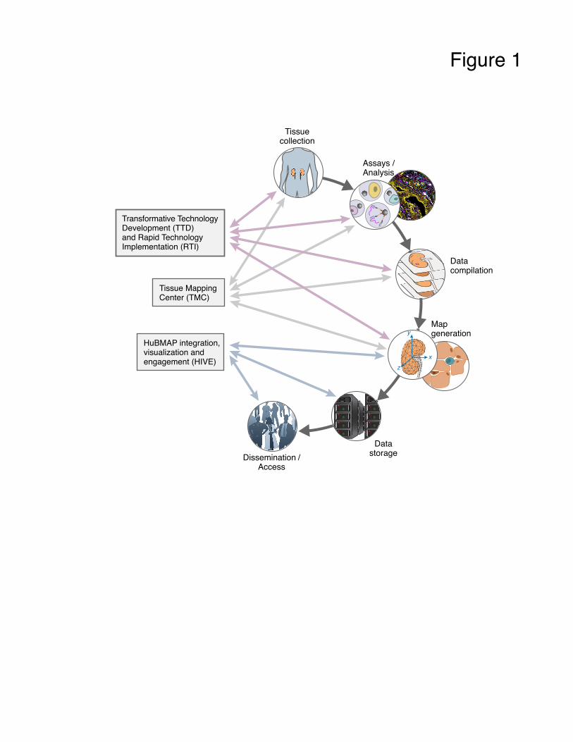
Figure 1
Assays /Analysis
Tissuecollection
Datacompilation
Mapgeneration
DatastorageDissemination /
Access
xz
y
Tissue MappingCenter (TMC)
Transformative TechnologyDevelopment (TTD)and Rapid TechnologyImplementation (RTI)
HuBMAP integration,visualization andengagement (HIVE)
TTT
TTT
TTT

Figure 1
Figure 2

Figure 3