mapping the minimal domain of hmsh-2 sufficient for binding mismatched oligonucleotides
TRANSCRIPT
BIOCHEMICAL AND BIOPHYSICAL RESEARCH COMMUNICATIONS 232, 10–13 (1997)ARTICLE NO. RC976211
Mapping the Minimal Domain of hMSH-2 Sufficient forBinding Mismatched Oligonucleotides
Adrian Whitehouse,1 Jayne Deeble,* Graham R. Taylor,* Pierre J. Guillou,† Simon E. V. Phillips,‡David M. Meredith, and Alexander F. MarkhamMolecular Medicine Unit, Clinical Sciences Building, *DNA Laboratory, Regional Genetics Service, †Division of Surgery,St. James’s University Hospital, and ‡Department of Biochemistry and Molecular Biology,University of Leeds, Leeds, United Kingdom
Received January 15, 1997
incidence of instability in microsatellite sequencesThe human MSH-2 gene product is a member of a which are prone to somatic mutations with the appear-
highly conserved family of proteins involved in post- ance of larger and/or smaller alleles (4). Also, cell linesreplication mismatch repair. Germline mutations in derived from HNPCC tumours are found to be geneti-this gene have been implicated in hereditary non-poly- cally unstable (5). These observations were indicativeposis colorectal cancer (HNPCC). Alterations in the of a failure in DNA repair. Subsequently, germlinescoding region of the hMSH-2 gene result in a mutator
mutations in HNPCC families were identified in fourphenotype with marked instability of microsatellitehuman genes, hMSH-2, hMLH1, hPMS-1 and hPMS-sequences, indicative of a deficiency in DNA repair. We2, all of which are homologous to bacterial genes in-have previously shown that a region of high homologyvolved in mismatch repair (6-12).between MutS proteins of different species containing
In Escherichia coli (E.coli), mismatch recognition isa nucleotide binding domain, is sufficient to bind DNAmediated by a single protein MutS, which binds to basecontaining specific mismatched residues. In order tomispairs or loops of up to 4 base pairs (13). However,determine the minimal domain of hMSH-2 necessaryin Saccharomyces cerevisiae, MSH2, (The MutS homo-for binding mismatch-containing oligonucleotides, de-logue) functions with either MSH3 or MSH6 in mis-letion analysis of the C-terminal region was per-
formed. We have constructed a 5* and 3* deletion series, match repair (14). The recognition of mismatched DNAexpressed each deletion as a bacterial fusion protein in humans is thought to be mediated by at least twoand assessed it for ATPase activity and its ability to MutS homologs, hMSH-2 and G/T binding proteinidentify mismatch containing DNA. Here we demon- (GTBP) forming the heterodimer, hMutSa (15-16). Thisstrate that a 585 bp fragment encoding 195 amino acids complex is believed to bind to any mismatches initiat-within the C-terminal domain of hMSH-2 is sufficient ing the repair of the heteroduplex. A third protein,to bind to DNA containing mismatches. q 1997 Academic hMSH-3 also forms a heterodimer with hMSH-2,Press hMutSb, which has been shown to bind loops of up
to 4 base pairs (17). The MutL homologs, hPMS2 andhMHL1, are then recruited to the complex and the mis-match is repaired. In bacteria, this repair process in-The two major inherited forms of colorectal cancervolves excision of the tract of single-stranded DNApredisposition are hereditary non-polyposis colorectalwhich contains the mismatched residue, resynthesis ofcancer (HNPCC) and familial adenomatous polyposisthe excised DNA and finally religation (13).(FAP). HNPCC is an autosomal dominant condition. It
hMSH-2 is of particular importance as mutations inis believed that 1 in 500 of the western population maythis gene are thought to underlie approximately 40%be heterozygotes for mutant alleles and predisposed toof HNPCC cases (18). hMSH-2 has also been shown tothis form of cancer (2-3). It leads to the developmentbind to DNA containing mismatches in vitro (19-20).of tumours of the colon, endometrium, stomach, small We have previously shown that the C-terminal domainintestine, hepatobiliary system, kidney, ureter and of hMSH-2 displays ATPase activity and is sufficientovary (4). HNPCC kindreds are characterised by a high to bind specific mismatched oligonucleotides (1). In thispaper, we have constructed a 5* and 3* deletion serieswithin the C-terminal domain of hMSH-2. These mu-1 Corresponding author. Fax: 044 113 2444475; Email: msjaw@
stjames.leeds.ac.uk. tants were expressed as bacterial fusion proteins, as-
0006-291X/97 $25.00Copyright q 1997 by Academic PressAll rights of reproduction in any form reserved.
10
AID BBRC 6211 / 691e$$$$61 02-17-97 10:17:38 bbrcg AP: BBRC
Vol. 232, No. 1, 1997 BIOCHEMICAL AND BIOPHYSICAL RESEARCH COMMUNICATIONS
sessed for ATPase activity and examined for the abilityto bind DNA containing mismatched residues.
METHODS
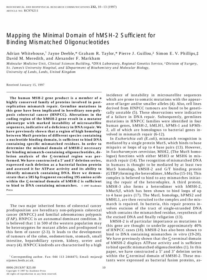
Construction of a hMSH-2 C-terminal 5* and 3* deletion series.The full length cDNA of hMSH-2 is contained in the vector,pBShMSH-2 (provided by Prof. B. Vogelstein). Deletion mutantswere constructed by digesting pBShMSH-2 with the restriction en-zymes shown (Fig.1), blunt ending with T4 polymerase or KlenowDNA polymerase and ligated with pFlag.CTC (IBI), previously di-gested with HindIII and blunt-ended to derive, pAWD1-6, respec-tively.
Expression of hMSH-2 C-terminal deletion mutants as bacterialfusion proteins. Each deletion construct cloned into the bacterialexpression vector pFlag.CTC, was used to transform E. coli strainDH5a. A fresh overnight culture of transformed E. coli was diluted1 in 20 with LB medium containing ampicillin (100 mg/ml). Aftergrowth at 377C for 2 hours, the culture was induced with IPTG (1mM) and grown at 377C for a further 5 hours. The cells were har-vested by centrifugation at 3200g for 10 minutes, resuspended in 0.1volume lysis buffer (100mM Tris, pH 8.0, 1mM EDTA) and incubatedon ice with 3 mg/ml of lysozyme for 30 minutes. The cells were then
FIG. 1. Diagrammatic representation of the deletion series fromsonicated and lysed by the addition of Tween 20 lysis buffer (100the C-terminal domain of hMSH-2. A series of 5* and 3* mutants wasmM Tris-HCl, pH 8.0, 200 mM NaCl, 1 mM EDTA, 0.3 mg/ml phenyl-constructed by digesting pBShMSH-2 with the restriction enzymesmethylsulphonyl fluoride, 0.8 mg/ml pepstatin, 1 mM DTT, 1% Tweenshown, blunt ending with T4 polymerase or Klenow enzyme and20). Cellular debris was pelleted by centrifugation at 4,000g.ligating with pFlag.CTC.
Detection of fusion proteins by Western blot analysis. Protein ex-tracts of each deletion mutant were mixed with 21 reducing samplebuffer (50mM Tris-HCl, pH 6.8, 4% sodium dodecyl sulphate (SDS),5 mM EDTA, 10% b-mercapthoethanol, 1 mM DTT and 0.01% bromo- RESULTS AND DISCUSSIONphenol blue). After boiling for 3 minutes, samples were fractionatedon a 12% SDS polyacrylamide gel. After electrophoresis the gel was
Cloning of 5* and 3* deletion mutants. A DNA frag-soaked for 10 min in transfer buffer (25 mM Tris, 192 mM glycine,20% methanol (v:v), and 0.1% SDS), and the proteins were trans- ment encoding the C-terminal domain of hMSH-2 hasferred to nitrocellulose membranes by electroblotting for 3 hours at been previously shown to be sufficient to bind mis-250 mA. After transfer, the membranes were soaked in PBS and matched oligonucleotides (1). To further analyse theincubated for 2 hr in blocking buffer (PBS containing 5% nonfat dry
sequences within this domain which are required formilk). Membranes were incubated with a 1/3000 dilution of the M2mismatch recognition a 5* and 3* deletion series withinmonoclonal antibody (IgG1, IBI), washed with PBS and incubated
for 1 hr at 377C with a 1/1000 dilution of rabbit anti-mouse immuno- the C-terminal domain of hMSH-2 was produced (Fig.globulin conjugated with horseradish peroxidase in blocking buffer. 1). Deletions within the C-terminal domain of hMSH-After five washes with PBS the nitrocellulose membranes were devel- 2 were constructed by digesting pBShMSH-2 with re-oped in PBS containing 0.02% 1-chloro-4-naphthol and 0.006% hy-
striction enzymes which progressively delete largerdrogen peroxide.fragments from the 5* and 3* regions of the gene. These
ATPase assay. The assay was performed at 377C in 20 mM Tris-fragments were ligated into the bacterial expressionHCl, pH 7.6, 0.5 mM CaCl2, 5 mM MgCl2, 1 mM DTT, 100 mg/mlvector, pFlag.CTC, in phase with respect to the ATGBSA, 0.1 mM EDTA and 150 ng of hMSH-2 domain protein. Assays
were performed using 2, 2.5, 3.3, 5 and 10 mM ATP. Hydrolysis of translational start codon immediately upstream of the[a-32P]ATP by the carboxy terminal domains of hMSH-2 was assayed multiple cloning site (MCS) and also in frame with theby thin layer chromatography. The radioactive counts for ATP and C-terminal coding sequence immediately downstreamits hydrolysis products were quantified using a scintiallation counter
of the MCS to ensure proper fusion to the C-terminal(Packard).Flag peptide (Asp Tyr Lys Asp Asp Asp Asp Lys), to
Functional binding assay. Mismatch binding was detected by aderive D1-6.nitrocellulose binding assay of labelled oligonucleotides followed by
autoradiography as previously described (1). Briefly, oligonucleotides Expression of the hMSH-2 C-terminal deletion mu-(dCGG ATC CGG ATG TXA TGG AAT TCC and dGGA ATT CCA tants. Expression of the deletion mutants resulted inTXA CAT CCG GAT CCG) were synthesised and annealed to produce
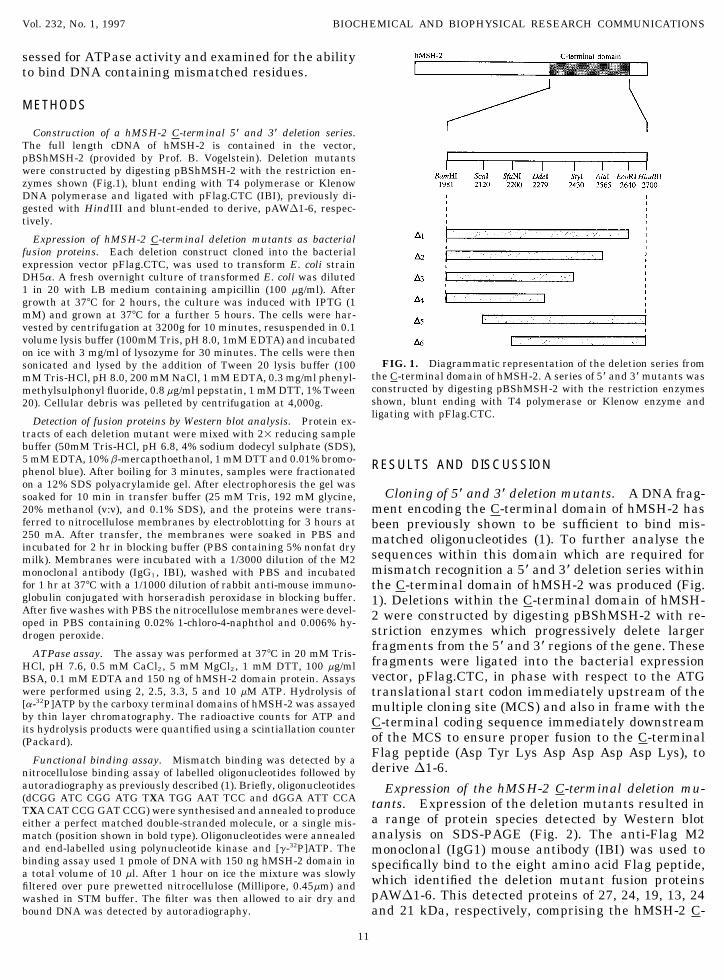
a range of protein species detected by Western bloteither a perfect matched double-stranded molecule, or a single mis-analysis on SDS-PAGE (Fig. 2). The anti-Flag M2match (position shown in bold type). Oligonucleotides were annealed
and end-labelled using polynucleotide kinase and [g-32P]ATP. The monoclonal (IgG1) mouse antibody (IBI) was used tobinding assay used 1 pmole of DNA with 150 ng hMSH-2 domain in specifically bind to the eight amino acid Flag peptide,a total volume of 10 ml. After 1 hour on ice the mixture was slowly which identified the deletion mutant fusion proteinsfiltered over pure prewetted nitrocellulose (Millipore, 0.45mm) and
pAWD1-6. This detected proteins of 27, 24, 19, 13, 24washed in STM buffer. The filter was then allowed to air dry andbound DNA was detected by autoradiography. and 21 kDa, respectively, comprising the hMSH-2 C-
11
AID BBRC 6211 / 691e$$$$61 02-17-97 10:17:38 bbrcg AP: BBRC

Vol. 232, No. 1, 1997 BIOCHEMICAL AND BIOPHYSICAL RESEARCH COMMUNICATIONS
at position 11 within the context of a double-stranded24-mer oligonucleotide pair. We found that the wildtype C-terminal domain of hMSH-2 selectively boundall specific mismatches apart from A/A and C/C, inagreement with results described previously (1). Thecontrol vector did not bind to any labelled oligonucleo-tide pair. Applying this assay to the mutant proteins,we found that D1 and D2 bound the same specific mis-matches as the wild type domain albeit to a slightlyFIG. 2. Western blot analysis of mutant fusion proteins. Extractslesser extent. This may be due to the deletion of se-were resolved by SDS PAGE, transferred to nitrocellulose, and incu-
bated with M2 monoclonal antibody (IgG1) and immune complexes quences leading to reduced recognition of the mis-were detected by using rabbit anti mouse immunoglobulin conju- matches or a reduced affinity of these proteins oncegated with horseradish peroxidase. The nitrocellulose membranes bound to a mismatch resulting in separation from thewere developed in PBS containing 0.02% 1-chloro-4-naphthol and
mismatch in the washing procedures of the assay. D30.006% hydrogen peroxide. Lane 1-2, hMSH-2 C-terminal domain-and D4 were essentially unable to bind mismatchesuninduced and induced; Lane 3, D1-induced; Lane 4, D2-induced;
Lane 5, D3-induced; Lane 6, D4-induced; Lane 7, D5-induced; Lane indicating that sequences downstream of 2430 bp (of8, D6-induced. the published cDNA sequence) are essential for mis-
match binding. D5 and D6, which have the nucleotidebinding domain deleted, are also unable to bind mis-matches demonstrating that ATPase activity is essen-terminal domain coupled to the Flag peptide at its car-tial for mismatch binding (Fig. 4).boxy terminus. As reported earlier (1), these domains
We have previously reported that a large C-terminalonly remain soluble in the presence of greater than 1domain of hMSH-2 exhibits ATPase activity and is suf-mM DTT, and as yet this protein has proved refractoryficient to bind mismatched oligonucleotides (1). In thisto complete purification (1). Purification studies of thereport we have produced and studied a range of dele-deletion mutant fusion proteins produced here weretion mutants to map function in the carboxy terminalagain complicated by insolubility problems. However,domain. We have shown that the minimal region forbinding to mismatched oligonucleotides and ATPasemismatch binding is contained within a 585 bp frag-activities, analogous to those previously observed ofment, which encodes a protein fragment of 195 aminoextracts from induced cultures of the carboxy terminalacids. The minimal 585 base pair fragment is highlydomains demonstrated the specificity of the enzymeshomologous between hMSH-2 and its homologues inas no activity was detected in uninduced cells. Thisother species and contains the type A nucleotide bind-suggests that these activities are generated from theing consensus sequence. The inability of D5 and D6,constructs alone.which have the nucleotide binding sequence removed,
ATPase analysis of mutant fusion proteins. It has to bind mismatches is consistent with our hypothesisbeen shown that the carboxy terminal domain ofhMSH-2 contains an ATPase activity (1). In order todetermine whether these mutants hydrolyse ATP, toADP and Pi, [a-32P]ATP was incubated with each mu-tant fusion protein and the products separated usingTLC. To determine Km and kcat values for the mutants,ATPase activity was measured in the presence of vari-ous concentrations of ATP (Fig. 3). The results showthat the D1 and D2 deletions have limited effects onthe ATPase activity of the domain. Wild type Km andkcat values were 7.96 mM and 0.5 s01, respectively. D3and D4 display low ATPase activities. However, D5and D6 had no detectable activity, probably due to theremoval of the nucleotide binding domain. In controlexperiments, nonenzymatic hydrolysis of ATP in theabsence of the wild type expressed domain was lessthan 5%.
Functional analysis of the mutant fusion proteins.FIG. 3. ATPase analysis of the mutant bacterial fusion proteins.A mismatch binding assay was developed to measure
Hydrolysis of various substrate concentrations of [a-32P]ATP by thethe hMSH-2 C-terminal domains’ activity (1). Mis- carboxy terminal domain of hMSH-2, pFlag and D1-6 were assayedmatch recognition was detected by nitrocellulose bind- by thin layer chromatography, and quantified using a scintillation
counter.ing of labelled oligonucleotides containing a mismatch
12
AID BBRC 6211 / 691e$$$$61 02-17-97 10:17:38 bbrcg AP: BBRC

Vol. 232, No. 1, 1997 BIOCHEMICAL AND BIOPHYSICAL RESEARCH COMMUNICATIONS
2. Dunlop, M. G. (1992) Br. J. Surgery 79, 488.3. Parker, S., Tong, T., Bolden, S., and Wingo, P. (1996). CA Cancer
J. Clin. 46, 5–27.4. Lynch, H. T., Smyrk, T. C., Watson, P., Lanspa, S. J., Lynch,
J. F., Cavalieri, R. J., and Bolland, C. R. (1993) Gastroentrology104, 1535–1549.
5. Loeb, L. A. (1994) Cancer Res. 54, 5059–5063.6. Parsons, R., Li, G. M., Longleym, M. J., Fang, W., Papadopoulos,
M., Jen, J., de la Chapelle, A., Kinzler, K. W., Vogelstein, B.,and Modrich, P. (1993) Cell 75, 1227–1235.
7. Leach, F. S., Nicolaides, N. C., Papadopoulos, N., Liu, B., Jen,J., Parsons, R., Peltomaki, P., Sistonen, P., Aaltonen, L. A., Ny-strom-Lahti, M., Guan, X.-Y., Zhang, L., Meltzer, P. S., Yu,J.-W., Kao, F.-T., Chen, D. J., Cerosaletti, K. M., Fournier,R. E. K., Todd, S., Lewis, T., Leach, R. J., Naylor, S. L., Weissen-bach, J., Mecklin, J.-P., Jarvinen, H., Petersen, G. M., Hamilton,S. R., Green, J., Jass, J., Watson, P., Lynch, H. T., Trent, J. M.,FIG. 4. Functional analysis of the mutant bacterial fusion pro-de la Chapelle, A., Kinzler, K. W., and Vogelstein, B. (1993) Cellteins. Oligonucleotides containing either a perfect match or a range75, 1215–1225.of single mismatches were radiolabelled using polynucleotide kinase.
8. Bronner, C. E., Baker, S. M., Morrison, P. T., Warren, G., Smith,One pmole of labelled DNA was incubated for 1 hour with proteinL. G., Lescoe, M. K., Kane, M., Earabino, C., Lipford, J., Lind-extracts of hMSH-2, pFlag or D1-6. After the incubation period thebolm, A., Tannergard, P., Bollag, R. J., Godwin, A. R., Ward,mixtures were slowly filtered over prewet nitrocellulose. Washed andD. C., Norden-skjold, M., Fishel, R., Kolodner, R., and Liskay,bound DNA was detected using autoradiography.R. M. (1994) Nature 368, 258–261.
9. Kolodner, R. D., Hall, N. R., Lipford, J., Kane, M. F., Rao,M. R. S., Morrison, P., Wirth, L., Finan, P. J., Burn, J., Chap-
that this ATPase capability is essential. Mutational man, P., Earabino, C., Merchant, E., and Bishop, D. T. (1994)Genomics 24, 516–526.analysis of this domain has previously demonstrated
10. Kolodner, R. D., Hall, N. R., Lipford, J., Kane, M. F., Morrison,that a variant protein containing an alteration withinP., Wirth, L., Finan, P. J., Burn, J., Chapman, P., Earabino, C.,the nucleotide binding consensus sequence of Lys675 toMerchant, E., and Bishop, D. T. (1994) Cancer Res. 55, 242–248.
an alanine residue severely impairs mismatch binding11. Liu, B., Nicolaides, N. C., Markowitz, S. S., Willson, J. K. V., Par-
(21), suggesting that this sequence plays a critical role sons, R. E., Jen, J., Papadopoulos, N., Peltomaki, P., de la Chap-in mismatch recognition. elle, A., Hamilton, S. R., Kinzler, K. W., and Vogelstein, B. (1995)
Nature Genetics 9, 48–55.It will be of interest to examine other regions of12. Papadopoulos, N., Nicolaides, N. C., Wei, Y.-F., Ruben, S. M.,hMSH-2 in the future to identify other separate func-
Carter, K. C., Rosen, C. A., Haseltine, W. A., Fleischmann, R. D.,tions or specific interactions. Mismatch recognition isFraser, C. M., Adams, M. D., Venter, J. C., Hamilton, S. R., Pet-thought to be mediated by the heterodimers, hMutSa ersen, G. M., Watson, P., Lynch, H. T., Peltomaki, P., Mecklin,
and hMutSb (15-16). Deletion analysis of the N-termi- J.-P., de la Chapelle, A., Kinzler, K. W., and Vogelstein, B. (1994)Science 263, 1625–1629.nal region of hMSH-2 may help in identifying specific
13. Modrich, P. (1991) Annu. Rev. Genet. 25, 229–253.domains necessary for the formation of these hetero-14. Johnsen, R. E., Kovvali, G. K., Prakash, L., and Prakash, S.dimers by interaction with GTBP or hMSH-3, respec-
(1996). J. Biol. Chem. 271, 7285–7288.tively.15. Drummond, J. T., Li, G.-M., Longley, M. J., and Modrich, P.
(1995) Science 268, 1909–1912.ACKNOWLEDGMENTS 16. Palambo, F., Gallinari, P., Iaccarino, I., Lettieri, T., Hughes,
M., D’Arrigo, A., Truong, O. J., Hsuan, J., and Jiricny, J. (1995)We thank the Yorkshire Cancer Research Campaign and West Science 268, 1912–1914.
Riding Medical Research Trust for financial support for this project. 17. Palambo, F., Iaccarino, I., Nakajima, E., Ikejima, M., Shimada,Research in the authors’ laboratory is also supported by the Medical T., and Jiricny, J. (1996) Current Biology 6, 1181–1184.Research Council, Northern and Yorkshire Regional Health Author- 18. Lui, B., Parsons, R., Papadopoulos, N., Nicolaides, N. C., Lynch,ity and the Wellcome Trust. SEVP is an International Research H. T., Watson, P., Jass, J. R., Dunlop, M., Wyllie, A., Peltomaki,Scholar of the Howard Hughes Medical Institute. We are particularly P., de la Chapelle, A., Hamilton, S. R., Vogelstein, B., and Kin-grateful to Bert Vogelstein for originally providing the clone zler, K. W. (1996) Nature Medicine 2, 169–174.pBShMSH-2.
19. Fishel, R., Ewel, A., Lee, S., and Lescoe, M. K. (1994) CancerRes. 54, 5539–5542.
REFERENCES 20. Fishel, R., Ewel, A., Lee, S., Lescoe, M. K., and Griffith, J. (1994)Science 266, 1403–1405.
21. Whitehouse, A., Parmar, R., Deeble, J., Taylor, G. R., Phillips,1. Whitehouse, A., Deeble, J., Taylor, G. T., Phillips, S. E. V., Mere-dith, D. M., and Markham, A. F. (1996) Biochem. Biophys. Res. S. E. V., Meredith, D. M., and Markham, A. F. Biochem. Biophys.
Res. Commun. 229, 147–153.Commun. 225, 289–295.
13
AID BBRC 6211 / 691e$$$$61 02-17-97 10:17:38 bbrcg AP: BBRC