march 2011 - university of toronto t-space · e-mail: [email protected] received for...
TRANSCRIPT
Original Article
Braz J Oral Sci. 10(1):74-78
Original Article Braz J Oral Sci.January | March 2011 - Volume 10, Number 1
Metabolic activity of Streptococcus mutansbiofilms after treatment with different
mouthwash formulationsTaciano R. Cardoso1, Alexandre S. Carvalho1, Marcelo E. Beletti2, Marcelo H. Napimoga3, Geraldo Thedei Jr1
1Laboratory of Biochemistry of Microorganisms, University of Uberaba, Uberaba, Brazil2College of Veterinary Medicine, Federal University of Uberlandia, Brazil
3Laboratory of Biopathology and Molecular Biology, University of Uberaba, Brazil
Correspondence to:Geraldo Thedei Jr.
Pró-reitoria de Pesquisa, Pós-Graduação eExtensão - Universidade de Uberaba. Avenida
Nenê Sabino, 1801 Bloco R. Bairro UniversitárioUberaba - MG - CEP 38055-500
Phone: +55-34-3319-8959Fax: +55-34-3314-8910
E-mail: [email protected]
Received for publication: November 16, 2010Accepted: March 22, 2011
Abstract
Aim: The aim of this study was to investigate the metabolic activity of Streptococcus mutans biofilmsafter treatment with mouthwashes with different compositions. Methods: S. mutans biofilms weregrowth on polystyrene plates during 18 h, washed with sterile saline and treated with the followingmouthwashes during 1 min: Listerine®, Oral B®, Parodontax® and Periogard® with and withoutalcohol. After the treatment, the biofilms were incubated with complete medium containing sucroseduring 60, 120 or 180 min, and then samples were collected for pH measurements. In addition,biofilms were grown in microscope coverslips treated as described above, followed by stainingwith Propidium Iodide and Fluoresceine for visualization with a confocal laser scanning microscopy.Results: For all mouthwashes evaluated, treatment was deleterious to cell metabolism, since littleor no acidification was observed at least 60 min after treatment. Mouthwashes containing 0.2%chlorhexidine (Parodontax®) or essential oils (Listerine®) induced a significant reduction in themetabolic activity of biofilms during the tested time points (120 and 180 min after treatment), beingthus more effective than the mouthwashes containing 0.12% chlorhexidine (Periogard®) orcetylpyridinium plus fluoride (Oral B®). The confocal analysis overall confirmed the results observedin the analysis of metabolic activity. Conclusions: The treatment of biofilms with mouthwashescontaining 0.2% chlorhexidine or essential oils induced significant reduction in S. mutans metabolism.
Keywords: Streptococcus mutans, mouthwashes, chlorhexidine, biofilm.
Introduction
Dental caries is a chronic contagious disease caused by several interactingfactors, which results in the irreversible destruction of the mineralized structuresof teeth, compromising their vitality and fixation in the maxillomandibularcomplex1, 2.
The Gram positive bacteria Streptococcus mutans are a substantial part of theoral microbiota and their importance in the dental caries etiology is unquestionable3.The carbohydrates present in the diet are the main energy source in an anaerobicprocess (mainly lactic fermentation) resulting in the production of organic acids.These acids decrease the pH to around 4.5 on the tooth surface, thus inducing itsdemineralization4.
One important characteristic of S. mutans in promoting caries development is
75
Braz J Oral Sci. 10(1):74-78
the ability to adhere firmly to the tooth surface in the presenceof sucrose. This adherence is mediated mainly by the actionof the GTF enzymes, which are considered fundamental to thevirulence of S. mutans in the pathogenesis of dental caries 5-7.
Biofilm formation occurs as a result of a sequence ofevents: microbial surface attachment, cell proliferation,matrix production and detachment8. This process is partiallycontrolled by quorum sensing, an interbacterial communicationmechanism that is dependent on population density and isassociated with radical changes in protein expressionpatterns8. Mature biofilms demonstrate a complex three-dimensional structure with numerous microenvironmentsdiffering with respect to osmolarity, nutritional supply andcell density. Many antimicrobial agents that are effectiveagainst planktonic cells turn out to be ineffective againstthe same bacteria growing in a biofilm state9,10. Planktonicand biofilm cells also exhibit different susceptibilities to acertain antimicrobial concentration.
Several studies focusing on the efficacy of mouthwasheswith diverse chemical composition demonstrated thatcombination of sodium fluoride and sodium lauryl sulfateas well as essential oils is able to diminish the metabolicactivity of microorganisms present in the dental biofilm11-13.
Foster, et al.14 (2004) studied the effects of mouthwashescontaining essential oils, triclosan, cetylpyridinium chlorideand chlorhexidine against Streptococcus gordonii biofilms.The confocal laser scanning microscopy analysis demonstratedthat all mouthwashes, except for cetylpyridinium chloride,were able to cause membrane damage after 60 s of incubationwith S. gordonii biofilms.
Zhang, et al.15 (2004) evaluated the effect of a mouthwashwith and without fluoride over metabolic activity of S. mutansbiofilms and demonstrated that essential oil-containingmouthwashes, with or without 100 ppm of fluoride reducedthe metabolic activity and the consequent acid productionby approximately 36-44%. A significant reduction on totalcolony forming units (CFU) was observed in saliva of healthyvolunteers after a single mouthwash with 0.2% or 0.12%chlorhexidine, but only the highest concentration showedbactericidal activity against salivary obligate anaerobes16.Furthermore, an in vivo study showed that both essentialoils and alcohol-free chlorhexidine mouthwashes were ableto reduce plaque acidogenicity after a sucrose challenge,with no difference between both solutions17.
Although several studies have been undertaken, littledata are available about the action of mouthwashes withdifferent active principles on bacterial biofilm metabolism,especially S. mutans biofilms, and the effects of thosemouthwashes on three-dimensional structure of biofilms.
Material and methods
MouthwashesThe following mouthwashes were evaluated in the present
study: Parodontax® (Composition: 0.2% chlorhexidinegluconate (w/v), Batch: 168F, SmithKline Beecham ConsumerHealthcare, United Kingdom); Listerine Cool Mint®
(Composition: 0.092% eucalyptol (w/v), 0.042% menthol(w/v), 0.060% methyl salicylate (w/v), 0.064% thymol (w/v), Batch: 3558B01, Johnson & Johnson, SP, Brazil); Oral-B® (Composition: water, glycerin, polysorbate 20, flavor,methylparaben, 0.053% monohydrated cetylpyridiniumchloride, 0.050% sodium fluoride (226 ppm fluoride), sodiumsaccharine, sodium benzoate, propylparaben, ci 42090, ci 47005batch: 8114852516, Rety Laboratories, Barranquilla, Colombia)and Periogard® with or without alcohol (Composition: 0.12%chlorhexidine gluconate (w/v), batch BR123A and BR112A,respectively, Colgate-Palmolive, São Bernardo do Campo, SP,Brazil). Positive and negative controls were 70% ethanol (v/v) and sterile 0.9% (w/v) saline, respectively.
Streptococcus mutans growth conditionsThe ATCC 25175 strain of S. mutans was purchased
from the André Tosello Foundation, Campinas, SP, Brazil.The lineage was kept stored at -20ºC in 40% (v/v) glycerol(Sigma, St. Louis, MO, USA) medium and checked for puritybefore being grown in broth.
The frozen S. mutans cultures were reactivated in 5 mLof Triptic Soy Broth (TSB - Soybean-casein digest medium;Difco, Sparks, MD, USA) and incubated at 37°C, undermicroaerophylic conditions for 18 h. The cultures wereadjusted to A620
nm= 0.2 using a photocolorimeter (Analyser
Com & Ind. LTDA. São Paulo, SP, Brazil) and 750 mL ofthis suspension was transferred to a tube containing 30 mLof previously autoclaved complete medium18 (10 g/L tryptone,5 g/L yeast extract, 60 µmol/L MgSO
4, 1.3 µmol/L FeSO
4,
1.5 µmol/L MnCl2, 0.2 mmol/L KH
2PO
4, 0.3 mmol/L K
2HPO
4,
0.7 mmol/L KCl, pH 7.0) supplemented with 50 mMol/Lsucrose as carbon source. Then, 600 mL of this suspensionwas inoculated in a 24-well cell culture plate (Corning Costar3524, flat bottom) and incubated at 37°C, under microaerophilia,during 18 h. for biofilm formation as previously described7.
Effects of mouthwashes on S. mutans metabolismAll procedures were carried out in as a blind experiment.
After biofilm formation as described above, the culturemedium of each well was removed and the pH was measuredusing a PG 1800 pH meter associated with a microelectrode(Gehaka, São Paulo, SP, Brazil). The formed biofilms werewashed 3 times with sterile 0.9% (w/v) saline and 1 mL ofeach the mouthwashes was added to each well. After 1 minof incubation, the mouthwashes were removed and the wellswashed with abundant sterile 0.9% (w/v) saline. Then, toeach well was added 1 mL sterile complete medium suppliedwith 50 mMol/L sucrose as carbon source. The treatedbiofilm was incubated at 37ºC under microaerophilicconditions and samples were taken at 60, 120 and 180 minfor further pH analysis.
The positive control used was ethanol 70% (v/v) andthe negative control was sterile 0.9% (w/v) saline.
Confocal Laser Scanning Microscopy (CLSM)For the CLSM study, glass coverslips were inserted in
previously autoclaved Falcon Tubes with 30 mL of complete
Metabolic activity of Streptococcus mutans biofilms after treatment with different mouthwash formulations
76
Braz J Oral Sci. 10(1):74-78
60 min 120 min 180 min
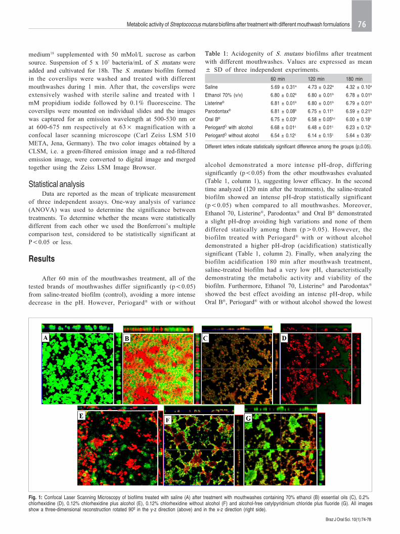
Saline 5.69 ± 0.31a 4.73 ± 0.22a 4.32 ± 0.10a
Ethanol 70% (v/v) 6.80 ± 0.02b 6.80 ± 0.01b 6.78 ± 0.01b
Listerine® 6.81 ± 0.01b 6.80 ± 0.01b 6.79 ± 0.01b
Parodontax® 6.81 ± 0.08b 6.75 ± 0.11b 6.59 ± 0.21b
Oral B® 6.75 ± 0.03b 6.58 ± 0.05b,c 6.00 ± 0.18c
PeriogardÒ with alcohol 6.68 ± 0.01c 6.48 ± 0.01c 6.23 ± 0.12c
PeriogardÒ without alcohol 6.54 ± 0.12c 6.14 ± 0.15c 5.64 ± 0.35c
Table 1: Acidogenity of S. mutans biofilms after treatmentwith different mouthwashes. Values are expressed as mean± SD of three independent experiments.
Different letters indicate statistically significant difference among the groups (p,0.05).
medium18 supplemented with 50 mMol/L sucrose as carbonsource. Suspension of 5 x 10⁷ bacteria/mL of S. mutans wereadded and cultivated for 18h. The S. mutans biofilm formedin the coverslips were washed and treated with differentmouthwashes during 1 min. After that, the coverslips wereextensively washed with sterile saline and treated with 1mM propidium iodide followed by 0.1% fluoresceine. Thecoverslips were mounted on individual slides and the imageswas captured for an emission wavelength at 500-530 nm orat 600-675 nm respectively at 63× magnification with aconfocal laser scanning microscope (Carl Zeiss LSM 510META, Jena, Germany). The two color images obtained by aCLSM, i.e. a green-filtered emission image and a red-filteredemission image, were converted to digital image and mergedtogether using the Zeiss LSM Image Browser.
Statistical analysisData are reported as the mean of triplicate measurement
of three independent assays. One-way analysis of variance(ANOVA) was used to determine the significance betweentreatments. To determine whether the means were statisticallydifferent from each other we used the Bonferroni’s multiplecomparison test, considered to be statistically significant atP<0.05 or less.
Results
After 60 min of the mouthwashes treatment, all of thetested brands of mouthwashes differ significantly (p<0.05)from saline-treated biofilm (control), avoiding a more intensedecrease in the pH. However, Periogard® with or without
alcohol demonstrated a more intense pH-drop, differingsignificantly (p<0.05) from the other mouthwashes evaluated(Table 1, column 1), suggesting lower efficacy. In the secondtime analyzed (120 min after the treatments), the saline-treatedbiofilm showed an intense pH-drop statistically significant(p<0.05) when compared to all mouthwashes. Moreover,Ethanol 70, Listerine®, Parodontax® and Oral B® demonstrateda slight pH-drop avoiding high variations and none of themdiffered statically among them (p>0.05). However, thebiofilm treated with Periogard® with or without alcoholdemonstrated a higher pH-drop (acidification) statisticallysignificant (Table 1, column 2). Finally, when analyzing thebiofilm acidification 180 min after mouthwash treatment,saline-treated biofilm had a very low pH, characteristicallydemonstrating the metabolic activity and viability of thebiofilm. Furthermore, Ethanol 70, Listerine® and Parodontax®
showed the best effect avoiding an intense pH-drop, whileOral B®, Periogard® with or without alcohol showed the lowest
Metabolic activity of Streptococcus mutans biofilms after treatment with different mouthwash formulations
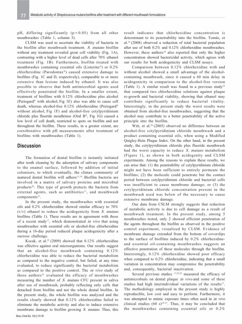
Fig. 1: Confocal Laser Scanning Microscopy of biofilms treated with saline (A) after treatment with mouthwashes containing 70% ethanol (B) essential oils (C), 0.2%chlorhexidine (D), 0.12% chlorhexidine plus alcohol (E), 0.12% chlorhexidine without alcohol (F) and alcohol-free cetylpyridinium chloride plus fluoride (G). All imagesshow a three-dimensional reconstruction rotated 90º in the y-z direction (above) and in the x-z direction (right side).
77
Braz J Oral Sci. 10(1):74-78
pH, differing significantly (p<0.05) from all othermouthwashes (Table 1, column 3).
CLSM was used to ascertain the viability of bacteria inthe biofilm after mouthwash treatment. S. mutans biofilmwithout any treatment revealed great cell viability (Fig. 1A),contrasting with a higher level of dead cells after 70% ethanoltreatment (Fig. 1B). Furthermore, biofilm treated withmouthwashes containing essential oils (Listerine®) or 0.2%chlorhexidine (Parodontax®) caused extensive damage tobiofilms (Fig. 1C and D, respectively), comparable to or moreextensive than lesions induced by ethanol. It was alsopossible to observe that both antimicrobial agents usedeffectively penetrated the biofilm. In a smaller extent,treatment of biofilms with 0.12% chlorhexidine plus alcohol(Periogard® with alcohol, Fig 1E) also was able to cause celldeath, whereas alcohol-free 0.12% chlorhexidine (Periogard®
without alcohol, Fig 1F) and alcohol-free cetylpyridiniumchloride plus fluoride mouthrinse (Oral B®, Fig 1G) caused alow level of cell death, restricted to spots on biofilm and notthroughout the biofilm. These results, in a greater extent, arecorroborative with pH measurements after treatment ofbiofilms with mouthwashes (Table 1).
Discussion
The formation of dental biofilm is instantly initiatedafter tooth cleaning by the adsorption of salivary componentsto the enamel surface, followed by addition of initialcolonizers, to which eventually, the climax community ofmatured dental biofilm will adhere11,19. Biofilm bacteria areinvolved in a matrix of salivary proteins and microbialproducts20. This type of growth protects the bacteria fromexternal agents, such as antibiotics11, and mouthwashcomponents21.
In the present study, the mouthwashes with essentialoils and 0.2% chlorhexidine showed similar efficacy to 70%(v/v) ethanol to reduce the acidogenicity from S. mutansbiofilms (Table 1). These results are in agreement with thoseof a recent study17, which demonstrated in vivo that usingmouthwashes with essential oils or alcohol-free chlorhexidineduring a 16-day period reduced plaque acidogenicity after asucrose challenge.
Kocak, et al.22 (2009) showed that 0.12% chlorhexidinewas effective against oral microorganisms. Our results suggestthat an alcohol-free mouthwash containing 0.12%chlorhexidine was able to reduce the bacterial metabolismas compared to the negative control, but failed, at any timeevaluated, to reduce significantly the bacterial metabolismas compared to the positive control. The in vivo study ofthose authors22 evaluated the efficacy of mouthwashesmeasuring the number of S. mutans CFU present in salivaafter use of mouthwash, probably reflecting only cells thatdetached from biofilm and not the whole dental biofilm. Inthe present study, the whole biofilm was analyzed and theresults clearly showed that 0.12% chlorhexidine failed toeliminate the metabolic activity and also to induce extensivemembrane damage to biofilm growing S. mutans. Thus, this
result indicates that chlorhexidine concentration isdeterminant to its penetrability into the biofilm. Tomás, etal.16 (2008) observed a reduction of total bacterial populationafter use of both 0.2% and 0.12% chlorhexidine mouthwashes.However, these authors16 also reported that only the higherconcentration showed bactericidal activity, which agrees withour results for both acidogenicity and CLSM assays.
Comparison between 0.12% chlorhexidine with andwithout alcohol showed a small advantage of the alcohol-containing mouthwash, since it caused a 60 min delay inacidogenicity in comparison to the alcohol-free version(Table 1). A similar result was found in a previous study23
that compared two chlorhexidine solutions against plaquere-growth and bacterial viability, showing that ethanol maycontribute significantly to reduce bacterial vitality.Interestingly, in the present study the worst results wereobtained from alcohol-free mouthwashes, suggesting that thealcohol may contribute to a better penetrability of the activeprinciple into the biofilm.
Witt, et al.24 (2005) observed no difference between analcohol-free cetylpyridinium chloride mouthwash and aproduct containing essential oils, when using a ModifiedQuigley-Hein Plaque Index. On the other hand, in the presentstudy, the cetylpyridinium chloride plus fluoride mouthwashhad the worst capacity to reduce S. mutans metabolism(Figure 1), as shown in both acidogenity and CLSMexperiments. Among the reasons to explain these results, wecan arise that: (1) the penetrability of cetylpyridinium chloridemight not have been sufficient to entirely permeate thebiofilms; (2) the molecule could penetrate but the contactperiod between cetylpyridinium chloride and bacterial cellswas insufficient to cause membrane damage; or (3) thecetylpyridinium chloride concentration present in themouthwash used was below of the necessary to causeextensive membrane damage.
Our data from CSLM strongly suggests that reductionof metabolic activity is due to cell damage as a result ofmouthwash treatment. In the present study, among 5mouthwashes tested, only 2 showed efficient penetration ofthe agents throughout the biofilm as observed in the positivecontrol experiment, visualized by CLSM. Evidence ofmembrane damage extended from the bottom of coverslipsto the surface of biofilms induced by 0.2% chlorhexidine-and essential oil-containing mouthwashes suggests aneffective penetration of these molecules through the biofilm.Interestingly, 0.12% chlorhexidine showed poor efficacywhen compared to 0.2% chlorhexidine, indicating that a smallvariation in concentration may compromise the penetrabilityand, consequently, bacterial inactivation.
Several previous studies 17,22,23 measured the efficacy ofantimicrobials on dental plaque in vivo and some of thesestudies had high interindividual variations of the results17.The methodology employed in the present study is highlyreproducible, low cost and easy to perform. Furthermore, itwas attempted to mimic exposure times often used in in vivoclinical studies (60 s)25-27. Thus, it may be concluded thatthe mouthwashes containing essential oils or 0.2%
Metabolic activity of Streptococcus mutans biofilms after treatment with different mouthwash formulations
78
Braz J Oral Sci. 10(1):74-78
chlorhexidine showed higher efficacy than those containingcetylpyridinium chloride plus fluoride or 0.12% chlorhexidine.
Acknowledgements
The authors would like to thank UNIUBE and FAPEMIGfor the continuous support given to our laboratories, andHilara N. Ruas for technical assistance. TRC and ASC wererecipients, respectively, of Master’s degree and undergraduatefellowships from FAPEMIG.
References
1. Krasse B. Caries risk: A pratical guide for a assessment and control.Chicago: Quintessence; 1985. p.113.
2. Marsh PD. Are dental diseases examples of ecological catastrophes?Microbiology. 2003; 149: 279-94.
3. Mikkelsen L, Jensen SB, Jakobsen J. Microbial studies on plaque fromcarious and caries-free proximal tooth surfaces in a population with highcaries experience. Caries Res. 1981; 15: 428-35.
4. Chestnutt IG, MacFarlane TW, Aitchison TC, Stephen KW. Evaluation ofthe in vitro cariogenic potential of Streptococcus mutans strains isolatedfrom 12-year-old children with differing caries experience. Caries Res.1995; 29: 455-60.
5. Loesche WJ. Role of Streptococcus mutans in human dental decay.Microbiol Rev. 1986; 50: 353-80.
6. Kuramitsu HK. Virulence factors of mutans streptococci: role of moleculargenetics. Crit Rev Oral Biol Med. 1993; 4: 159-76.
7. Mattos-Graner RO, Napimoga MH, Fukushima K, Duncan MJ, SmithDJ. Comparative analysis of Gtf isozyme production and diversity inisolates of Streptococcus mutans with different biofilm growth phenotypes.J Clin Microbiol. 2004; 42: 4586-92.
8. Sauer K, Camper AK, Ehrlich GD, Costerton JW, Davies DG.Pseudomonas aeruginosa displays multiple phenotypes during developmentas a biofilm. J Bacteriol. 2002; 184: 1140-54.
9. Drenkard E. Antimicrobial resistance of Pseudomonas aeruginosa biofilms.Microbes Infect. 2003; 5: 1213-9.
10. Fux CA, Costerton JW, Stewart PS, Stoodley P. Survival strategies ofinfectious biofilms. Trends Microbiol. 2005; 13: 34-40.
11. Petersen FC, Assev S, Scheie AA. Combined effects of NaF and SLS onacid- and polysaccharide-formation of biofilm and planktonic cells. ArchOral Biol. 2006; 51: 665-71.
12. Filoche SK, Soma K, Sissons CH. Antimicrobial effects of essential oilsin combination with chlorhexidine digluconate. Oral Microbiol Immunol.2005; 20: 221-5.
13. Takarada K, Kimizuka R, Takahashi N, Honma K, Okuda K, Kato T. Acomparison of the antibacterial efficacies of essential oils against oralpathogens. Oral Microbiol Immunol. 2004; 19: 61-4.
14. Foster JS, Pan PC, Kolenbrander PE. Effects of antimicrobial agents onoral biofilms in a saliva-coated flowcell. Biofilms. 2004; 1: 3-10.
15. Zhang JZ, Harper DS, Vogel GL, Schumacher G. Effect of an essential oilmouthrinse, with and without fluoride, on plaque metabolic acid productionand pH after a sucrose challenge. Caries Res. 2004; 38: 537-41.
16. Tomás I, Cousido MC, Tomás M, Limeres J, García-Caballero L, Diz P.In vivo bactericidal effect of 0.2% chlorhexidine but not 0.12% on salivaryobligate anaerobes. Arch Oral Biol. 2008; 53: 1186-91.
17. Albertsson WK, Persson A, Lingström P, van Dijken JW. Effects ofmouthrinses containing essential oils and alcohol-free chlorhexidine onhuman plaque acidogenicity. Clin Oral Investig. 2010; 14: 107-12.
18. Dashper SG, Reynolds EC. Characterization of transmembrane movementof glucose and glucose analogs in Streptococcus mutants Ingbritt. J Bacteriol.1990; 172: 556–63.
19. Costerton JW, Lewandowski Z, Caldwell DE, Korber DR, Lappin-ScottHM. Microbial biofilms. Annu Rev Microbiol. 1995; 49: 711-45.
20. Marsh PD, Martin MV. Dental plaque. In: Oral microbiology. 3.ed. London:Chapman and Hall; 1992. p.98-132.
21. Landa AS, van der Mei HC, Busscher HJ. Detachment of linking filmbacteria from enamel surfaces by oral rinses and penetration of sodiumlauryl sulphate through an artificial oral biofilm. Adv Dent Res. 1997; 11:528-38.
22. Kocak MM, Ozcan S, Kocak S, Topuz O, Erten H. Comparison of theefficacy of three different mouthrinse solutions in decreasing the level ofStreptococcus mutans in saliva. Eur J Dent. 2009; 3: 57-61.
23. Arweiler NB, Boehnke N, Sculean A, Hellwig E, Auschill TM. Differencesin efficacy of two commercial 0.2% chlorhexidine mouthrinse solutions: a4-day plaque re-growth study. J Clin Periodontol. 2006; 33: 334-9.
24. Witt JJ, Walters P, Bsoul S, Gibb R, Dunavent J, Putt M. Comparativeclinical trial of two antigingivitis mouthrinses. Am J Dent. 2005; 18:15A-17A.
25. Moran J, Addy M, Newcombe R. A 4-day plaque regrowth study comparingan essential oil mouthrinse with a triclosan mouthrinse. J Clin Periodontol.1997; 24: 636–9.
26. Fine DH, Furgang D, Barnett ML, Drew C, Steinberg L, Charles CH, et al.Effect of an essential oil-containing antiseptic mouthrinse on plaque andsalivary Streptococcus mutans levels. J Clin Periodontol. 2000; 7: 157–61.
27. Pan P, Barnett ML, Coelho J, Brogdon C, Finnegan MB. Determination ofthe in situ bactericidal activity of an essential oil mouthrinse using a vitalstain method. J Clin Periodontol. 2000; 27: 256–61.
Metabolic activity of Streptococcus mutans biofilms after treatment with different mouthwash formulations