marker in vivo after bone augmentation with a collagen
TRANSCRIPT

Page 1/19
A collagen membrane in�uences bone turnovermarker in vivo after bone augmentation withxenogenic boneHenning Staedt
Universitatsmedizin RostockMichael Dau
Universitatsmedizin RostockEik Schiegnitz
Johannes Gutenberg Universitat MainzDaniel G.E. Thiem
Johannes Gutenberg Universitat MainzOlga Tagadiuc
University of MoldovaVictor Palarie
University of MoldovaPeter Ottl
Universitatsmedizin RostockBilal Al-Nawas
Johannes Gutenberg Universitat MainzPeer W Kammerer ( [email protected] )
Johannes Gutenberg Universitat Mainz
Research
Keywords: Bone Regeneration, Membrane, Collagen, Bone Remodeling, Serological bone turnovermarkers, Animal study
Posted Date: September 17th, 2020
DOI: https://doi.org/10.21203/rs.3.rs-48773/v2
License: This work is licensed under a Creative Commons Attribution 4.0 International License. Read Full License

Page 2/19
Version of Record: A version of this preprint was published at Head & Face Medicine on December 7th,2020. See the published version at https://doi.org/10.1186/s13005-020-00249-9.

Page 3/19
AbstractBackground: The aim was to compare early biochemical and histological osseous healing of chronicmandibular defects regenerated with bovine bone substitute with and without collagen membrane in vivo.
Methods: Eight weeks after formation of a lateral full-thickness two-wall defect in the mandible of 40rabbits, bovine bone substitute with (“+”;n=20) and without (“-“;n=20) collagen membrane was applied.Blood and bone was collected 24, 72 hours, 7, 14 and 21 days after surgery. Total acid phosphatase, boneacid phosphatase, total alkaline phosphatase and bone alkaline phosphatase activities were comparedbetween groups. Formation of new bone was quanti�ed histologically for all time points.
Results: Twenty-four hours after surgery, bone alkaline phosphatase was signi�cantly elevated in “+”group when compared to “-“ (p=0.012). After 72 hours, all bone turnover markers except for total acidphosphatase (p=0.078) where signi�cantly elevated in “+” (all p<0.05). 14 days after surgery, thesigni�cant highest values for all bone turnover markers were detected in “-“ (all p<0.05). A signi�cantdifference in favor of group “-“ could also be detected after 3 weeks in terms of both acid phosphatases(p<0.05). In histology, no signi�cant differences could be detected.
Conclusion: Bone regeneration with bovine bone substitute material and collagen membrane shows asigni�cantly earlier bone remodeling activity but does not seem to in�uence formation of new bone inhistological samples.
BackgroundGuided bone regeneration (GBR) is an established treatment method for regeneration of osseous defectsof the jaws. GBR is based on a bone graft material and a barrier membrane to cover and stabilize theaugmented bone defect [1-3]. Membranes for GBR can be divided into non-resorbable and resorbable.Non-resorbable systems include titanium meshs and polytetra�uoroethylene membranes. Thosematerials have to be removed in a second procedure after completion of the regenerative bone healing [4],whereas absorbable membranes are dissolved by catalytic processes [2, 5]. These membranes mostlyconsist of porcine or bovine collagen and are characterized by a good barrier function [3, 6]. As abiomaterial, collagen has a number of properties including low immunogenicity, fast vascularization,promotion of wound healing and even bone regeneration [7-11]. It was shown that the material allowssu�cient diffusion of nutrients for cellular proliferation and differentiation [12]. Deproteinized bovinebone is a well-documented natural hydroxyapatite that promotes bone healing and implantosseointegration during osseous regeneration procedures in the jaws [3, 13-17]. Besides, it has shown tobe resistant to resorption [18]. The combination of resorbable collagen membranes with underlyingbovine bone substitute material successfully led to osseous regeneration in animal models as well as inhuman jaws [3, 19-21].
Radiological examination is the standard method for estimating the osseous healing and consolidationprocess after augmentation. However, correlations between healing status and callus resistance are

Page 4/19
assumed to be very low [22]. Bone turnover markers are products of bone cell activity, have a prognosticvalue for early detection of osseous healing [22, 23] and might be an interesting alternative for evaluationof graft remodeling. Bone turnover markers can be divided into bone resorption and formation markers.Tartrate-resistant acid phosphatase is mainly present in osteoclasts and - to a lesser extent - inosteoblasts and osteocytes [24-27]. Alkaline phosphatase is attached to the outer surface of cells andmatrix vesicles and has an important role in development and mineralization of bone. In brief, acidphosphatase may facilitate bone mineralization in the osteocyte lacunae and alkalic phosphatasepositively in�uences osteoblast-derived mineralization. Both bone remodeling enzymes exhibit signi�cantactivity versus the mineralization inhibitor inorganic pyrophosphate and regulate osteopontin, that couldinhibit de novo bone formation [28, 29]. However, there are no studies examining activity of bone turnovermarkers after bone augmentation procedures in the jaws so far.
Therefore, this experimental project was designed to evaluate differences in the sequential osseoushealing events that occur during early stages of bone regeneration in mandibular chronic lateral ridgedefects in rabbits using bovine bone substitute material covered or non-covered with a collagenmembrane. The outcomes of bone regeneration were measured by the activities of acid phosphatase andalkaline phosphatase in peripheral blood and within bone as well as by histological measurement of newformed bone within the augmented defect.
MethodsStudy materials
Deproteinized bovine bone substitute material (BBSM; Bio-Oss; Geistlich Pharma AG, Wolhusen,Switzerland; granularity 1-2 mm), as well as resorbable, non-cross-linked collagen membranes (Bio-Gide;Geistlich Pharma AG, Wolhusen, Switzerland) were used. BBSM is deproteinized bovine cancellous bonewith a structure similar to human bone and osteoconductive characteristics. In brief, it consists of anatural, non-antigenic, porous bone mineral matrix and it is produced by removal of all organiccomponents from bovine bone [3, 30]. The porcine-derived type I and III collagen membrane has a bilayerstructure, consisting of a compact outer layer and a porous inner layer of collagen �ber bundles [10]. Bothmaterials, alone and in combination by means of GBR-procedures, are frequently used in preclinical aswell as clinical regenerative maxillofacial surgery [3, 18, 31-33].
Experimental animal model
The study was planned prospectively in accordance to the ARRIVE guidelines [34] and the EU Directive2010/63/EU for animal experiments. The animal experiments were approved by the Research EthicsCommittee for Laboratory Animals at the University of Medicine and Pharmacy “Nicolae Testemitanu”,Chisinau, Moldova. Fourty, 9 months old, 4–5 kg, female New Zealand white rabbits were used for the invivo experiments. All animals were treated in accordance with both policies and principles of laboratoryanimal care and with the European Union guidelines. The rabbits were housed in individual cages in ananimal room maintained at 22°C and 55% relative humidity with ventilation 18–20 times/h and a 12-h

Page 5/19
light–dark cycle. They were allowed free access to diet and water. The treatment consisted of twosurgical approaches under general anesthesia (intramuscular injections of a combination of a dose of 35mg/kg body weight ketamine and a dose of 5 mg/kg body weight xylazine) each. Prior to any surgicalintervention, local anesthetic was applied (4% articaine with 1:200.000 epinephrine (Ultracaine DS, Sano�,Frankfurt am Main, Germany)) followed by disinfection using chlorhexidine (Chlorhexamed FORTE 0.2%,GlaxoSmithKline Consumer Healthcare, Bühl, Germany). At the �rst surgical step, a full thickness criticalsize defect removing both cortical plates and the trabecular bone [35] (1 x 1 cm) was created at the rightside of the mandible in all animals after incision of the skin and elevation of the periosteum. The woundswere closed with absorbable sutures (Vicryl 4–0 (Ethicon GmbH, Norderstedt, Germany)). Eight weeksafter formation of the defect, the regeneration procedure was carried out. In brief, using the same surgicalapproach as in the �rst step, the bone was carefully skimmed with a straight �ssure carbide bur undercopious irrigation with sterile 0.9% physiological saline to remove remaining soft tissue and to lay openfresh bone tissue (�gure 1). In no defect, osseos healing was seen and each defect was augmented usingBBSM. In a randomized approach using a computerized list, the animals either received a collagenmembrane to cover the BBSM-containing defect (group +; n=20) or none such membrane (group -; n=20).The membranes were put under the periosteum and no further �xation was conducted. Themucoperiosteal �aps, muscles, subcutaneous tissue and skin were advanced, repositioned anatomicallyand �xed via interrupted and mattress sutures with Vicryl 4–0. Postoperatively, Ibuprofen (2-10 mg/kgbody weight orally) was used for analgesia.
Measurements
Bone turnover marker
The outcomes of bone regeneration were measured by activities of total acid phosphatase out of theperipheral blood (TAcPh; �gure 2), bone acid phosphatase out of the ground bone from the augmentedsite (BAcPh), total alkaline phosphatase (TAlPh) as well as bone alkaline phosphatase (BAlPh). Theperipheral blood (TAcPh; (TAlPh) as well as the bone (BAcPh; BAlPh) samples were collected immediatelyafter 24 and 72 hours, 7 days, 2 weeks and 3 weeks after surgery with n=4 samples per group. Bloodsamples were obtained from anesthetized animals before sacri�ce. Animals were sacri�ced with anintravenous overdose of pentobarbital (100 mg/kg body weight). Half of the augmented sites wereremoved en bloc, ground into particles and processed for enzyme analysis, using standard kits andfollowing the manufacturer’s protocol (Acid Phosphatase Assay Kit CS0740, Sigma-Aldrich, Taufkirchen,Germany; Alkaline Phosphatase Detection Kit AFP, Sigma-Aldrich, Taufkirchen, Germany). The serum wasseparated by centrifugation at 3000g for 10 min at 37 °C. Aliquots were stored at 80°C in appropriatecuvettes.
Histology
Using the other half of the samples, histological analysis on formation of new bone was carried out. Forthis purpose, following �xation in 4% buffered formaldehyde and dehydration, the specimens wereembedded in a 1:1 combination of glycol-methacrylate and ultraviolet light-activated polymethyl-

Page 6/19
methacrylate (Technovit 7200® VLC; Heraeus Kulzer, Hanau, Germany) for 5 days. After penetration ofthe whole specimen by Technovit 7200® VLC, the slides were carefully photopolymerized and processedapplying the sawing and grinding technique [36] by using the microgrinding system EMS (Exact,Norderstedt, Germany) to a thickness of 10 – 20 μm and stained with toluidine blue. Finally, �verepresentative cut and ground sections from the core for each defect were digitized using a color scannerwith a resolution of 2400 dpi. Additionally, an empty histology slide with millimeter scale was scanned forcalibration. The pictures were digitally edited with imaging software (Adobe Photoshop CS, AdobeSystems Software Ireland Ltd., Dublin, Ireland) in order to amplify the contrast between remaining bonegraft material and soft tissue by color-coding. The slides were evaluated histomorphometrically using thecomputer software Analysis® (Soft-Imaging-Systems, Münster, Germany). For each slide, the amount ofnew-formed bone within the augmented matrix (%) was analyzed as described before [16]. For eachdefect, mean values were created out of 3 slides and used for further calculations. All measurementswere therefore performed in triplicates. Examiners were blinded to the kind of augmentation.
Statistics
The study was carried out as a pilot study as there was no prior analysis on enzymatic activities afterguided bone regeneration procedures in the literature. The case number of n=4 per time point and groupis comparable to other animal studies reporting GBR-procedures [3, 37, 38]. Comparisons were conductedbetween the membrane groups and between the different time periods for each group. A non-parametricKruskal–Wallis test was used to identify statistical differences between the experimental groups or thetime points. Whenever a statistical difference was found, the Mann–Whitney test was applied. Analyseswere made using SPSS Version 24 software (SPSS, Inc., Chicago, USA) and the signi�cance level was setat p<0.05. The data are presented as the mean ± standard deviation. P-values <0.05 were described as‘‘statistically signi�cant’’, although no adjustment for multiple tests has been applied and the p-values arereported descriptively only.
ResultsThe post-operative healing was generally uneventful. All animals completed the study and could beincluded in the descriptive statistical analysis. No complications such as fractures, allergic reactions,swellings, abscesses, or infections were noticed throughout the entire study period.
Bone turnover marker
24 hours after surgery, bone alkaline phosphatase (BAlPh) was signi�cantly elevated in the collagen (+)group when compared to the non-collagen samples (p=0.012; table 1). After 72 hours, all bone turnovermarkers except for TAcPh (p=0.078) where signi�cantly elevated in the group treated with collagenmembranes (BAcPh: p=0.005, TAlPh: p=0.013, BAlPh: p=0.06; table 1, �gure 3). 7 days after surgery, nonesuch differences were seen anymore (table 2). 14 days after surgery, the signi�cant highest values for allbone turnover markers were detected in the group without collagen membrane (TAcPh: p=0.016, BAcPh:

Page 7/19
p=0.031, TAlPh: p=0.038, BAlPh: p=0.036; table 2, �gure 4). A signi�cant difference in favor of group “-“could also be detected after 3 weeks in terms of acid phosphatase (TAcPh: p=0.01, BAcPH: p=0.026; table2).
Histology
At 24 hour, 72 hours and 7 days (�gure 5) after surgery, no new bone formation was seen in both groups.After 14 days, a decent osseous growth coming from the residual bone could be detected, that did notshow any signi�cant difference between groups (group “+”: 5.5% (standard deviation: 4.2%), group “-“:3.3% (standard deviation: 3.1%); p=0.12; �gure 6). After 3 weeks, there was no signi�cant differencebetween groups as well (group “+”: 54.4% (standard deviation: 7.7%), group “-“: 49.3% (standarddeviation: 5.9%); p=0.25).
DiscussionTo the best of our knowledge, this is the �rst study that investigated the enzymatic activity in blood andbone after augmentation of bone defects with a bovine bone substitute material, covered or not coveredwith a collagen membrane. In the early healing phase 24 hours after surgery, bone alkaline phosphatase(BAlPh) was signi�cantly elevated in the group that received a collagen membrane. After 72 hours, theinvestigated enzymes bone acid phosphatase (BAcPH) as well as total alkaline phosphatase (TAlPh) andBAlPh were signi�cantly higher in the collagen group. In contrast, in the later healing phases - after twoand three weeks - all enzymes in the group without a membrane showed higher activity. The porcinecollagen membrane consists of a thick bilayer structure resulting in a considerable amount of type I andIII collagen that might in�uence early healing activities. In accordance, the modi�cation of the samexenogenic bone substitute with collagen has already shown to increase initial platelet consumptiontogether with a higher release of VEGF, PDGF and TGF-beta when compared to the bone substitutewithout collagen adjunct [30]. Also, the superiority of allogenic bone over xenogenic bone substitutesboth in pre- as well as clinical studies has been attributed to its incorporated growth factors and collagen[33, 39, 40].
TAcPh and BAcPH are seen as bone resorption marker whereas both TAlPh and BAlPh are assumed toplay a role in osteoid formation and bone mineralization [41]. Therefore, the use of a membrane inosseous regeneration procedures of the jaws signi�cantly enhances bone remodeling activity in the earlyhealing phase and accelerates bone regeneration. These results are in accordance to the literature. Forexample, it could be shown that guided bone regeneration (GBR) procedures result in an increased peri-implant bone growth even if a group without membrane was missing in this study [15]. Turri et al.examined the molecular and structural pattern of bone healing in rat femurs bone defects with andwithout naturally derived resorbable membranes. In this study, in contrast to our study, histomorphometryshowed that the presence of the membrane promoted bone formation in early and late periods. Inconcordance, upregulation of cell recruitment and coupled bone remodeling genes in the defect wereseen. Cells recruited into the membrane expressed signals for bone regeneration like BMP-2, FGF-2, TGF-

Page 8/19
b1 and VEGF. Western blot and immunohistochemistry analysis demonstrated that the single nativemembrane contained FGF-2 but not BMP-2. However, an accumulation of FGF-2 and BMP-2 proteins andimmunoreactive cells were demonstrated in the implanted membrane in vivo. Though, the authors used arat femur model without bone substitute materials. In the present study, a full thickness critical sizedefect [35] reconstructed with a xenogenic bone substitute material was assessed; therefore, the resultsof Turri et al. cannot be extrapolated to the present study [2]. Several studies have shown that serum andurinary bone turnover markers are able to re�ect the healing process depending on the location, type andsize of the defect [42]. In an animal study by Komnenou et al., as well as in a study by Singh Ajai et al. inhuman patients, alkaline phosphatase (AlPh) activity was determined throughout the healing process offractures. In the group of patients who had a normal bone healing compared to the delayed healinggroup, signi�cantly higher serum AlPh activity levels were found. The serum levels of AlPh are the sum ofthe iso-enzymes from the intestine, placenta, liver and bone. The bone AlPh and liver isoforms representthe most relevant fraction of total AlPh activity, with an almost equal contribution to about 95% of thisenzyme. In the absence of pregnancy and liver or intestinal disorders, AlPh activity could be a low-costmarker for monitoring the bone fracture healing process [42-44]. Plagnat et al. even suggested thatlongitudinal monitoring of AlPh in peri-implant crevicular �uid has a potential to be a marker for dentalimplant failure [45]. In consideration of the change over time of the mean values of the AlPh-level, theAlPh-level decreased in the test group after 1-4 weeks and then increased after 6, 8, 10 and 12 weeks.These results are similar to those of an animal study on gene expression of AlPh during theosseointegration period [46]. However, Tirachaimongkol and colleagues noted no signi�cant differencesin AlPh levels over time [47]. Piattelli and colleagues analyzed the histochemical characterization of AlPhand acid phosphatase (AcPh) at the bone-implant interface after the insertion of smooth screw-shapedthreaded titanium implants in rabbit tibia [48]. It was found that there is a strong decrease in AlPh activityfrom the third week. After 2 months it could be noticed that the AlPh and AcPh activities were similar,possibly in terms of bone remodeling.
In the present study, both BAlPh and TAlPh reached their peak after 72 hours and rose again between the�rst and second postoperative week, both in the membrane group and in the non-membrane group. Thishas been demonstrated in earlier studies as well [23, 49-52]. As for the bone speci�c isoform BAlPh,Emami et al. demonstrated lower values in patients with delayed healing earlier in the fracture healingprocess than patients with normal bone union [49].
Though, even if signi�cant differences in bone turnover marker between the groups with and withoutcollagen membrane were seen in our study, this did not seem to in�uence the formation of new bone intoluidine blue stained histological sections. Lateral wall defects with three to four intact bone walls havea comparable high biological capacity for regeneration [53]. For those defects in the jaws - if the bonesubstitute material can be stabilized properly - the clinical advantageous effect of barrier membranescould not be proven yet [17, 54]. On contrary, more demanding defects have shown to bene�t from GBR-techniques [55], maybe also because of the early increase platelet-derived growth factors [30] incombination with the (later) increase of bone turnover markers as found in the present study. Therefore, acritical size, full thickness defect with limited healing properties [35] was chosen. Even so, for example, at

Page 9/19
the lingual cortical bone plate, no collagen membrane was applied and therefore no effect can beassumed at this site. In addition, a more elaborated histological analysis including more parameter mightbe needed that should be focused at in future studies. Also, as there in an increased demand ofalloplastic as well as allogeneic materials, these bone substitutes in combination with collagen should beaddressed as well.
ConclusionHigher levels of enzyme activity indicate a more intense bone remodeling. The results of this study giveshints that GBR with bone substitute particles and collagen membrane show a desirable, signi�cantlyearlier bone remodeling activity when compared regeneration procedures with bone substitute particlesonly. Therefore, the membrane during GBR potentially aczs like a bioactive compartment rather than justa passive barrier. Even so, these enzymatic results could not be veri�ed in terms of new bone formation.
AbbreviationsGBR: Guided bone regeneration; BBSM: bovine bone substitute material; TAcPh: total acid phosphatase;BAcPh: bone acid phosphatase; TAlPh: total alkaline phosphatase; BAlPh: bone alkaline phosphatase;AlPh: alkaline phosphatase
DeclarationsEthical Approval and Consent to participate:
The study was planned prospectively in accordance to the ARRIVE guidelines [34] and the EU Directive2010/63/EU for animal experiments. The animal experiments were approved by the Research EthicsCommittee for Laboratory Animals at the University of Medicine and Pharmacy “Nicolae Testemitanu”,Chisinau, Moldova.
Consent for publication:
Not applicable
Availability of data and materials:
The dataset used and/or analyzed during the current study are available from the corresponding authoron reasonable request.
Competing interests:
The authors declare that they have no competing interests.
Funding:

Page 10/19
No funding was obtained
Author contributions:
Conceptualization, V.P., H.S. and P.W.K.; Methodology, V.P., H.S., O.T. and P.W.K.; Formal Analysis, H.S., V.P.,O.T.; Animal Experiments, V.P., H.S., O.T.; Resources, B.A., P.O., H.S.; Writing – Original Draft Preparation,H.S., M.D., E.S., D.G.E.T., V.P., P.W.K.; Supervision, P.O., B.A., P.W.K.; Project Administration, V.P., D.G.E.T.
Acknowledgements:
Not applicable
Authors’ information:
1Private Practice, Germany. 2Department of Prosthodontics and Materials Science, University MedicalCenter Rostock, Strempelstraße 13, 18057 Rostock, Germany. 3Department of Oral, Maxillofacial PlasticSurgery, University Medical Center Rostock, Schillingallee 35, 18057 Rostock, Germany. 4Department ofOral, Maxillofacial Plastic Surgery, University Medical Center Mainz, Augustusplatz 2, 55131 Mainz,Germany. 5Laboratory of Biochemistry, State University of Medicine and Pharmacy “NicolaeTestemitanu”, Stefan cel Mare si Sfant Boulevard 165, Chisinau 2004, Moldova. 6Laboratory of TissueEngineering and Cell Cultures, State University of Medicine and Pharmacy “Nicolae Testemitanu”, Stefancel Mare si Sfant Boulevard 165, Chisinau 2004, Moldova.
References1. Retzepi M, Donos N. Guided Bone Regeneration: biological principle and therapeutic applications.
Clin Oral Implants Res. 2010;21(6):567-76.
2. Turri A, Elgali I, Vazirisani F, Johansson A, Emanuelsson L, Dahlin C, et al. Guided bone regenerationis promoted by the molecular events in the membrane compartment. Biomaterials. 2016;84:167-83.
3. Kämmerer PW, Palarie V, Schiegnitz E, Nacu V, Draenert FG, Al-Nawas B. In�uence of a collagenmembrane and recombinant platelet-derived growth factor on vertical bone augmentation in implant-�xed deproteinized bovine bone--animal pilot study. Clin Oral Implants Res. 2013;24(11):1222-30.
4. Hartmann A, Hildebrandt H, Schmohl JU, Kämmerer PW. Evaluation of Risk Parameters in BoneRegeneration Using a Customized Titanium Mesh: Results of a Clinical Study. Implant Dent.2019;28(6):543-50.
5. Machtei EE. The effect of membrane exposure on the outcome of regenerative procedures inhumans: a meta-analysis. J Periodontol. 2001;72(4):512-6.
�. von Arx T, Broggini N, Jensen SS, Bornstein MM, Schenk RK, Buser D. Membrane durability and tissueresponse of different bioresorbable barrier membranes: a histologic study in the rabbit calvarium. IntJ Oral Maxillofac Implants. 2005;20(6):843-53.

Page 11/19
7. Wang HL, Carroll MJ. Guided bone regeneration using bone grafts and collagen membranes.Quintessence Int. 2001;32(7):504-15.
�. Bunyaratavej P, Wang HL. Collagen membranes: a review. J Periodontol. 2001;72(2):215-29.
9. Parrish LC, Miyamoto T, Fong N, Mattson JS, Cerutis DR. Non-bioabsorbable vs. bioabsorbablemembrane: assessment of their clinical e�cacy in guided tissue regeneration technique. Asystematic review. J Oral Sci. 2009;51(3):383-400.
10. Dau M, Volprich L, Grambow E, Vollmar B, Frerich B, Al-Nawas B, et al. Collagen membranes ofdermal and pericardial origin - in vivo evolvement of vascularization over time. J Biomed Mater ResA. 2020.
11. Blatt S, Burkhardt V, Kämmerer PW, Pabst AM, Sagheb K, Heller M, et al. Biofunctionalization ofporcine-derived collagen matrices with platelet rich �brin: in�uence on angiogenesis in vitro and invivo. Clin Oral Investig. 2020.
12. Schwarz F, Rothamel D, Herten M, Sager M, Becker J. Angiogenesis pattern of native and cross-linkedcollagen membranes: an immunohistochemical study in the rat. Clin Oral Implants Res.2006;17(4):403-9.
13. Fontana F, Santoro F, Maiorana C, Iezzi G, Piattelli A, Simion M. Clinical and histologic evaluation ofallogeneic bone matrix versus autogenous bone chips associated with titanium-reinforced e-PTFEmembrane for vertical ridge augmentation: a prospective pilot study. Int J Oral Maxillofac Implants.2008;23(6):1003-12.
14. Maiorana C, Sigurta D, Mirandola A, Garlini G, Santoro F. Sinus elevation with alloplasts or xenogenicmaterials and implants: an up-to-4-year clinical and radiologic follow-up. Int J Oral MaxillofacImplants. 2006;21(3):426-32.
15. Kämmerer PW, Scholz M, Baudisch M, Liese J, Wegner K, Frerich B, et al. Guided Bone RegenerationUsing Collagen Scaffolds, Growth Factors, and Periodontal Ligament Stem Cells for Treatment ofPeri-Implant Bone Defects In Vivo. Stem Cells Int. 2017;2017:3548435.
1�. Dau M, Kämmerer PW, Henkel KO, Gerber T, Frerich B, Gundlach KK. Bone formation in mono corticalmandibular critical size defects after augmentation with two synthetic nanostructured and onexenogenous hydroxyapatite bone substitute - in vivo animal study. Clin Oral Implants Res.2016;27(5):597-603.
17. Pabst A, Kämmerer PW. Collagen matrices: opportunities and perspectives in oral hard and softtissue regeneration. Quintessence Int. 2020;51(4):318-27.
1�. Klein MO, Kämmerer PW, Götz H, Duschner H, Wagner W. Long-term bony integration and resorptionkinetics of a xenogeneic bone substitute after sinus �oor augmentation: histomorphometric analysesof human biopsy specimens. Int J Periodontics Restorative Dent. 2013;33(4):e101-10.
19. Hammerle CH, Lang NP. Single stage surgery combining transmucosal implant placement withguided bone regeneration and bioresorbable materials. Clin Oral Implants Res. 2001;12(1):9-18.
20. Zitzmann NU, Scharer P, Marinello CP. Long-term results of implants treated with guided boneregeneration: a 5-year prospective study. Int J Oral Maxillofac Implants. 2001;16(3):355-66.

Page 12/19
21. Merli M, Moscatelli M, Mariotti G, Pagliaro U, Raffaelli E, Nieri M. Comparing membranes and bonesubstitutes in a one-stage procedure for horizontal bone augmentation. Three-year post-loadingresults of a double-blind randomised controlled trial. Eur J Oral Implantol. 2018;11(4):441-52.
22. Klein P, Bail HJ, Schell H, Michel R, Amthauer H, Bragulla H, et al. Are bone turnover markers capableof predicting callus consolidation during bone healing? Calcif Tissue Int. 2004;75(1):40-9.
23. Seebeck P, Bail HJ, Exner C, Schell H, Michel R, Amthauer H, et al. Do serological tissue turnovermarkers represent callus formation during fracture healing? Bone. 2005;37(5):669-77.
24. Halling Linder C, Ek-Rylander B, Krumpel M, Norgard M, Narisawa S, Millan JL, et al. Bone AlkalinePhosphatase and Tartrate-Resistant Acid Phosphatase: Potential Co-regulators of BoneMineralization. Calcif Tissue Int. 2017;101(1):92-101.
25. Kirstein B, Chambers TJ, Fuller K. Secretion of tartrate-resistant acid phosphatase by osteoclastscorrelates with resorptive behavior. J Cell Biochem. 2006;98(5):1085-94.
2�. Lau KH, Baylink DJ. Osteoblastic tartrate-resistant acid phosphatase: its potential role in themolecular mechanism of osteogenic action of �uoride. J Bone Miner Res. 2003;18(10):1897-900.
27. Solberg LB, Brorson SH, Stordalen GA, Baekkevold ES, Andersson G, Reinholt FP. Increased tartrate-resistant Acid phosphatase expression in osteoblasts and osteocytes in experimental osteoporosis inrats. Calcif Tissue Int. 2014;94(5):510-21.
2�. Linder CH, Ek-Rylander B, Krumpel M, Norgard M, Narisawa S, Millan JL, et al. Bone alkalinephosphatase and tartrate-resistant acid phosphatase: potential co-regulators of bone mineralization.Calcif Tissue Int. 2017;101:92-101.
29. Millán JL, Whyte MP. Alkaline phosphatase and hypophosphatasia. Calcif Tissue Int. 2016;98:398–416.
30. Kämmerer PW, Schiegnitz E, Alshihri A, Draenert FG, Wagner W. Modi�cation of xenogenic bonesubstitute materials--effects on the early healing cascade in vitro. Clin Oral Implants Res.2014;25(7):852-8.
31. Palachur D, Prabhakara Rao KV, Murthy KR, Kishore DT, Reddy MN, Bhupathi A. A comparativeevaluation of bovine-derived xenograft (Bio-Oss Collagen) and type I collagen membrane (Bio-Gide)with bovine-derived xenograft (Bio-Oss Collagen) and �brin �bronectin sealing system (TISSEEL) inthe treatment of intrabony defects: A clinico-radiographic study. J Indian Soc Periodontol.2014;18(3):336-43.
32. Camelo M, Nevins ML, Lynch SE, Schenk RK, Simion M, Nevins M. Periodontal regeneration with anautogenous bone-Bio-Oss composite graft and a Bio-Gide membrane. Int J Periodontics RestorativeDent. 2001;21(2):109-19.
33. Kyyak S, Blatt S, Pabst A, Thiem D, Al-Nawas B, Kämmerer PW. Combination of an allogenic and axenogenic bone substitute material with injectable platelet-rich �brin - A comparative in vitro study. JBiomater Appl. 2020;35(1):83-96.
34. Kilkenny C, Browne WJ, Cuthill IC, Emerson M, Altman DG. Improving bioscience research reporting:The ARRIVE guidelines for reporting animal research. J Pharmacol Pharmacother. 2010;1(2):94-9.
Page 13/19
35. Young S, Bashoura AG, Borden T, Baggett LS, Jansen JA, Wong M, et al. Development andcharacterization of a rabbit alveolar bone nonhealing defect model. J Biomed Mater Res A.2008;86(1):182-94.
3�. Donath K, Breuner G. A method for the study of undecalci�ed bones and teeth with attached softtissues. The Sage-Schliff (sawing and grinding) technique. J Oral Pathol. 1982;11(4):318-26.
37. He H, Huang J, Ping F, Sun G, Chen G. Calcium alginate �lm used for guided bone regeneration inmandible defects in a rabbit model. Cranio. 2008;26(1):65-70.
3�. Chen TL, Lu HJ, Liu GQ, Tang DH, Zhang XH, Pan ZL, et al. Effect of autologous platelet-rich plasmain combination with bovine porous bone mineral and bio-guide membrane on bone regeneration inmandible bicortical bony defects. J Craniofac Surg. 2014;25(1):215-23.
39. Bauer TW, Muschler GF. Bone graft materials. An overview of the basic science. Clin Orthop RelatRes. 2000(371):10-27.
40. Moussa NT, Dym H. Maxillofacial Bone Grafting Materials. Dent Clin North Am. 2020;64(2):473-90.
41. Albeshri S, Alblaihess A, Niazy AA, Ramalingam S, Sundar C, Alghamdi HS. Biomarkers asIndependent Predictors of Bone Regeneration around Biomaterials: A Systematic Review ofLiterature. J Contemp Dent Pract. 2018;19(5):605-18.
42. Sousa CP, Dias IR, Lopez-Pena M, Camassa JA, Lourenco PJ, Judas FM, et al. Bone turnover markersfor early detection of fracture healing disturbances: A review of the scienti�c literature. An Acad BrasCienc. 2015;87(2):1049-61.
43. Komnenou A, Karayannopoulou M, Polizopoulou ZS, Constantinidis TC, Dessiris A. Correlation ofserum alkaline phosphatase activity with the healing process of long bone fractures in dogs. Vet ClinPathol. 2005;34(1):35-8.
44. Singh Ajai AS MA, Srivastava RN. Evaluation of serum alkaline phosphatase as a biomarker ofhealing process progression of simple diaphyseal fractures in adult patients. Int Res J Biol Sci.2013(2):40-3.
45. Plagnat D, Giannopoulou C, Carrel A, Bernard JP, Mombelli A, Belser UC. Elastase, alpha2-macroglobulin and alkaline phosphatase in crevicular �uid from implants with and withoutperiimplantitis. Clin Oral Implants Res. 2002;13(3):227-33.
4�. Monjo M, Ramis JM, Ronold HJ, Taxt-Lamolle SF, Ellingsen JE, Lyngstadaas SP. Correlation betweenmolecular signals and bone bonding to titanium implants. Clin Oral Implants Res. 2013;24(9):1035-43.
47. Tirachaimongkol C, Pothacharoen P, Reichart PA, Khongkhunthian P. Relation between the stability ofdental implants and two biological markers during the healing period: a prospective clinical study. IntJ Implant Dent. 2016;2(1):27.
4�. Piattelli A, Scarano A, Piattelli M. Detection of alkaline and acid phosphatases around titaniumimplants: a light microscopical and histochemical study in rabbits. Biomaterials. 1995;16(17):1333-8.

Page 14/19
49. Emami A, Larsson A, Petren-Mallmin M, Larsson S. Serum bone markers after intramedullary �xedtibial fractures. Clin Orthop Relat Res. 1999(368):220-9.
50. Joerring S, Krogsgaard M, Wilbek H, Jensen LT. Collagen turnover after tibial fractures. Arch OrthopTrauma Surg. 1994;113(6):334-6.
51. Kurdy NM. Serology of abnormal fracture healing: the role of PIIINP, PICP, and BsALP. J OrthopTrauma. 2000;14(1):48-53.
52. Lammens J, Liu Z, Aerssens J, Dequeker J, Fabry G. Distraction bone healing versus osteotomyhealing: a comparative biochemical analysis. J Bone Miner Res. 1998;13(2):279-86.
53. Terheyden H. [Bone augmentation in implantology] Deutsche Zahnärztliche Zeitung [German].2010;6:320-30.
54. Alkanan A, Greenwell H, Patel A, Hill M, Shumway B, Lowy J. Ridge Preservation Comparing theClinical and Histologic Healing of Membrane vs No-Membrane Approach to Buccal Overlay Grafting.Int J Periodontics Restorative Dent. 2019;39(5):643-50.
55. Jepsen S, Schwarz F, Cordaro L, Derks J, Hammerle CHF, Heitz-May�eld LJ, et al. Regeneration ofalveolar ridge defects. Consensus report of group 4 of the 15th European Workshop onPeriodontology on Bone Regeneration. J Clin Periodontol. 2019;46 Suppl 21:277-86.
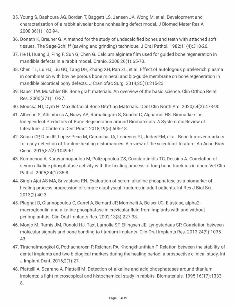
TablesTable 1:Mean values as well as standard deviations for all bone turnover markers at24 h and 72 h afteraugmentation of the defect.
Table 2:Mean values as well as standard deviations for all bone turnover markers 7 d, 14 d and 21 d afteraugmentation of the defect.
Page 15/19
Figures
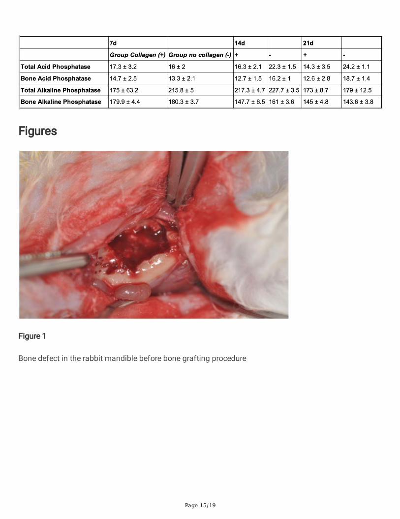
Figure 1
Bone defect in the rabbit mandible before bone grafting procedure

Page 16/19
Figure 2
Collection of peripheral blood from the ear vein of a rabbit
Figure 3
Bar charts showing expression of all four bone remodeling markers after 72 h. Signi�cant differences infavor of the group with collagen membrane were detected in case of bone acid phosphatase, as well astotal and bone alkalic phosphatase (all p<0.05).

Page 17/19
Figure 4
Bar charts showing expression of all four bone remodeling markers after 2 weeks. Signi�cant differencesin favor of the non-collagen group were detected in case of all four bone remodeling markers (all p<0.05).

Page 18/19
Figure 5
Histology (toluidine blue, original magni�cation x20) of a specimen (group with collagen membrane)after 7 days. No new bone formation can be seen. * = residual bone at the edge of the defect; + = bovinebone substitute particles

Page 19/19
Figure 6
Histology (toluidine blue, original magni�cation x20) of a specimen (group with collagen membrane)after 14 days. New osseous growth coming from the residual bone onto the grafted material can be seen.* = residual bone at the edge of the defect; + = bovine bone substitute particles; - = new-formed bonegrowing from the residual bone.