markus neuper technology exploitation in the field of ... · markus neuper technology exploitation...
TRANSCRIPT

Markus Neuper
Technology Exploitation in the Field of
Brain Computer Interface
Diploma Thesis
Area of Study:
Mechanical Engineering and Business Economics
Production Science and Management
Graz University of Technology
Written on Behalf of:
Institute for Industrial Management and Innovation Research
o.Univ.-Prof. Dipl.-Ing. Dr.techn. Josef W. Wohinz
Graz 2011

Acknowledgment
At this point I would like to especially thank my professor o.Univ. Prof. Dipl.-Ing.
Dr.techn. Josef W. Wohinz, who supervised my thesis and was supportive during the
stay abroad. Furthermore, Professor Wohinz provided, as curator of the study course
in Production Science & Management, a major contribution to the development of this
English branch of study. The PSM study program was a perfect preparation for the stay
abroad. I would also like to thank Dipl.-Ing. Dr.techn. Sonja Embst for the support,
helpful suggestions and proposal for amendment.
My special thanks go to my professor abroad, Gerwin Schalk Ph.D., who inspired me
with enthusiastic supervision and many long talks. Furthermore, all my colleagues at
Wadsworth Center made me feel welcome and they give me supporting input for this
work. Especially, Xiao-mei Pei Ph.D., Dipl.-Ing. Peter Brunner, Aysegul Gunduz Ph.D.,
Disha Gupta Ph.D., Jeremy Hill Ph.D. and Bill Baxter Ph.D. helped me with proof
reading and they had always time for me. Jonathan R. Wolpaw, M.D. and Theresa M.
Vaughan laid the foundation for my time at the Wadsworth Center and I wish to thank
them for that.
This work was supported by the Marshall Plan Scholarship.
I would like to dedicate this thesis to my parents because they have not only financed my
studies for the most part, but always showed great interest in my work and supported me
as best as they could.
i

Abstract
Epilepsy is an incurable disease that afflicts approximately three million Americans of all
ages, and roughly 612,000 persons were living with a diagnosis of primary brain and central
nervous system tumor in the US. Treatment of these diseases may require resection of tar-
geted brain areas and thus preoperative intraoperative localization of their primary brain
functions is required to preserve language and memory. Currently, the Wada test and elec-
trocortical stimulation are the golden standard used in medical practice for lateralization
and localization of these essential brain functions, respectively. However, these techniques
are time consuming and costly. A promising alternative for lateralization and localization
utilizes Brain-Computer-Interfaces (BCI) to map human brain functions by measuring and
analyzing brain signals directly. Recently, the BCI group at the Wadsworth center has
developed two novel clinical diagnostic tests based on this technology: Electroencephalog-
raphy Lateralization Index text (EEG LI test) for the purpose of language lateralization
and, SIGFRIED for functional brain mapping. SIGFRIED stands for “SIGnal modeling
For Realtime Identification and Event Detection”, and both clinical diagnostic tests con-
tain innovative signal processing software.
These BCI system softwares have been validated at the research level, but their com-
mercial prospects have not been explored. Because certification for commercialization
is missing, it is currently not possible to obtain reimbursement for these development
results. Even if these clinical diagnostic tests were approved for commercialization, this
would not indicate that the medicare companies automatically reimburse these clinical
diagnostic tests. The amount, which will be reimbursed by the medicare companies, plays
an import rule in the success of a development result in the medical device field. The
insurance companies decide independently whether a service or product is covered and
they also define the amount which will be reimbursed. In summary, both the missing
certification for communalization and the missing reimbursement coverage determination
limit wider use of the two clinical diagnostic tests. The time and effort to acquire these
two milestones are unclear as well as the tests’ market demand at this stage.
This work highlights the essential steps necessary to launch a BCI technology successfully
onto the market after the technology has been validated. This theoretical analysis is ap-
plied to two case studies that address EEG LI test and SIGFRIED. These case studies
include both a detailed comparison against the current gold standards and an evaluation of
its cost-effectiveness. Furthermore, these case studies address their market demand, reg-
ulatory environment, and Medicare reimbursement. A detailed time line of the necessary
steps to market these technologies concludes this topic.
ii

Contents
1 Introduction 11.1 Initial situation . . . . . . . . . . . . . . . . . . . . . . . . . . . . . . . . . 31.2 Problem . . . . . . . . . . . . . . . . . . . . . . . . . . . . . . . . . . . . . 41.3 Aim . . . . . . . . . . . . . . . . . . . . . . . . . . . . . . . . . . . . . . . 41.4 BCI2000 . . . . . . . . . . . . . . . . . . . . . . . . . . . . . . . . . . . . . 51.5 BCI2000 group at Wadsworth Center . . . . . . . . . . . . . . . . . . . . . 61.6 Procedure and document structure . . . . . . . . . . . . . . . . . . . . . . 7
2 Background 82.1 BCI systems . . . . . . . . . . . . . . . . . . . . . . . . . . . . . . . . . . . 8
2.1.1 Signal acquisition . . . . . . . . . . . . . . . . . . . . . . . . . . . . 92.1.2 Signal processing . . . . . . . . . . . . . . . . . . . . . . . . . . . . 112.1.3 Device command . . . . . . . . . . . . . . . . . . . . . . . . . . . . 112.1.4 Operating environment . . . . . . . . . . . . . . . . . . . . . . . . . 11
2.2 Functional brain mapping . . . . . . . . . . . . . . . . . . . . . . . . . . . 112.2.1 Electrocortical stimulation . . . . . . . . . . . . . . . . . . . . . . . 142.2.2 Functional magnetic resonance imaging (fMRI) . . . . . . . . . . . 152.2.3 Methods for language lateralization . . . . . . . . . . . . . . . . . . 16
2.3 Application field for functional brain mapping . . . . . . . . . . . . . . . . 192.3.1 Epilepsy surgery . . . . . . . . . . . . . . . . . . . . . . . . . . . . 202.3.2 Brain tumor surgery . . . . . . . . . . . . . . . . . . . . . . . . . . 21
2.4 Summary of functional brain mapping . . . . . . . . . . . . . . . . . . . . 23
3 Development results at Wadsworth Center 243.1 General description of the innovation procedure . . . . . . . . . . . . . . . 253.2 Innovation procedure at Wadsworth Center . . . . . . . . . . . . . . . . . . 263.3 Development result SIGFRIED . . . . . . . . . . . . . . . . . . . . . . . . 29
3.3.1 Components of SIGFRIED . . . . . . . . . . . . . . . . . . . . . . . 303.3.2 Technical comparison against the established method . . . . . . . . 313.3.3 Economical comparison against the established method . . . . . . . 32
3.4 Development result EEG LI test . . . . . . . . . . . . . . . . . . . . . . . . 363.4.1 Components of EEG LI test . . . . . . . . . . . . . . . . . . . . . . 373.4.2 Comparison against established methods . . . . . . . . . . . . . . . 383.4.3 Cost-effectiveness analysis . . . . . . . . . . . . . . . . . . . . . . . 39
3.5 Summary of development results . . . . . . . . . . . . . . . . . . . . . . . . 42
4 Evaluation of development results’ market 434.1 Valuation of existing market studies . . . . . . . . . . . . . . . . . . . . . . 45
4.1.1 American Association of Neurological Surgeons AANS . . . . . . . 46
iii

4.1.2 Association of American Medical College AAMC . . . . . . . . . . 484.1.3 National Association of Epilepsy Centers NAEC . . . . . . . . . . . 494.1.4 National Brain Tumor Society NBTS . . . . . . . . . . . . . . . . . 514.1.5 Central Brain Tumor Registry of the United States CBTRUS . . . 524.1.6 American Academy of Neurology AAN . . . . . . . . . . . . . . . . 53
4.2 Summary of development results’ available market . . . . . . . . . . . . . . 53
5 Regulatory environment of a medical device in US 565.1 Investigational Device Exemption . . . . . . . . . . . . . . . . . . . . . . . 585.2 Food and Drug Administration . . . . . . . . . . . . . . . . . . . . . . . . 60
5.2.1 Premarket approval (PMA) . . . . . . . . . . . . . . . . . . . . . . 625.2.2 Premarket notification . . . . . . . . . . . . . . . . . . . . . . . . . 645.2.3 Software in the context of medical device . . . . . . . . . . . . . . . 71
5.3 Sequence of action . . . . . . . . . . . . . . . . . . . . . . . . . . . . . . . 795.4 Summary of regulatory environment . . . . . . . . . . . . . . . . . . . . . . 87
6 Reimbursement policy of a medical device in US 886.1 Center of Medicare & Medicaid Services . . . . . . . . . . . . . . . . . . . 90
6.1.1 Local coverage determination . . . . . . . . . . . . . . . . . . . . . 936.1.2 National coverage determination . . . . . . . . . . . . . . . . . . . . 946.1.3 Sequence of action . . . . . . . . . . . . . . . . . . . . . . . . . . . 976.1.4 Summary of the reimbursement coverage determination . . . . . . . 98
6.2 Reimbursement code . . . . . . . . . . . . . . . . . . . . . . . . . . . . . . 996.2.1 International Statistical Classification of Diseases 9th rev code . . . 1006.2.2 Current Procedural Terminology CPT . . . . . . . . . . . . . . . . 1016.2.3 Relative value unit . . . . . . . . . . . . . . . . . . . . . . . . . . . 1116.2.4 Summary of reimbursement code . . . . . . . . . . . . . . . . . . . 114
7 Competitive advantage 1157.1 Time line and cost estimation . . . . . . . . . . . . . . . . . . . . . . . . . 1187.2 Benefits of development results . . . . . . . . . . . . . . . . . . . . . . . . 1197.3 Summary of competitive advantage . . . . . . . . . . . . . . . . . . . . . . 121
8 Conclusion 122
9 Indices 1289.1 Abbreviations . . . . . . . . . . . . . . . . . . . . . . . . . . . . . . . . . . 1289.2 List of Figures and Charts . . . . . . . . . . . . . . . . . . . . . . . . . . . 1319.3 List of Tables . . . . . . . . . . . . . . . . . . . . . . . . . . . . . . . . . . 1339.4 Bibliography . . . . . . . . . . . . . . . . . . . . . . . . . . . . . . . . . . . 133
10 Appendix 142
iv

1 Introduction
A Brain-Computer Interface (BCI) is a special human-machine interface that allows a
connection between the brain and a computer without any activity from the peripheral
nervous system. Instead, a BCI records the electrical activity either non-invasively from
the surface of the scalp, or invasively with the help of implanted electrodes. While numer-
ous different BCI systems have been validated for many purposes, BCIs typically required
a major development effort to create and test the customized software for each specific
application and user. Recently, this problem has been solved with a software platform
called BCI2000, discussed below. Concurrently, advances in electrode technology, elec-
tronics, signal processing, and other fields have paved the way for new BCI applications
for new user groups.
As a result, the very definition of “BCI” is expanding. While conventional articles define
a BCI as a device used strictly for communication Wolpaw et al. (2002), new articles
have expanded the definition to include a broader range of passive monitoring devices
and medical applications. Similarly, various research articles and new products reflect the
growing enthusiasm and opportunity surrounding BCI systems.
BCIs for healthy users Several different market segments are interested in this new
technology. Neuromarketing has received a lot of attention in the popular media Lewis
and Brigder (2005). As the name implies, neuromarketing involves advanced analysis of
customer behavior based partly on brain imaging technologies like electroencephalogra-
phy (EEG) and functional magnetic resonance imaging (fMRI). One of the most popular
studies in this field was done by the group of Read Montague. They evaluated the differ-
ences in consumer responses to either Coke and Pepsi McClure et al. (2004). New BCI
products are also being sold within the gaming industry.
The company Neurosky released their MindSet BCI system in 2009. This package includes
a headset that records brain signals from the scalp, and these signals provide information
about the users’ mental states. The famous game manufacturer Mattel also sells a BCI
product based on Neurosky technology called the Star Wars Force Trainer, which allows
players to move a ball via concentration. Both systems are available for under USD100.
1

Similar technology as been used in other games and in a safety devices that track the at-
tention of vehicle operators and warn drivers if they are falling asleep Huang et al. (2008),
Muller et al. (2008).
These two systems underscore both the opportunities and challenges of developing BCIs
for new user groups. Until recently, BCI users were typically “locked-in” patients, such
as people in the last stage of ALS, who have no muscle control left. When developing
applications for new user groups, the critical question is: why use a BCI? In the case of
game or neuromarketing applications, the answer may be that the BCI provides a more
entertaining or convenient means of communication. That is, the BCI provides informa-
tion that might be otherwise available (such as through a joystick or questionnaires), but
in a different way.
However, another reason why people might use a BCI is need. A BCI could provide
information or opportunities that are not otherwise possible. In particular, if BCIs can
provide a superior mechanism to diagnose or treat a disorder, then they might be partic-
ularly useful to some users, who might pay considerably more than about US$100. Some
recent research has already explored BCI systems for medical applications such as rehabil-
itation of movement disorders resulting from stroke or amelioration of autistic symptoms
Birbaumer and Cohen (2007). We focus here on a related emerging application: BCI
technology for functional brain mapping to facilitate surgery.
BCIs for functional brain mapping Many people with drug resistant epilepsy (i.e.,
epilepsy that cannot be controlled by drugs) or with brain tumors consider invasive brain
surgery. In such surgery, the affected areas are surgically removed. This type of surgery
is extremely effective because it reduces or eliminates seizures or tumor growth in these
patients.
As part of the pre-surgical preparations, experts must identify the affected areas of the
brain, as well as the areas that correspond to important functions such as motor or
language function. The surgery will then maximize the amount of affected tissue that is
removed while simultaneously minimizing the removal of areas important for key functions.
At present, this functional mapping requires electrodes that are implanted on the surface
of the brain, together with a relatively old technique called electrocortical stimulation,
which is very time-consuming and has many other problems.
One of the most disconcerting risks of electrocortical stimulation is that it can induce
seizures, particularly in patients who already have such severe epilepsy that they require
surgery. Electrocortical stimulation is also expensive, slow, and inaccurate. All other
more modern techniques, such as functional magnetic resonance imaging (fMRI), are
impractical or have other severe limitations (Wolpaw et al. (2006)). Thus, they have not
2
replaced electrocortical stimulation, which remains the gold standard despite its severe
problems.
1.1 Initial situation
The BCI group at the Wadsworth center has developed a new clinical diagnostic test
that uses the electrodes that are already implanted in all these patients. Using modern
signal processing techniques, they can detect and visualize (in real time) the brain signal
changes associated with a particular function, such as when the patient speaks or moves a
limb. This results in a functional mapping technique that can be accomplished in minutes.
The new technology is faster as well as more accurate, and avoids some other problems
with conventional techniques. “In particular, since the system relies on passive recording
from electrodes that are already implanted, there is no electrocortical stimulation and
hence no risk of seizure. The Wadsworth group just published a multi-center study that
demonstrates the efficacy of this new clinical diagnostic test, and shows that the results
generally agree with the results achieved using the present gold standard, electrocortical
stimulation” cf. Brunner et al. (2009).
In a related development result, the Wadsworth group has also explored new ways to iden-
tify the brain regions responsible for language using EEG based electrodes. These nonin-
vasive electrodes do not require surgery or significant preparation effort. The Wadsworth
group’s new technology can identify which hemisphere is dominant for language function.
This information is important because doctors need to know where to implant electrodes;
implanting an electrode grid in a non-dominant hemisphere entails unnecessary time, ex-
pense, and risk.
Hence, doctors will often apply a test called the Wada test to identify the language dom-
inant hemisphere. This test was developed many decades ago, and also remains the gold
standard despite several problems. Notably, the test requires injecting a chemical such as
sodium amytal into the carotid artery to effectively shut down one brain hemisphere prior
to the language test. Obviously, this is a nontrivial procedure, and can produce scarring,
infections, seizures, strokes, anaphylactic reactions, and other problems Loddenkemper
et al. (2004). The new approach from the Wadsworth team is completely noninvasive,
requiring no invasive electrodes, drugs, or injections, and thus avoids these problems. This
new approach is safe and requires much less time and expense.
3

1.2 Problem
At this stage, it is unclear whether these two clinical diagnostic tests present an eco-
nomically feasible innovation. It is unclear because no business strategy evaluating the
suitability, cost-effectiveness and acceptability has been developed. The development
results are certified only for research purpose and for commercial distribution, every med-
ical device must be approved by the Food and Drug Administration (FDA). Currently the
FDA approval conditions for the two diagnostic tests are unknown as well as the required
documentation to apply for FDA approval.
The field of medical device has an unique character that the end-user (patient) does not
pay directly for service. Instead a medicare company normally covers the expenses, hence
the goal of the development results is the receive a positive Medicare coverage determi-
nation. For the two clinical diagnostic tests, the Medicare coverage decision procedure is
ambiguous and also whether the innovations are valid for reimbursement. Furthermore
expenditure of time and costs for the FDA approval and Medicare coverage determina-
tion is not evaluated yet. Also the appraisal of the development results’ market demand
is missing, which is one of the most imported factors to evaluate the feasibility. This
inhibits the venture of commercializing these new clinical diagnostic tests.
1.3 Aim
The mentioned novel diagnostic techniques exist currently as prototypes that are only
used for research purpose. The main task of this master’s thesis is to investigate the eco-
nomic feasibility of these development results. Therefore, this work estimates the market
demand and the effort, which are necessary for the commercialization of the new clinical
diagnostic tests. In particular, the required certification and the volume of the necessary
tests and documentation to obtain the certification should be clarified
Furthermore, the goal of this work is to develop a strategy to obtain Medicare reimburse-
ment, which is an fundamental milestone of medical device innovation. The interest of
every Medicare company is to reimburse the most cost-effective treatment and one object
of this work is to address this issue. The interest of a manufacturer is to sell the clinical
diagnostic tests and hence this work points out the purchasing decision impact factors of
customer (hospital).
The final outcome of this work should be a time line and cost estimation that includes all
necessary step of the clinical diagnostic tests pre-commercial development.
4

1.4 BCI2000
“BCI2000 is a general-purpose research and development platform that greatly facilitates
implementation, evaluation, and comparison of different BCI options”Schalk et al. (2004).
BCI2000 has been in development for over 10 years and it has clearly emerged as the dom-
inant software platform in BCI research. Over 450 labs have downloaded it, and it has
been referenced in very many articles Schalk (2009). The Wadsworth group has received
multiple grants to support further development of BCI2000, which not only improved
the software itself, but also fostered BCI2000 support mechanisms such as workshops,
conference talks, published articles, and a website with helpful instructions and other
documentation. Indeed, the work described in this master’s thesis is based on BCI2000,
and would not have been possible otherwise without substantially more effort.
Fig. 1.1 illustrates the basic modular structure of BCI2000. The four modules (Opera-
Operator
Source Signal
Processing
User
Application
control signals
event markers
brain signals
event markers event markers
Storage
system configuration visualization
Figure 1.1: BCI2000 design. “The four modules in BCI2000 are called Operator,Source, Signal Processing, and Application” cf. Schalk et al. (2004).
tor, Source, Signal Processing and Application) correspond to the four components of a
BCI. These four modules communicate using TCP/IP sockets, and thus can be placed
on different machines or even in different locations. The central configuration is provided
by the Operator module. The interfaces are well defined and thus each module is inter-
changeable. One goal of this thesis was to provide a SIGFRIED signal processing module
for BCI2000.
BCI2000 was developed primarily by Gerwin Schalk at the Wadsworth group. While many
people have contributed as well, the software is patented and owned by the Wadsworth
group, and thus is protected against infringement.
5

1.5 BCI2000 group at Wadsworth Center
Our team is part of the Wadsworth Center. The Wadsworth Center is a comprehen-
sive state public health laboratory that is unique among state public health laboratories
for its commitment to basic and applied biomedical and environmental research. The
Wadsworth Center has a staff of 1,100 (including more than 175 doctoral-level scientists)
and is housed in 900,000 square feet of state-of-the art facilities. It maintains a num-
ber of core facilities for all investigators, including a core of specialized research facilities
for state-of-the-art microscopy, advanced biochemical techniques, molecular neuroscience,
and nanofabrication of devices, as well as an AAALAC-accredited animal facility, a com-
puter support center, and a large biomedical library. Wadsworth investigators receive
substantial research funding (i.e., >$35m in 2006) from outside research sponsors.
The BCI research group, headed by Jon Wolpaw, has grown so much over the past few
years that it has been subdivided, with different research teams focusing on different chal-
lenges. One of the research teams, headed by Prof. Dr. Schalk, focuses on developing
BCI2000.
Figure 1.2: BCI2000 group at the Wadsworth Center.
While Schalk and other colleagues also conduct experimental research involving both
invasive and non-invasive systems, their primary focus is on improving BCI2000 and its
6

applicability to new paradigms, with a strong emphasis on the functional brain mapping
paradigms described in this thesis. The functional principle of the BCI at the Laboratory
of Nervous System Disorders at the Wadsworth Center in Albany, NY is illustrated in Fig.
2.1. Early Wadsworth BCIs (see Wolpaw et al. (1991) and Wolpaw and McFarland (1994)
for a comprehensive review) relied on noninvasive imaging tools. Our team at Wadsworth
began exploring ECoG based BCIs several years ago, and adapted BCI2000 and other
tools accordingly.
1.6 Procedure and document structure
Chapter I introduces Brain-Computer Interfaces (BCI) and explains the work of the BCI
group at the Wadsworth Center in detail. It provides a short introduction to current BCI
applications and BCI2000.
Chapter II extends our review of the state of the art with an overview of basic neuro-
science, functional brain mapping technologies, and relevant application fields.
Chapter III details the two recently developed clinical diagnostic tests described in this
thesis, including an analysis of cost effectiveness relative to conventional methods and
technologies.
Chapter IV assesses the market demand for the two clinical diagnostic tests based on
existing marketing studies.
Chapter V explains the regulatory environment for the two development results with a
focus on the Food and Drug Administration (FDA) approval process. The FDA plays a
major role in the certification process of a medical device in the US.
Chapter VI describes the reimbursement process between the hospitals and insurance
companies, because in the medical device market, the end user (the patient) does not
usually pay directly for the treatment. Instead, the patient’s insurance reimburses the
hospital and also decides how much money can be reimbursed.
Chapter VII reviews the purchasing-decision process between the innovation’s manufac-
turer and the hospital. The purchasing process is the last step at the innovation process
and, obviously, it is the overall goal of the manufacturer.
Chapter VIII presents a conclusion, including a time line and further directions.
7

2 Background
This chapter provides a general overview about BCI Systems, functional brain mapping
and language lateralization because the innovative clinical diagnostic tests, which are
introduced in chapter development results at the Wadsworth Center, are novel techniques
for functional brain mapping and language lateralization. Thus this chapter lists the
different methods for both functional brain mapping and language lateralization. The
current established method for functional brain mapping, Electrocortical stimulation, and
the gold standard for language lateralization, Wada test, are explained particularly in
order to compare them against the recently developed clinical diagnostic tests in chapter
3. The two application fields of the development results, epilepsy- and brain tumor, are
explicated in the end of this part.
2.1 BCI systems
Brain-Computer Interfaces (BCI) use Electroencephalography (EEG) or other neurophys-
iological methods to extract specific features of brain activity (e.g., sensorimotor rhythms,
slow cortical potentials, event-related potentials) and translate them into an output sig-
nal that can be regulated by the specific user. All BCI systems require four processes, as
shown in 2.1. First of all, brain activity is recorded, which is called Signal Acquisition.
Next, a Signal Processing module must categorize the user’s brain activity after learning
the individual’s subjective signal features. Third, the user’s brain activity must produce
some effect in real-time, such as moving a cursor or wheelchair. Finally, the BCI must
provide some feedback as part as an overall operating system that controls how the dif-
ferent modules interact with each other and with the user Wolpaw et al. (2002).
In the case of a conventional BCI - a device for communication and control - the user
receives real-time feedback. As noted above, this thesis addresses a new type of BCI in
which the goal is not to provide communication, but rather to provide functional brain
mapping. Hence, the user does not receive any real-time feedback. Instead, real-time
feedback is sent to the operator, who can use the information to learn more about the
patient’s brain. Otherwise, the system described here is identical to a conventional BCI -
8

Figure 2.1: BCI components. “Basic design and operation of any BCI system. All BCIshave four components: Signal Acquisition (measuring brain activity); Signal Process-ing (translating brain activity into outputs); Device Commands (executing the desiredcommands; and an Operating Protocol (communication among modules and the user)”Wolpaw et al. (2002).
all four processes are necessary and rely on very similar hardware and software.
2.1.1 Signal acquisition
There are many ways to study brain function in real-time. The different brain imaging
approaches used in BCIs are generally divided into two categories: invasive and non-
invasive. Invasive techniques require a neurosurgical procedure to implant electrodes on
or in the brain. Non-invasive techniques rely on electrodes placed outside the head, often
in an electrode cap, and thus do not require surgery, drugs, injections, or other invasive
procedures.
Non-invasive: Over 85% of BCIs rely on non-invasive methods (Mason et al., 2007).
Brain activity can be detected outside of the scalp with different technologies. The most
common non-invasive neuroimaging technology is the electroencephalogram (EEG), shown
in Figure 2.2 on page 10 (A). This is a measure of the electrical activity over a certain
area of the brain. While non-invasive electrodes do not provide as much information
9

CORTEX
DURA
SKULL
BC
A
5 mm
SCALP
SOFTTISSUE
Figure 2.2: BCI sensor types. “BCIs may rely on invasive or non-invasive electrodes.(A) shows a non-invasive electrode placed outside the scalp. (B) and (C) show twotypes of non-invasive electrodes. Electrodes may be placed on the surface of the brain(B), or may penetrate the brain (C)” Brunner (2005).
as invasive electrodes, they are adequate for many purposes, including identifying the
language dominant hemisphere.
Invasive: Invasive electrodes are subdivided into two categories. Some electrodes, called
ECoG electrodes, measure the electocorticogram, which reflect’s the activity on the surface
of the brain. These electrodes do need to be surgically implanted, but entail less risk and
damage because they never penetrate the brain (see part (B) of Figure 2.2 on page 10.
Other depth electrodes do penetrate the brain’s surface (see part (C) of Figure 2.2 on
page 10. Since these different types of electrodes provide different information, they are
better suited to different BCIs. The invasive approach described in this thesis relies on
ECoG electrodes.
Invasive methods have obvious drawbacks, including the added time, cost, risk, and ethical
issues inherent in neurosurgery. However, invasive electrodes can provide a more detailed
picture of brain activity, with less interference from external noise sources. Invasive BCIs
are also always available. That is, there is no need to prepare the subject for each session of
BCI use, nor to wash electrodes afterward. The consensus of most BCI researchers is that
neither approach is generally superior; the choice of neuroimaging technology depends on
each subject’s situation and needs.
10

2.1.2 Signal processing
One of the most heavily researched facets of BCIs is Signal Processing. Many differ-
ent linear and nonlinear approaches have been used. In addition, various preprocessing
techniques are common, such as improved spatial filters Krusienski et al. (2008) or di-
mensionality reduction techniques. Hence, this master’s thesis does not focus heavily on
development of new signal processing algorithms, as they have been well explored, and
additional research is unlikely to yield a major breakthrough.
2.1.3 Device command
Early BCIs were used to control simple monitor based applications such as spellers Farwell
and Donchin (1988), Wolpaw et al. (1991). More recent work showed that BCIs could
control many other devices, such as a wheelchair, mobile robot, or orthosis Millan et al.
(2010), Pfurtscheller et al. (2010). This component of a BCI is not emphasized here, since
this thesis does not focus on conventional BCIs for communication and control.
2.1.4 Operating environment
Any BCI requires an operating environment. The operating environment might specify
details such as how the modules pass information to each other, how to present feedback,
and how to handle errors. Until fairly recently, this was a major challenge in the BCI field.
Many groups developed their own operating environments, which entailed many problems.
An accurate real-time EEG data collection and analysis system is hard to develop, even
for groups with a very strong computer science background, leading to some failed projects
and other problems Bayliss, Inverso and Tentler (2004).
In the past few years, some groups have proposed or tried to develop some kind of universal
platform that any group could use as a BCI operating system. For example, the OpenVibe
system from the INRIA group in France is publicly available, and has been downloaded by
some groups. However, the most trenchant and widely used program is called BCI2000,
discussed below.
2.2 Functional brain mapping
This chapter introduces the different methods for functional brain mapping and in par-
ticular it explains language lateralization. The term functional brain mapping can be
defined as, “the attempt to specify in as much detail as possible the localization of func-
tion in the human brain” Savoy (2001). This chapter starts with a short description of the
11

biological structure of the human brain followed by functional brain mapping’s historical
development and an overview of the established methods for language lateralization and
functional brain mapping.
The human brain is a very complex organ as well as the central command unit for the
nervous system. If you take a look at a human brain, you can recognize three different
areas:
1 Cerebrum
2 Cerebellum
3 Metencephalon, which passes into the spinal cord
The cerebrum, the largest part of the brain, is divided in the middle into two halves called
left and right hemispheres. Between the hemispheres, there is a thick nerve cord known as
the corpus callosum. The cerebrum’s outer layer, which is 2–4mm thick, is called cerebral
cortex or grey matter, hence every brain consists of a left and right cerebral cortex. The
neurons and unmyelinated fibers in cerebral cortex have a large field of responsibility, for
example language, memory and attention. Each hemisphere is divided into four lobes
and Figure 2.3 on page 12 illustrates them. Each lobe has specific character, which are
Figure 2.3: Human brain. “One hemisphere of the human brain is depict in this figureand the hemisphere’s outer layer is called the cerebral cortex, which plays a major rolein e.g., language, attention, thought and memory. The cortex of each hemisphere canbe divided on the basis of gross topographical conventions into four lobes: Frontal-,Parietal-, Temporal and Occipital lobe” Macmillan Cancer Support (2009).
explained below.
Frontal lobe is situated at the front of the brain and normally in authority of both lan-
guage skills and motor functions. The body movements are carried out by the motor
12
cortex, which is located in the back of the frontal lobe.
Parietal lobe is placed in the center of the brain and it processes the sensorial information
like pain and touch. The body’s senses are handled at the somatosensory cortex, which
is a part of this lobe.
Temporal lobe is situated at the bottom of the brain and it contains the primary auditory
cortex, amygdala and hippocampus. The hippocampus is responsible for long-term mem-
ory and the amygdala dominates the responses linked with arousal, fear and emotional
secretions.
Occipital lobe the occipital lobe comprises the visual cortex, which processes the infor-
mation from the eyes, at brain’s back.
However, for many purposes it is not satisfactory to distinguish only between four lobes
and many scientists have been working on methods and technology to create a more de-
tailed map of the human brain. Still nowadays, the brain is one of the most un-explored
parts of the human body and plenty of further research work is needed to understand
how the human brain is working. The following section gives a historical overview on
development of functional brain mapping and out if it the resultant brain function maps.
History of functional brain mapping In the last century the electrical stimulation was
the most important method to explore and discover the different areas of the brain. The
Italian scientist Luigi Galvani (1737-1798) discovered that muscles and nerves are elec-
trically excitable around 1786. In 1802, Giovanni Aldini’s experiments showed that also
the human brain is electrically excitable and the next milestone was the development of
the ”Homunculi” map (see Figure 2.4 on page 14) by Wilder G. Penfield. With the use of
electrocortical stimulation, Penfield and his group created a detailed map, that represents
the localization of the different body parts on the cerebral cortex, which is known as the
outermost layer of the cerebrum. During brain surgeries for the purpose of treating severe
epilepsy, Penfield and his group stimulated the patient’s cerebral cortex with electrical
probes. Because of the use of local anesthesia, they could monitor the different response
behaviors and characteristics of the patient according to the different stimulated brain
areas. The “Homunculus” is still up to date and also electrocortical stimulation is the
state of art for functional brain mapping.
The composition of the human brain is morphologically bisymmetric. Although this sym-
metry indicates a substantially similar construction, it is known from the many studies
and experiments that several functions are located asymmetrically. In other words, some
brain functions e.g., language and memory, can be found on either the left or right hemi-
sphere and in a few cases in both hemispheres. Right handed persons have the language
function normally on the left hemisphere. “The specialization of the left hemisphere for
13

homunculus.jpg (JPEG Image, 1894x983 pixels) - Scaled (68%) http://www.ahs.uwaterloo.ca/~ranney/Images/homunculus.jpg
1 of 1 7/6/2010 11:34 AM
Figure 2.4: Motor homunculus. “Detailed picture of the motor strip, showing what issometimes called the motor homunculus, which is a depiction of how motor functionsare localized along the motor strip. The portion adjacent to Broca’s area controls theface and mouth” Sabbatini (2003).
language was one of the earliest observations of brain asymmetry. Reported in the 19th
century by Broca and Wernicke, language was found to be more severely impaired in
response to tumors or strokes in the left hemisphere. Language production and some
aspects of syntactic processing have subsequently been localized primarily to areas of the
anterior left hemisphere.” Toga and Thompson (2003). Unfortunately the correlation
between handedness and language lateralization, which stem from the neuroanatomical
inequality, functional division, and specialization of the cerebral hemispheres, does not
apply to every person. Hence different methods for language lateralization have been
developed and Section 2.2.3 discuss them in detail.
However, every human brain looks slightly different and thus the exact position of the
brain functions are also variable from human to human. Thus it is not possible to predict
the exact location of brain functions but the brain maps provide a good indicator, where
a brain function is expected. Because of that it is necessary to locate the brain functions
for every individual person.
2.2.1 Electrocortical stimulation
As the name indicated, the paper “Functional brain mapping and its applications to neu-
rosurgery” explains the methods for functional brain mapping and content of the next
paragraph is based on this paper.
“Since the 1930s, direct electrocortical stimulation (ECS) testing has been the gold stan-
dard method for mapping brain function in preparation for surgical resection. The motor
14
cortex is mapped intraoperatively by stimulating the pre- and postcentral gyri, as well
as the premotor area and supplementary motor area. ECS for motor mapping may be
performed under general anesthesia without muscle relaxants and in motor mapping, ECS
is used as an activation technique. To test language functions, it is necessary that the
patient remain awake and able to perform certain tasks such as counting or naming.
Awake craniotomy for language mapping is typically performed using a combination of
local anesthetic field block and short acting general agents to induce a rapidly reversible
hypnotic state. Once the scalp, cranium, and dura are opened, the sedation is allowed
to wear off so that the patient may cooperate with behavioral testing. During the cor-
tical stimulation testing, the patient is awake and asked to perform language tests such
as counting or naming while the surgeon stimulates the cortical surface. Areas in which
cortical stimulation induces speech arrest or paraphasic errors are considered essential for
language function. In this case, ECS is used as an inhibition technique causing disruption
in normal neuronal firing. Because the bipolar stimulating electrodes have a 5-mm tip
separation, a 1-cm margin is generally respected during the subsequent resection. In a
study of 40 patients undergoing removal of gliomas in the dominant temporal lobe, among
patients without preoperative language deficits, 87% had no deficits postoperatively using
the above methods. ECS can also be performed extraoperatively. This option is used pri-
marily for epilepsy surgery for the mapping of the seizure focus through the chronic ( < 1
wk) implantation of intracranial electrodes. In addition to electrocorticography, cortical
stimulation for the determination of eloquent cortex may also be performed during this
time period. When indicated, this technique has the advantage of allowing significant
time and a sufficiently relaxed and cooperative patient to allow detailed cognitive testing”
cf. Tharin (2007).
2.2.2 Functional magnetic resonance imaging (fMRI)
Functional MRI is considered an indirect method to assess brain activity, because it is
based on the change of the blood oxygenation level dependent (BOLD) signal. “The fMRI
techniques are based on changes in regional blood flow that occur in response to neuronal
activity. Local increases in neuronal activity lead to greater blood flow to the cortical
tissue parenchyma” cf. Belliveau et al. (1991).
Theoreticly, functional magnetic resonance imaging can be used for functional brain map-
ping but in practice only determination of cerebral dominance is executed with fMRI.
“Functional MRI is noninvasive and, therefore, repeatable, both for many runs in a single
session (unlike the Wada test) and on multiple occasions to follow patients over time. Be-
cause of its non-invasiveness and safety, fMRI is suitable for use in children. Unlike Wada
testing, fMRI can provide localization and not merely lateralization of critical functions
15

such as language and memory. Finally, fMRI is able to demonstrate functional activa-
tions in the depths of cortical sulci, not just at the cortical surface, an advantage over
the “gold standard” electrocortical stimulation. Disadvantages of fMRI include sensitivity
to motion-related artifacts, including those arising from the heartbeat, breathing, and
head motion. This has proven particularly problematic for language mapping, generally
precluding the use of tasks involving overt spoken language. fMRI also does not have the
proven clinical track record of the Wada test” Tharin (2007).
2.2.3 Methods for language lateralization
The article “An Update on determination of language dominance in screening for epilepsy
surgery: the Wada test and new noninvasive alternatives” Abou-Khalil (2007b) gives a
great detailed overview about all methods. The paper’s conclusion is that “Several meth-
ods provide sufficiently good lateralization of language dominance that they could be
considered alternatives to the Wada test. It is likely that fMRI will be the most widely
used method, obviating the need for the Wada test in the majority of patients with clearly
lateralized language. However, the best testing paradigm and the best method of image
analysis still have to be defined” Abou-Khalil (2007a). The Table 2.1 evaluates all meth-
ods for language lateralization and based on the conclusion, we picked the Wada test and
functional MRI as the current methods used for language lateralization.
Wada test
“Most epilepsy patients considering a surgery undergo the Wada test. This test is officially
known as the intracarotid sodium amobarbital procedure (ISAP), but the nickname Wada
test is commonly used. The name comes from the physician who first performed it,
Dr. Juhn Wada” Weiner (2004). “Juhn Wada introduced the intracarotid amobarbital
procedure to lateralize language in 1949, and soon thereafter, Brenda Milner included
memory testing during the procedure to help determine risk for postoperative amnesia”
Meador and Loring (2005). Since developed in 1949, the Wada test is the gold standard
for language and memory lateralization. The next two subsections elucidate the procedure
and involved doctors of the ISAP.
Involved doctors and required equipment The Wada test procedure usually de-
mands three doctors: Neuroradiologist, Epileptologist and Neuropsychologist.
Neuroradiologist deals with the radioactive substances and imaging devices and his
task during the test is to study the patient’s brain structure. However, a Radiology
resident must run though four months of learning neuroradiology to get a radiology
16

Method Physiologic basis Directness ofmeasurement
Reliability rel-ative to Wadatest
Availability
Wada test Deactivation by anesthe-sia
Direct NA ++
Dichotic listeningand divided vi-sual field test
Directness of access tolanguage cortex
Indirect ++ +++
rTMS Deactivation by electricalinterference
Direct ++ +
sMRI Association with domi-nance
Very indirect + +++
ERP Electrophysiologic ex-pression of activation
Direct + +++
MEG Magnetic flux directly as-sociated with activation
Direct +++ +
fTCD Hemodynamic responseto activation
Indirect +++ +++
NIRS Increased oxygenation Indirect +++ ++PET Hemodynamic response
to activationIndirect +++ +
SPECT Hemodynamic responseto activation
Indirect ++ +++
fMRI Increased oxygenation Indirect +++ +++ERP: event-related potentials; fMRI: functional MRI; fTCD: functional transcranial Doppler; MEG: magnetoencephalography
NIRS: near infrared spectroscopy; PET: positron emission tomog raphy; rTMS: repetitive transcranial magnetic stimulation;
sMRI: structural MRI; SPECT: single photon emission computed tomography.
Table 2.1: Methods for determining cerebral dominance. “It is worth noticing that theWada test is the only invasive method but the current gold standard for language andmemory lateralization. The comparison concludes that fMRI is the most promisingemerging technology for determining cerebral dominance” cf. Abou-Khalil (2007b).
board certification in the United States. The following fellowship program takes one
or two years and then an additional one or two years of training are required for
interventional neuroradiology.
Epileptologist has special training in treating epilepsy patients, especially in the cases
with difficult controllable seizures. They also are also experts in handling children
and pregnant women with epilepsy. Their special training starts with three years
of residency in Pediatrics after medical school and then a fellowship in Pediatric
Neurology comes after that. The last part of the training are two years of particular
concentration on pediatric epilepsy.
Neuropsychologist “A clinical neuropsychologist is a professional within the field of
psychology with special expertise in the applied science of brain-behavior relation-
ships. The clinical neuropsychologist uses psychological, neurological, cognitive,
17

behavioral, and physiological tests to evaluate a patient’s neurocognitive, behav-
ioral, and emotional strengths and weaknesses and their relationship to normal and
abnormal central nervous system functioning. The clinical neuropsychologist uses
this information and information provided by other medical/healthcare providers to
identify and diagnose neurobehavioral disorders, and plan and implement interven-
tion strategies” Hersen (2004). Based on Hersen (2004), the neuropsychologist has
to fulfill the following qualifications, after the doctoral degree in psychology:
• An internship, or its equivalent, in a clinically relevant area of professional
psychology.
• The equivalent of two years of experience and specialized training.
• A license in his or her state or province to practice psychology and/or clinical
neuropsychology independently, or is employed as a neuropsychologist by an
exempt agency.
Sequence of actions This paragraph describes in detail the procedure of the Wada
test and the cerebral angiogram, which has to be executed prior to of the Wada test. The
cerebral angiogram depicts the blood flow within the brain to make sure that there are no
obstacles to the ISAP. The neuroradiologist inserts a catheter (a long, narrow tube) into
an artery, usually in the leg. The catheter is directed to the right or left internal carotid
artery in the neck, which supplies the brain with blood. Once the catheter is in place, a
dye is injected and can be seen on a special x-ray machine. This machine takes pictures
of the dye as it flows through the blood vessels of the brain. Once the angiogram is done,
the catheter will stay in place for the Wada test. The neuroradiologist starts the course of
action with injection of sodium amobarbital into the either the left or right carotid artery.
“The left and right carotid arteries form together the common carotid artery and supplies
the neck and head with blood. Each artery, left or right, supplies one side of the upper
body, hence only either the right or left hemisphere of the brain” cf. Ashrafian (2007).
“The injection of sodium amobarbital has the pharmacological effect, that one hemisphere
falls asleep for a short time period. During the time the patient is in the hemiplegia
phase, the neuropsychologist begins with the language test that includes several tasks e.g.,
naming the calender days, following commands and naming of different objects shown on
pictures. Especially for patients, who will undergo an operation on the temporal lobe the
memory test is very important and therefore the neuropsychologist shows five to twelve
memory objects during the anesthesia. Hence, the awake half of the brain has the job
to remember, recognize and direct the language process of the brain. The language and
memory test during the hemiplegia phase usually takes 10 to 15 minutes and when it is
18

over the doctors ask the patient to retrieve the shown objects and pictures. They record
the answers word for word and then start with the procedure for the other side. The
delay between the two injections is normally 30 to 60 minutes and the second routine is
the same as the first, except that now different pictures and objects are shown” cf. Weiner
(2004).
2.3 Application field for functional brain mapping
As mentioned earlier, many scientists have been working on exploring the human brain,
but what are the application fields for this knowledge? Before we talk about application
fields, we have to specify the term brain mapping more in detail. Brain mapping can be
split into two main parts, conventional structural imaging and functional brain mapping.
Based on Engel (1996), test for structural imaging are:
• X-ray films, computed tomography, and other radiographic studies
• MRI
• Magnetic resonance spectroscopy
On the other hand, functional MRI or electrocortical stimulation are considered func-
tional brain mapping techniques. This work deals with both functional brain mapping
and language lateralization, and these methods should not to be confused with techniques
for identifying seizure foci.
Functional brain mapping and structure mapping methods jointly provide information
about the relationship between the location of lesions and primary brain functions for
operative planning. Functional brain mapping is performed to treat two specific diseases,
brain tumor and epilepsy surgery. Therefore, lateralization and localization of brain func-
tion is normally a required preparation for epilepsy and brain tumor surgery, because
these surgeries remove a part of the brain. The overall goal of any surgery is to avoid
irreparable damage to the patient, so a brain surgery should not derogate any functions
of the brain. Thus, the medical team has to make sure that the removed part contains
no fundamental brain function. If the doctors know the exact locations of brain functions
and infected brain areas before a surgery, they can plan the position of the transections to
ensure the best outcome. Therefore, brain function lateralization and localization provide
very important information for the surgical team, and the following sections describe the
surgeries in detail.
19

2.3.1 Epilepsy surgery
This paragraph gives a short overview about epilepsy surgery. First, we define epileptic
seizure and epilepsy.
“Epileptic seizure is a transient occurrence of signs and/or symptoms due to abnormal
excessive or synchronous neuronal activity in the brain” Fisher et al. (2005). “Epilepsy is
a disorder of the brain characterized by an enduring predisposition to generate epileptic
seizures and by the neurobiologic, cognitive, psychological, and social consequences of
this condition. The definition of epilepsy requires the occurrence of at least one epileptic
seizure and enduring alteration in the brain that increases the likelihood of future seizures”
Fisher et al. (2005).
“Based on 1995 data, seizures and epilepsy are estimated to affect approximately 2.3
million people with 181,000 new cases per year in the United States” Epilepsy Foundation
(2003), so epilepsy is one of the most common neurological disorders worldwide. “80% of
the patients can be successfully treated with antiepleptic drugs, but for the other 20 %
only epilepsy surgery can help. However, currently just a small portion of the potential
surgical candidates are getting a surgery” cf. Engel (2003). Under general anesthesia,
a multidisciplinary team of specialists usually operates the patient in an epileptic center
and the operation itself takes a few hours. “Under normal circumstance, the patient can
leave the hospital after a few days and the operation’s procedure normally includes the
removal of the hippocampus’s anterior part, a small part of the temporal pole and the
amygdala” cf. Engel (1996).
Modern epilepsy surgery methods aim to remove only the brain areas that contain seizure
foci and to avoid damaging fundamental brain functions such as language, memory and
motor functions. Therefore, the position of the seizure prone area as well as the position of
the fundamental brain functions must be known. Different epilepsy excitability detection
methods have been developed, based on Engel (1996):
• Noninvasive:
– Routine interictal EEG
– Video EEG, long-term monitoring EEG
– Ictal single-photon-emission computed tomography
– Interictal and ictal magnetoencephalography
– Functional MRI
• Invasive
– Intraoperative electrocorticography
20

– Stereotactic-depth-electrode, long-term recording
– Subdural grid electrode, long-term recording
However, some evaluation procedures are performed during the surgery and some are part
of the presurgical preparation.
Presurgical preparation The surgical team decides how much information and which
evaluation methods are required. The normal presurgical preparation contains interictal
and long term EEG monitoring, MRI imaging and position emission tomography. These
methods seek to find abnormalities and seized in the brain areas. One the other hand,
memory and language laterialization are also typically explored before an epilepsy surgery.
If the focus of the seizure is close to conjectural language cortex, an accurate topographic
localization of the language area is required to specify boundaries for surgical excision,
because the doctor can make sure to avoid any damage in the language and memory re-
gions.
In summary, if the methods for determining cerebral dominance, such as Wada test, show
that the fundamental brain functions are located on the same hemisphere where seizure
foci were found, functional brain mapping is then performed to localize brain functions
near the seizure foci. In this mapping procedure, an awake craniotomy is executed as part
of the presurgical preparation. In this procedure, doctors implant a Subdural Grid Elec-
trode (see Figure 3.2 on page 30) above the seizure foci that was identified by noninvasive
tests for epilepsy excitability. This Subdural Grid Electrode is used for two purposes.
First, the data recorded from the Subdural Grid Electrode during an epileptic seizure can
help precisely localize the seizure foci, because the invasive tests for epilepsy excitabil-
ity have a higher resolution than a noninvasive test. Second, electrocortical stimulation,
which localizes brain function, requires the Subdural Grid Electrode to stimulate the
brain, and the Subdural Grid Electrode is implanted in an average for 15 days.
2.3.2 Brain tumor surgery
Besides epilepsy surgery, brain tumor surgery is the second application field for brain
mapping. This paragraph provides general information, and then the subsection presurgi-
cal preparation explains the used methods. “Primary malignant brain tumors account for
2 percent of all cancers in U.S. adults. The American Cancer Society estimates that there
are more than 18,000 new diagnoses of brain and nervous system cancers causing more
than 12,000 deaths each year in the United States.” Sreenivasa R. Chandana (2008). The
procedure of a brain tumor surgery is slightly different than an epileptic surgery, because
the patient’s condition is stabile over time. In other words, an epilepsy patient must
21

have a seizure to detect the brain area prone to seizures, and it is not possible to predict
or control the epileptic seizure. Thus, long-term monitoring is necessary to record and
analyze the epileptic seizure.
Presurgical preparation for a brain tumor surgery does not include monitoring over
time, but the goal of functional brain mapping is the same for epilepsy and brain tumor
patients. Like epilepsy surgery, the medical team decides whether electrocortical stimula-
tion is necessary to ensure the best outcome of the surgery, based on the result from the
presurgical methods mentioned in table 2.2.
Modality UsesCT Localizing the tumor and defining its dimensions, morphologyMRI Localizing the tumor and surrounding structures with a high-resolution im-
age, diagnosis of supra- and subtentorial tumors, diagnosis of extra- andintra-axial tumors, presurgical planning with three-dimensional imaging,stereotactic biopsy, radiotherapy
DTI Establishing spatial relationships between tumor border and white matter,assessing the progression and regression of white matter tracts caused bytumor growth or resection
fMRI Neurosurgical planning and neurologic risk assessment by localizing thecortical regions that control language, motor, and memory functions
MRA Understanding tumor vascularity and identifying the anatomic relation-ship between the tumor and blood vessels MRS Obtaining biochemicaland metabolic information about the tumor, determining tumor type andgrade by assessing the cellular contents, differentiating tumor from radia-tion necrosis PET Metabolic assessment of tumor aggressiveness (grade),assessing the highly metabolic areas within the tumor, differentiating be-tween tumor recurrence and radiation necrosis, functional localization ofcortical regions, predicting patient survival and prognosis
CT = computed tomography; DTI = diffusion tensor imaging; fMRI = functional magnetic resonance imaging
MRA = magnetic resonance angiography; MRI = magnetic resonance imaging; MRS = magnetic resonance spectroscopy
PET = positron emission tomography
Table 2.2: Imaging Modalities for the Management of Primary Brain Tumors. “Theseimaging modalities can be helpful when performing the initial work-up and follow-upand in the evaluation and treatment of brain tumors” Sreenivasa R. Chandana (2008).
If yes, the doctors will first implant an electrical grid above the area where the brain
tumor is diagnosed, and then electrical stimulation is performed to ensure that no primary
brain functions like memory or language are situated in this area. The second part of the
surgery is the removal of the affected section of the brain.
22

2.4 Summary of functional brain mapping
During preparation for epilepsy and brain tumor surgery, a surgical team may need to
identify which brain regions are responsible for primary brain functions such as memory
or language. Currently intracarotid amobarbital, or Wada test, has been the standard
for language and memory lateralization. If the seizure focus is close to primary brain
functions, then the physician implants a Subdural Grid Electrode for approximately 15
days, and executes both functional brain mapping and precise localization of the brain
tumor or epileptic foci. Electrocortical stimulation is the establish method for functional
brain mapping. Afterwards, the tumor or epileptic foci and the Subdural Grid Electrode
are both removed in a second surgery. The need to replace the Wada test and electrical
stimulation mapping with less invasive and more reliable techniques has long been recog-
nized, both procedures are invasive and entail risks and discomfort.
Alternative brain imaging approaches include functional MRI and EEG. fMRI, which de-
tects a blood oxygenation level dependent (BOLD) signal, can be used for lateralization
and for functional brain mapping. fMRI has the advantages of excellent spatial resolution
and safety, because it is a noninvasive procedure. However, fMRI systems have numerous
practical problems. Due to the high purchase price, a fMRI device is only available in a
few hospitals, and hospitals are not likely to purchase an fMRI just to support lateraliza-
tion and localization efforts. fMRI systems require a very powerful magnetic field, which
makes them impossible to use with persons who have pacemakers, neurostimulators, metal
implants, or other special circumstances. Also, since fMRI analysis require patients to lie
very still for extended times in a very noisy environment, they are unpleasant for most
patients and impossible for some, such as persons with claustrophobia. Furthermore, lat-
eralization and especially functional brain mapping are very scientific and complex tasks
with fMRI, and therefore very few physicians are qualified. There also many concerns
about the reliability of the fMRI for functional brain mapping, and hence most physicians
verify the results from fMRI with the established methods anyway.
In conclusion, fMRI could theoretically be used for lateralization and functional brain
mapping. However, in practice, there are too many disadvantages for it to be practical in
the foreseeable future. Hence, fMRI is far away from becoming the new gold standard for
language lateralization and functional brain mapping. Brain computer interface methods,
on the other hand, suffer none of the disadvantages described here; instead, the main
barrier to wider adoption is the absence of a well-known software platform that can make
EEG-based mapping easy and effective.
23
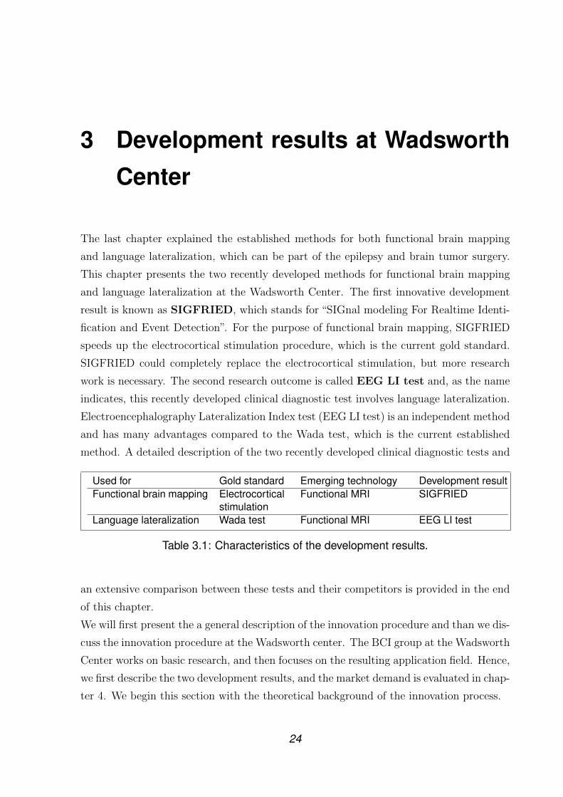
3 Development results at WadsworthCenter
The last chapter explained the established methods for both functional brain mapping
and language lateralization, which can be part of the epilepsy and brain tumor surgery.
This chapter presents the two recently developed methods for functional brain mapping
and language lateralization at the Wadsworth Center. The first innovative development
result is known as SIGFRIED, which stands for “SIGnal modeling For Realtime Identi-
fication and Event Detection”. For the purpose of functional brain mapping, SIGFRIED
speeds up the electrocortical stimulation procedure, which is the current gold standard.
SIGFRIED could completely replace the electrocortical stimulation, but more research
work is necessary. The second research outcome is called EEG LI test and, as the name
indicates, this recently developed clinical diagnostic test involves language lateralization.
Electroencephalography Lateralization Index test (EEG LI test) is an independent method
and has many advantages compared to the Wada test, which is the current established
method. A detailed description of the two recently developed clinical diagnostic tests and
Used for Gold standard Emerging technology Development resultFunctional brain mapping Electrocortical
stimulationFunctional MRI SIGFRIED
Language lateralization Wada test Functional MRI EEG LI test
Table 3.1: Characteristics of the development results.
an extensive comparison between these tests and their competitors is provided in the end
of this chapter.
We will first present the a general description of the innovation procedure and than we dis-
cuss the innovation procedure at the Wadsworth center. The BCI group at the Wadsworth
Center works on basic research, and then focuses on the resulting application field. Hence,
we first describe the two development results, and the market demand is evaluated in chap-
ter 4. We begin this section with the theoretical background of the innovation process.
24

3.1 General description of the innovation procedure
The literature provides many variation of the definition of the term innovation and we
use the definition by Schumpeter. “Innovation, that is the process of finding economic
application for the inventions” of Economics, Schumpeter and Opie (1934). The following
aspects of the term innovation are based on of Economics, Schumpeter and Opie (1934):
1. The introduction of a new good or a new quality of the good
2. The introduction of a new method of production
3. The opening of a new market
4. The conquest of a new source of supply
5. The carrying out of the new organization of an industry
This definition shows that there is a significant difference between the term invention and
innovation. A invention is defined as the development result itself, the term innovation
goes beyond that. It includes all the development stages of development result, beginning
with an idea and ending at its introduction in the usage area. Generally a innovation can
be understand as a change process undertaken by an organization for the first time.
The Organization for Economic Co-Operation and Development’s document ”The Mea-
surement of Scientific and Technological Activities, Proposed Guidelines for Collecting
and Interpreting Technological Innovation Data”, also known as the Oslo Manual, defines
four types of innovations, which are listed below.
“Product innovation is the introduction of a good or service that is new or significantly
improved with respect to its characteristics or intended uses. This includes significant
improvements in technical specifications, components and materials, incorporated soft-
ware, user friendliness or other functional characteristics. Product innovations can utilize
new knowledge or technologies, or can be based on new uses or combinations of existing
knowledge or technologies” Bloch (2007).
“Process innovation is the implementation of a new or significantly improved production
or delivery method. This includes significant changes in techniques, equipment and/or
software. Process innovations can be intended to decrease unit costs of production or de-
livery, to increase quality, or to produce or deliver new or significantly improved products”
Bloch (2007).
“Marketing innovation is the implementation of a new marketing method involving signif-
icant changes in product design or packaging, product placement, product promotion or
pricing. Marketing innovations are aimed at better addressing customer needs, opening
25

up new markets, or newly positioning a firm’s product on the market, with the objective
of increasing the firm’s sales” Bloch (2007).
“Organizational innovation is the implementation of a new organizational method in the
firm’s business practices, workplace organization or external relations. Organizational
innovations can be intended to increase a firm’s performance by reducing administrative
costs or transaction costs, improving workplace satisfaction, gaining access to non-tradable
assets or reducing costs of supplies” Bloch (2007).
3.2 Innovation procedure at Wadsworth Center
The Wadsworth group based their innovation procedure on the State-Gate process. De-
veloped by Robert G. Cooper, State-Gate process is an operational model to develop a
product consistently from idea generation to market entry. The State-Gate process di-
vides the innovation process into pre-determined sequentially continuous stages, which
consist of a subset of cross-functional and parallel activities. accordingly to the idea of
process orientation, employees of different functional divisions perform the wide range of
technical, market and value-related activities.
Each new section is entered through a gate that has the goal to evaluate and control
the step-by-step realized results of innovation process. After each gate, the innovator
team decides whether they move on with the process or to refuse the idea. The structure
of all gates are similar and consist of result’s allowance, appraisal factors and approved
decisions. For the next gate, the innovation team leader always appoints the result’s al-
lowance at the previous gate and the leader communicants them through the whole team.
The appraisal factors consist of necessary and desirable criteria, which can be fulfilled
in varying degrees and forms a basis for the classification of priorities between different
development projects. Approved decisions are the results of each gate, which results in
three outcomes: continuation, adjusting, or revising the considered development projects.
The decision also includes a plan of action, that organizes resources such as personnel,
budget and time limit for the next stage.
The structure of stages and gates is characteristic for the State-Gate process and has led
to its naming. The Figure 3.1 on page 27 is an overview of the idealized Stage Gate
process developed by Robert G. Cooper, that is adapted and adjusted to each situation
depending on the individual company.
26

Figure 3.1: Stage gate process. “Stage gate process divides the innovation procedurein gates and stages and at each gate, the responsible project members decide whethercontinuation, adjusting, or revising the considered development project” Cooper (1990).
The basic concept by cooper of product innovation process is divided into five stages,
“Preliminary Assessment”, “Detailed Investigation Preparation”, “De.velopment”“Testing
& Validation” and “Full Production and Market Launch” as referred to Cooper. Each
state is designed so that both all required information are collected and conditions are
met, which are necessary in order to be able to pass the following gate. The first gate in
front is the phase development that is not explicitly included in the basic concept as a
separate stage. However, Cooper points out that many companies treat this phase as a
formal stage, because of its high importance for them.
Gate 1: Initial Screen The first gate assesses the rough idea and assigns both required
and desirable criteria to the project. Financial or monetary measurable criteria are not
given in this early stage, because in practice a correct estimation is to complex in the
most cases.
Stage 1: Preliminary Assessment The aim of this stages is to determine the technical
and market advantages of the development project, which will be a final product at a later
date, in the form of a first overview.
Gate 2: Second Screen The second gate assesses much more concrete the idea on the
basis of the first stage’s obtained information and the on the appraisal factors established
at first gate. In addition, financial aspects are considered in the idea evaluation.
Stage 2: Detailed Investigation Preparation Subject of the second stage is a detailed
analysis of the development project that includes a market-based and technical research.
The goal is to create an entrepreneurial framework for the project and a definition of the
final product. The development project is justified in terms of market and technology
27

perspective, which is normally reached with an income statement and a detailed financial
analysis of the project.
Gate 3: Decision on Business Case The third gate is the last point before the
physical development starts of the future product. According to Cooper, it is the last
point at which the development project can be canceled without the occurrence of massive
costs, but it is worth to notice that also costs incurred until this gate. Generally, this Gate
deals with the review of the second stage’s results and to assess whether the development
projects should be pursued. The assessment is done on the basis of required and desirable
criteria established at the second gate.
Stage 3: Development In the third stage is the implementation of the development
plan and the physical development of the product. The focus is on the technical work,
however, other activities such as customer surveys and economic analysis take place par-
allel, and the results can lead to iterative loops in the development process.
Gate 4: Post-Development Review Subject of the fourth gate is the update control
of the unchanged technical and economic attractiveness of the product. The results of
the third stages are compared with the goals, which were ascertained at the third gate.
The primary gate four output comprises of an inspection and test plans for the stage four
and a review of the marketing plans as well as checking of the production schedule, which
would enter into force in case of product launching.
Stage 4: Testing & Validation In this stage the product is rigorously tested and val-
idated. In addition to testing, the actual products include the testing and validation of
the production process, the acceptance by the customer as well as a review of the already
made cost-analysis based on accurate data on revenues, costs and payment information.
Gate 5: Pre-commercialization Business Analysis This gate is focused on the re-
sults of the preceding test and validation section as well as to the adequacy of the pro-
duction and marketing plans.
Stage 5 :Full Production & Market Launch In this final section, the production and
marketing strategy are realized into practice. After a defined period the formed innovation
project team will be dissolved and a product management team takes over.
Post Implementation review Before dissolution of the project team, it seems advisable
to assess the project and its expiry review, to be able to learn from the strengths and
28

weaknesses of project implementation were made for future development projects. As
shown, the Stage Gate process provides a concept to structure the development plan.
Furthermore the Stage Gate process improves the predictability of the development result
procedure and it supports a consistent implementation from idea to market. The results
of previous sections affect the objectives of the follow-up phase, hence the overall context
of the development project is emphasized explicitly.
In summary, the Wadsworth group follows the structure of the State Gate process and
it is worth noticing that some development projects require minor adjustments of this
structure.
3.3 Development result SIGFRIED
The method “SIGnal modeling For Realtime Identification and Event Detection”
(SIGFRIED) was invented by the BCI group at the Wadsworth Center in cooperation
with the Albany Medical College and SIGFRIED is a novel technique for functional brain
mapping. In 2009, the first paper was published in the Journal of Epilepsy & Behavior
with the title “A practical procedure for real-time functional mapping of eloquent cortex
using electrocorticographic signals in humans” and since then, functional brain mapping
was performed approximately 60 times with SIGFRIED. It is important to reiterate that
SIGFRIED was reviewed by an Institutional Review Board (IRB) (see Section 5.1) and
the (IRB) decided that SIGFRIED can only be used for research purposes. For communal-
ization, SIGFRIED must pass the approval process of the Food and Drug Administration
(see Section 5.2) and this work describes the regulatory environment in chapter 5. This
section explains the operating mode and component of SIGFRIED.
However, SIGFRIED records data from subdural grid, which is normally implanted in
brain tumor or epileptic patient for the intention of electrocortical stimulation. The im-
plantation of the Subdural Grid Electrode is called craniotomy, which indicates a surgical
operation where a bone flap is temporary removed to access the skull (see Figure 3.2).Both
SIGFRIED and electrocortical stimulation execute functional brain mapping, but the es-
sential technology difference between them is that SIGFRIED records the electrical brain
waves and does not stimulate the brain at all. In other word, both methods require the
same implanted grid and have the same intend use, which is functional brain mapping,
but SIGFRIED never actively stimulates the brain.
Mode of operation “The subject is presented with visual cues shown on a computer
monitor while electrocorticographic signals are recorded. Both the patient screen and the
data acquisition device are interfaced with a laptop computer running BCI2000. BCI2000
29

Figure 3.2: Implanted subdural grid. “Example of an implanted subdural grid in apatient. (A) Subdural grid placed over frontoparietal areas. (B) Lateral radiographindicating the position of the grid” Brunner et al. (2009).
acquires signals from the device, submits these signals in real time to the SIGFRIED
method, and presents the results visually in a topographical display to the investigator”
Brunner et al. (2009). Dr. Schalk and his team improves SIGFRIED continuously and
Author's personal copy
movements, with red bars. Interleaved rest periods are shown inwhite. The SIGFRIED trace in the upper �gure detects hand move-ments but not tongue movements, whereas the bottom trace de-tects tongue but not hand movements.
Finally, for each grid contact and task, the distribution of thenegative log-transformed likelihood values was further re-refer-enced to those values calculated during the resting period betweenthe tasks by calculating the value of r2, that is, the proportion ofvalues that was accounted for by the task. This resulted in a value
between 0 (not di�erent) and 1 (very di�erent) for each grid con-tact and task.
2.5. Interface to the investigator
The results from the signal analyses described above were pre-sented to the investigator in real-time using a topographic interface(Fig. 5 ). The interface contained, for each task (i.e., hand or tongue), adisplay of the r2 values at each location. Each display contained one
Fig. 3. SIGFRIED-based mapping procedure: After an initial 6-minute baseline period, an automated routine generates a statistical signal model for that baseline period foreach electrode (this automated procedure takes less than 1 minute). The subject then alternated between hand and tongue movement tasks interspersed with rest periods.
Fig. 4. Output of the SIGFRIED procedure for two locations recorded from subject VAH2. Locations for hand (top) and tongue (bottom) electrodes are each marke d in Fig. 7 bya star and rectangle, respectively.
Fig. 5. Equipment setup and interface to the investigator. The subject is presented with visual cues shown on a computer monitor while electrocorticographic signals arerecorded. Both the patient screen and the data acquisition device are interfaced with a laptop computer running BCI2000. BCI2000 acquires signals from the device, submitsthese signals in real time to the SIGFRIED method, and presents the results visually in a topographical display to the investigator.
P. Brunner et al. / Epilepsy & Behavior 15 (2009) 278–286 281
Figure 3.3: “Equipment setup of SIGFRIED” Brunner et al. (2009).
the last version includes a three dimensional topographical map of the detected brain
functions.
3.3.1 Components of SIGFRIED
The Table 3.2 on page 31 contains the required hardware to run SIGFRIED, and each
hardware component can be used without any modification. This point is very important
30

for the further pre-commercial development, because the hardware components Subdural
Grid Electrode and g.USBamp are already FDA approved for that intended use.
Item Company name Prize [USD]Subdural Grid Electrode Ad-Tech Medical Instrument Corporation 1000g.USBamp g.tec medical engineering GmbH 20,000Inspiron 1545 Dell Inc. 499Laser printer 1110 Dell Inc. 69Cables Miscellaneous 70Cart Miscellaneous 400Total hardware costs of SIGFRIED 22,138
Table 3.2: Compilation of all hardware components for a SIGFRIED.
The software that is the innovative part of the SIGFRIED system runs on the BCI2000
platform and is programmed in MatlabTM. To execute the SIGFRIED, the Visual Studio
2010 Professional software package is required, and SIGFRIED’s results are depicted as a
PDF document. The required software for a SIGFRIED system is shown in Table 3.3 on
page 31.
In conclusion, SIGFRIED software coupled with BCI2000 can lead to a system for func-
tional brain mapping and Figure 3.3 on page 30 displays the complete system without
the cart.
Item Company name Prize [USD]BCI2000 Wadsworth Center NASIGFRIED software Wadsworth Center NAWindow 7 Home Premium Microsoft 0*Visual Studio 2010 Professional Microsoft 1,200Adobe Reader 9.4 Adobe Systems Inc. 0Total software costs of SIGFRIED 1,200
Table 3.3: Compilation of all software components for SIGFRIED. * The operating sys-tem (Windows 7 Home Premium) comes with the laptop (an Inspiron 1545) and isincluded in the price of the laptop.
Since SIGFRIED can only be used for research purposes, the Albany Medical Center
performs SIGFRIED in addition to electrocortical stimulation. The results of SIGFRIED
can thus be verified and evaluated with the outcome of electrocortical stimulation, which
is the current gold standard in functional brain mapping.
3.3.2 Technical comparison against the established method
SIGFRIED’s biggest advantages are the short duration time of two hours and patient
31

safety, because our system relies on passive recording of the brain waves without actively
stimulating the brain. Currently SIGFRIED is performed upfront electrocortical stimu-
lation, and based on the results of SIGFRIED, the order of electrocortical stimulation’s
procedure is adjusted. In particular, the Neurologist starts stimulating at the areas where
SIGFRIED already detected relevant brain function, and hence the electrocortical stimu-
lation procedure is optimized with SIGFRIED’s results. SIGFRIED has many advantages
over electrocortical stimulation, which are listed in 3.4.
Electrocortical stimulation SIGFRIEDTime consuming Yes NoRisk of seizure induction Yes NoDifficulty in observing inhibit response Yes NoNecessity for antiepileptic drugs Yes NoVariable property of stimulation current Yes NoProcedural variability Yes NoNonphysiological model Yes NoPatient compliance necessary Yes Currently yesProven by clinical studies Yes Not yet
Table 3.4: Comparison of electrocortical stimulation and SIGFRIED]. “Comparison ofthe properties of electrocortical stimulation and SIGFRIED” Brunner et al. (2009).
In summary, SIGFRIED has serval advantages compared to electrocortical stimulation.
3.3.3 Economical comparison against the established method
Beside clinical effectiveness, cost-effectiveness plays a major role in the innovation process
of a medical device. Functional brain mapping has two applications field brain tumor
and epilepsy surgery. First we show the cost-effectiveness of these surgeries, and then we
discuss the cost-saving potential of SIGFRIED compared to the current gold standard
electrocortical stimulation.
Epilepsy surgery According to the paper “Cost-Effectiveness of Anterotemporal Lobec-
tomy in Medically Intractable Complex Partial Epilepsy”, functional brain mapping costs
$29,286 on average per patient. On average means that for only 45% of all Anterotem-
poral lobectomy (ATL) cases, functional brain mapping is required and $29,286 reflects
discounted costs per patient. Furthermore, this paper evaluates the cost-effectiveness of
ATL compared to with standard medical management in medically-intractable epilepsy.
“Anterotemporal lobectomy (ATL) is an effective treatment for selected patients with
medically intractable complex partial temporal lobe seizures. The per patient cost of
presurgical evaluation and surgery has been estimated at $25,000–$100,000 depending on
the complexity of the case. ATL is the most frequently and successfully performed epilepsy
32

surgery. Base case and sensitivity analysis suggest that under the most plausible clinical
circumstances, evaluation for ATL is likely to be both more costly and more effective than
standard medical management over the lifetime of the patient, from the perspective of
the provider. Only under the most optimistic assumptions of the model was evaluation
associated with long-term cost savings” cf. Langfitt (1997).
The Chart 3.4 shows that “inpatient depth monitoring, which equals functional brain
mapping, costs $55,364 per hospitalization, thus this is by far the most expensive part of
the epilepsy treatment costs” cf. Begley et al. (2000).
33

Chart 3.4: Medical care items captured in the treatment of epilepsy. “As indicated inthe last column of the table, nationally representative unit costs were used wheneverpossible (i.e., the national Medicare payment rate for physician visits, procedures, andlaboratory tests, and the national wholesale drug price for AEDs). Inpatient depthmonitoring is by far the most expensive part of epilepsy treatment” cf. Begley et al.(2000).
The paper “A cost-effectiveness analysis of anterior temporal lobectomy for intractable
temporal lobe epilepsy” determines the inpatient depth monitoring costs precisely for ATL
patients. The average total protocol cost for evaluation and treatment per ATL patient
is $38,500. This number can be spilt into 4 parts. Inpatient depth electrode video-EEG
34

Task Cost [USD]Initial outpatient evaluation 2,120Inpatient surface video monitoring 11,106Inpatient depth electrode video-EEG monitoring 49,296Admission for Anterior temporal lobectomy (ATL) with mean length of stay 6days
16,079
Table 3.5: ATL average cost per patient. “The table lists the average cost per anteriortemporal lobectomy for intractable temporal lobe epilepsy patient. As we have noted,not all patients undergo each stage of the evaluation and treatment protocol, so theaverage total procedure cost per patient is $38,500” King et al. (1997).
monitoring was necessary in 29% of all patients, which explains why the average total
cost is lower than the sum of the enumerated costs. Part of the Inpatient depth electrode
video-EEG monitoring procedure is functional brain mapping, and SIGFRIED can be
used for this purpose. The Table 3.6 on page 35 breaks down the costs for inpatient
depth electrode video-EEG monitoring.
Item CPT Cost [USD]Neurology initial 9954 123Neurology daily 99262 587Neurology discharge 99238 55Video-EEG-professional ($184.46/24hr) 95951–26 2,764EEG - mapping/stimulation (1 st hr) 9561–26 155Wada test - neurology professional 95958–26 174Wada test - radiology professional 75671 300Neurosurgeon - professional (stereotactic depth electrodes) 61760 1,532Neurosurgeon - professional (subdural strip electrodes) 61531 688Anesthesiologist N/A 712Arterial line - professional 36620 68MR imaging (90% of patients @$84.92) 70450–26 76CT,no contrast - professional (10% of patients @$48.86) 70250–26 5Skull x-ray films 70250–26 14Hospital reimbursement (median cost) NA 42,043Total costs of inpatient depth electrode video-EEG monitoring 49,296
Table 3.6: Admission for depth electrode video-EEG monitoring. “Hospital reimburse-ment causes for most of the costs. King et al. reported a mean length of stay 15days and SIGFRIED could significant lower the mean length of stay and the EEG -mapping/stimulation time” King et al. (1997).
The last three papers proved that most of the cost in epilepsy surgeries result from
functional brain mapping, in particular the hospital reimbursement (median cost) for the
15 of stay mainly due the high costs.
In conclusion, SIGFRIED’s shorter duration time will result in large cost savings compared
35

to electrocortical stimulation. In general, functional brain mapping has a high cost saving
potential, because it substantially reduces the largest epilepsy treatment cost. There are
no adequate cost-effectiveness studies available for brain tumor surgery. However, since
epilepsy and brain tumor surgeries involve similar procedures, SIGFRIED could reduce
costs of brain tumor surgery.
3.4 Development result EEG LI test
The BCI group at the Wadsworth Center developed the novel technology“EEG LI test” in
2009. As the name indicates, this non-invasive technology performs language lateralization
based on electrical activity recorded outside the scalp. At this stage the BCI group at
the Wadsworth Center conducted one study in accordance with the guidelines for the use
of human subjects and approved by the Review Board of Albany Medical College. The
results of this study will be published in the near future.
Mode of operation In this study, the BCI group at the Wadsworth Center used 64-
channel EEG and the BCI 2000 platform (see Section 1.4) to evaluate hemispheric lan-
guage dominance during four language tasks in 22 right- and left-handed healthy volun-
teers and one left-handed epilepsy patient. The language tasks included word repetition,
oddball recognition, complex listening, and animation description and data was recorded
form the scalp’s surface overlying Wernicke’s and Broca. The study was carried out in
two experimental sessions lasting approximately 1 h each. The sessions were conducted in
a quiet room with dimmed lights. BCI2000 was also used to analyze the resulting data,
and a significant correlation was found between the EEG signals and language dominance.
Based on this correlation, a new parameter is defined, Lateral Index (LI), to characterize
the lateralization of language. If the language is dominant over right hemisphere, the LI
has positive value, and the LI is negative if language is dominant over the left hemisphere.
In summary, this study demonstrated that 64-channel EEG coupled to the BCI2000 plat-
form could be used to assess language lateralization in humans.
36

3.4.1 Components of EEG LI test
The Table 3.7 lists the required software components for a EEG LI test system.
Item Company name Prize [USD]BCI2000 Wadsworth Center NAEEG LI test software Wadsworth Center NAWindow 7 Home Premium Microsoft 0*Visual Studio 2010 Professional Microsoft 1,200Adobe Reader 9.4 Adobe Systems Inc. 0Total software costs of EEG LI test 1,200
Table 3.7: Compilation of all software components for a EEG LI test system. * Theoperating system, Windows 7 Home Premium, comes with the laptop, Inspiron 1545and it is included in the prize of the laptop.
The core of the EEG LI test system is the novel signal processing software algo-
rithm, which detects and calculates the Lateral Index (LI) and runs on the BCI2000
platform. The novel signal processing software algorithm is written in the computer lan-
guage MatlabTM, and Visual Studio 2010 Professional is required to run the application.
The signal processing software algorithm’s result, Lateral Index, is reported as a pdf doc-
ument. Both the BCI2000 platform and the EEG LI test’s processing software algorithms
were developed by the BCI group at the Wadsworth Center, but currently these software
tools are only approved for research purposes. For commercial use, both software systems
need an Food and Drug Administration approval, which necessitates further development
and expenses. At this point, the effort for the approval process is unclear, and thus an
realistic estimation of the expenses is too complex.
All required hardware components are off the self products and each element is used for
its intended purpose without any modification.
Item Company name Prize [USD]g.EEGcap g.tec medical engineering GmbH 300g.USBamp g.tec medical engineering GmbH 20,000Inspiron 1545 Dell Inc. 499Laser printer 1110 Dell Inc. 69Cables Miscellaneous 70Cart Miscellaneous 400Total hardware costs of EEG LI test 21,338
Table 3.8: Compilation of hardware components for a EEG LI test system.
The software and hardware components jointly form the EEG LI test system.
37

3.4.2 Comparison against established methods
Compared to the current gold standard, the Wada test, EEG LI test system is not yet able
to determine memory lateralization, but EEG-based memory lateralization is conceivable
in the future. On the other hand, EEG LI testprovides many advantages compared to the
Wada test, which has numerous problems.
“The Wada test is an invasive test that involves an arteriogram and the risks associ-
ated with an arteriogram. In one study, the risk of carotid artery dissection was 0.7%
(Loddenkemper, Morris and Perl (2002)). Other potential complications of the Wada
test include cerebral infarction, transient femoral neuropathy, and arterial spasm with
potential transient deficits. The invasive nature of the Wada test makes it unsuitable to
study language dominance in normal volunteers or in any group where the results are not
essential for treatment” cf. Abou-Khalil (2007b). The Table 3.9 on page 38 summaries
the differences between the Wada test and EEG LI test. In conclusion, EEG LI test has
Wada test EEG LI testInvasive procedure Yes NoRisk of carotid artery dissection Yes (0.7%) NoRisk of complications Yes NoTime consuming Yes NoIntracarotid injection of an anesthetic Yes NoAngiography suite required Yes NoProven by clinical studies Yes Not yetRequires medical expert Yes NoEstimated cost per procedure High Low
Table 3.9: Comparison of the properties of Wada test mapping and EEG LI test.
many advantage compared to Wada test.
Functional Magnetic Resonance Imaging (fMRI) is an emerging technology for the
assessment of cerebral language dominance, and this paragraph points out the significant
differences between EEG and fMRI-based assessment of language lateralization supported
by the article “Methods for determination of language dominance: the Wada test and pro-
posed noninvasive alternatives”.
“It has an excellent spatial resolution and it can provide both localization and lateral-
ization. fMRI can be repeated many times without risk to the patient, because it is a
non-invasive technology. However, fMRI is not possible in patients who are claustropho-
bic, excessively obese, and who cannot lie still. The need to be still and keep the head in
the same position limits the monitoring of performance. Most of the fMRI tests are covert,
38

and it is difficult to be sure that the patient is performing the tasks required. fMRI is not
possible in individuals who have metal in their head or those who have a pacemaker. “The
presence of vascular malformations with high flow or large structural lesions can result
in false lateralization and localization of language functions” Lehericy et al. (2002). In
such cases, MEG can prove to be superior because it directly measures neurophysiologic
activation rather than the hemodynamic response to cerebral activation. MEG also has a
very high temporal resolution, making it possible to exclude from consideration activation
of primary sensory cortex, which occurs within 200 ms from the stimulus. In addition,
MEG can be complementary to fMRI in some instances, such that a combination of the
two tests has perfect concordance with the Wada test Kamada et al. (2007). However,
MEG is not widely available and it is expensive, making it an option only in major medical
centers” cf.Abou-Khalil (2007a).
3.4.3 Cost-effectiveness analysis
The primary goal for a development result is to be more cost-effective than the estab-
lished devices on the market and the paper “Functional MR Imaging versus Wada test for
Evaluation of Language Lateralization: Cost Analysis” evaluated the cost-effectiveness of
the Wada test versus fMRI, published in the year 2004. This paragraph highlights the
findings of this paper and we compare them with EEG LI test system costs.
The result of the paper is that “The total direct costs of the Wada test
$1,130.01 ± $138.40
and of functional MR imaging
$301.82 ± $10.65
were significantly different (P < .001). The cost of the Wada test was 3.7 times higher
than that of functional MR imaging” Medina et al. (2004).
39

sis model. Laborers included physicians,technologists, nurses, and assistants. Forsalaried workers, labor cost calculationswere based on total annual compensa-tion, including bene �ts and salary, dividedby the estimated number of billable laborhours per year (15). The time spent by ra-diologists in image interpretation also wasmeasured. All images were interpretedand reported by the attending radiologistor neuroscientist. Teaching time was notincluded as part of the analysis.
Statistical Analysis
Total direct costs were tabulated foreach examination by two of the authors(L.S.M., E.A.) and classi �ed as either �xed
or variable costs (17). Variable directcosts were further subdivided into vari-able costs o� abor, supplies, and contrastmaterial. Table 3 shows the unit cost es-timates used in the analysis.
Means ( SDs) for each cost categorywere calculated separately for the patientgroup that underwent the Wada test andthe group that underwent functional MRimaging; 95% CIs also were calculated(18 –20). Sensitivity analyses were per-
formed for important cost variables.
Spreadsheet software (Excel; Microsoft,Redmond, Wash) was used in the statis-tical analysis.
RESULTS
The direct �xed and variable costs of ex-amination with the Wada test and withfunctional MR imaging are shown in Ta-ble 4 and Figure 3. The mean total direct
cost of the Wada test was $1,130.01
$138.40 (95% CI: $1,066.07, $1,193.95),and the mean total direct cost o� unc-tional MR imaging was $301.82 $10.65(95% CI: $297.27, $306.38). There was a
statistically signi �cant di�erence inmean total direct cost between the Wadatest and functional MR imaging ( P.001). The mean total cost of the Wadatest relative to that o� unctional MR im-aging was 3.74 (95% CI: 3.30, 4.25).
TABLE 3Unit Cost Estimates for Base Case
*tsoCecruoseR
000,005,1regamiRMT-5.1foesahcruP000,008,1etiusyhpargoignafoesahcruP000,02tinuGEEfoesahcruP
Purchase o� unctional MR imaging audiovisual hardware and processingsoftware 27,680
000,59regamiRMT-5.1roftcartnocecnanetniamdnaecivresylraeY000,021etiusyhpargoignaroftcartnocecnanetniamdnaecivresylraeY000,4tinuGEEroftcartnocecnanetniamdnaecivresylraeY
Yearly service and maintenance contract for functional MR imaging hardwareand software 5,500
07)h(regamiRMT-5.1foesuylkeeW04)h(etiusyhpargoignafoesuylkeeW
3esacRMlanoitcnufrepsegamifotsoC –761esaccihpargoignarepsegamifotsoC –24
021tsigoloidarrepyralasylruoH52tsigolonhcetygoloidarrepyralasylruoH05tsitneicsgnigamiRMlanoitcnufrepyralasylruoH58tsigoloruenrepyralasylruoH61tsigolonhcetGEErepyralasylruoH62esrunderetsigerrepyralasylruoH
46.05yartyhpargoignAIntroducer set 20.7552.02)owtoteno(sretehtaC –40.50
00.81)owtoteno(seriwediuG –51.5557.43)Lm001(losrevoI49.98)Lm001(loxehoI
* Cost in U.S. dollars unless otherwise speci �ed.
TABLE 4Comparison of Direct Fixed and Variable Costs for Wada Test versus FunctionalMR Imaging
Cost Category Wada Test*Functional MR
Imaging †Mean Relative
Cost ‡
91.992)$(tsocdexiF 86.01 124.83 2.41 2.40Variable costs ($) 830.82 105.83 176.99 9.40 4.70Total physician labor 440.83 95.20 147.63 4.96 2.99Radiologists 324.02 100.34 147.63 4.96 2.19Neurologists 116.81 8.00 NA . . .
Total nonphysician labor § 185.47 20.32 28.11 6.70 6.60Radiology nonphysicians 138.14 23.14 28.11 6.70 4.91Neurology nonphysicians 47.33 5.00 NA . . .
Total variable supplies 204.52 6.81 1.25 163.62Variable contrast material 63.43 28.46 0 . . .
Total direct cost ($) 1,130.01 138.40 301.82 10.65 3.74
Note. —Data are means SDs unless otherwise indicated. NA not applicable.* Wada test was performed in 18 patients with a mean age SD of 19.2 years 5.4.† Functional MR imaging was performed in 21 patients with a mean age of 15.5 years 8.9.‡ Ratio of the cost of Wada test to the cost of evaluation with functional MR imaging.§ Costs o� abor by technologists and nurses.
Figure 2. Flowchart o� unctional MR imag-ing procedure.
52 Radiology January 2004 Medina et al
Rad
iology
Chart 3.5: Total direct cost of Wada test and fMRI. “Fixed direct costs included thecosts of equipment purchase, depreciation, maintenance, and service. Variable directcosts included the costs of labor and materials directly attributable to the performanceof the procedures. Because indirect costs are incurred regardless of the procedureperformed, they were excluded from the statistical analysis” Medina et al. (2004).
The analysis is based on the unit costs displayed in Chart 3.6, and a closer look at
the numbers shows that the fixed costs, which include equipment purchase, depreciation,
maintenance, and service, are slightly more expensive for the Wada test than for the fMRI,
but the labor cost for the Wada test procedure are much greater than for the fMRI.
40

sis model. Laborers included physicians,technologists, nurses, and assistants. Forsalaried workers, labor cost calculationswere based on total annual compensa-tion, including bene �ts and salary, dividedby the estimated number of billable laborhours per year (15). The time spent by ra-diologists in image interpretation also wasmeasured. All images were interpretedand reported by the attending radiologistor neuroscientist. Teaching time was notincluded as part of the analysis.
Statistical Analysis
Total direct costs were tabulated foreach examination by two of the authors(L.S.M., E.A.) and classi �ed as either �xedor variable costs (17). Variable directcosts were further subdivided into vari-able costs o� abor, supplies, and contrastmaterial. Table 3 shows the unit cost es-timates used in the analysis.
Means ( SDs) for each cost categorywere calculated separately for the patientgroup that underwent the Wada test andthe group that underwent functional MRimaging; 95% CIs also were calculated(18 –20). Sensitivity analyses were per-
formed for important cost variables.Spreadsheet software (Excel; Microsoft,Redmond, Wash) was used in the statis-tical analysis.
RESULTS
The direct �xed and variable costs of ex-amination with the Wada test and withfunctional MR imaging are shown in Ta-ble 4 and Figure 3. The mean total direct
cost of the Wada test was $1,130.01$138.40 (95% CI: $1,066.07, $1,193.95),and the mean total direct cost o� unc-tional MR imaging was $301.82 $10.65(95% CI: $297.27, $306.38). There was astatistically signi �cant di�erence inmean total direct cost between the Wadatest and functional MR imaging ( P.001). The mean total cost of the Wadatest relative to that o� unctional MR im-aging was 3.74 (95% CI: 3.30, 4.25).
TABLE 3Unit Cost Estimates for Base Case
*tsoCecruoseR
000,005,1regamiRMT-5.1foesahcruP000,008,1etiusyhpargoignafoesahcruP000,02tinuGEEfoesahcruP
Purchase o� unctional MR imaging audiovisual hardware and processingsoftware 27,680
000,59regamiRMT-5.1roftcartnocecnanetniamdnaecivresylraeY000,021etiusyhpargoignaroftcartnocecnanetniamdnaecivresylraeY000,4tinuGEEroftcartnocecnanetniamdnaecivresylraeY
Yearly service and maintenance contract for functional MR imaging hardwareand software 5,500
07)h(regamiRMT-5.1foesuylkeeW04)h(etiusyhpargoignafoesuylkeeW
3esacRMlanoitcnufrepsegamifotsoC –761esaccihpargoignarepsegamifotsoC –24
021tsigoloidarrepyralasylruoH52tsigolonhcetygoloidarrepyralasylruoH05tsitneicsgnigamiRMlanoitcnufrepyralasylruoH58tsigoloruenrepyralasylruoH61tsigolonhcetGEErepyralasylruoH62esrunderetsigerrepyralasylruoH
46.05yartyhpargoignAIntroducer set 20.75
52.02)owtoteno(sretehtaC –40.5000.81)owtoteno(seriwediuG –51.55
57.43)Lm001(losrevoI49.98)Lm001(loxehoI
* Cost in U.S. dollars unless otherwise speci�ed.
TABLE 4Comparison of Direct Fixed and Variable Costs for Wada Test versus FunctionalMR Imaging
Cost Category Wada Test*Functional MR
Imaging †Mean Relative
Cost ‡
91.992)$(tsocdexiF 86.01 124.83 2.41 2.40Variable costs ($) 830.82 105.83 176.99 9.40 4.70Total physician labor 440.83 95.20 147.63 4.96 2.99Radiologists 324.02 100.34 147.63 4.96 2.19Neurologists 116.81 8.00 NA . . .
Total nonphysician labor § 185.47 20.32 28.11 6.70 6.60Radiology nonphysicians 138.14 23.14 28.11 6.70 4.91Neurology nonphysicians 47.33 5.00 NA . . .
Total variable supplies 204.52 6.81 1.25 163.62Variable contrast material 63.43 28.46 0 . . .
Total direct cost ($) 1,130.01 138.40 301.82 10.65 3.74
Note. —Data are means SDs unless otherwise indicated. NA not applicable.* Wada test was performed in 18 patients with a mean age SD of 19.2 years 5.4.† Functional MR imaging was performed in 21 patients with a mean age of 15.5 years 8.9.‡ Ratio of the cost of Wada test to the cost of evaluation with functional MR imaging.§ Costs o� abor by technologists and nurses.
Figure 2. Flowchart o� unctional MR imag-ing procedure.
52 Radiology January 2004 Medina et al
Rad
iology
Chart 3.6: The unit cost of fMRI and Wada test. “The fMRI procedure generates di-rect fixed for equipment purchase, depreciation, maintenance, and service of the fMRIimager and variable costs that are mainly labor costs. The EEG related and variablesupply costs that are Angiography tray, Introducer set, Catheters, Guidewires,Ioversoland Iohexol count to the Wada test procedure” Medina et al. (2004).
We next compare the costs of EEG LI test with the costs of fMRI for language later-
alization. At this early stage of the innovation process, the unit cost of EEG LI test are
rough estimations based on assumptions. Hence, the goal of this comparison is to provide
a first indication of EEG LI test’s cost-effectiveness. The following assumptions are made:
Device related costs assumptions The total hardware costs are $21,338 and we as-
sume a retail price of $30,000. A prise estimation of the EEG LI test software prize is
difficult, because the volume of the certification is currently unclear, but through sev-
eral interviews with software engineers, there is evidence that $30,000 is a realistic price
for EEG LI test software. The next assumption is that the yearly service and mainte-
nance contract hardware and software expenses should be the same for fMRI and EEG LI
41

test. The Table 3.10 summaries the device related costs between EEG and fMRI-based
assessment of language lateralization.
Resource fMRI [USD] EEG LI test [USD]Purchase of device 1,500,000 30,000Purchase of software 27,680 30,000Yearly service and maintenance contract for device 95,000 4,000Yearly service and maintenance contract hardwareand software
5,500 5,500
Sum of device related costs for a hospital 1,628,180 69,500
Table 3.10: Comparison of device related cost,which occur for a hospital.
Variable labor costs assumptions “The mean duration of patient examinations with
functional MR imaging was 41 minutes +/- 8 minutes” Medina et al. (2004). Currently,
the duration of EEG LI test was approximately 2 hours, but our team is confident that
we can reduce the duration. We expect that our revised EEG LI test procedure will be
as fast as fMRI, and also the number of physicians involved in both procedure should be
equivalent. Hence there should not be a significant difference between the variable labor
costs of fMRI or EEG LI test.
3.5 Summary of development results
The BCI2000 Group at Wadsworth Center developed two novel clinical diagnostic tests,
SIGFRIED and EEG LI test. Both tests have significant advantages in terms of patient
safety, time- and cost-effectiveness compared to the current established methods. Both
development results are only accredited for research purposes and for commercial distri-
bution additional certification is required. Currently, the complexity of the additional
certification is unknown, thus an estimation of the certification’s effort and expenses is
not possible at the current stage. But obviously it makes only sense to obtain and pay for
the additional certification, if there is an adequate market demand for the innovative tests.
The chapter 4 evaluates the clinical diagnostic tests’ market demand and the chapter 5
shows the effort and time for the certification. In addition, the chapter 6 and 7 complete
the feasibility study of the two clinical diagnostic tests.
42

4 Evaluation of development results’market
The last chapter described the two recently developed clinical diagnostic tests at the
Wadsworth center, and a critical step in the innovation process is the evaluation of the
market demand. The two development results are in an early stage of the innovation pro-
cess, and the BCI group at the Wadsworth Center has never executed any investigation
to determine the development outcome’s market demand. Thus, this chapter focuses on
the validation of existing market studies to find a first indication of the market demand,
which is sufficient for now. The market for a medical innovation has a special charac-
teristic: the end user (the patient) does not usually pay directly for the treatment. The
patient’s insurance reimburses the hospital or private physician. Hence, the hospital or
private physicians purchase the device from the medical device companies, and the pa-
tient’s insurance companies are the hospital or private physician’s customers. In order
to assess coherent market demand, we evaluate two issues for each novel test. First, we
address the number of hospitals and private physicians with the interest and qualifications
to use the development result. Second, we explore the number of patients that benefit
from the clinical diagnostic test. The subsequent chapter discusses the medical device’s
regulatory environment and is followed by the chapter on reimbursement, which explains
the reimbursement process of the insurance companies and the reimbursement codes e.g.,
CPT codes, which dictate the amount that can be reimbursed.
Target customer To evaluate the market demand, the innovation’s customer must be
defined. The end customers of a medical device are the patients, who profit from the
medical device development result. The next paragraph summaries a relevant interview
with a practicing neurologist, Anthony Ritaccio MD, who is director of the epilepsy and
human functional brain mapping program at the Albany Medical Center.
Dr. Ritaccio assumes that an EEG LI test device can be operated by every psychologist
after the psychometric tests. Every epileptic and brain tumor patient undergoes at least
one EEG monitoring session during the therapy. In his opinion, since the phases of
43

introducing and accepting new medicare methods like EEG LI test take a long time, only
the hospitals that specialize in epilepsy and brain tumor patients will be interested in
purchasing this new device in the beginning. After the development result is an established
technique in some hospitals, private neurologists will become viable target customers.
According to Dr. Ritaccio, the target customers of SIGFRIED are the hospitals that
currently provide electrocortical stimulation, and obviously all patients who are subject
to electrocortical stimulation.
Market demand The literature provides many different ways to measure the market
demand, and the terminology for each market demand varies from book to book. The
next paragraph presents the terminology of market demands from the book “Marketing
Management” by Philip Kotler and Kevin Lane Keller, which is a very well established
source.
“Companies can prepare as many as 90 different types of demand estimates (see Figure
4.1 on page 44. Demand can be measured for six different product levels, five different
WorldU.S.A.
RegionTerritory
Customer
SpaceLevel
Product line sales
Company sales
Industry sales
All sales
Product form sales
Product item sales
ProductLevel
Time Level
Short run Medium run Long run
Figure 4.1: “Types of market demand” Philip Kotler (2006).
space levels, and three different time levels. Each demand measure serves a specific
purpose. A company might forecast short-run demand for a particular product for the
purpose of ordering raw materials, planning production, and borrowing cash. It might
forecast regional demand for its major product line to decide whether to set up regional
distribution. Forecasts also depend on which type of market is being considered. The size
44

of a market hinges on the number of buyers who might exist for a particular market offer.
But there are many productive ways to break down the market:” Philip Kotler (2006)
“The potential market is the set of consumers who profess a sufficient level of interest in
a market offer. However, consumer interest is not enough to define a market. Potential
consumers must have enough income and must have access to the product offer.
The available market is the set of consumers who have interest, income, and access to a
particular offer. For some market offers, the company or government may restrict sales
to certain groups. For example, a particular state might ban motorcycle sales to anyone
under 21 years of age. The eligible adults constitute the qualified available market–the
set of consumers who have interest, income, access, and qualifications for the particular
market offer.
The target market is the part of the qualified available market the company decides to
pursue. The company might decide to concentrate its marketing and distribution effort
on the East Coast. The company will end up selling to a certain number of buyers in its
target market
The penetrated market is the set of consumers who are buying the company’s product”
Philip Kotler (2006).
4.1 Valuation of existing market studies
The goal of this section is to find an expedient estimation of the market demand for EEG
LI test and SIGFRIED, both of which can be part of the presurgical procedure for either
brain tumor or epilepsy surgery. In the US, different associations represent different topics,
and typically publish an annual report. This report usually contains statistical data and
specific information about this field. The next subsections present data from :
• American Association of Neurological Surgeons AANS
• Association of American Medical College AAMC
• American Academy of Neurology AAN
• National Association of Epilepsy Centers NAEC
• National Brain Tumor Society NBTS
• Central Brain Tumor Registry of the United States CBTRUS
45

4.1.1 American Association of Neurological Surgeons AANS
In 2008 the AANS published an interesting survey that sought to outline the practice of
neurosurgery in the United States in 2006. The survey questionnaire was sent to 4.482
neurosurgeons, 748 of whom returned it, which is a return rate of 17%. After the question-
naire was evaluated, the mean was calculated for the sample by each procedure audited. In
2006, 3.443 neurosurgeons were certified by the American Board of Neurological Surgery,
so the procedural mean was multiplied by this number. Since a previous study was con-
ducted in 1999, every table in the 2006 survey included information about trends. The
only significant difference between the two surveys is that the latter survey differentiates
categories based on the CPT codes, described in Section 6.2.2. In other words, every
category consists of several specific CPT codes, and thus the content of the categories are
very precisely defined.
The report contains data about physician and practice demographic profiles, procedures
conducted in 2006, and other topics. Moreover, it includes an analysis and trend estima-
tion as well as a “percentage performing” statistic. “Percentages were used in the 2006
and 1999 comparison columns. The “Percent Performing” statistics reflect the percent-
age of sampled neurosurgeons that performed each respective procedure. For example, of
the 442 neurosurgeons whom responded to Spine/Laminectomy/Cervical, (63001, 63015,
63045) for 2006, 380 stated that they performed these cervical procedures, correspond-
ing to 86.0% of the population sampled” American Association of Neurological Surgeons
(2008). According to the report, the results should be accurate within five percentage
points.
Craniotomy for seizures Table 4.1 on page 46 presents the number of lesion removal
and mapping procedures performed. As mentioned earlier, the categories lesion removal
Craniotomy for Seizures 2006 Total 2006 1996Procedures % Performing % Performing
Lesion removal (61490, 61534, 61536through 61543, 61566, 61567)
6,655 23.7% 12.5%
Mapping (61531, 61533, 61535, 61760) 5,084 15.7% 6.1%
Table 4.1: Number of performed lesion removal and mapping. “Both lesion removaland mapping consist of several CPT codes, which are listed in the tables 4.2 and 4.3”cf. American Association of Neurological Surgeons (2008).
and mapping both consist of several CPT codes, which are listed in the tables 4.2 and 4.3.
The next two tables list the CPT codes for the categories lesion removal and mapping.
46

code Description Facility61490 Craniotomy for lobotomy, including cingulotomy $1658.5161534 Craniotomy with elevation of bone flap; for excision of epilepto-
genic focus without electrocorticography during surgery$1432.57
61536 Craniotomy with elevation of bone flap; for excision of cerebralepileptogenic focus, with electrocorticography during surgery (in-cludes removal of electrode array)
$2267.52
61537 Craniotomy with elevation of bone flap; for lobectomy, temporallobe, without electrocorticography during surgery
$2140.16
61538 Craniotomy with elevation of bone flap; for lobectomy, temporallobe, with electrocorticography during surgery
$2306.78
61539 Craniotomy with elevation of bone flap; for lobectomy, other thantemporal lobe, partial or total, with electrocorticography duringsurgery
$2059.44
61540 Craniotomy with elevation of bone flap; for lobectomy, other thantemporal lobe, partial or total, without electrocorticography duringsurgery
$1912.84
61541 Craniotomy with elevation of bone flap; for transection of corpuscallosum
$1876.43
61542 Craniotomy with elevation of bone flap; for total hemispherectomy $2007.7761543 Craniotomy with elevation of bone flap; for partial or subtotal
(functional) hemispherectomy$1884.40
61566 Craniotomy with elevation of bone flap; for selective amygdalo-hippocampectomy
$1975.78
61567 Craniotomy with elevation of bone flap; for multiple subpial tran-sections, with electrocorticography during surgery
$2255.51
Table 4.2: CPT code for lesion removal. “The American Medical Association main-tains the Current Procedural Terminology (CPT) codes, which describe medical, surgi-cal, and diagnostic services, and the reimbursement process between physicians andinsurance companies are based on the CPT code” Abraham and American MedicalAssociation (2010).
code Description Facility61531 Subdural implantation of strip electrodes through 1 or more burr
or trephine hole(s) for long-term seizure monitoring$1058.47
61533 Craniotomy with elevation of bone flap; for subdural implantationof an electrode array, for long-term seizure monitoring
$1327.38
61535 Craniotomy with elevation of bone flap; for removal of epiduralor subdural electrode array, without excision of cerebral tissue(separate procedure)
$862.78
61760 Stereotactic implantation of depth electrodes into the cerebrumfor long-term seizure monitoring
$1357.08
Table 4.3: CPT code for functional brain mapping. “The table lists all CPT codes, whichdescribe medical, surgical, and diagnostic services, for the purpose of functional brainmapping. For functional brain mapping, the implantation of electrodes is a basic pre-requisite. Also, SIGFRIED requires an implanted grid” Abraham and American MedicalAssociation (2010).
47

Procedure setting The Table 4.4 on page 48 provides statistics about the different
facilities where the surgeries were performed. This statistic shows that almost every
Facility Percentage 2006 Percentage 1996Hospital 95% 97%Freestanding Surgical Center 3% 1%Other 2% N.A.Office Facility 0% 1%
Table 4.4: Procedure setting. “National Neurosurgical Procedural Statistics from 2006assess that 95% of the procedure settings are performed in a hospital environment. Es-pecially for SIGFRIED, this number states that only hospitals perform functional brainmapping” American Association of Neurological Surgeons (2008).
procedure was performed in a hospital. We will use this result evaluate the number of
hospitals that can perform functional brain mapping.
4.1.2 Association of American Medical College AAMC
In the United States, medical schools have state of the art equipment and technologies,
since their responsibility is to educate medical students. The most common way for
medical schools to provide students with the best training environment is to collaborate
with different medical hospitals, such as an epilepsy and brain tumor center. Furthermore,
these collaborations provide opportunities for introducing new and/or high scientific tech-
nologies because of the unique combination of research, education and clinical practise.
In other words, the medical schools are the target customers for SIGFRIED and EEG LI
test and if the medical school contains both an epilepsy center and brain tumor center
then it is most likely that the medical school purchases two products. Thus, the next
two sections explains and assess the precise number of epilepsy and brain tumor centers,
which have the knowledge to perform functional brain mapping.
Number Item133 Accredited M.D.-granting U.S. medical schools
17 Accredited Canadian medical schools150 Total number of medical schools in the US and Canada
Table 4.5: “Number of medical schools in the US and Canada” Association of AmericanMedical College (2010).
48

4.1.3 National Association of Epilepsy Centers NAEC
A epilepsy surgery (see Section 2.3.1) is a complex procedure, and hence many hospitals
specialize in treating seizure disorders. In 1989, the National Association of Epilepsy
Centers published the first set of guidelines for epilepsy centers under the title “Guidelines
for essential services, personnel, and facilities in specialized epilepsy centers”. The last
update is dated January 12 2010 and available through the homepage of the association
(www.naec-epilepsy.org). This guideline distinguishes between:
• Third–Level Medical Center for Epilepsy
• Fourth–Level Medical Center for Epilepsy
A third–level epilepsy center offers primary treatment and diagnostic services. The di-
agnostic services include noninvasive validation for a surgery, which could also consider
EEG LI test. “In addition, many level 3 centers offer noninvasive evaluation for epilepsy
surgery, straightforward resective epilepsy surgery and implantation of devices, such as
the vagus nerve stimulator. Knowledge and experience with epilepsy surgery has become
sufficiently widespread that lesionectomy and anterior temporal lobectomy in the presence
of clear-cut mesiotemporal sclerosis can be performed at level 3 epilepsy centers” Labiner
et al. (2010).
The more complex invasive functional brain mapping is performed exclusively at fourth–
level epilepsy centers, because the surgery requires the best available conditions. Hence,
the SIGFRIED technology would be operated only in level fourth centers. Moreover, the
guideline provides a precise list of necessary specifications of provided services, personnel,
outpatient video–EEG monitoring units, and inpatient units. In the appendix, the Table
9.1 on page 142 and Table 9.2 on page 143 contain a detailed description of the provided
services with a focus on electrodiagnostic epilepsy surgery and imaging for a third and
fourth level medical center. The next itemization outlines the provided services of a third
level epilepsy center and is based on Labiner et al. (2010):
• Electrodiagnostic EEG services including long term monitoring
• Epilepsy Surgery including VNS (routine lesional surgeries and those not requiring
invasive monitoring)
• Neuroimaging
• Neuropsychological and psychological services
• Pharmacological expertise
49

• Nursing support (specific to epilepsy)
• Rehabilitation (in patient and outpatient) including physical, occupational and
speech therapy
• Consultative expertise in multiple fields: neurosurgery, psychiatry, internal medicine,
pediatrics, general surgery, obstetrics/ gynecology
The next itemization shows the services that distinguish a fourth level epilepsy center
from a third level epilepsy center and is based on Labiner et al. (2010):
• Functional cortical mapping by stimulation of subdural electrodes either extra–
operatively or intraoperatively.
• Evoked potential recording capable of being used safely with intracranial electrodes.
• Electrocorticography.
• Placement of intracranial electrodes.
• Resection of epileptogenic tissue in the absence of structural lesions.
• Adequate clinical experience by both the neurosurgeon and neurologist/epileptologist.
• Specialized neuroimaging either on site or by established arrangement including
interictal positron emission tomography (PET) and/or ictal single photon emission
computed tomography (SPECT)
The database of the NAEC includes a list of all registered Epilepsy Centers in the US,
and an outline is presented below in table 4.6.
Hospital NumberThird–Level Medical Center for Epilepsy 12Fourth–Level Medical Center for Epilepsy 115Not attributed to a specific level 28Total number of registered Epilepsy Centers 145
Table 4.6: Number of epilepsy centers. “The registered epilepsy centers are slitted intothree categories and for SIGFRIED, the number of Fourth–Level Medical Center forEpilepsy can be counted as available market. On the other hand, the available marketfor EEG LI test includes all three categories” National Association of Epilepsy Centers(2010).
50

4.1.4 National Brain Tumor Society NBTS
In addition to epilepsy centers, brain tumor treatment centers also benefit from the new
technologies. Therefore, Table 4.7 concentrates on the number and size of brain tumor
treatment centers in the US based on information provided by the National Brain Tumor
Society (http://www.braintumor.org).
Hospital NumberBrain Tumor Centers, that perform functional brain mapping 94Brain Tumor Centers, that not perform functional brain mapping 31Brain Tumor Centers, that provide no information whether the perform func-tional brain mapping
9
Total number of Brain Tumor Centers 134
Table 4.7: Number of Brain Tumor Centers. “The table categorizes the Brain TumorCenters into three groups and for SIGFRIED, the number of Brain Tumor Centers, thatperform functional brain mapping are suitable as available market. On the other hand,the available market for EEG LI test includes the total number of Brain Tumor Centers”National Association of Epilepsy Centers (2010).
At present, the society counts 134 brain tumor treatment centers in their database.
The next pie diagram illustrate the sizes of the brain tumor treatment centers.
Fewer than 25
3%
25-50
9%
More than 300
24%
N/A
1%
51-100
22%
101-150
20%
151-200
10%
201-300
11%
24%
Figure 4.2: Contribution of performed brain surgery. The pie diagram shows the per-centage of performed brain tumor surgeries per year at all 134 hospitals. For example,24 per cent of all Brain Tumor Centers operate more than 300 patients per year.
51

The diagram is based on the number of performed brain surgeries at each hospi-
tal per year and these numbers are categorized in eight groups: Fewer than 25, 25–50,
51–100,...,more than 300 brain surgeries performed per year. The percentage number
represents the treatment center quantity of each category divided by the total number of
brain tumor treatment centers.
4.1.5 Central Brain Tumor Registry of the United States CBTRUS
The CBTRUS published a statical report with the title: “Primary brain and central
nervous system tumors diagnosed in the United States in 2004–2006 and Figure 4.3 on
page 52 is a part of this report.
Figure 4.3: Incidence rate of brain tumor per year. “Average annual age-adjusted in-cidence rates, age-adjusted to the 2000 United States standard population, of primarybrain and CNS tumors by age and behavior. In general, functional brain mapping isdifficult to perform at children, because the brain functions’ location changes duringthe childhood” Kruchko (2010).
“CBTRUS expects 62.930 new cases of primary non–malignant and malignant brain
and central nervous system tumors to be diagnosed in the United States in 2010” Kruchko
(2010). The Figure 4.3 on page 52 shows the compares incidence rate between children
and adults. In general, the knowledge about functional brain mapping in children limited,
because the location of the brain functions changes during childhood and the changing
process is not well explored. Thus every established methods has limitation in functional
brain mapping for children.
52

4.1.6 American Academy of Neurology AAN
The following chart lists the number of AAN members in each category in 2009 and 2008.
Each total represents membership as of December 31. Because EEG-based technologies
AAN Members Category Year 2008 Year 2009Active (certified by qualifying board) 9782 9668Junior (In training) 3596 3878Associate(Not certified by qualifying board) 2579 2741Fellow (elected based on contributions) 1702 1741Senior (Retired or disabled) 1258 1326Students (Medical and PhD candidate students) 1305 1536Corresponding Active (Certified outside of US and Canada) 563 666Non-physician neurology professionals 933 1083Corresponding Fellow (Outside the US/Canada elected based oncontributions)
269 253
Honorary (elected based on meritorious contributions) 66 65Total number of American Academy of Neurology’s Members 22053 22957
Table 4.8: AAN Members. “The chart lists the number of American Academy of Neu-rology members in each category in 2009 and 2008” American Academy of Neurology(2010).
are safe and easy to use, it can be performed at the doctor’s practice. After EEG LI test
is established in medical schools, the available market for EEG LI test expands to the
9668 active neurologies.
4.2 Summary of development results’ available market
Based on the information from the four associations presented above, this section ascer-
tains the market demand for both recently developed clinical diagnostic tests SIGFRIED
and EEG LI test. The aim of this thesis is to assess whether these development results
are economically meaningful. Hence, a detailed market study is not currently necessary.
This section and the following section evaluate the available market for the two new tech-
nologies, because this is the most significant number for the next step of the innovation
process. The available market contains two numbers. The first is the number of annually
performed procedures. The second number is the quantity of hospitals and doctors who
meet the categories of the available market.
Market demand of SIGFRIED The SIGFRIED method only can be utilized for patients
who have a grid implanted. Furthermore there must be a medical need to localize of brain
functions. Hence, the available market can be estimated based on the number of patients
who get the grid implanted for purpose of brain mapping. Based on CPT codes, the
53

section Section 4.1.1 identifies the number of functional brain mapping, which is the
available market for SIGFRIED, as following:
In the year 2006 brain mapping was performed 5,084 times.
The next table lists the quantity of hospitals that perform functional brain mapping,
which include the Fourth–level Epilepsy Centers (see Section 4.1.3), and Brain Tumor
Treatment Center that execute functional brain mapping (see Section 4.1.4).
Medical Centers that perform functional brain mapping NumberFourth–Level Medical Center for Epilepsy 115Brain Tumor Center that perform functional brain mapping 94Available market demand for SIGFRIED 209
Table 4.9: Available market of SIGFRIED. The Table lists all hospital, which performfunctional brain mapping. Thus the sum of them is the available market for SIGFRIED.
Market demand of EEG LI test The market demand for EEG LI test is much larger
than for SIGFRIED for two reasons. First, EEG LI test is non invasive, and thus there
no concerns about the patient’s safety. Second, EEG is a widely used technology that
is less expensive that invasive alternatives. In the interview (see Section 4; paragraph
Target customer),Dr. A.L. Ritaccio said that every new patient is evaluated with EEG
measures once, and it would reasonable to do a EEG LI test for epileptic and brain tumor
patients. In other words, the available market can be estimated based on the number of
new epileptic and brain tumor patients per year.
Patients, who benefit from EEG LI test NumberNew cases of epilepsy (see Section 2.3.1) 181,000New cases of primary non–malignant and malignant brain and centralnervous system tumors (see Section 4.1.5)
62,930
Sum of new cases of epilepsy and brain and central nervous systemtumors
243,930
Table 4.10: Incidence rate of epilepsy and brain tumor. Per year, all this patients wouldbenefit from the EEG LI testin the United States.
As mentioned above, all specialized Medical Center in epilepsy and brain tumor are
the available market for EEG LI test and the Table 4.11 lists them.
54

Medical Centers that perform language lateralization NumberAll Epilepsy Centers 145All Brain Tumor Centers 134Available market for EEG LI test 279
Table 4.11: Available market of EEG LI test as a pre-surgical procedure for brainsurgery. The Table lists the hospital, which are considered as available market for EEGLI test at the introduction phase. When the technique is established in the hospitals,than the available market will expand to private physicians.
According to Dr. Ritaccio, the available market for EEG LI test as a neurological
assessment battery is provided in Table 4.12.
Hospital and physicians that benefit from EEG LI test NumberActive certified AAN members) 9782All Epilepsy Centers 145All Brain Tumor Centers 134Available market for EEG LI test as a neurological assessment battery 10,061
Table 4.12: Available market of EEG LI test as a neurological assessment battery. TheTable lists the hospital, which are considered as available market for EEG LI test atthe introduction phase. When the technique is established in the hospitals, than theavailable market will expand to private physicians.
The available market for EEG LI test as a neurological assessment battery is approx-
imately 35 times larger than as a presurgical procedure for brain surgery.
55

5 Regulatory environment of a medi-cal device in US
This chapter explain and form the statutory framework pertaining to software in a BCI
system. In the United States are different types of certification necessary during the
innovation process that must be considered. We discuss in this chapter the regulatory
environment and the subsequent chapter deals with reimbursement process. The chapter 7
focuses on the purchasing-decision process between the development result’s manufacturer
and the hospital, which is the last step at the innovation process.
There are three big organizations that play a major role in regulatory environment and
reimbursement process, and they are listed below. The documentation for both topics
may overlap or correspond to each other and furthermore, working on both applications
simultaneously saves time.
1. Food and Drug Association (FDA)
2. Centers for Medicare & Medicaid Services (CMS)
3. American Medical Association (AMA), in particular Current Procedural Terminol-
ogy (CPT)
The FDA’s (see Section 5.2) responsibility is to ensure the safety and effectiveness of
any medical device to be launched, and thus each medical device needs an FDA approval
certificate to be distributed in the United States. The Centers for Medicare & Medicaid
Services are the biggest health insurers in the United States: they decide if the new treat-
ment is reimbursed by them, and most other insurance companies follow their decision.
The determination hinges on claims of cost-effectiveness and clinical effectiveness rather
than on safety concerns, because these are already checked by the FDA. The next step is
to obtain a reimbursement code for the billing system of the hospital and private physi-
cian. The CPT codes are administrated by the AMA and the code itself has an impact
on the amount that can be reimbursed for the treatment. A high reimbursement value
is one key element in the customers’ purchasing decision process Raab and Parr (2006c),
56

and in the chapter 7, we look into the purchasing decision of a hospital. In particular,
we point out the position of the different decision-making units (DMU) and the relevant
factors of influence.
The regulatory environment comes into play during the implementation phase of the in-
novation process, because the first safety concerns occur at the testing phase. The FDA
strives to be involved very early in the innovation process, and Figure 5.1 displays the
time line of the recommended collaboration. This document is organized in the order
of the milestones presented in Figure 5.1 on page 57. The first chance to contact the
2
Background
Prior to the passage of FDAMA, the Office of Device Evaluation (ODE) had encouraged IDEsponsors to obtain Agency advice on their proposed bench/animal testing as well as on the
design of their clinical trials before submitting an investigational device exemption (IDE)application. Such interactions, referred to as “pre-IDEs,” allow the Agency to provide earlyfeedback to sponsors and, in so doing, help to facilitate approval of the IDE. Pre-IDE meetingsare informal, and the feedback is non-binding on either party. FDA encourages applicants tocontinue to take advantage of these informal communications, as outlined in IDE Guidances
#D95-1, “Goals and Initiatives for the IDE Program” and #D99-1, “Pre-IDE Program: Issues and
Answers.” FDAMA, for the first time, provides for formal collaboration meetings(Determination and Agreement) where the results of the meetings are now binding.
The Determination and Agreement Meetings should take place early in the development of thedevice so the applicant may use the meeting results to plan efficiently for the appropriate study.FDAMA is silent as to whether these early collaboration meetings are individual events or partof an ongoing dialogue with the applicant. Experience and practicality suggest that, particularlywith novel device/technologies, prior dialogue is critical to the effectiveness of these formal
binding meetings. Preliminary discussions can help clarify and focus on the key issues to beaddressed in the pre-submission materials and in the meetings themselves. Preliminarydiscussions may also help an applicant decide between the use of formal binding meetings andinformal meetings. Depending on FDA’s familiarity with the device and the degree to which
relevant precedents exist, one or more informal meetings may be useful before a Determination
or Agreement meeting can be productive.
A graphical illustration of how early collaboration meetings fit into this communicationcontinuum is presented below. The x-axis represents the device development timeline, while they-axis represents the amount known about the device.
Figure 1: Graphical illustration of a typical approach to communication about a new device.Vertical arrows illustrate typical timing of potential communications between the sponsor and theAgency; not all will be applicable to every device. Informal communications are also encouragedthroughout the device development process.
Time
Am
ount
kno
wn
abou
t dev
ice
“Pre-IDE” Determination Agreement IDE PMA or 100-day Marketsubmission meeting meeting 510(k) meeting approval
Figure 5.1: Market approval time line. “Graphical illustration of a typical approach tocommunication about a new device. Vertical arrows illustrate typical timing of poten-tial communications between the sponsor and the Agency. The x-axis represents thedevice development time line, while the y-axis represents the amount known about thedevice” Food and Drug Administration (2001).
FDA is the Pre-IDE Process (IDE stands for Investigational Device Exemption and is ex-
plained in Section 5.1), which is especially recommended for either new sponsor or novel
technologies. The early corporation should speed up the whole application process and
support the understanding of the FDA regulations. There are two possible mechanisms
for the inventor and FDA to communicate:
1. Pre-IDE meeting
2. Pre-IDE submission
Further information about the Pre-IDE Process is provided on the homepage of the FDA
(www.fda.gov). The next two milestones in the time line are the determination meeting
and agreement meeting.
57

Determination meeting According to the FDA: “A Determination Meeting is available
to anyone anticipating submitting a PMA (see Section 5.2.1) and is intended to provide
the applicant with the Agency’s determination of the type of valid scientific evidence that
will be necessary to demonstrate that the device is effective for its intended use. Based
on this meeting, the FDA will determine whether clinical studies are needed to establish
effectiveness and, in consultation with the applicant, determine the least burdensome way
of evaluating device effectiveness that has a reasonable likelihood of success” cf. Food and
Drug Administration (2001).
Agreement meeting The FDA describes the agreement meeting as : “The other op-
portunity for a meeting established by FDA is an Agreement Meeting, which is open to
any person planning to investigate the safety or effectiveness of a class III (see Section 5.2)
product or any implant. Thus, unlike the Determination Meeting, the Agreement Meeting
is available to submitters of 510(k)s (see Section 5.2.2 for eligible devices. The purpose
of this meeting is to reach agreement on the key parameters of the investigational plan,
including the clinical protocol” cf. Food and Drug Administration (2001).
5.1 Investigational Device Exemption
To execute a clinical study, an investigator must have approval for Investigational Device
Exemption (IDE). This approval must be issued before the clinical study takes place.
However, a clinical study is required for a Premarket Approval (PMA) (see Section 5.2.1)
application and for a few Premarket Notification 510(k) (see Section 5.2.2) submissions,
but there could be also other motives for a clinical study American Medical Association
(2009b). The IDE rules differentiate between significant and nonsignificant risk device
studies. IDE’s for a significant risk study must be submitted to the FDA. However, FDA
regulations allow an Institutional Review Board (IRB) to act in FDA’s stead for non-
significant risk studies. So if you presented your investigational plan, informed consent
forms, etc. to the IRB, and the IRB concluded that it was a nonsignificant risk device
studies (NSR) study, you in effect have an “approved” IDE. The majority of IDE’s are
NSR, and the FDA does not know about them until a firm submits a premarket submission
to the agency. Thus, the data gathered under your IDE is to support the safety and
effectiveness of the device and will be included in the premarket approval submission,
if one is required. The Flowchart in Figure 5.2 on page 59 summarize the whole IDE
application process.
58

Fits in: used eitherwith modification or off-label
in the study
Is your product amedical device?
Yes
No
Determine in which of the three categories the device fits in.
Fits in: Diagnosticdevice
Fits in: customdevice
No IDEneeded
No IDE needed
No IDE needed
Is the purpose of the clinical study tocollect data to evaluate either the
effectiveness or safety of the device?
Yes
Is the whole device package FDA approved ?
Yes
No
Is the whole device packageexempt from FDA approved ?Yes
No
NoNo IDE needed
Classify the device in non-significantrisk or significant risk
Significant riskNon- significant risk
Justify if the devicemeets all requirements
listed in subsection
Justify if the devicemeets all requirements
listed in subsection
Justify if the devicemeets all requirements
listed in subsection
NoYes
No IDE needed
A IDE number is necessaryto start the clinical study Go for abbreviated IDE
Figure 5.2: Flowchart IDE. Graphical illustration of the Investigational Device Exemp-tion submission process.
59

For the purposes of assisting the Center of Medicare & Medicaid Services (see Sec-
tion 6.1) in determining Medicare coverage, the FDA will place all approved IDEs in one
of two categories:
Category A:“Experimental - Innovative devices believed to be in class III for which
absolute risk of the device type has not been established (i.e., initial questions of safety
and effectiveness have not been resolved, and the FDA is unsure whether the device type
is safe and effective)” cf. Center of Medicare & Medicaid Services (2010).
Category B:“Nonexperimental and/or investigational devices believed to be in classes I
or II, or devices believed to be in Class III, where the incremental risk is the primary risk
in question (i.e., underlying questions of safety and effectiveness of that device type have
been resolved), or it is known that the device type can be safe and effective because, for
example, other manufacturers have obtained FDA approval for that device type” Center
of Medicare & Medicaid Services (2010).
5.2 Food and Drug Administration
The Food and Drug Administration (FDA) takes care of public health in the United States
through supervision and regulations. It is a part of the United States federal executive
department of Health and Human Services, headquartered in Rockville, Maryland. One
of the agency’s responsibilities is the regulatory environment for medical devices and the
certification of devices for the US market. Every medical device needs an FDA approval
certification before it can be commercially sold in the US. “The FDA estimates that more
than 8,000 new medical devices are marketed each year in the United States” Raab and
Parr (2006a). This section highlights the effective regulations for software used in a BCI
systems and in particular for the two clinical diagnostic tests SIGFRIED and EEG LI test.
First all relevant regulations for both tests are discussed in general and then Section 5.3
shows in detail what the submitter must do for a successful application. The regulations
are spilt into two parts and are organized in the following order.
1. Guidelines for medical devices in general
2. Guidelines for software used in context of medical devices
Each part is based on information provided by the Food and Drug Administration. Gen-
erally, there are two types of documents published by the FDA. First, title 21 of the Code
of Federal Regulations (CFR) contains all rules of the Food and Drug Administration and
is promulgated in the Federal Register by agencies of the Federal Government and the
Executive departments. In this work, we use the term 21 CFR to reference this text of
60
law. Second, the FDA releases guidance documents, which represent the Agency’s current
thinking, but are not legally binding. In other words, the 21 CFR is the text of a law
and difficult to understand for a layman, so the FDA provides further explanations in
form of guidance documents. Most of the citations in this chapter are the from guidance
documents, and they are marked as usual.
Before we describe the regulations for a medical device in detail, we have to talk about the
term “medical device”. Hence, the first question is: does the FDA consider the diagnostic
tests as an medical device? A medical device is defined as:
“an instrument, apparatus, implement, machine, contrivance, implant, in vitro reagent, or
other similar or related article, including a component part, or accessory which is” Food
and Drug Administration (2010c):
• “recognized in the official National Formulary, or the United States Pharmacopoeia,
or any supplement to them” Food and Drug Administration (2010c),
• “intended for use in the diagnosis of disease or other conditions, or in the cure,
mitigation, treatment, or prevention of disease, in man or other animals, or” Food
and Drug Administration (2010c)
• “intended to affect the structure or any function of the body of man or other animals,
and which does not achieve any of it’s primary intended purposes through chemical
action within or on the body of man or other animals and which is not dependent
upon being metabolized for the achievement of any of its primary intended purposes”
Food and Drug Administration (2010c).
However, if the definition is not precise enough to categorize the development result, then a
look into the“CDRH product classification database”can give you support. This database
contains all medical devices with a short description and is only available at the Web site
http://www.accessdata.fda.gov/scripts/cdrh/cfdocs/cfpcd/classification.cfm.
If an almost similar device is listed in the database, it is predictable that the develop-
ment result will also be considered a medical device. The final method for answering this
question is to contact the FDA.
Guidelines for medical devices in general The FDA distinguishes between two prin-
cipal routes for marketing approval, and the classification for one route has a large impact
on the further marketing approval application process.
1. Premarket notification (510k) is available for development results that are considered
“substantially equivalent” (see 5.2.2 for a precise definition) to an already legally
launched class I or II medical device.
61

2. Pre-marketing approval process (PMA) is required for all class III medical devices
and for novel devices which are ”not substantially equivalent” to existing medical
devices legally launched on the market.
Class I, II and III medical devices are discussed in the next section.
Medical device classification The FDA uses three categories, class I to III, to classify
a medical device, and this classification affects the scope and nature of the approval
process. The classification depends on the:
• intended use
• indications for use
• the risk the device poses to the patient
A class I medical device has the lowest safety concerns and a class III has a highest
concerns. The fastest way to find out in which class a development result fits is to look
for a similar device in the already mentioned “CDRH product classification database”
of the FDA. The database is organized in parts with the official reference numbers 21
CFR 862 through 892. 21 CFR stands for Title 21 of the Code of Federal Regulations
(CFR), and (for example) 862 defines the part “Anesthesiology”. As mentioned earlier,
everyone who wants to launch a class III device, or a novel device for human use, has
to obtain Premarket Approval. Novel is this context means that it is not possible to
find an already-approved “substantially equivalent” device. By default, any novel device
is treated at the same risk level as class III device. However, a novel medical device
manufacturer can go for a “Evaluation of automatic class III designation provision” (see
section 5.2.2) if the safety concerns of the novel device would fit into class I or II. Devices
for which a“substantially equivalent”medical device for human use can be found require a
510(k) certification, unless exempt from it. The PMA has a lengthier submission process
than a 510(k) approval process and the Sections 5.2.1 and 5.2.2 explain both application
procedures in detail.
5.2.1 Premarket approval (PMA)
All class III medical devices need to apply for the PMA, which it is the most strict approval
process. “Fewer than 100 of the 8,000 new medical devices that come to market in the
United States in any given year undergo full “premarket approval” review to determine
their safety and effectiveness” Raab and Parr (2006a). The application has to be support
by much data. The FDA provides a description of the required information on the Web
site and the next paragraphs are cited from the FDA.
62

Data Requirements “A Premarket Approval (PMA) application is a scientific, regula-
tory documentation to the FDA to demonstrate the safety and effectiveness of the class
III device. There are administrative elements of a PMA application, but good science and
scientific writing are key to the approval of a PMA application. If a PMA application
lacks elements listed in the administrative checklist, the FDA will refuse to file a PMA
application and will not proceed with the in-depth review of scientific and clinical data.
If a PMA application lacks valid clinical information, and appropriate scientific analysis
based on sound scientific reasoning, it will delay FDA’s review and approval. PMA ap-
plications that are incomplete, inaccurate, inconsistent, omit critical information, and/or
poorly organized have resulted in delays in the approval or denial of PMA applications.
Manufacturers should perform a quality control audit of a PMA application before send-
ing it to FDA to assure that it is scientifically sound and presented in a well organized
format” Food and Drug Administration (2010d).
Technical sections: “The technical sections containing data and information should
allow the FDA to determine whether to approve or disapprove the application. These
sections are usually divided into non-clinical laboratory studies and clinical investigations”
Food and Drug Administration (2010d).
Non-clinical laboratory studies’section: “Non-clinical laboratory studies’ section in-
cludes information on microbiology, toxicology, immunology, biocompatibility, stress, wear,
shelf life, and other laboratory or animal tests. Non-clinical studies for safety evaluation
must be conducted in compliance with 21CFR Part 58 (Good Laboratory Practice for
Nonclinical Laboratory Studies)” Food and Drug Administration (2010d).
Clinical investigations’section: “Clinical investigations’ section includes study proto-
cols, safety and effectiveness data, adverse reactions and complications, device failures and
replacements, patient information, patient complaints, tabulations of data from all indi-
vidual subjects, results of statistical analysis, and any other information from the clinical
investigations. Any investigation conducted under an Investigational Device Exemption
(IDE) must be identified as such” Food and Drug Administration (2010d).
The last paragraphs showed that a PMA is both very time consuming and expensive.
Therefore, a company should decide very carefully whether it makes sense to apply for
PMA for a given development result. For both research outcomes, a less time-consuming
premarket notification should be the goal.
63

5.2.2 Premarket notification
A medical device is qualified for the premarket notification (510k) application process if
it is exempt from a PMA process. Typically this is the case for a class I and II medical
device, but there also a few class III devices that qualify for the premarket notification.
The next paragraphs deal with the different submission process methods and address
when you have to submit 510k. First, we will explain in more detail when the premarket
notification is applicable for a new medical device.
The goal of every 510k application is to receive a letter from the FDA that judges a device
as “substantially equivalent”, because this letter allows the submitter to market the device
in the United States. The term “substantially equivalent” plays a major role in the 510(k)
application process. The idea behind the 510(K) application process is to demonstrate
that the new device is as safe as an already approved medical devices. “substantially
equivalent” is defined as follows based on Food and Drug Administration (2010e):
• has the same intended use as the predicate; and
• has the same technological characteristics as the predicate; or
• has the same intended use as the predicate; and
• has different technological characteristics and the information submitted to FDA;
– does not raise new questions of safety and effectiveness; and
– demonstrates that the device is at least as safe and effective as the legally
marketed device
Hence an inventor can submit a premarket notification if his innovation is not categorized
as class III and he can find a “substantially equivalent” approved medical device. The
FDA can also refuse the submission with the reason that they define the device as “not
substantially equivalent”. We discuss this scenario in the paragraph “Evaluation of auto-
matic class III designation provision”(5.2.2).
In the last paragraph we assumed that we have a novel device, but the regulatory envi-
ronment for a change or modification of an existing device is another matter. The FDA
requires a 510 k submission for the following cases:
• “You propose a different intended use for a device which you already have in com-
mercial distribution. The 510(k) regulation (21 CFR 807) specifically requires a
510(k) submission for a major change or modification in intended use. Intended use
is indicated by claims made for a device in labeling or advertising. Most, if not all
changes, in intended use will require a 510(k). Please note that prescription use to
64

over the counter use is a major change in intended use and requires the submission
of a new 510(k)” Food and Drug Administration (2010e).
• “There is a change or modification of a legally marketed device and that change
could significantly affect its safety or effectiveness. The burden is on the 510(k)
holder to decide whether or not a modification could significantly affect safety or
effectiveness of the device. Any modifications must be made in accordance with the
Quality System regulation, 21 CFR 820, and recorded in the device master record
and change control records. It is recommended that the justification for submitting
or not submitting a new 510(k) be recorded in the change control records” Food and
Drug Administration (2010e).
In our case, we may be classified as a“change or modification of a legally marketed device”
and the FDA published the guideline “Deciding When to Submit a 510(k) for a Change
to an Existing Device” to provide additional information. In this guideline, the section
“Is it a change in software or firmware?” deals with software changes and the following
itemization is based on Food and Drug Administration (1997).
• Does the change affect the indications for use? As with an explicit labeling change, if
the change affects the indications for use, i.e., if it creates an implied new indication
for use, then a new 510(k) should be submitted.
• Are clinical data necessary to evaluate safety and effectiveness to determine substan-
tial equivalence? Whenever a manufacturer recognizes that clinical data are needed
because bench testing or simulations are not sufficient to assess safety and effec-
tiveness, and thus to establish the substantial equivalence of a new design, a 510(k)
should be submitted. In the case of in vitro diagnostic devices, however, clinical
samples may be collected and analyzed to demonstrate that the device continues
to conform to performance specifications as contained in a voluntary standard or
as described in a previous 510(k). A new 510(k) is normally not necessary in this
situation.
• Do results of design validation raise new issues of safety and effectiveness? All
changes to device design will require some level of design validation or evaluation
to assure that the device continues to perform as intended. The successful appli-
cation of routine design validation activities will logically result in manufacturers
documenting their efforts and proceeding with the design change, i.e., assuring that
no issues of safety or effectiveness are raised. Occasionally, however, either routine
design validation activities produce unexpected results or otherwise prove to be in-
adequate to validate the design change. In such instances, questions of safety and
65

effectiveness may be associated with the design change, and the manufacturer may
need to submit a new 510(k).
Now we know when we have to submit a 510k, and hence the next step is to look more
closely at the submission process itself and at the different submission methods.
510k submission methods
The FDA recently developed the new 510k paradigm that offers two additional submission
methods to the established traditional 510k.
1. Traditional 510k
2. Special 510k
3. Abbreviated 510k
The workflow diagram below gives an overview of the three submission methods and the
next paragraphs discuss them in more detail.
66

Figure 5.3: 510(k) submission methods. “The flowchart diagram displays the 510kparadigm, which splits up into three types: Special, Abbreviated and Traditional. Every510k application is based on a “Substantially Equivalent” (SE) device” Food and DrugAdministration (2010a).
67

Before the different methods are explicated in particular, we present some general
guidelines that apply to all types. Based on Food and Drug Administration (2010b), the
article“content of a 510k”, which contains a list of the necessary elements that a submitter
needs up front
1. Classification of your device
2. Predicate device(s)
3. Final draft labeling
4. Specifications including engineering drawings, photos, etc.
5. Performance data such as bench, animal, or clinical testing (if applicable)
6. Sterilization information (if applicable)
7. Guidance document(s) specific to your device type, if it exists
When writing the application, you should ensure that the data are presented in a logical
order, and that the data analysis exhibits appropriate scientific rigor. The application
should also show why the test program makes sense, and should include a complete sum-
mary of the test results. The content of the application should be organized in the
following order, based on Food and Drug Administration (2010b):
1. Table of Contents
2. 510(k) Screening Checklist
3. Statement of Indications for Use
4. 510(k) Statement or Summary
5. Truthful and Accuracy Statement
6. Proposed Labeling
7. Specifications
8. Substantial Equivalence Comparison
9. Performance
68

As mentioned earlier, the article “content of a 510k” Food and Drug Administration
(2010b) provides a definite description of each single component of the context. Also the
article “510(k) Format Tips” gives support to write an application and is also available on
the FDA webpage. In this section, we concentrate on the regulatory environment for a
BCI system, with particular focus on the regulations for software used in a BCI system.
The article “Special Considerations” in the context of premarket notification expresses the
current thinking of the FDA on this topic, and the paragraph below is cited from this
article.
“If the device contains software or is controlled by a computer, the submission should
contain documentation of software development and validation appropriate to the level
of risk of the software. The submission should include any information, prompts, and
cautions displayed by the system. The software documentation should support all perfor-
mance and safety claims” Food and Drug Administration (2010b).
The following guidance documents provide guidance on the recommended software docu-
mentation for a premarket submission and on software validation.
1. Guidance for the Content of Premarket Submissions for Software Contained in Med-
ical Devices
2. General Principles of Software Validation
3. Guidance for Off-the-Shelf Software Use in Medical Devices
4. Cybersecurity for Networked Medical Devices Containing Off-the-Shelf (OTS) Soft-
ware
These four guidance documents are explained in the section 5.2.3, and we now address
the three submission methods.
Traditional 510k The FDA provides the following explanation: “The Traditional 510(k)
method can be used under any circumstances. There is no Premarket Notification 510(k)
”form” to complete. A 510(k) is a document containing information required under 21
CFR 807 Subpart E. All 510(k)s are based on the concept of substantial equivalence (SE)
to a legally marketed (predicate) device. All 510(k)s provide a comparison between the
device to be marketed and the predicate device or devices”Food and Drug Administration
(2009a)
Special 510(k): device modification If a device modification does not affect the in-
tended use or alter the fundamental scientific technology of the device, the manufacturer
qualifies for the “Special 510k” application. The “Special 510k” requires less documenta-
tion than the traditional 510k.
69

Abbreviated 510(k) The FDA provides the manufacturers a shorter premarket notifica-
tion application process for medical devices, where already recognized standards, guidance
documents and special controls are established. However, the submission must contain the
elements represented in 21 CFR 807.87, but under certain conditions, test data may not
be necessary for an FDA approval certification. The FDA recommends the abbreviated
510(k) for the following circumstances.
1. a guidance documents exists
2. a special control has been established, or
3. FDA has recognized a relevant consensus standard.
The application should include an explanation for the circumstance above used.
Evaluation of automatic class III designation provision At the beginning of this
subsection, we define the goal of the 510k to receive a letter from the FDA which deter-
mines the device as “substantially equivalent”. But what can be done if you the letter says
that the FDA considers our device as “not substantially equivalent” (NSE)? This letter
places automatically the device into class III. Then, the inventor has two options:
1. to apply for a premarket approval (PMA) or
2. to apply for “Evaluation of automatic class III designation provision” also know as
“de novo reclassification”
The PMA application is discussed in the section 5.2.1, and we now address the second
option because it is very realistic for our innovative diagnostic tests. After receiving the
NSE letter, the submitter has 30 days to request a risk-based classification determination.
This request must include a statement that points out that the device is entitled for a
lower medical device class than level III. Within sixty days, the FDA responds on the
request and decides the class. If the FDA concludes that the device is suitable for class
I or II the submitter can go for a 510k application. The “de novo reclassification” has
many advantages and disadvantages, and we will first address the positive aspects. A
510k application is less extensive, and the requirements after commercialization are less
onerous. Second, the approval process is lighted for iterations and upgrades, but on the
other hand the reclassification helps competitive companies to launch a “substantially
equivalent” device. A PMA approval requires more stringent review and testing. Hence,
this procedure could improve the product, and also helps to protect from liability. The
inventor has to decide which aspect is more important for the overall goal of the company.
70
5.2.3 Software in the context of medical device
Both recently developed diagnostic tests SIGFRIED and EEG LI test contain novel signal
processing software (see chapter 3). In our case, we use an already FDA approved device
for the signal acquisition (see Section 2.1.1 and Figure 2.2 on page 10), which consists of
hardware and software. Hence there are two possible scenarios to obtain FDA approval
for both tests.
Scenario I: to cooperate with a signal processing device manufacturer and sell the diag-
nostic test as an upgrade of the existing software operating system.
Scenario II: to sell the clinical diagnostic test as stand alone software.
In both cases, an FDA approval certificate is required, and the FDA published four guide-
lines related to software in the field of medical device, which are named in Section (5.2.2).
These guidance documents are explained in the following subsection, and we highlight the
important information for the two development results.
Guidance for the Content of Premarket Submissions for Software Contained in
Medical Devices
Based on Food and Drug Administration (2005), he content of this guideline fits for the
following products:
• firmware and other means for software-based control of medical devices
• stand-alone software applications
• software intended for installation in general-purpose computers
• dedicated hardware/software medical devices.
• accessories to medical devices when those accessories contain or are composed of
software.
This guidance is valid for Premarket Notification (510k) (including Special, Traditional
and Abbreviated), Premarket Approval Application and Investigational Device Exemp-
tion.“This guidance applies to software devices regardless of the means by which the
software is delivered to the end user, whether factory-installed, installed by a third-party
vendor, or field installed or upgraded” Food and Drug Administration (2005).
Level of Concern The FDA distinguishes between major, moderate or minor level of
concern, which is not related to the medical device classification (see 5.2). The level
of concern makes an huge impact on volume of the application process, and therefore
71

the application documentation should explain the level used. The FDA provides several
questions which support the determination of the level of concern and the relevant ones
for a BCI product are listed below.
“If the answer to the question below is Yes, the Level of Concern for the Software
Device is likely to be Major.
Does the Software Device provide diagnostic information that directly drives a decision
regarding treatment or therapy, such that if misapplied it could result in serious injury or
death? Is the Software Device an accessory to a medical device that has a Major Level of
Concern?
If the answer to the question below is Yes, the Level of Concern for the Software Device
is likely to be Moderate.
Could a malfunction of, or a latent design flaw in, the Software Device lead to an erroneous
diagnosis or a delay in delivery of appropriate medical care that would likely lead to Minor
Injury? Is the Software Device an accessory to a medical device that has a Moderate Level
of Concern?
If the answers the above questions are No, the Level of Concern is Minor” Food and
Drug Administration (2005).
The determination of the level of concern should be clear before the investor moves on
with the next steps. Therefore, if any doubts are left, the inventor should contact the FDA.
The table 5.4 shows the required documentation for minor, moderate and major levels of
concern based on the document “Guidance for the Content of Premarket Submissions for
Software Contained in Medical Devices”. This guidance also provides more information
for each element in the list and this guideline also recommends risk management, which
is explained in Section 5.2.3.
72

Contains Nonbinding Recommendations
9
Table 3. Documentation Based on Level of Concern
SOFTWAREDOCUMENTATION
MINORCONCERN
MODERATECONCERN
MAJORCONCERN
Level of Concern A statement indicating the Level of Concern and a description of the rationale for that level.
Software Description A summary overview of the features and software operating environment.
Device Hazard Analysis Tabular description of identified hardware and software hazards, including severity assessment and mitigations.
Software Requirements Specification (SRS)
Summary of functionalrequirementsfrom SRS.
The complete SRS document.
Architecture Design Chart
Nodocumentation is necessary in the submission.
Detailed depiction of functional units and software modules. May include state diagrams as well as flow charts.
Software Design Specification (SDS)
Nodocumentation is necessary in the submission.
Software design specification document.
Traceability Analysis Traceability among requirements, specifications, identified hazards and mitigations, and Verification and Validationtesting.
Software Development Environment Description
Nodocumentation is necessary in the submission.
Summary of software life cycledevelopmentplan, including a summary of the configurationmanagement and
Summary of software life cycle development plan. Annotated list of control documents generated during developmentprocess. Include the
73
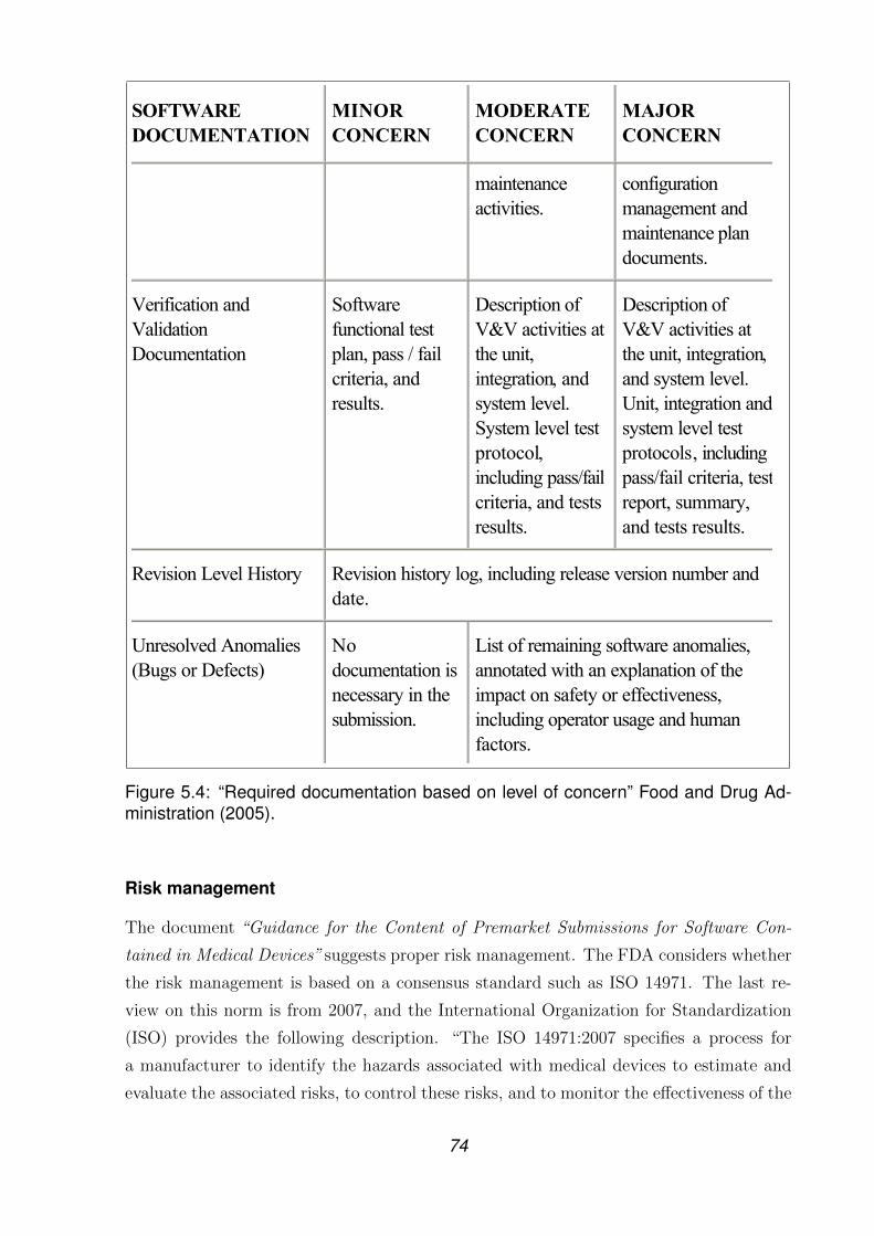
Contains Nonbinding Recommendations
10
SOFTWAREDOCUMENTATION
MINORCONCERN
MODERATECONCERN
MAJORCONCERN
maintenanceactivities.
configurationmanagement and maintenance plan documents.
Verification and ValidationDocumentation
Softwarefunctional test plan, pass / fail criteria, and results.
Description of V&V activities at the unit, integration, and system level. System level test protocol,including pass/failcriteria, and tests results.
Description of V&V activities at the unit, integration,and system level. Unit, integration and system level test protocols, including pass/fail criteria, test report, summary, and tests results.
Revision Level History Revision history log, including release version number and date.
Unresolved Anomalies (Bugs or Defects)
Nodocumentation is necessary in the submission.
List of remaining software anomalies, annotated with an explanation of the impact on safety or effectiveness, including operator usage and human factors.
Level of ConcernWe recommend that you indicate the Level of Concern for your Software Device, determined before the effects of any mitigations. We recommend that you clearly state which one of the three levels of concern is appropriate for your device and include documentation of the rationalefor your decision. We also recommend that your documentation make your decision-makingprocess apparent to FDA.
Software DescriptionWe recommend that you provide a comprehensive overview of the device features that are controlled by software, and describe the intended operational environment. Generally, we
Figure 5.4: “Required documentation based on level of concern” Food and Drug Ad-ministration (2005).
Risk management
The document “Guidance for the Content of Premarket Submissions for Software Con-
tained in Medical Devices” suggests proper risk management. The FDA considers whether
the risk management is based on a consensus standard such as ISO 14971. The last re-
view on this norm is from 2007, and the International Organization for Standardization
(ISO) provides the following description. “The ISO 14971:2007 specifies a process for
a manufacturer to identify the hazards associated with medical devices to estimate and
evaluate the associated risks, to control these risks, and to monitor the effectiveness of the
74

controls. The requirements of ISO 14971:2007 are applicable to all stages of the life-cycle
of a medical device” International Organization for Standardization (2007).
ISO 14971:2007(E)
6 © ISO 2007 – All rights reserved
NOTE 2 A schematic representation of the risk management process is shown in Figure 1. Depending on the speci�c life-cycle phase, individual elements of risk management can have varying emphasis. Also, risk management activities can be performed iteratively or in multiple steps as appropriat e to the medical device. Annex B contains a more detailed overview of the steps in the risk management process. Compliance is checked by inspection of appropriate documents.
Figure 1 — A schematic representation of the risk management process Figure 5.5: Risk management. “A schematic representation of the risk managementprocess” British Standards Institute Staff, British Standards Institution and InternationalOrganization for Standardization (2009).
Risk management is an extensive topic, and we only present the risk management
process graphically in this work to provide an overview. For more detailed information,
75

we refer to specialist literature, such as the ISO 14971:2007 norm, and the Figure 5.5 is
cited form this standard.
Quality system regulation The FDA has published regulations that address Quality
system regulations that concern all manufacturers of medical devices. Part 820 of the
FDA regulation deals with Quality system regulations, and is also known as current good
manufacturing practice (CGMP). The regulations are only legal for device manufactur-
ers who sell a finished device. Paragraph 820.30(l) defines the term “finished device” as:
“Finished device means any device or accessory to any device that is suitable for use
or capable of functioning, whether or not it is packaged, labeled, or sterilized.” Every
manufacturer must hold a quality system manual, and hence established medical device
producers already have one, and the quality system manual should include the design
control process. The design input and output are two important elements in the design
control process, and the manufacturer has to comprise them. The depth of the quality
system implementation is up to the manufacturer, but the quality system manual should
disclose how the company reaches its quality goals. Therefore, the goals should be de-
scribed for the design and building phase, and the goals for quality control at the end of
production should be specified in the manual. It is important to note that the quality
system should be well documented in case the FDA audits the manufacturer. The con-
clusion for research departments like the Wadsworth Center is that it is compulsory for
them to develop a quality system manual if they sell a medical device. Also, if they intend
is to develop and test prototypes, and the device itself is manufactured by someone else,
a quality system manual is required for them. Therefore, the next paragraph deals with
software validation, which includes design control process.
General Principles of Software Validation
This guidance is valid for almost every software package used in the context with a medi-
cal device. The guidance documents also address stand-alone software that is considered
a medical device. Software validation is part of Design control, which is a part of Qual-
ity system regulation. In other words, “TITLE 21 of FOOD AND DRUGS; CHAPTER
I-PART 820 QUALITY SYSTEM REGULATION” says that: “each manufacturer shall
establish and maintain a quality system that is appropriate for the specific medical de-
vice(s) designed or manufactured, and that meets the requirements of this part” Food and
Drug Administration (2009b). The subpart C “Design Controls” includes under 820(g)
“Design validation”, which every manufacturer shall establish, and the next paragraph
explains this topic in more detail.
The guideline aims to support the requirements for software validation system. Software
76

validation is an important part of guidance ”Software in the context of medical device” in
Section 5.2.3, and hence the document contains itemization of the acceptable elements for
the validation of software. ”Planning, verification, testing traceability, configuration man-
agement and many other aspects of good software engineering discussed in this guidance
are important activities that together help to support a final conclusion that software is
validated” Food and Drug Administration (2002).
Design control After the concept phase, the product development process reaches the
design control phase, and therefore the ideas resulting from the concept phase must be
documented and formalized to form a safe product. This process is called design con-
trol and should take place after the concept was developed and before clinical trails are
starting. The FDA published the manual “Design Control Guidance For Medical Device
Manufacturers” in 1997 and the following paragraph gives an overview of the guidance.
The figure 5.6 depicts the design control process, which consists of two closed loop pro-
cesses.
1. Verification loop: design input Ü design process Ü design output
2. Validation loop: user needs Ü verification loop Ü medical device
Design control forms a controlled method to key out the product requirements and inputs.
These input parameters are converted into engineering language by using the product
specifications, and these specifications are transformed into prototypes and or implemen-
tations. The design outputs of the software are created either from the prototypes or
implementations, and the inputs and outputs are linked up in a loop structure. The Fig-
ure 5.6 depicts this closed loop system, which contains design input, design output and
verification.
77

...070mcu/egami/stnemucod/cilbup-vogadf/spuorg/mcu/vog.adf.www//:ptth)slexip 304x575 ,egamI FIG( fig.536070mcu
1 of 1 8/23/2010 4:42 PM
Figure 5.6: Design Control. “The figure illustrates the influence of design controls on adesign process, which is an example for a traditional waterfall model” Food and DrugAdministration (2009b).
The documentation of these reviews in the product development process are funda-
mental and mandatory for the manufacturer. The verification process ensures that the
suitability of the design satisfied the needs of the health care personnel and patients. The
FDA requires reporting of the iterative steps, and the validation must include a complete
testing series that shows that the specifications and requirements matches each other. The
next step after verification is software validation, which assures that the prototype meets
the requirements of the user needs. Hence, the term validation is used for testing trials
performed with a prototype to evaluate whether the prototype matches the user needs.
Therefore, the validation should involve the target user and simulate the environment
normally used. When the software validation is finished, the design can be transferred to
manufacturing phase. This step also needs ongoing reviews and documentation.
78

5.3 Sequence of action
This section addresses the guidance documents and regulations regarding both diagnostic
test SIGFRIED and EEG LI test. In particular, we define three classifications for each
development result.
1. Medical device class
2. Predicate devices (“substantially equivalent”)
3. Level of concern
In our case, we already use an FDA approved device for signal acquisition, but two clini-
cal diagnostic tests involve new signal processing software. Both development results use
FDA approved signal acquisition devices that consist of hardware and software; hence,
there are two possible ways to obtain an FDA approval for both clinical diagnostic tests.
Scenario I to cooperate with a signal processing device manufacturer and sell the devel-
opment result as an upgrade of the existing software operating system.
Scenario II to sell the development result as stand alone software.
Both cases need an FDA approval certificate. As described earlier, a class I and II medi-
cal device is qualified for the 510k application process. First, we will determine the class
of each innovation and then we will look for a “substantially equivalent” device on the
market (predicate devices). The level of concern is the last topic, which is discussed for
each research outcome.
FDA approval requirements for EEG LI test As the title suggests, this development
result is based on the Electroencephalograph (EEG) (see Section 2.1.1, which is available
in many hospitals and through private physicians. EEG recording has been well known
for many years, and there are already several EEG-based devices on the market. Also,
the FDA developed part 882.1400 exclusively for EEG, and thus the development result
fits into this part, which is displayed in Figure 5.7.
79

Quick Links: Skip to main page content Skip to Search Skip to Topics Menu Skip toSection Content Menu Skip to Common Links
510(k) | Registration &Listing
| AdverseEvents
| Recalls | PMA | Classification | Standards
CFR Title21
| Radiation-EmittingProducts
| X-RayAssembler
| MedsunReports
| CLIA
New Search Help |
More About 21CFR[Code of Federal Regulations][Title 21, Volume 8][Revised as of April 1, 2009][CITE: 21CFR882.1400]
TITLE 21--FOOD AND DRUGSCHAPTER I--FOOD AND DRUG ADMINISTRATIONDEPARTMENT OF HEALTH AND HUMAN SERVICES
SUBCHAPTER H--MEDICAL DEVICES
PART 882 -- NEUROLOGICAL DEVICES
Subpart B--Neurological Diagnostic Devices
Sec. 882.1400 Electroencephalograph.
(a)Identification. An electroencephalograph is a device used tomeasure and record the electrical activity of the patient's brainobtained by placing two or more electrodes on the head.
(b)Classification. Class II (performance standards).
.www//:ptth12 eltiT snoitalugeR laredeF fo edoC - RFC accessdata.fda.gov/scripts/cdrh/cfdocs/cfcfr/CFRSearch.cfm?...
1 of 1 8/25/2010 3:16 PM
Figure 5.7: Medical device class of electroencephalograph. Electroencephalograph iscategorized as class 2 medical device.
We know now that EEG LI test is most likely considered a class II medical device,
and the next step is to look for “substantially equivalent” devices. The FDA provides
the“ 510(k) premarket notification database”online (http://www.accessdata.fda.gov/
scripts/cdrh/cfdocs/cfpmn/pmn.cfm) and there are two search options available, ad-
vanced and simple. The outcome of a search is a list with all premarket notifications that
match the keywords, and the list consists of four columns. The last column includes a
link to the summary or statement of the 510(k), which is typically a separate PDF file.
The summary or statement always contains the paragraph “indications for use” and this
description should be “substantially equivalent” to the research outcome.
For the scenario I, we need to find a relevant 510(k) description of a signal acquisition
device. The data which supported the development of EEG LI test is recorded with a
signal acquisition device from the company g.tec Guger Technologies OG. The BCI lab
at the Wadsworth has a strong relationship with this company through many years of
collaboration. Hence, the lab has a solid foundation to cooperate with g.tec to launch this
diagnostic test on the market. The figure 5.8 shows the intended use of the signal acquisi-
tion device g.USBamp, and the description of the intended use opens up the possibility of
including the EEG LI test as an additional feature to the existing software of the device.
The regulations listed in section 5.2.2 require a submission of a premarket notification in
that specific case.
80
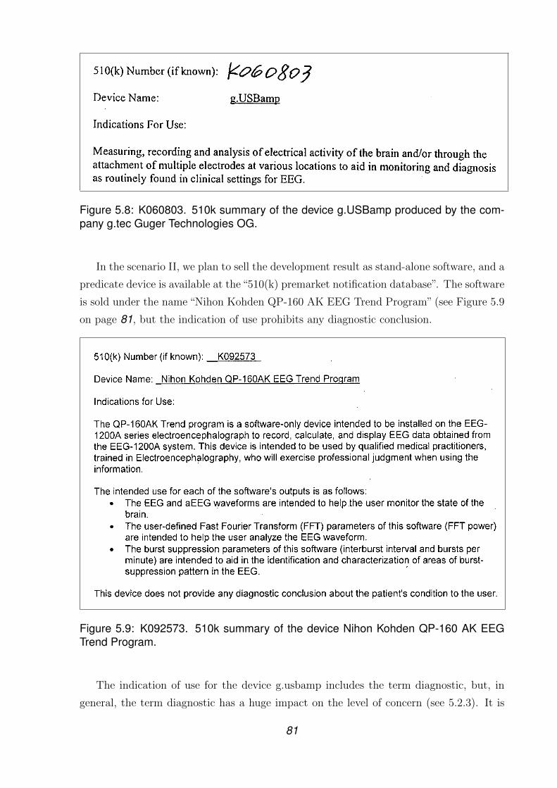
Figure 5.8: K060803. 510k summary of the device g.USBamp produced by the com-pany g.tec Guger Technologies OG.
In the scenario II, we plan to sell the development result as stand-alone software, and a
predicate device is available at the“510(k) premarket notification database”. The software
is sold under the name “Nihon Kohden QP-160 AK EEG Trend Program” (see Figure 5.9
on page 81, but the indication of use prohibits any diagnostic conclusion.
Figure 5.9: K092573. 510k summary of the device Nihon Kohden QP-160 AK EEGTrend Program.
The indication of use for the device g.usbamp includes the term diagnostic, but, in
general, the term diagnostic has a huge impact on the level of concern (see 5.2.3). It is
81

clear that the diagnostic test does not have a major level of concern, but the distinction
between moderate versus minor level of concern needs a closer explanation. EEG LI test
aims to provide information for a medical doctor, and it is up to the doctor to what extent
the information influences the diagnosis. We recommend that the application include a
statement from a medical doctor that answers this question, and the inventor should
decide carefully whether or not the indication of use includes a diagnostic purpose. The
second question for determining the moderate level of concern deals whether the software
accessorizes a moderate level of concern device. The innovation does not control any
device function, and does not provide input information for any medical device. The
outcome of the software is displayed on a third party monitor, so this question can be
answered in the negative.
Figure 5.10: K002631. 510k summary of the device eemagine EEG software.
During our premarket notification database research, we found two additional 510ks
that can support the submission for EEG LI test. They are presented in Figures 5.10 and
5.11.
82

Figure 5.11: K991054. 510k summary of the device Natus Neuroworks, Model 104196.
FDA approval requirements for SIGFRIED The novel functional brain mapping tech-
nology SIGFRIED uses data recorded from an implanted electrode, and the implantation
procedure is exclusively performed in a hospital environment. The signal acquisition pro-
cess (see Figure 2.2 on page 10) consists of two class II medical devices. The first device
is the aforementioned data amplifier g.USBamp (see Figure 5.8 on page 81), and the
second is the implanted cortical electrode used to record data from the cortex. For the
first recordings, we used subdural electrodes from the company Ad-tech, and the 510k of
these devices is depicted in Figure 5.12 on page 84.
83

Figure 5.12: K053363. 510k summary of the device subdural electrodes.
The FDA created the section 882.1310 for cortical electrodes which categorizes a cor-
tical electrode as a class II device and the Figure 5.13 on page 84 illustrates this section.
Quick Links: Skip to main page content Skip to Search Skip to Topics Menu Skip toSection Content Menu Skip to Common Links
510(k) | Registration &Listing
| AdverseEvents
| Recalls | PMA | Classification | Standards
CFR Title21
| Radiation-EmittingProducts
| X-RayAssembler
| MedsunReports
| CLIA
New Search Help | More About 21CFR
[Code of Federal Regulations][Title 21, Volume 8][Revised as of April 1, 2009][CITE: 21CFR882.1310]
TITLE 21--FOOD AND DRUGSCHAPTER I--FOOD AND DRUG ADMINISTRATIONDEPARTMENT OF HEALTH AND HUMAN SERVICES
SUBCHAPTER H--MEDICAL DEVICES
PART 882 -- NEUROLOGICAL DEVICES
Subpart B--Neurological Diagnostic Devices
Sec. 882.1310 Cortical electrode.
(a)Identification. A cortical electrode is an electrode which istemporarily placed on the surface of the brain for stimulating thebrain or recording the brain's electrical activity.
(b)Classification. Class II (performance standards).
.www//:ptth12 eltiT snoitalugeR laredeF fo edoC - RFC accessdata.fda.gov/scripts/cdrh/cfdocs/cfcfr/CFRSearch.cfm?...
1 of 1 8/25/2010 4:16 PM
Figure 5.13: Medical device class of cortical electrode. Cortical electrode is catego-rized as class 2 medical device.
The last paragraph verifies that there are only class II used in combination with
SIGFRIED. The innovative functional brain mapping technique SIGFRIED contains sig-
nal processing software, like the already described EEG LI test. There are no differences
evident in the content of the classification of the medical device between these two diag-
nostic tests, so SIGFRIED is eligible for the category medical device class II.
The next important classification for software in content with a medical device is the“level
of concern”(see 5.2.3), so we explain the usage of the software more precisely. Currently,
SIGFRIED is used as a pre-detection of brain functions and the result of the pre-detection
supports the sequence of the Electrocortical stimulation(see Section 2.2.1. In other words,
84

SIGFRIED is not used for diagnosis, and no conclusions about the patient’s conditions
are based on the outcome of SIGFRIED. The outputs of “Sigfied” are colored figures of
brain function, which are displayed on a third party monitor, and the innovation does not
support information for any device. These facts indicate that SIGFRIED can be catego-
rized as Minor level of concern.
The last step is to assess whether the research outcome is authorized for the premarket
notification (see Section 5.2.2). Therefore, the key element is to show that the innova-
tion is not considered a novel technology by the FDA, because then the inventor must go
trough the more complex pre-marketing approval process or through the “Evaluation of
automatic class III designation provision” process. The best way to verify that the de-
velopment result is not a novel technique is to find matching predicate approved medical
devices. Every named predicate device for the EEG LI test is applicable for SIGFRIED
too. In particular, the indication of use for the device eemagine EEG software (see Figure
5.10 on page 82) sounds qualified as a predicate device.
Figure 5.14: K050833. 510k summary of the device Electrode Junction Box, JE-921 Aserie.
We also note the 510k of the device Electrode Junction Box (see Figure 5.14) to
provide a complete overview of all relevant predicated devices for the research outcome
SIGFRIED.
85

Time line and cost estimation The table 5.1 list all task to prepare a FDA approval
application and DI Brunner, software engineer at the Wadsworth center estimated the
required times to accomplish the tasks.
Task EEG LI test SIGFRIEDOverhaul the GUI 0.30 0.25Level of Concern 0.30 0.20Software Description 0.10 0.10Device Hazard Analysis 0.15 0.30Software Requirements Specification 0.20 0.20Traceability Analysis 0.15 0.15Verification and Validation Documentation 0.35 0.35Revision Level History 0.10 0.10Total effort to prepare diagnostic tests’s SW for the FDA ap-proval
1.65 1.65
All number have the unit: [years/employee]
Table 5.1: Software development effort estimation. The Table estimates the develop-ment effort to prepare the software SIGFRIED and EEG LI test for the Food and DrugAdministration approval application. The numbers represent Person Years requiredfrom software engineers.
For both development results 1.65 Person Years are necessary to prepare the Premarket
notification application.
The next citation shows the duration time of a Premarket notification application. “FDA’s
fiscal year 2009 goal is to review and decide on 90 percent of 510(k) submissions within
90 days and 98 percent of them within 150 days. The comparable goals for a Premarket
Approval Application (PMA) is to review and decide upon 60 percent of original PMA
submission in 180 days and 90 percent of them within 295 days” Crosse (2009). 180 day
equal 0.25 years thus the total time effort for the FDA approval is 1.9 Person years.
The table 5.2 shows the total mount of money, that the Wadsworth center spends to
employ a software engineer with the grade P25. The specification for a p25 grade software
engineer are a Master degree and several years of experience.
Software engineerBase Salary Fringe rate Fringe cost Indirect cost Total90,844 37.50% 34,067 85,064 209,975
Table 5.2: Software engineer salary. All numbers have the unit USD per year.
The required time multiplied by the salary equals the total personal cost of the certi-
fication.
$209,975 ∗ 1.9 = $398,953
86

“For the applicant, the standard fee provided to FDA at the time of submission is also
significantly lower for a 510(k) submission than for a PMA submission. In fiscal year
2009, for example, the standard fee for 510(k) submissions is $3,693 while the standard
fee for original PMA submissions is $200,725” Crosse (2009).
$398,953 + $3,693 = $402,646
The last equation sums the personal cost and the applications fee that are the total
expenses for the certification of one clinical diagnostic test.
5.4 Summary of regulatory environment
For commercialization, every medical device must be approved by the Food and Drug
Administration. The FDA categories the medical device into class, which decide the
volume of certification process. However, both clinical diagnostic tests are considered
as class two medical device and they are qualified for a Premarket Notification (510k)
application, which is based on a predicate device. This chapter list suitable predicate
devices for both development results.
For devices that contain software, the FDA requires additional documentation and the
volume is based on the level of concern, which is independent of the medical device
class. For both development results, the classification in either minor or moderate level
of concern is unclear. If these tests are performed in addition to the established methods
than minor concern is reasonable. If these tests replace the established methods than
moderate level of concern is suitable, which required more documentation and detailed
safety tests.
In conclusion, the FDA guidelines are precise thus the whole application process and
required documentation are clear and transparent for both tests. The total costs for the
certification of one test are $402,646.
87

6 Reimbursement policy of a medicaldevice in US
Chapter 4 evaluated the market demand, particularly the available market of each de-
velopment result. Chapter 5 explained all necessary milestones to gain market clearance
for a medical device which allows the manufacturer to commercially distribute the device
within a US market. This chapter deals with the reimbursement policies of the US medi-
care companies, which cover the treatment costs of their clients and the chapter’s aim is
to clarify whether it is possible to obtain reimbursement of the development results. Fur-
thermore, this chapter talk about the Medicare coverage determination in general. The
final chapter of this thesis discusses the purchasing process between manufacturer and
hospital, which is the overall goal of the manufacturer.
The chapter market of development results explained the singularity of the health care
field, which basically means that the payer for the treatment expenses is typically not the
patient. Instead, a private or government-run insurance plan reimburses the hospital or
physician for the performed medical, surgical, and diagnostic services. Hence, the success
of an innovation in the medical device field depends heavily on whether the insurance com-
panies cover the treatment that is performed with the recently developed medical device.
There is a large time and cost-saving potential if the manufacturer considers this aspect
early in the innovation process. Hence, the inventor should be aware of relevant coverage
regulations during the development phase of an novel medical device so that the design of
the development result meets the reimbursement policy. Also, the documentation of each
design step and trial should match the coverage requirements in the first place to ensure
a fast and cost-effective innovation process.
This chapter highlights the important factors for the medicare coverage decision in the
United States. In particular, we address the coverage conditions of Center of Medicare
and Medicaid Services, which is the nation’s largest health insurance program, as ex-
plained in Section 6.1. The Section 6.2 outlines the three different reimbursement codes
e.g., International Statistical Classification of Diseases 9th rev. and Current Procedural
Terminology. The Section 6.2.2 focuses on the Current Procedural Terminology codes,
88

because this reimbursement code is adequate for SIGFRIED and EEG LI test
In the reimbursement field, the three major objectives for an innovative medical device,
which are essential for the overall success of research outcome, are:
1. Positive coverage decision
2. Assigned reimbursement code
3. Adequate amount that can be reimbursed
At first glance, these goals may give the impression that reimbursement coverage is simple
and straightforward. In reality, it is a complex and time-consuming undertaking. Gre-
gory Raab states these reimbursement challenges very well in the paper “From Medical
Invention to Clinical Practice: The Reimbursement Challenge Facing New Device Proce-
dures and Technology-Part 1: Issues in Medical Device Assessment”, and the following
paragraph is cited from this article.
“Insurer coverage and payment processes not only determine whether current tech-
nologies will be made available to patients; they also create a climate that can provide
incentives or disincentives for manufacturers to innovate in the first place. This was
documented in a 2000 a medical device industry study conducted by the Lewin Group,
which was based on a survey of device manufacturers in the United States, an analysis
of secondary research information, and confidential interviews with industry executives,
security analysts, and other informed observers. The Lewin survey identified Medicare
coverage and payment processes as often being “inconsistent and confusing” and noted
that, although manufacturers express similar views about private health insurer coverage
and payment processes, “concerns regarding Medicare are particularly acute, reflecting
the program’s size and scope, as well as the program’s influence on payment policy in all
sectors of the health care market”.
In examining Medicare’s coding, coverage, and payment processes, the Lewin Group’s
study found that the systems for making these decisions are “separate and largely un-
coordinated”; manufacturers are required to negotiate “multiple, distinct, and complex
processes”. As a result, Lewin found that it can take the Centers for Medicare & Med-
icaid Services officials from 15 months to 5 years to add new medical technologies to
the Medicare program. The time it takes manufacturers to manage these reimbursement
hurdles is particularly troublesome because most medical devices have life spans of only
12 to 18 months. Most device manufacturers surveyed by Lewin felt that the Medicare
coverage and payment processes were not clear, transparent, predictable, or consistently
and fairly applied” Raab and Parr (2006a).
89

6.1 Center of Medicare & Medicaid Services
The Center of Medicare & Medicaid Services (CMS) plays a major role in the medicare
coverage process because it administers the medicare program of the US. The CMS is part
of the Department of Health and Human Services and is considered a federal agency. TI-
TLE XVIII-HEALTH INSURANCE FOR THE AGED AND DISABLED set up the legal
basis for the medicare program, which is the nation’s largest health insurance program
covering approximately 41 million Americans. “Beneficiaries consist primarily of individ-
uals 65 years of age or older, some disabled people under 65 years of age, and people with
end-stage renal disease (permanent kidney failure treated with dialysis or a transplant)”
Department of Health and Human Services (2003a).
The Center of Medicare and Medicaid Services established a coverage decision process for
devices and treatments. However, the private insurers normally follow the coverage deci-
sions of the CMS. The Center of Medicare and Medicaid Services distinguishes between
two coverage determinations.
1. Local coverage determination (LCD)
2. National coverage determination (NCD)
The local coverage determinations are decided by local contracts, and the vast majority
of determinations are LCD. “Currently, there are in total 6000 LCDs established, but na-
tionwide, only approximately 300 NCDs are installed” Schoonmaker, Bagley and Scanlan
(2002). Especially for a new device, it is common to apply first for local coverage deter-
mination and then consider pursuing an NCD in the future. First, we describe generally
valid aspects for both coverage decisions, then we explain each in detail.
Coverage criteria and requirements In the beginning of this paragraph, the different
interests of the FDA and CMS are explained and afterwards, the coverage criteria and
requirements for a BCI-based medical device are discussed. The FDA approval process
verifies that a service or device is safe and effective, but it does not check whether there is
a more efficient device or service on the market.Generally the CMS accepts only a request
for a device that is FDA approved. In other words, a medical device must have one of
these FDA approvals as a basic prerequisite for a coverage determination:
• Premarket approval (see Section 5.2.1
• Premarket Notification 510(k) (see Section 5.2.2)
• Category B IDE devices (see Section 5.1)
90

• Hospital Institutional Review Board (IRB) approved IDE devices (see Section 5.1)
Unfortunately, the Category A IDE devices are excluded from the coverage decision, as
FDA explains on their Web site, http://www.fda.gov. The relevant parts of the exclu-
sion are summarized in the next paragraph.
“The Medicare program has historically interpreted the statutory terms ”reasonable and
necessary” to mean that a service or medical device must be safe and effective, medically
necessary and appropriate, and not experimental in order to qualify for reimbursement.
For Medicare coverage purposes, the term ”experimental” has been used synonymously
with the term ”investigational.” Therefore, with rare exceptions, an FDA-approved Inves-
tigational Device Exemption (IDE) application served as an indication that the device
was not ”reasonable and necessary” within the meaning of the Medicare program. Thus,
Medicare coverage was denied for devices which were under an IDE and had not yet re-
ceived premarket notification clearance and or premarket approval. There was increasing
recognition, however, that there are devices that are refinements of existing technologies
or replications of existing technologies made by other manufacturers. Many of these de-
vices are under an FDA-approved IDE as a means of gathering the scientific information
needed for the FDA to establish the safety and effectiveness of that particular device, even
though there is evidence that the device type can be safe and effective. Such devices could
be viewed as ”reasonable and necessary” by Medicare and thus be reimbursed if it were
possible to identify these devices to Health Care Financing Administration” cf. American
Medical Association (1995). Therefore the FDA categorizes IDE certifications into two
classes, and only a IDE Category B device can be reimbursed:
• IDE Category A - Experimental
• IDE Category B - Investigational; Non-experimental
What means “reasonable and necessary”? The Medicare program aims to reim-
burse the most efficient treatment, and therefore the CMS bases their decision on “What
is reasonable and necessary” over the last 35 years Department of Health and Human
Services (2003b). However, the CMS made several attempts to describe “reasonable and
necessary” more precisely, but they could not agree on one definition. “Medicare officials
tried to include “cost-effectiveness” and “added value” as coverage criteria in formal regu-
lations governing the Medicare coverage review process. These regulatory attempts were
controversial, because manufacturers and medical groups strongly objected to introducing
cost considerations into coverage decision making. As a result, neither of these regulations
was finalized, and the CMS has decided to forgo overt efforts to add economic consid-
eration to nation coverage decision making. Nevertheless, it is generally acknowledged
91

by CMS staff members that economic and cost considerations are factors considered in
evaluation new technologies and that they are an implicit, if not an explicit, factor in the
decision-making process itself” Raab and Parr (2006a). Which devices are covered by the
Medicare program?
G.Gregory Raab answered the question with the following statement: “To be covered by
Medicare, new medical procedures and technologies must fit within a benefit category
set out in the Medicare statute and be found to be “reasonable and necessary” for the
diagnosis or treatment of illness or injury” Raab and Parr (2006b). As mentioned earlier,
the CMS does not provide a precise description of the term “reasonable and necessary”,
but the CMS published a list of services and devices that are not covered by them. This
list was released as 42 Code of Federal Regulations (CFR) 411.15 in 2004, and the next
list outlines the relevant exclusions for the field BCI:
1. Routine physical checkups such as:
(a) performed for a purpose other than treatment and diagnosis
(b) required by insurance companies or government agencies.
2. Personal comfort service, except as necessary for the palliation or management of
terminal illness.
3. Any services that are not reasonable and necessary for following purpose
(a) For the diagnosis or treatment of illness or injury or to improve the functioning
of a malformed body member.
(b) In the case of hospice services, for the palliation or management of terminal
illness.
4. Experimental or investigational devices, except for certain devices-
(a) Categorized by the FDA as a nonexperimental/investigational (Category B)
device
(b) Furnished in accordance with the FDA-approved protocols governing clinical
trials.
5. Services furnished to SNF residents: Any physical, occupational, or speech-language
therapy services, regardless of whether the services are furnished by (or under the su-
pervision of) a physician or other health care professional, and regardless of whether
the resident who receives the services is in a covered Part A stay.
92

Maintained by the CMS, the Medicare Coverage Database (MCD) contains all Local
Medical Review Policies (LMRPs), Local and National Coverage Determinations. The
CMS’s Web site, http://www.cms.gov/mcd/search.asp?from2=search.asp&, provides
access to this database, which is the official source of all local payment policies around the
US. The Medicare Coverage Database also contains all local medicare contractors that
follow the Local Coverage Determinations and Local Medical Review Policies.
Published study In general, published studies have a large impact on the coverage
determination. The CMS and also the local medicare companies review the relevant
published papers and the conclusion of the papers are considered in the decision. Hence
published papers support the coverage application and the Figure 6.1 depicts hierarchy
of the paper’s impact.
"Gold Standard"
controlled study published
in US peer - reviewed journal
Randomized placebo - controled study accepted for publication
Open trial, retrospective published peer - reviewed journal
Registry data published US peer - reviewed journal
Figure 6.1: Hierarchy of papers. This figure displays the impact factor of publishedstudies in the medicare coverage determination.
The gold Standard are controlled study published in US peer-reviewed journals and
the coverage determination statement form the insurance companies contain a list of
references, which shows that papers
6.1.1 Local coverage determination
According to the CMS, a Local coverage determination is defined as: “Local coverage de-
termination (LCD) means a decision by a fiscal intermediary or a carrier under Medicare
Part A or Part B, as applicable, whether to cover a particular service on an intermediary-
wide or carrier-wide basis in accordance with section 1862(a)(1)(A) of the Act. An LCD
93

may provide that a service is not reasonable and necessary for certain diagnoses and/ or
for certain diagnosis codes. An LCD does not include a determination of which procedure
code, if any, is assigned to a service or a determination with respect to the amount of
payment to be made for the service” Department of Health and Human Services (2003a).
Medicare Part A, B and C are also defined by the CMS, but the Medicare program reim-
burses only treatments that are contained in Part A or B.
Part A:“The hospital insurance program covers certain care provided to inpatients in
hospitals, critical access hospitals, skilled nursing facilities, as well as hospice care and
some home health care” cf. Department of Health and Human Services (2003a).
Part B:“The supplementary medical insurance program covers certain physicians’ ser-
vices, outpatient hospital care, and other medical services that are not covered under
Part A” cf. Department of Health and Human Services (2003a).
Part C:“Known as the Medicare+Choice (M+C) program, this provides beneficiaries
with various options, including the right to choose a Medicare managed care plan or a
Medicare private fee-for-service plan. Under the M+C program, an individual is entitled
to those items and services (other than hospice care) for which benefits are available under
Part A and Part B. An M+C plan may provide additional health care items and services
that are not covered under the original Medicare program” cf. Department of Health and
Human Services (2003a).
“CMS has delegated authority to Medicare contractors to develop and issue contractor-
specific policy that identify the circumstances under which particular items or services will
be covered in a geographic area. Medicare contractors typically have developed policies in
the form of local medical review policies (LMRPs), which include four different types of
provisions-coding, benefit category, statutory exclusions, and medical necessity provisions
(i.e., those provisions interpreting the reasonable and necessary provision of the Medicare
statute)” Scherb and Kurlander (2006)
6.1.2 National coverage determination
National Coverage Determination is a long-lasting application process, and uncommon for
a new technology or device. Hence, the usual route for a new device is to apply for a Local
Coverage Determination and subsequently for a National Coverage Determination. This
paragraph explains only the basics of the NCD process, because a manufacturer normally
does not consider an NCD for an innovation.
The Center of Medicare and Medicaid Services formulated a guideline for the National
Coverage Determination, and this guideline is modeled on the guidance documents from
the FDA. The Federal Register volume 68, number 187 published this document under
the title: “Medicare Program; Revised Process for Making Medicare National Coverage
94

Determinations” in 2003, and the purpose of this document is to outline the NCD pro-
cess. Hence these documents contain information that is relevant for the diagnostic tests,
including a definition of the term National Coverage Determination: “(NCD) means a
decision that CMS makes regarding whether to cover a particular service nationally under
title XVIII of the Act. An NCD does not include a determination of what code, if any, is
assigned to a service or a determination with respect to the amount of payment to be made
for the service ” Department of Health and Human Services (2003a). Next, we describe
the necessary components of an NCD submission. The application must be submitted
in written and electronic form, and it must be declared as “formal request for an NCD”.
Furthermore, the submission must include a statement from the requestor that explains
which benefit category of the Medicare program is appropriate for the device. In addition,
there are several required documents, displayed in the next list based on Department of
Health and Human Services (2003b):
1. Detailed description of the treatment or device
2. Estimation of the target Medicare population and explanation of the considered
population
3. Any information regarding the medical benefit of the treatment or device
4. Explanation of whether the device is operated by a physician or patient, and details
of the procedures that involve the device (design, purpose and method)
5. A statement from the requestor (in cases in which there is an aggrieved party, the
statement must be from that party) containing the following:
(a) An explanation of the relevance of the evidence selected
(b) Rationale for how the evidence selected demonstrates the medical benefits for
the target Medicare population
(c) Information that examines the magnitude of the medical benefit
(d) Reasoning for how coverage of the item or service will help improve the medical
benefit to the target population
(e) In the case of an aggrieved party, how that party is “in need” of the item or
service.
6. A description of any clinical trials or studies currently underway that might be
relevant to a decision regarding coverage of the item or service
95

7. Information involving the use of a drug or device subject to FDA regulation as well
as the status of current FDA regulatory review of the drug or device involved. An
FDA regulated article would include the labeling submitted to the FDA or approved
by the FDA for that article, together with an indication of whether the article for
which a review is being requested is covered under the labeled indication(s)
8. In the case of items that are eligible for a 510(k) clearance by the FDA, identification
of the predicate device to which the item is claimed to be substantially equivalent.
However, a positive NCD was issued for the awake craniotomy, which implants an electrode
grid in the patient’s brain, followed by extended monitoring to diagnose resectable seizure
foci. The statement’s title is “Steroetaxic Depth Electrode Implantation”, and the Figure
6.2 displays the statement.
in the same patient when a satisfactory result has not been achieved with the first cingulotomy. Stereotactic cingulotomy is not covered under Medicare because the procedure is considered to be investigational. 160.5 - Steroetaxic Depth Electrode Implantation (Rev. 1, 10-03-03) CIM 50-40 Stereotaxic depth electrode implantation prior to surgical treatment of focal epilepsy for patients who are unresponsive to anticonvulsant medications has been found both safe and effective for diagnosing resectable seizure foci that may go undetected by conventional scalp electroencephalographs (EEGs). The procedure employs thin wire electrodes which are implanted in the brain of the focal epileptic patient for EEG monitoring. By taking several readings during seizure activity, the location of the epileptic focus may be found, so that better informed decisions can be made regarding the surgical treatment of persons with intractable seizures. 160.6 - Carotid Sinus Nerve Stimulator (Rev. 1, 10-03-03) CIM 65-4 Implantation of the carotid sinus nerve stimulator is indicated for relief of angina pectoris in carefully selected patients who are refractory to medical therapy and who after undergoing coronary angiography study either are poor candidates for or refuse to have coronary bypass surgery. In such cases, Medicare reimbursement may be made for this device and for the related services required for its implantation. However, the use of the carotid sinus nerve stimulator in the treatment of paroxysmal supraventricular tachycardia is considered investigational and is not in common use by the medical community. The device and related services in such cases cannot be considered as reasonable and necessary for the treatment of an illness or injury or to improve the functioning of a malformed body member as required by §1862(a)(1) of the Act. Cross-reference: The Medicare Bene�t Policy Manual, Chapter 15, “ Covered Medical and Other Services,” §120 The Medicare Bene�t Policy Manual, Chapter 1, “Inpatient Hospital Services,” §40 and §120. 160.7 - Electrical Nerve Stimulators
Figure 6.2: Medicare national coverage determination. “The document Medicare Na-tional Coverage Determinations Manual Chapter 1, Part 2 (Sections 90 - 160.26) Cov-erage Determinations includes the coverage determinations dealing with electrocorti-cal stimulation. Hence, this document contains also the statement about electrocorticalstimulation for the purpose of functional brain mapping in content of epilepsy surgery”cf. Center of Medicare & Medicaid Services (2010).
As mentioned in the Section 2.2.1 electrocortical stimulation is well established around
the USA, so the CMS decided to cover this treatment. The document also includes
decisions about the use of electrocortical stimulation for another purpose: functional
brain mapping. For example, “Treatment of Motor Function Disorders with Electric
Nerve Stimulation” is not covered by the CMS, and this statement and other relevant
statements can be found in the appendix.
96
6.1.3 Sequence of action
Our plan to seek reimbursal is as follows. Notably, no further technical development or
preparatory research is needed. Hence, our first step is to get our two development results
classified as IDE Category B devices, and then seek approval for an approved clinical
trial. We will also need to demonstrate that the procedure does not cost more than a
conventional procedure, which is already demonstrated in the chapter 3. Next, we will
approach local insurance companies to see if they will reimburse us for our clinical trials.
This plan is based partly on “Medicare Benefit Policy Manual Chapter 14 - Medical De-
vices”, which describes the items that may influence an insurer’s decision regarding reim-
bursal. This text is reviewed below, along with our approach to addressing each item.
For dates of service on or after November 1, 1995, Medicare may cover certain FDA-
approved and Institutional Review Board (IRB) approved investigational devices and
services, provided the investigational device meets certain conditions. The box below
copies the text describing these conditions, followed by italicized text that describes how
we will address them.
• “Appears on the listing of devices eligible for coverage/payment on CMS’ master file
of IDE devices” Center of Medicare & Medicaid Services (2010). We will ask the
FDA to add our device to this list;
• “Is reasonable and necessary for the individual patient” Center of Medicare & Medi-
caid Services (2010). We have published articles that prove that our clinical diagnos-
tic tests are reasonable and necessary. Determinations for individual patients will
be made through practicing neurosurgeons who work with many potential patients,
such as Dr. Ritaccio;
• “The device or services associated with the use of a device were provided to the
beneficiary within the start and end dates contained in the master file” Center of
Medicare & Medicaid Services (2010). We will be responsible for ensuring that our
work is conducted within the appropriate time span
• “There is no national coverage policy that would otherwise prohibit Medicare cover-
age” Center of Medicare & Medicaid Services (2010). We have already explored this
question, but will reconfirm it as needed.
However, the preceding list only describes conditions that must be met before Medicare
may cover relevant expenses. That is, insurers are not required to cover costs relating to
any device or study that meets those conditions. The same document then lists criteria
that “must also be applied when making coverage determinations on FDA-approved IDE
Category B devices.” This list is reproduced here, along with italicized text that we added:
97

• The device must be used within the context of the FDA-approved clinical trial. We
will seek this FDA approval ;
• The device must be used according to the clinical trial’s approved patient protocols.
We will develop and heed appropriate protocols, drawing on our extensive experience
with clinically oriented research;
• There may be an established national policy as contained in existing manual in-
structions, e.g., National Coverage Determinations Manual instructions, etc. We
noted existing tests above, but there is nothing unique to our work;
• In the absence of national policy, there may be a local policy for similar FDA-
approved devices. We are not aware of any such local policies, but will check again
as needed;
• There may be Policy/Position papers or recommendations made by pertinent na-
tional and/or local specialty societies. We are not aware of any such national or
local societies that would issue relevant policies, but will check again as needed. In-
deed, there has been some discussion of forming a BCI Society that could establish
relevant policies, and we are in contact with the organizers. ”
As noted, these criteria must be considered, but reimbursal still depends on local
contractors’ decisions. This is addressed in the book “Overview of Medicare Coverage of
Clinical Trials”: “Devices used pursuant to an FDA-approved IDE that are classified as
Category B are eligible for full Medicare coverage. Unlike the limited Medicare coverage
afforded by the routine costs policy, the Category B IDE policy authorizes Medicare
coverage for the investigational item itself as well as for routine costs associated with the
clinical trial. The investigational nature of Category B devices is neither sufficient reason
to grant nor deny Medicare coverage. Such devices are covered only if they meet all other
Medicare coverage requirements and coverage is not precluded by a national noncoverage
policy. Although a Medicare contractor may deny coverage for several reasons, it cannot
refuse to cover routine costs because of the investigational nature of the use of the device”
cf. Becker and Whyte (2006)
6.1.4 Summary of the reimbursement coverage determination
Because the market for an medical device in the BCI field is limited, it may not be suitable
to apply for a NCD, which requires elaborate documents. For SIGFRIED, the NCD of
the Steroetaxic Depth Electrode Implantation (see Figure 6.2 on page 96) ensures that
physician will be reimbursed for the implantation of grid electrodes in epilepsy patients’
98

brains. Thus, additional functional brain mapping with SIGFRIED takes little effort, and
SIGFRIED’s result can significantly improve the outcome of the epilepsy surgery.
Because the local insurance companies never developed a detailed and generally valid
guideline that applies to all local insurance companies, it is very difficult to predict the
outcome of a typical LCD and to be on top of the LCD procedure. Thus, for the two
diagnostic tests, it is necessary to contact the local insurance companies to determine
whether they would cover the tests.
6.2 Reimbursement code
The last section discussed the medicare coverage process. Notably, a coverage decision
does not determine the amount of reimbursal. The reimbursement code assigns the amount
of money which can be reimbursed by a Medicare carrier, and thus the reimbursement
code process has a large impact on the innovation process. For any innovation, a key goal
is establishing an adequate amount of money that the hospital or physician receives for
a treatment or diagnosis. “If a new procedure involves more costly equipment, is more
difficult to perform, or requires more skill than current procedures, new codes are a nec-
essary precondition for the new procedure’s securing a higher payment rate. New codes
also spur insurers to consider whether the new procedure should be covered and, if it is
covered, to spell out whether the coverage is limited in terms of patient indications, sites
of care, or qualified providers” Raab and Parr (2006a).
A standardized coding system should also ensure an effective and less bureaucratic reim-
bursement process between insurance companies and health care facilities and practition-
ers. In order to reach this goal, the reimbursement codes contain two essential features.
First, the code can be processed in an online billing system, and hence the whole process
can be done via computer. Second, the reimbursement code states precisely the treat-
ment’s procedure and the amount that can be reimbursed.
The American Medical Association (AMA) plays an important role in the reimbursement
code process, because they are in charge of the Current Procedural Terminology CPT
codes, which are used in the whole country. In addition to the CPT codes, two other
billing codes exist, and all of these codes are shown below.
1. Current Procedural Terminology CPT (identify physician procedure and payment
for outpatient, laboratory and ambulatory surgical fees)
2. The International Statistical Classification of Diseases 9th rev (ICD-9) code (mainly
for inpatient payment)
99

3. Healthcare Common Procedure Coding System (HCPCS) code set (mainly for Medi-
care physician fee and hospital outpatient prospective payment)
Before explaining each payment code system, we present a study that shows the timing
of an application. “The coding decisions for ICD-9 codes, CPT codes, and HCPCS codes
are generally made on an annual basis. The Lewin Group found that it takes a minimum
of 15 months to secure a new code because of filing requirements. However, depending
on the timing of the product launch, requirements associated with requesting a code, and
the time required for new codes to become effective once decisions have been made, it
may take as long as 27 months after a new technology has been cleared by the FDA for
a new code to become effective” Raab and Parr (2006a). First, we address the ICD-9
code system, particularly the term “diagnosis related group (DRG)”. Second, we state the
application process for a new CPT code, and we also provide a selection of CPT codes
that are relevant for our development results.
6.2.1 International Statistical Classification of Diseases 9th rev
code
“Based on the World Health Organization disease classification system, the International
Statistical Classification of Diseases 9th rev code (ICD-9-CM) are the official method
of coding diagnoses and procedures associated with hospital utilization in the United
States. ICD-9-CM codes, Volumes 1 and 2, are used to report a patient’s diagnosis or
condition and are used by third-party payors to determine whether the service or product
is warranted based on the patient’s diagnosis or symptoms. For Medicare billing purposes,
ICD-9-CM Volume 3 codes classify hospital inpatient procedures. These codes are also
included on Medicare claims and drive the payment methodology for acute care hospital
inpatient services. The National Center for Health Statistics and CMS maintain and
annually update the ICD-9-CM procedure codes.” Scherb and Kurlander (2006).
Diagnosis-related group (DRG) The DRG classification system is the most common
system for categorizing urgent care inpatients and evaluating case mix. Case mix is an
average of determining and evaluating the kind of patients a hospital treats. Diagnosis-
related groups cases that are clinically the same and require identical resources. One DRG
is linked to every inpatient stay and DRGs are designated using the principal procedure,
principal diagnosis, age, discharge status and sex. Diagnoses and procedures assigned
by using ICD-9-CM codes determine the DRG assignment. Thus, precise and complete
ICD-9-CM coding by professionals is fundamental for adequate DRG naming and poste-
rior reimbursement”. “Before 1983, Medicare payments for hospital inpatient care were
100

based on a retrospective reasonable cost system, which meant hospitals received 80 per-
cent of reasonable charges. Since 1983, when the patient prospective payment system
was implemented, Medicare has reimbursed hospitals for inpatient hospital services ac-
cording to a predetermined rate for each discharge. Each discharge is categorized into
a diagnosis-related group, which is based on the patient’s principal and secondary diag-
noses (comorbidities and complications) as well as principal and secondary procedures
(if performed). The DRG determines how much payment the hospital receives and it is
worth noting that ICD-9 codes directly affect DRG assignment and CPT codes play no
role in DRG assignments. Diagnosis-related groups are organized into mutually exclusive
categories called major diagnostic categories (MDCs), which are loosely based on body
system (e.g., nervous system)” Green and Rowell (2007).
6.2.2 Current Procedural Terminology CPT
”CPT codes are a coding system, defined in the publication Current Procedural Terminol-
ogy, for medical procedures that allows for comparability in pricing, billing, and utilization
review.” Dictionary (2009) The American Medical Association (AMA) is maintaining the
Current Procedural Terminology codes and holds also the registered trademarks rights of
the CPT. Now a day it is the most common medical nomenclature to account medical
services and treatments under private and public health insurance companies. American
Medical Association (2010a)
“Although Medicare hospital inpatient payment rates are based on ICD-9 codes, Medi-
care’s hospital outpatient payment system uses CPT codes to identify medical services.
These outpatient services are grouped into ambulatory payment classifications, and CMS
sets payment rates for each of these ambulatory patient classifications. The CPT codes
also serve as the basis for other Medicare payment systems, including the Medicare physi-
cian fee schedule, the clinical laboratory fee schedule, and ambulatory surgical center
payments. Payment rates are assigned to individual CPT codes under the physician and
clinical laboratory fee schedules.” Raab and Parr (2006a).
Based on American Medical Association (2010d), the CPT code includes procedure set
for:
• Physician services.
• Physical and occupational therapy services.
• Radiological procedures.
• Clinical laboratory tests.
101
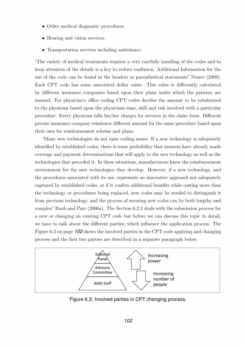
• Other medical diagnostic procedures.
• Hearing and vision services.
• Transportation services including ambulance.
“The variety of medical treatments requires a very carefully handling of the codes and to
keep attention of the details is a key to reduce confusion. Additional Information for the
use of the code can be found in the headers or parenthetical statements” Nuwer (2009).
Each CPT code has some associated dollar value. This value is differently calculated
by different insurance companies based upon their plans under which the patients are
insured. For physician’s office coding CPT codes decides the amount to be reimbursed
to the physician based upon the physicians time, skill and risk involved with a particular
procedure. Every physician bills his/her charges for services in the claim form. Different
private insurance company reimburse different amount for the same procedure based upon
their own fee reimbursement scheme and plans.
“Many new technologies do not raise coding issues. If a new technology is adequately
identified by established codes, there is some probability that insurers have already made
coverage and payment determinations that will apply to the new technology as well as the
technologies that preceded it. In these situations, manufacturers know the reimbursement
environment for the new technologies they develop. However, if a new technology, and
the procedures associated with its use, represents an innovative approach not adequately
captured by established codes, or if it confers additional benefits while costing more than
the technology or procedures being replaced, new codes may be needed to distinguish it
from previous technology, and the process of securing new codes can be both lengthy and
complex” Raab and Parr (2006a). The Section 6.2.2 deals with the submission process for
a new or changing an existing CPT code but before we can discuss this topic in detail,
we have to talk about the different parties, which influence the application process. The
Figure 6.3 on page 102 shows the involved parties in the CPT code applying and changing
process and the first two parties are described in a separate paragraph below.
Editorial
Panel
Advisory
Committee
AMA Staff
Increasing
power
Increasing
number of
people
Figure 6.3: Involved parties in CPT changing process.
102

Afterwards, the content of the CPT Manual is explained and all three different cat-
egories of CPT codes are described in a separate paragraph. In end of this Section, we
display the CPT code application process and also the relevant existing CPT code for
SIGFRIED and EEG LI test.
CPT Editorial Panel The CPT Editorial Panel meets tree times per year and deal with
new technologies and upgrades of existing codes. “In total the Panel consists of 17 persons,
11 of these are physicians recommended by the Nation Medical Specialty Societies and
accepted by the AMA Board of Trustees. The CPT Health Care Professionals Advisory
Committee nominates two members the CPT Editorial Panel. Each of the following
Association also designate one physician: the Blue Cross and Blue Shield Association,
America’s Health Insurance Plans, the American Hospital Association, and the Centers
for Medicare and Medicaid Services” cf.American Medical Association (2010d).
The Advisory CPT Committee The Advisory CPT Committee assists the CPT Edi-
torial Panel during the Meetings and is preparing the details and special aspects of topics
in forefront of the meetings. The Committee mainly consists of physicians selected by the
various of specially national medical societies and at present the societies are restrict to
members of AMA Health Care Professionals Advisory Committee (HCPAC) and AMA
House of Delegates. Also professionals with a performance measures background are sup-
porting the CPT Editorial Panel, if it is necessary and requested. Based on American
Medical Association (2010d), the next itemization lists the several objective, which are
under the responsibility of the Advisory Committee.
• The most important task is to give advices on the nomenclature and procedure
coding.
• The continuously documentation of the advices in particularly with regard to the
adequacy use of the CPT code for the various of surgical and medical procedures.
• In an annually meeting the Committee discuss and review codes, which are getting
a bit long in the tooth and need an update.
• The Advisory CPT Committee reviews the articles published by the AMA concern-
ing the CPT code to provide information material particularly for new emerging
technologies.
Current Procedural Terminology Manual
Since 1966, the American Medical Association published a CPT Manual every year, which
comprehend updates and changes of CPT codes as a result of significant improvement of
103

medical practices and technology. The AMA released the CPT 2011 Professional Edition
in October 2010 and the 760 pages dick spiralbound book costs about $107.95. The book
contains corresponding guidelines to the three different CPT categories and we explain
every category in detail after a brief executive summary of them, based on OpenClinical
(2010):
Category I Codes are designated for services (or procedures) common in contemporary
medical practice and being performed by many physicians in clinical practice in
multiple locations.
Category II Codes are used on performance measurement: Category II CPT codes are
intended to facilitate data collection by coding certain services and/or test results
that are agreed upon as contributing to positive health outcomes and quality patient
care.
Category III Codes deal with emerging technology. The purpose of this category of
codes is to facilitate data collection on and assessment of new services and proce-
dures. These codes are intended to be used for data collection purposes to substan-
tiate widespread usage or in the Food and Drug Administration (see Section 5.2)
approval process.
Category I CPT code is used for well-established procedures and services. The category
one codes are the bigger part of the CPT codes and people usually identify with this code
when using the term CPT code. The code consist of five digits and starts with the number
00100 and ends with 99607. The Advisory CPT Committee the Category I codes ones a
year and the codes are cut into six areas:
1 Evaluation and Management
2 Anesthesiology
3 Surgery
4 Radiology
5 Pathology and Laboratory
6 Medicine
As a condition, a procedure should be practices by various doctors in several locations and
up to date. The requirements for a CPT I codes are given by the Advisory Committees
and the Editorial Panel and are listed below based on American Medical Association
(2010d):
104
• that the service/procedure has received approval from the Food and Drug Admin-
istration for the specific use of devices or drugs
• that the suggested procedure/service is a distinct service performed by many physi-
cians/practitioners across the United States
• that the clinical efficacy of the service/procedure is well established and documented
in U.S. peer review literature
• that the suggested service/procedure is neither a fragmentation of an existing pro-
cedure/service nor currently reportable by one or more existing codes; and
• that the suggested service/procedure is not requested as a means to report extraordi-
nary circumstances related to the performance of a procedure/service already having
a specific CPT code.
Category II CPT code hold the codes concerning the evaluation for a physical process
and the performance of services. A typically appropriation of a category II codes is to
account the assessment from clinical laboratory and radiology tests. Also the reimburse-
ment for clinical or management services report the code and hence it should minimize
the bureaucratic work for the medical staff. The code itself consists out of four digits
followed by an F, but they are not designed for replacement of a regularly category I
code. CPT Category II codes are arranged according to the following categories based on
Merion Publications (2004):
• Composite Measures 0001F
• Patient Management 0500F-0503F
• Patient History 1000F-1002F
• Physical Examination 2000F
• Diagnostic/Screening Processes or Results 3000F
• Therapeutic, Preventive or Other Interventions 4000F-4011F
• Follow-up or Other Outcomes 5000F
• Patient Safety 6000F
105

Category III CPT code covers new rising technologies and comprises temporary codes
for emerging procedures and services. The main idea behind the category III is to provide
a billing basis for the face of data acquisition for the purpose of evaluation of a medical
treatment. This evaluation can support the FDA certification process or show the clinic
efficacy of the emerging technology. For this special purpose a few exceptions compere to
the CPT code I requirements are done and listed below.
• The FDA approval must not be present
• Clinical efficacy has not to be proven
• A locally use of the treatment is accepted
As mentioned above, this conditions are especially helpful for an innovation hence this is
the most likely category for our two diagnostic tests. Thus we will talk about this category
more closer and in particular we precisely explain the Relative Value Unit (RVU) in a
separate paragraph.
The American Medical Association provides the document “CPT Category III codes” on
their Web site and this document gives a good overview about category III codes. The
next paragraph highlights the most important parts of this document.
“CPT Category III codes are assigned an alphanumeric identifier with a letter in the
last character (e.g., 1234T) and are located in a separate section of the CPT manual,
following the Medicine section. It is worth noticing, that the inclusion of a service or
procedure in a category III neither implies nor endorses clinical efficacy, safety, or the ap-
plicability to clinical practice, but the service/procedure must have relevance for research,
either ongoing or planned. Because CPT Category III codes are intended to be used for
data collection purposes to substantiate widespread usage or to provide documentation
for the FDA approval process, they are not intended for services or procedures that are
not accepted by the CPT Editorial Panel due to an incomplete proposal, the need for
more information, or a lack of CPT Advisory Committee support.
The CPT Editorial Panel is in charge to set up a new CPT category III code and will
publish them on a half-year basis on the official Web site. The releasing days are January
1st and July 1st. After the code is released the 6 month time period of implementation
starts and so the code will be effective six month after published on the AMA Web site.
This new codes will be print in the next CPT cycle edition. In Order to approve the new
technology the CPT Editorial Panel needs information about the emerging technology
and especially the CPT Advisory Committee plays a major role in the decision process.
In the main after five years the codes are file away, unless it can show that it is still used
in an appositely purpose” cf. American Medical Association (2009a).
106
Appendix After the category 3 codes, the CPT manual contains an appendix, that
is numbered from A to M. The Appendix A consists of a list of 2 digit CPT codes
called modifiers. Modifiers are appended to regular CPT codes in order to show the
circumstances which cause deviation form the exact code description of a regular CPT
code or to show some additional information to the insurance company so that correct
dollar amount will be reimbursed according to the service rendered. The Appendix B
consists of a summary of additions, deletions and revisions in the CPT manual of the
current edition over that of CPT manual of the last year. The Appendix C consists of a
list of clinical examples to ensure the understanding of certain CPT codes and Appendix
D contains a summary of add-on codes.
Add-on codes Codes with a + symbol in the category I CPT codes are all add-on codes.
Certain listed procedures of CPT are commonly carried out in addition to the primary
procedures performed. These additional or supplemental procedures are designated as
add-on codes. For Example the CPT code 19000 is for Puncture aspiration of cyst of
breast. If more than one cyst us aspirated then add-on code + 19001 is used to report
aspiration of each additional cyst as many cyst aspirated. Here the primary code is 19000
and its add-on code is 19001. Add-on codes are never reported as stand-alone or primary
code, because they always have to used with their designated primary codes together.
Applying for a CPT code
In the last section we talked about the CPT Manual and we explained the Manual’s
content. Generally, it is not expected to find an existing CPT code that matches for a
new technology. Therefore a development result normally needs a new or modified CPT
code to be entered into the payment billing system. Thus this subsection deals with the
application process for a new or to modify an existing CPT code and we stress on the
category I and III codes, because they are relevant for our two development results. This
subsection is spilt up into two main parts: who decides the request and which factors
count for the decision. Second, we talk about the Coding Change Application Form and
about additional information which should be submitted with this form.
During the Editorial Panel meeting, the CPT application are discussed by the panel and
also they come to a decision, whether a new CPT code application is approved or de-
clined. The Editorial Panel gets support for the CPT/HCPAC Advisors, who review the
code change requests. Also relevant clinical literature is considered in the final decision.
The whole meeting is organized and managed by the CPT staff and also the necessary
information for a request of CPT codes change must be submitted to the CPT staff 30
days in advanced of the Meeting.
107

In the most cases it is very useful to submit the application for a code change in corpo-
ration with the relevant medical societies, because the societies support the code change
request in terms of consistency and coherency. In addition, the societies make sure that
the requested code is not contradictory to existing codes and we discuss the role of the
American Academy of Neurology in a separate paragraph below. It is important to men-
tion, that the American Medical Association also formulated a clear statement that the
supporting role of the medical societies and especially of their advisors has nothing to do
with lobbing. But they explicit forbid any kind of unsolicited communication both with
the CPT/HCPAC Advisors and members of the Editorial Panel. Because of these, every
information should be submitted the CPT staff and not directly to the CPT/HCPAC
Advisors or members of the Editorial Panel. Only during the open meeting a comment
in front of the full Editorial Panel or a directly request of a Panel member is permitted.
The Role of the American Academy of Neurology (AAN) The support of the cor-
responding society is essential for the success of a changing or new code request and for
both SIGFRIED and EEG LI test, the American Academy of Neurology is the expedient
society. An early cooperation with the society can be beneficial and the next citation
expresses, how the AAN participates in the application process. “The American Academy
of Neurology will send one or two so called CPT Advisors to the CPT Editorial Panel,
when a new relevant code application is deposited. The CPT Advisors are members of the
Academy’s Coding Subcommittee of the Medical Economics and Management Commit-
tee, which oversees reimbursement, billing, and coding processes. These advisors will act
as topic experts in particular during the oral discussion when the code request is debated”
Nuwer (2009).
Coding Change Application Form The AMA provides a form to submit an appli-
cation for a new code or either a change of an existing code. The form is available at
the AMA Web site (http://www.ama-assn.org/). The AMA suggests to make yourself
first familiar with the Fourth Edition of the Current Procedural Terminology before filling
out the application form. Their are also several guidelines developed by the AMA which
should simplify the requesting process. The article “Applying for CPT Codes” formulates
the criteria for development and evaluation of CPT Category I and Category III codes
and is cited below.
In developing new and revised Category I codes the CPT Advisory Committee and the
CPT Editorial Panel require the following conditions based on American Medical Associ-
ation (2010b):
• that the service/procedure has received approval from the Food and Drug Admin-
108

istration (FDA) for the specific use of devices or drugs;
• that the suggested procedure/service is a distinct service performed by many physi-
cians/practitioners across the United States;
• that the clinical efficacy of the service/procedure is well established and documented
in U.S. peer review literature;
• that the suggested service/procedure is neither a fragmentation of an existing pro-
cedure/service nor currently reportable by one or more existing codes; and
• that the suggested service/procedure is not requested as a means to report extraordi-
nary circumstances related to the performance of a procedure/service already having
a specific CPT code.
The following is used as formalized criteria by the CPT Advisory Committee and the
CPT Editorial Panel for evaluating Category III code requests and includes identification
of the following elements as guidelines for establishment of a Category III code based on
American Medical Association (2010b):
• A protocol of the study or procedures being performed;
• Support from the specialties who would use this procedure;
• Availability of United States peer-reviewed literature for examination by the Edito-
rial Panel;
• Descriptions of current United States trials outlining the efficacy of the procedure.
The submitter of an application should contemplate the following aspects. First, the can-
didate should make clear if there is any currently existing code, where the new procedure
or service could be included without significantly changes to the extent of the code. Sec-
ond, the submitter should define whether it is a variation or fragmentation of an existing
code or either a complete new code. In this context it is very important to document why
an existing code in not adequate for the service or procedure and also keep in mind the
opportunity to use several existing codes to bill the new technology. Hence, this should
include a statement which verify that new technology is a distinct service. In this regard
an estimation of how many physician might use this procedure or service and where it is
performed can back up the argumentation. The following list contains all necessary in-
formation, which should be submitted to the CPT staff in addition to the coding change
application form and the itemization is based on American Medical Association (2010c):
109

• a complete description of the procedure/service (eg, describe in detail the skill and
time involved. If this is a surgical procedure, include an operative report that
describes the procedure in detail);
• a clinical vignette which describes the typical patient and work provided by the
physician/practitioner; the diagnosis of patients for whom this procedure/service
would be performed;.
• a copy of the diagnosis from potential patients for the new procedure or service
• a copy(s) of peer reviewed articles published in US journals indicating the safety and
effectiveness of the procedure, as well as the frequency with which the procedure is
performed and/or estimation of its projected performance; and
• additional literature, which support the request relating to policy statements and
guidelines.
• evidence of FDA approval of the drug or device used in the procedure/service if
required.
• national statistical data about the procedure or service
Clinical Vignette For every code change request a clinical vignette must be submitted
with the application. The clinical vignette should show which persons typically will receive
the new treatment. The guideline “Sample Format for Required CPT Clinical Vignettes”,
which is available on the AMA Web site, contain the following example.
“Current Procedural Terminology code á 61863: Twist drill, burr hole, craniotomy, or
craniectomy with stereotactic implantation of neurostimulator electrode array in subcorti-
cal site (eg, thalamus, globus pallidus, subthalamic nucleus,periventricular, periaqueductal
gray), without use of intraoperative microelectrode recording; first array.
Typical patient: A 66-year-old woman presents with a ten-year history of idiopathic
Parkinson’s disease that has caused progressive disability. She continues to respond to an-
tiparkinson medications, however; her symptoms are no longer adequately controlled with
medications and she is experiencing disability for many activities of daily living. Medi-
cation adjustments and different medication regimens have failed to improve her level of
disability.
Description of procedure(s)/service(s): She undergoes implantation of a stereotactically-
guided deep brain stimulator electrode array in the subthalamic nucleus using macro
stimulation for targeting and confirmation of electrode placement” American Medical As-
sociation (2010e).
110

6.2.3 Relative value unit
“CPT Category III codes are not referred to the AMA-Specialty RVS Update Committee
(RUC) for valuation because no relative value units (RVUs) are assigned to these codes.
Payment for these services or procedures is based on the policies of payers and not on
a yearly fee schedule” American Medical Association (2009a). “Fortunately, the CMS is
mandated to publicly announce, through the Federal Register, the anticipated fee schedule
for the following year. This is done first in a“proposed rule,”usually distributed during the
summer (June or July). A comment period follows, allowing specialty societies, individual
physicians, and others to review any proposed changes in the fee schedule and the rationale
behind those changes. The CMS staff must then consider these comments and construct
a “final rule” that is published in November or December, which establishes the actual
fee schedule for the following year. Using the RVU values in that schedule, each of the
Medicare carriers and private payers can construct an actual payment schedule for the
coming year” Thorwarth (2004).
In to the “Federal Register / Vol. 66, No. 212 / Final rule with comment period”, the
CMS states their position regarding to the reimbursement policies of payers. “Commenters
expressed appreciation for our recognition of these new categories of CPT codes. However,
one commenter believed that we should refrain from categorically denying payment for
category III (emerging technology) CPT codes, because these CPT codes may sometimes
warrant payment. Another commenter believed that we were proposing not to pay for
these codes at all. The commenter recommended that we clarify in the final rule that
carriers may determine if payment should be made for a particular emerging technology
code. Response: We believe that these codes will serve an useful purpose. We regret that
some commenters believed that the discussion in the proposed rule implied that these
services should not be covered. We only intended to indicate that by publishing these
codes we are not indicating that we would pay for these services in all instances. As the
commenter indicates, coverage of emerging technologies and payment for these services is
at the discretion of the carriers. We also want to clarify that our carriers will be able to
incorporate these codes only after they are entered into our system during our regularly
scheduled updates and not as soon as the AMA posts them on the CPT web site” Centers
for Medicare & Medicaid Services (2001).
Time line and cost estimation The goal for an innovation is to be assigned with a re-
imbursement code that guaranties adequate amount that can be reimbursed. The ICD-9
codes are used to reimburse hospital utilization for inpatient diagnoses and procedures.
The CPT code is used for both hospital utilization for outpatient diagnoses and pro-
cedures, and for physicians fees. The flowchart diagram (Figure 6.4) summarizes and
111

displays the CPT codes applying process.
Does theTreatment �t in aexisting code ?
The CPT AdvisoryCommittee reviews the
new request
Is a new codeneeded ?
The CPT AdvisoryCommittee inform how
to use a existing code toreport the procedure
The CPT Editorial Panelreviews the request.
The CPTEditorial Panel can
result in 3 outcomes
Add a new code orrevise existingnomenclature
Postpone/table an itemto obtain further
informationReject the request
If not
If not
If yes
If not
The existing code canbe used.
If yes
If yes
Does the theAMA sta� have any
suggestions ?
A new procedure,technology, orperformance is
introduced.
A coding request form issubmitted
Figure 6.4: Flowchart diagram of CPT code application process.
112

According to Table 6.1 on page 113, an expansion of existing technology needs one to
two years post-FDA approval.
Similar to anotherproduct
Expansion of exist-ing technology
Truly new and innovative
Reimbursementcomponentsthat must bedeveloped
Confirm existing codeand inclusion for cov-erage of this product
Alter coverage,coding, and pay-ment to include thisproduct
Create new coverage,coding, and payment forthis product
Science required Usually FDA approvalfor same indicationssuffice for inclusion inexisting coverage
Publication ofcontrolled studies(usually 1-2)
Publication of controlledstudies (usually 2-4)and cost-effectivenessdata (publication and/orregistry data)
Typical time linefor these compo-nents post-FDAapproval
6 months to 1 year 1 to 2 years 2 to 5 years
Table 6.1: Overview of reimbursement’s time line for development results. “The last rowestimates the time line for obtaining both coverage determination and reimbursementcode, after the product is approved by the Food and Drug Administration (FDA)” Beckerand Whyte (2006).
“Indication expansion of an existing technology often requires altering coverage, cod-
ing, and payment to address the new indication. Published studies supporting the pro-
posed additional indication and revisions of established medical policy will be necessary
to create coverage. Codes may need to be revised, including new code descriptions, which
can trigger different payments rates” Becker and Whyte (2006).
Both development results match this description, and hence both are in a range between
one and two years, but there are minor differences in the argumentation of the terms
reasonable and necessary. These terms are the major criteria for the local coverage deter-
mination, and papers that support the argumentation for these criteria are helpful in this
regard. For the EEG LI test, several papers call the current gold standard, Wada-test
into question. For examples, in 2002, the journal NEUROLOGY published the article
“Is it time to replace the Wada test?” by Abou-Khalil and Schlaggar (2002). In 2003,
an article with same title was published in the same Journal, but with different authors
Baxendale et al. (2003). We assume that the EEG LI test will need 1.25 years to pass the
reimbursement process.
Currently, SIGFRIED improves the electrocortical stimulation procedure, and the value
of the improvement is not as widely accepted as for EEG LI test. Thus, more detailed
documentation of the improvements is required, and we assume that the reimbursement
113
process will need 2 years for SIGFRIED.
For accomplish reimbursement coverage, the ideal qualification of the responsible person
is a master in software engineering and the Table 5.2 on page 86 provide the expenses
to employ a software engineer. For EEG LI test, the product of the software engineer
personal cost multiplied by the necessary time results in $262,469 and for SIGFRIED, the
reimbursement process cost is $419,950.
6.2.4 Summary of reimbursement code
The next two paragraphs summarizes the relevant reimbursement code issues for the two
clinical diagnostic tests.
SIGFRIED is performed in an inpatient setting and it takes place in a hospital such
as a Fourth–Level Epilepsy Center. For inpatient care, the Medicare companies do not
pay for every device separately, instead the hospital receives a bundled payment amount
based on the diagnosis related group (DRG). DRG payment are linked to ICD-9 codes,
which identify surgical procedures. If a ICD-9 code that describes electrocortical stimu-
lation is sufficient to include SIGFRIED into the procedure than the hospital can obtain
reimbursement for SIGFRIED. In this case, SIGFRIED does not need a separate code to
be identified. If not sufficient ICD-9 codes exists, the manufacturer needs to seek a new
ICD-9 code, which includes appropriate DRG assignments for the new ICD-9 code.
EEG LI test is executed in the hospital outpatient setting, which requires to use CPT
codes to identify the procedure. The Medicare companies also combined the CPT codes
into payment bundles, which are called ambulatory payment classification. This classi-
fication pays the hospital a presumably amount for the facilities costs, which equals the
procedure’s technical item fees for the performed procedure.
The physicians, who execute the procedure, also need to use the CPT code to identify the
performed procedure because their fee is also based on the CPT code. It is worth noticing
that hospitals are paid separately from physicians.
114
7 Competitive advantage
At this stage, our development results already went trough the FDA approval, Medicare
coverage decision and applying for a reimbursement code procedure. In other words,
the manufacturer owns a product that can be sold to a hospital and private physician
and these health care practitioners would be reimbursed by medicare companies for their
treatments. Thus, the last missing step is the actual purchasing-decision process between
the hospital or private physician and the manufacturer. Economically, the success of
a development result is measured in profit and the revenue has a linear impact on the
profit. The number of sales are affected by the outcome of purchasing-decision process
and the purchasing process can be complex and time consuming because several parties
are involved the purchasing decision. Especially in large organizations such as a hospital,
it is difficult to determinate every influencing party and factor of the purchasing-decision
process. This chapter deals with the decision-making units and impact factors of the
medical device purchasing process.
Aspects of hospital purchasing-decision The paper focuses on the cognitive and
organizational factors and The article “Institutional decision-making to select patient care
devices: identifying venues to promote patient safety”by Keselman reports the purchasing-
decision process in a large urban hospital for the item infusion pump and we reference
to this article trough the whole section. In the paper’s introduction, the author gives an
overview of the relevant influence factors and their connections. The Figure 7.1 on page
116 outlines potential (hypothesized) relationships among various factors that may affect
the process of institutional medical device selection and the purchasers perception of the
process.
115

studying the infusion pump selection process. The studyinvolved: (a) semi-structured interviews with partici-pants in the latest infusion pumps purchase in a majorurban hospital system and (b) analysis of documentsrelevant to the purchase. The interview data were usedto construct participants representation of the processand information �ow. Document analysis was used toreconstruct the actual sequence of process-relatedevents.
5. Methodological design
5.1. Overview of the setting
The study setting was a comprehensive urban hospi-tal composed of two medical centers, which had merged
a few years prior to the beginning of the selection pro-cess. The merger resulted in consolidation of all majoradministrative departments under supervision of bi-campus General Services. Prior to the merger, the twocenters had pumps manufactured by two di�erent ven-dors. One of the goals of this selection process was tostandardize infusion devices across the centers by pur-chasing a common model.
5.2. Instrumentation
As discussed above, one method of data collection forthe study involved semi-structured interviews. Design ofthe interview instrument was based on Miles and Hu-berman s guidelines for qualitative analysis [26]. Prior todesigning the questions, the investigators outlined con-ceptual framework for data collection. According to
Miles and Huberman [26], a conceptual framework is agraphic or narrative representation of main constructs
and variables to be studied, and hypothesized relation-
ships among them. The basis for the design of the con-ceptual framework can be both theoretical (based on thetheories and hypotheses guiding the study) and empiri-cal (based on initial observations in the setting). Thebene�ts of outlining the conceptual framework prior tothe study are twofold. First, it helps researchers tocontain the area of the study and focus their questionson the factors and relationships within that area. Sec-ond, it explicates researchers assumptions that will betested and possibly altered in the course of the study.
The initial conceptual framework for the presentstudy is depicted in Fig. 1. In the �gure, arrows repre-sent direction of the in�uence among the factors. The�gure outlines potential (hypothesized) relationships
among various factors that may a�ect the process ofinstitutional medical device selection and the purchasersperception of the process. As suggested by Miles and
Huberman [26], the design of the framework involvedboth theoretical and empirical processes. Following asystems approach to medical errors, we chose to focuson the institutional structure, communication patterns,and �nancial factors as contributors to the process. Thestudy s focus on patient safety and research on medicaldecision-making lead us to include purchasers knowl-edge and attitudes towards patient safety, as well as theirperception of the process and the outcome [27,28]. Thedirectionality of the hypothesized relationships in theframework was �nalized after an informal discussionwith a clinician who was familiar with typical deviceacquisition procedures in the hospital.
Based on the conceptual framework, interview ques-tions covered the following aspects of the process: trig-gers (what triggered the process), process, safetyevaluation, and decision and process quality. An interview
instrument containing key questions for each of theseaspects was developed (see Appendix A). Appropriateprobes and clari�cations followed each key question.
Fig. 1. Conceptual framework for the study. The boxes represent aspects of the decision-making process and factors that in�uenced it. The arrowsrepresent the directionality of causal relationships among the di�erent factors.
34 A. Keselman et al. / Journal of Biomedical Informatics 36 (2003) 31–44
Figure 7.1: Aspects of hospital purchasing-decision. “The boxes represent aspectsof the decision-making process and factors that influenced these. The arrows showthe directionality of causal relationships among the different factors” Keselman et al.(2003).
“The design of the framework involved both theoretical and empirical processes. Fol-
lowing a systems approach to medical errors, we chose to focus on the institutional struc-
ture, communication patterns, and financial factors as contributors to the process. The
study’s focus on patient safety and research on medical decision-making lead us to include
purchasers’ knowledge and attitudes towards patient safety, as well as their perception of
the process and the outcome” Keselman et al. (2003).
Decision-making unit Every decision to purchase a product or service is influenced
by one or more members of the so-called decision-making unit (DMU). Based on Morse
(1998), this comprises:
User: the person who will use the product or service
Influencer: usually someone whose advice is requested or offered which as a result can
influence the brand, the manufacturer, price, timing,etc, of the purchase.
Decision maker: the ultimate maker of the decision to purchase the service or prod-
uct(remember that it can be a committee).
Buyer: the purchasing officer who has responsibility for the purchasing budget and there-
fore of getting the best value for money.
Gatekeeper: literally he or she who controls access to the rest of the decision-making
unit: perhaps an information provider, a secretary or a security manager.
Keselman reported in his article the following DMUs: “Professionals with three types
of expertise-administrative, engineering, and clinical-were involved in the selection pro-
116

cess. Three administrative/technical departments played a major role in device selection:
Purchasing, Support Services, and Engineering and they are displayed in Figure 7.2
audibility, �ow accuracy and frequency o� ams). Theadjunctive ‘‘and’’ pronoun used in the statement indi-cates equal status of the �nancial and the clinical fac-tors. The participant s statement used in this exampleprovides a part of the basis for the semantic represen-tation of an administrator s view of relevant factors, asdepicted in Fig. 4.
The process documentation and interview data wereused to describe organizational structure and informa-tion �ow. Finally, survey evaluation forms were con-
trasted with established design usability heuristics. The14 heuristics used were developed by Zhang et al. [31] intheir evaluation of an online courseware system, on thebasis of 10 principles developed by Nielsen and Molich[32]. These heuristics were also employed by Patel andco-workers [33] in the evaluation of the infusion pumpthat was purchased as a result of this decision-makingprocess.
Design heuristics are often used in device and/or in-terface usability evaluation. This method, in which ateam of evaluators note a device s violations of a num-ber of usability design principles, is a frequent method ofchoice in usability engineering. We therefore used this asa ‘‘gold standard’’ for evaluating the sensitivity of thesurvey forms from the infusion pumps purchasing pro-cess to design principle violations.
7. Characterization of the decision-making process
This section is organized in the following way. First,
we describe various groups that participated in theprocess and the structure o� nformation �ow amongthese groups. Next, we describe the decision-making
process as: (a) represented in participants
recollectionsand (b) captured in the minutes of the Committee for
Technology in Clinics meetings. Following that, we
present analysis of survey questionnaires that were usedto assess user satisfaction during clinical evaluation ofthe pumps. Finally, we analyze and discuss di�erences
between administrators and clinicians
perceptions of
driving forces of the selection process.
7.1. Participating groups
Professionals with three types of expertise—
adminis-
trative, engineering, and clinical—
were involved in theselection process. Three administrative/technical de-partments played a major role in device selection: Pur-
chasing, Support Services, and Engineering. The
function of the Purchasing is to supervise and negotiate
�nancial agreements. Support Services deals with thedistribution of supplies and devices within the hospitaland provides liaison between top-level administratorsand clinical personnel. Engineering is responsible fortechnical maintenance of equipment. Individuals from
these departments formed the Core Project Manage-ment Group that led the selection process.
Two clinical groups participated in the selectionprocess. The Committee for Technology in Clinics is astanding committee that includes high-ranking physi-cians, engineers, and administrators. The committeereviews and formally approves all technology acquisi-tions in the hospital. The second clinical group was anad-hoc committee that included nurse managers frommajor hospital units that used infusion devices. The in-
fusion pump is a device that is primarily used by �oornurses. Since core project managers did not directly in-teract with �oor nurses, they relied on Nurse ManagersCommittee to provide the liaison with the users.
7.2. Flow o� nformation during the process
Fig. 2 describes information �ow among the playersin the decision-making process and is based on interviewdata. As we describe in the next section, interview andprocess documentation data produce somewhat di�erentrepresentations of the process. However, the �ow ofinformation in these two representations is comparable.In a diagram based on process documentation, sequence4–7 would loop twice.
Sequence 1–3 of the �gure refers to the initial stagesof the process when Purchasing, Support Services, andEngineering (1) identi�ed the need to purchase new
pumps, (2) formed core project management group thatwould coordinate the selection, (3) identi�ed vendorsand models to consider, and (4) appointed Nurse
Fig. 2. Information �ow during the process. The blocks in the �gure
represent di�erent groups and committees that participated in theprocess. The numbered arrows represent the direction o� nformation�ow (from the transmitter to the receiver of the primary information).The dashed line separates participating groups with administrative andengineering expertise from participating groups with clinical expertise.
36 A. Keselman et al. / Journal of Biomedical Informatics 36 (2003) 31–44
Figure 7.2: Decision-making flowchart. “The blocks in the figure represent differentgroups and committees that participated in the process. The numbered arrows repre-sent the direction of information flow (from the transmitter to the receiver of the primaryinformation). The dashed line separates participating groups with administrative andengineering expertise from participating groups with clinical expertise” Keselman et al.(2003).
The function of the Purchasing is to supervise and negotiate financial agreements.
Support Services deals with the distribution of supplies and devices within the hospital
and provides liaison between top-level administrators and clinical personnel. Engineering
is responsible for technical maintenance of equipment. Individuals from these departments
formed the Core Project Management Group that led the selection process.
Two clinical groups participated in the selection process. The Committee for Technology
in Clinics is a standing committee that includes high-ranking physicians, engineers, and
administrators. The committee reviews and formally approves all technology acquisitions
in the hospital. The second clinical group was an ad-hoc committee that included nurse
managers from major hospital units that used infusion devices. The infusion pump is
a device that is primarily used by floor nurses. Since core project managers did not
directly interact with floor nurses, they relied on Nurse Managers’ Committee to provide
the liaison with the users” Keselman et al. (2003).
117

7.1 Time line and cost estimation
The last two sections reviewed the numerous factors and parties involved in the decision
process. Figures 7.1 and 7.2 in particular highlight the complexity of the process, and
hospitals had to yet developed a standardized purchasing process. Hence, every hospital
has its own purchasing procedure, which is not transparent to the public and entails
unpredictable delays. As a Reference, we use the case study published by Keselman et
al., and the Table 7.1 on page 118 is cited from this study.
Event DateAnnouncement of Vendor B s new model release 11/18/96Price estimate negotiated with Vendor B; clinical trials planned at campus 1 1/13/97Pump B evaluation completed at campus 1 11/24/98First mention of Vendor A; plans to trial both pumps at both sites 1/12/98Both vendors present financial proposals 4/27/98Trials completed on both campuses 10/19/98Purchasing administration meets with the leadership on both campuses 3/22/99Pump B chosen by administration; committee endorses decision 5/17/99The total duration of the purchasing-decision process was approximately 2.5 years
Table 7.1: “Time of purchasing-decision process” Keselman et al. (2003).
“Figure 7.1 presents major selection process events and the process starts with consid-
eration of one option, Vendor B’s new model. The model is chosen from a familiar vendor,
and the purchasers quickly proceed to financial negotiations, and then clinical evaluations.
It is only after significant shortcomings are found in Pump B the second candidate comes
under consideration. Next, preliminary negotiations with both vendors establish that the
new candidate costs 8% more than Pump B. Both pumps are clinically evaluated on both
campuses and the Core group administrators selected the less expensive model” Keselman
et al. (2003).
In this study, the decision-making process needed approximately 30 months and both
products were almost similar. In the end, it came down to a financial decision. We as-
sume that the purchasing-making decision time for products, that have a clear advantage
compared to the concurrent, is shorter and less complex. As a rough estimation we assume
that:
118

Development result Purchasing-decision time [years]EEG LI test 1.2 yearsSIGFRIED 1.8 years
Table 7.2: Estimated purchasing-decision process’s time of the two diagnostic tests.Both numbers are rough estimations, because a details evaluation of these numbers isnot possible at this early stage of the innovation process. But, it is important to includepurchasing-decision time into the overall picture of the innovation process to have acomplete picture.
EEG LI test is an independent device, and it is based on a well established, non-
invasive, and safe technology (EEG recording). Furthermore the EEG LI test’s attributes
compared to the current gold standard, the Wada test, are significantly better (see Table
3.9 on page 38). Hence, we assume for EEG LI test a short purchasing-decision time of
1.2 years.
The invasive method SIGFRIED is more complex than the EEG LI test, so the purchasing-
decision process involves more parties. Thus, we estimate for SIGFRIED a purchasing-
decision time of 1.8 years.
cost A marketing manager is the right person to supervise the purchasing process, and
Table 7.3 presents the annual costs for a marketing manager at the Wadsworth Center.
Marketing managerBase Salary Fringe rate Fringe cost Indirect cost Total89,000 37.50% 33,375 85,064 207,439
Table 7.3: Marketing Manager salary. This table shows the total costs incurred by theWadsworth Center to employ a marketing manager with several years of experience.All numbers are in USD per year.
The product of the total personal cost per year times the required years equals the
total expenses of the hospital purchasing decision. Hence, the Wadsworth Center has to
spend for SIGFRIED $373,388 and for EEG LI test $248,927 to sell the diagnostic tests
to a hospital.
7.2 Benefits of development results
A development result should address the customer’s needs better than the existing com-
petitors on the market, and the customers for both development results are hospitals and
private physicians. At the current stage, a detailed evaluation of the purchasing-decision
119

process is not possible, because every hospital has a different process, and the first target
customers have not yet been identified. In general, the hospitals base their purchasing
decisions on the same factors and this section ranks factors that influence the hospital’s
purchasing decision. Afterwards this section shows that the two clinical diagnostic tests
address the influence factors of hospital purchasing decision better than the established
methods.
The article “The Role of Patient Safety in the Device Purchasing Process” by Johnson
et al. (2005) concludes that patient and user safety is the major impact factor followed by
clinical reasonable. Furthermore cost-effectiveness is more important than easy of opera-
tion. Note that ease of operation should not influence the correct handling of the device.
Especially for software, tools that are not easy to operate can lead to safety concerns.
For example, if the graphical user interface is not arranged clearly, then users may apply
incorrect settings that lead to erroneous outputs. In our ranking, ease of operation did
not interact with safety concern and is arranged behind being a technological leader. ”Be-
cause being a technological leader is graded as a strong factor in the purchasing-decision
process” cf.Teplensky et al. (1995).
1. Safety
2. Clinical reasonable
3. Cost-effectiveness
4. Being a technological leader
5. Easy of operation
Safety Because EEG LI test is a completely non invasive procedure, it raises no safety
concerns. “The established method, the Wada test had a 0.7% risk of carotid artery
dissection” cf. Loddenkemper, Morris and Perl (2002).
Compared to the electrocortical stimulation, SIGFRIED does not actively stimulate the
cortex, and hence SIGFRIED does not raise any risk of seizure induction.
Clinical reasonable For both diagnostic tests, the conducted studies demonstrated
that the tests results are in consensus with the established methods. Thus they are as
reasonable as the current gold standard.
Cost-effectiveness The Table 3.10 on page 42 points out that the costs, which occur
that the hospital’s costs are 23 times lower for an EEG LI test than for fMRI, and the
Wada test costs about 3.8 times more than fMRI. Thus, the EEG LI test has significant
120
cost advantages against both the established method (the Wada test) and the dominant
emerging technology, fMRI.
Being a technology leader Both development results are based on the novel technique
Brain Computer Interface and especially SIGFRIED already gained attention of different
journals and museums. For example, the American Museum of Natural History shows
SIGFRIED in a special brain exhibition and published an article about SIGFRIED in the
Fall 2010 edition of the museum’s journal Rotunda.
Easy of operation Because the final graphical user interface is in the development
phase, it is not possible to compare them yet. But the responsible software engineers
are aware of this point and they will stress on the easy to operation factor in further
improvements.
7.3 Summary of competitive advantage
This chapter demonstrated that the purchasing decision-making process is time consum-
ing, because hospitals are complex organizations and several different parties are involved
in the purchasing decision. The personnel cost estimations conclude for SIGFRIED
$373,388 and for EEG LI test $248,927 to accomplish the purchasing decision between the
manufacturer and the hospital. Furthermore, this chapter highlighted that these novel
clinical diagnostic tests address all hospital purchasing decision factors significantly better
than competing technologies. Hence, hospitals are likely to purchase these new clinical
diagnostic tests.
121

8 Conclusion
Two common medical procedures - language lateralization and functional brain mapping
- currently require procedures that are invasive, time consuming, expensive, and rely
extensively on medical experts. By capitalizing on rapid progress in BCI systems and
other related technologies, as well as the extensive experience developing BCIs for patients,
the BCI2000 group at Wadsworth center has developed new BCI technologies that can
perform these two procedures. The new technologies are noninvasive, faster, cheaper, and
much easier to use even for staff without medical training. We developed a plan to exploit
the two recently developed clinical diagnostic tests, which we summarize in the following
time line.
Time line Four of the chapters address four topics: results at the Wadsworth Center;
regulatory environment; reimbursement policies; and the hospital purchasing decision
process. These four topics correspond to four phases in the pre-commercial development
process for the two novel diagnostic tests.
Improvement of development results → overhaul the graphical user
interface and prepare the documentation for the FDA approval process
Regulatory environment → submit a Premarket Notification (510(k))
to obtain an FDA approval
Reimbursement coverage determination → apply for a local coverage
determination and request a reimbursement code
Competitive advantage → demonstrate the benefits of the clinical
diagnostic tests to the customer based on patient safety and cost-
effective evaluation
Figure 8.1: Outline of the pre-commercial development. The work’s structure is inaccordance with this figure.
122

Figure 8.1 summarizes these four phases and the Table 8.1 shows the total time effort
that is necessary to accomplish the four milestones in terms of person years.
Task EEG LI test SIGFRIEDImprovement of development result 1.65 years 1.65 yearsRegulatory environment 0.25 years 0.25 yearsReimbursement 1.25 years 2.00 yearsHospital purchasing decision 1.00 years 1.50 yearsTotal 4.15 years 5.40 years
Table 8.1: Total time effort. Estimated time line for SIGFRIED and EEG LI tests. Notethat some rows reflect variable time costs. For example, the top row reflects that 1.65years are necessary, but hiring additional staff could reduce this time considerably.Furthermore, some stages can overlap each other, which is called “simultaneous en-gineering” and is common in such projects. Hence, these estimates are quite conser-vative and reflect more the total effort required more than the minimum duration of theinnovation process.
The Figure 8.3 illustrates the time line for SIGFRIED and Figure 8.3 displays the time
line for EEG LI test to reach the market. These Figures are based on the assumption
that two software engineers are working at the same time to improve the development
result and only one person works on reimbursement and hospital purchasing decision
at the same time. The waiting time depict the time which is necessary to finish the
preconditions for the specific task. After the improvement of the development result is
finished it is possible to start with the FDA application and reimbursement coverage
determination at the same time. For both inventions, it is reasonable to start with the
hospital purchasing decision after a positive coverage determination is predictable and we
assume 1.5 years. Simultaneous engineering saves time but on the other hand it requires
additional workforce. The pre-commercial development time line of SIGFRIED and EEG
LI test uses simultaneous engineering, because of the short lifetime cycle of software it is
reasonable to cut off the pre-commercial development time.
123

Reimbursement
Hospital purchasing decision
0,0 0,5 1,0 1,5 2,0 2,5 3,0 3,5
Improvement of development result
Regulatory environment
Years
Waiting time
Duration time
Figure 8.2: Pre-commercial development time line of SIGFRIED.
Reimbursement
Hospital purchasing decision
0,0 0,5 1,0 1,5 2,0 2,5 3,0
Improvement of development result
Regulatory environment
Years
Waiting time
Duration time
Figure 8.3: Pre-commercial development time line of EEG LI test.
124

Pre-commercial development cost Based on the time lines, the Table 8.2 estimates
the pre-commercial development cost for SIGFRIED and EEG LI test.
Task EEG LI test SIGFRIEDImprovement of development result & regulatory environ-ment
$402,646 $402,646
Reimbursement $262,469 $419,950Hospital purchasing decision $248,927 $373,388Total pre-commercial development costs $914,042 $1,195,984
Table 8.2: Total pre-commercial development cost. This Table estimates the pre-commercial cost time for SIGFRIED and EEG LI tests to sell the clinical diagnostictest to hospitals.
In order to calculate the total pre-commercial development cost per unit, it is neces-
sary to estimate the total number of products that will be sold. In chapter 4, the available
market is estimated for both inventions and the following assumption is made: The pene-
trated market, which is the set of consumers who are buying the company’s product, is a
half of the available market. In other words, 50% of the available market customers actu-
ally purchase the medical device. The Table 8.3 contains the pre-commercial development
cost per unit based on the assumed penetrated market.
Clinical diagnostic test Pre-commercial developmentcosts per product
SIGFRIED $11,446EEG LI test as pre-surgical procedure for brain surgery $6,554EEG LI test as neurological assessment battery $182
Table 8.3: Pre-commercial development cost per unit based on estimated penetratedmarket.
For SIGFRIED and EEG LI test, the Table 8.4 forecasts the product cost that contains
pre-commercial development cost, software cost and hardware cost.
SIGFRIED EEG LI test as pre-surgicalprocedure for brain surgery
EEG LI test as neurologi-cal assessment battery
Pre-commercialdevelopment cost
$11,446 $6,554 $182
Software cost $1,200 $1,200 $1,200Hardware cost $22,138 $21,338 $21,338Total cost $34,784 $28,881 $22,720
Table 8.4: Estimated product cost.
The numbers in Table 8.4 does not include all arising expenses for example overhead,
125

marketing, assembly or installation cost are missing. But, the numbers provide a first
indication of the direct related product cost.
Summary of technology exploitation This thesis discusses the economical aspects of
the inventions EEG LI test and SIGFRIED, which have been developed by the Wadsworth
Center. EEG LI test provides benefits as neurological test battery and as a presurgical
procedure examination for resective brain surgery. On the hand, SIGFRIED can only be
sold as a presurgical procedure test for resective brain surgery The two clinical diagnostic
tests are ready for further development and technology exploitation. The development
results have been tested in both laboratory and field environments, by practicing research
scientists and medical experts, and upgraded accordingly. The conducted studies proved
that both clinical diagnostic tests are safer and less time consuming than the current
established methods. Both tests provide large cost-reducing potential compared to the
gold standards, because fewer medical personal are needed to perform them.
The next steps involve appropriate certification, reimbursement coverage determination
and final purchasing decisions of hospitals and private physicians. For EEG LI test, this
work estimates that it takes 2 12
years to accomplish the mentioned steps and these steps
cost $914,042, which include personnel expenditures and application fees. The available
market as neurological test battery contains 9668 private physicians and 279 hospitals.
Assuming that 50% of the available market customers purchase the development result
EEG LI test, pre-commercial development cost per unit is $182 as neurological test battery
and $6,554 as pre-surgical procedure for brain surgery. For SIGFRIED, this thesis assumes
that it takes 3 years to finish pre-commercial development and to launch the invention
at the market. The expenses for that are estimated at $1,195,984, includes personnel
expenditures and application fees. If 50% of the 209 hospitals that are forecasted as
available market buy the clinical diagnostic test SIGFRIED then the pre-commercial
development cost amounts $11,446 per unit. Both EEG LI test and SIGFRIED can fetch
a retail price of $40,000–$50,000 per software package, excluding the hardware, hence the
margin of profit is adequate to serve all stakeholders like inventor, manufacturer, Medicare
company and hospital.
This work proved that both inventions are economically feasible and that they have what
it takes to become accepted as innovations. The ongoing efforts will continue to show that
the development results are superior to the current gold standard technologies in various
ways, and hence - with an appropriate exploitation plan - could become the new de facto
standard for critical aspects of common neurosurgical procedures. As an FDA approved
and qualified for reimbursement diagnostic test, SIGFRIED and EEG LI test could be
offered by many hospitals and private physicians. Thus more patient would benefit from
126

the significant advantages which these cutting-edge development results provide.
127

9 Indices
9.1 Abbreviations
AANC Association of Amercian Medical College
AANS American Association of Neurological Surgeons
ALS Amyotrophic Lateral Sclerosis
ANN American Academy of Neurology
BCI Brain Computer Interface
BOLD Blood Oxygenation Level Dependent
CBTRUS Central Brain Tumor Registry of the United States
CFR Code of Federal Regulations
CGMP Current Good Manufacturing Practice
DMU Decision Making Unit
ECoG Electrocorticogram
EEG LI test Electroencephalogram Lateralization Index test
FDA Food and Drug Administration
fMRI Functional Magnetic Resonance Imaging
IDE Investigational Device Exemption
ISAP Intracarotid Sodium Amobarbital Procedure
ISO International Organization for Standardization
IRB Institutional Review Board
PMA Pre Market Approval
MEG Magnetoencephalography
NAEC National Association of Epilepsy Centers
PET Positron Emission Tomography
SEER Surveillance, Epidemiology, and End Results
SIGFRIED SIGnal modeling For Realtime Identification and Event Detection
128

List of Figures and Charts
1.1 BCI2000 design . . . . . . . . . . . . . . . . . . . . . . . . . . . . . . . . . 5
1.2 BCI group at the Wadsworth Center . . . . . . . . . . . . . . . . . . . . . 6
2.1 BCI components . . . . . . . . . . . . . . . . . . . . . . . . . . . . . . . . 9
2.2 BCI sensor types . . . . . . . . . . . . . . . . . . . . . . . . . . . . . . . . 10
2.3 Human brain . . . . . . . . . . . . . . . . . . . . . . . . . . . . . . . . . . 12
2.4 Motor homunculus . . . . . . . . . . . . . . . . . . . . . . . . . . . . . . . 14
3.1 Stage gate process . . . . . . . . . . . . . . . . . . . . . . . . . . . . . . . 27
3.2 Implanted subdural grid . . . . . . . . . . . . . . . . . . . . . . . . . . . . 30
3.3 Equipment setup of SIGFRIED . . . . . . . . . . . . . . . . . . . . . . . . 30
3.4 Medical care items captured in the treatment of epilepsy . . . . . . . . . . 34
3.5 Total direct cost of Wada test and fMRI . . . . . . . . . . . . . . . . . . . 40
3.6 The unit cost of fMRI and Wada test . . . . . . . . . . . . . . . . . . . . . 41
4.1 Market demand . . . . . . . . . . . . . . . . . . . . . . . . . . . . . . . . . 44
4.2 Contribution of performed brain surgery . . . . . . . . . . . . . . . . . . . 51
4.3 Incidence rate of brain tumor per year . . . . . . . . . . . . . . . . . . . . 52
5.1 Market approval time line . . . . . . . . . . . . . . . . . . . . . . . . . . . 57
5.2 Flowchart IDE . . . . . . . . . . . . . . . . . . . . . . . . . . . . . . . . . 59
5.3 510(k) submission methods . . . . . . . . . . . . . . . . . . . . . . . . . . . 67
5.4 Required documentation based on level of concern . . . . . . . . . . . . . . 74
5.5 Risk management . . . . . . . . . . . . . . . . . . . . . . . . . . . . . . . . 75
5.6 Design Control . . . . . . . . . . . . . . . . . . . . . . . . . . . . . . . . . 78
5.7 Medical device class of electroencephalograph . . . . . . . . . . . . . . . . 80
5.8 K060803 . . . . . . . . . . . . . . . . . . . . . . . . . . . . . . . . . . . . . 81
5.9 K092573 . . . . . . . . . . . . . . . . . . . . . . . . . . . . . . . . . . . . . 81
5.10 K002631 . . . . . . . . . . . . . . . . . . . . . . . . . . . . . . . . . . . . . 82
5.11 K991054 . . . . . . . . . . . . . . . . . . . . . . . . . . . . . . . . . . . . . 83
129

5.12 K053363 . . . . . . . . . . . . . . . . . . . . . . . . . . . . . . . . . . . . . 84
5.13 Medical device class of cortical electrode . . . . . . . . . . . . . . . . . . . 84
5.14 K050833 . . . . . . . . . . . . . . . . . . . . . . . . . . . . . . . . . . . . . 85
6.1 Hierarchy of papers . . . . . . . . . . . . . . . . . . . . . . . . . . . . . . . 93
6.2 Medicare national coverage determination . . . . . . . . . . . . . . . . . . 96
6.3 Involved parties in CPT changing process . . . . . . . . . . . . . . . . . . . 102
6.4 Flowchart diagram of CPT code application process. . . . . . . . . . . . . 112
7.1 Aspects of hospital purchasing-decision . . . . . . . . . . . . . . . . . . . . 116
7.2 Decision-making flowchart . . . . . . . . . . . . . . . . . . . . . . . . . . . 117
8.1 Outline of the pre-commercial development . . . . . . . . . . . . . . . . . . 122
8.2 Pre-commercial development time line of SIGFRIED . . . . . . . . . . . . 124
8.3 Pre-commercial development time line of EEG LI test . . . . . . . . . . . . 124
130

List of Tables
2.1 Methods for determining cerebral dominance . . . . . . . . . . . . . . . . . 17
2.2 Imaging Modalities for the Management of Primary Brain Tumors . . . . . 22
3.1 Characteristics of the development results. . . . . . . . . . . . . . . . . . . 24
3.2 Compilation of all hardware components for a SIGFRIED. . . . . . . . . . 31
3.3 Compilation of all software components for SIGFRIED . . . . . . . . . . . 31
3.4 Comparison of the properties of electrocortical stimulation and SIGFRIED 32
3.5 ATL average cost per patient . . . . . . . . . . . . . . . . . . . . . . . . . 35
3.6 Admission for depth electrode video-EEG monitoring . . . . . . . . . . . . 35
3.7 EEG LI test’s software . . . . . . . . . . . . . . . . . . . . . . . . . . . . . 37
3.8 Compilation of hardware components for a EEG LI test system. . . . . . . 37
3.9 Comparison of the properties of Wada test mapping and EEG LI test. . . . 38
3.10 Comparison of device related cost,which occur for a hospital. . . . . . . . . 42
4.1 Number of performed lesion removal and mapping . . . . . . . . . . . . . . 46
4.2 CPT code for lesion removal . . . . . . . . . . . . . . . . . . . . . . . . . . 47
4.3 CPT code for functional brain mapping . . . . . . . . . . . . . . . . . . . . 47
4.4 Procedure setting . . . . . . . . . . . . . . . . . . . . . . . . . . . . . . . . 48
4.5 Number of medical schools in the US and Canada . . . . . . . . . . . . . . 48
4.6 Number of epilepsy centers . . . . . . . . . . . . . . . . . . . . . . . . . . . 50
4.7 Number of Brain Tumor Centers . . . . . . . . . . . . . . . . . . . . . . . . 51
4.8 AAN Members . . . . . . . . . . . . . . . . . . . . . . . . . . . . . . . . . 53
4.9 Available market of SIGFRIED . . . . . . . . . . . . . . . . . . . . . . . . 54
4.10 Incidence rate of epilepsy and brain tumor . . . . . . . . . . . . . . . . . . 54
4.11 Available market of EEG LI test as a pre-surgical procedure for brain surgery 55
4.12 Available market of EEG LI test as a neurological assessment battery . . . 55
5.1 Software development effort estimation . . . . . . . . . . . . . . . . . . . . 86
5.2 Software engineer salary . . . . . . . . . . . . . . . . . . . . . . . . . . . . 86
131
6.1 Overview of reimbursement’s time line for development results . . . . . . . 113
7.1 Time of purchasing-decision process . . . . . . . . . . . . . . . . . . . . . . 118
7.2 Estimated purchasing-decision process’s time of the two diagnostic tests . . 119
7.3 Marketing Manager salary . . . . . . . . . . . . . . . . . . . . . . . . . . . 119
8.1 Total time effort . . . . . . . . . . . . . . . . . . . . . . . . . . . . . . . . . 123
8.2 Total pre-commercial development cost . . . . . . . . . . . . . . . . . . . . 125
8.3 Pre-commercial development cost per unit based on penetrated market . . 125
8.4 Estimated product cost . . . . . . . . . . . . . . . . . . . . . . . . . . . . . 125
9.1 Third–Level Medical Center for Epilepsy . . . . . . . . . . . . . . . . . . . 142
9.2 Offered services by a fourth–Level Medical Center for Epilepsy . . . . . . . 143
132

Bibliography
Abou-Khalil, B.: Methods for determination of language dominance: the Wada test and
proposed noninvasive alternatives, in: Curr Neurol Neurosci Rep, 7(6) (2007a), pp.
483–490.
Abou-Khalil, B.: An update on determination of language dominance in screening for
epilepsy surgery: the Wada test and newer noninvasive alternatives, in: Epilepsia,
48(3) (2007b), pp. 442–455.
Abou-Khalil, B.; Schlaggar, B.L.: Is it time to replace the Wada test?, in: Neurology,
59(2) (2002), pp. 160–161.
Abraham, M.; American Medical Association: CPT 2010: current procedural terminology,
American Medical Association 2010.
American Academy of Neurology: Membership Statistics, https://www.aan.com/go/
about/statistics, 2010. Timestamp: 10 Aug. 2010.
American Association of Neurological Surgeons: National Neurosurgical Procedural
Statistics, Tech. rep., American Association of Neurological Surgeons, 2008.
American Medical Association: Implementation of the FDA/HCFA Interagency
Agreement Regarding Reimbursement Categorization of Investigational De-
vices, http://www.fda.gov/MedicalDevices/DeviceRegulationandGuidance/
GuidanceDocuments/ucm080302.htm, 1995. Timestamp: 01 Jun. 2010.
American Medical Association: CPT Category III codes, http://www.ama-assn.org/
ama1/pub/upload/mm/362/cptcat3codes.pdf, 2009a. Timestamp: 06 Jun. 2010.
American Medical Association: Device Advice: Investigational Device Exemption
(IDE), http://www.fda.gov/MedicalDevices/DeviceRegulationandGuidance/
HowtoMarketYourDevice/InvestigationalDeviceExemptionIDE/default.htm,
2009b. Timestamp: 29 Jun. 2010.
133

American Medical Association: About CPT, http://www.ama-assn.org/
ama/pub/physician-resources/solutions-managing-your-practice/
coding-billing-insurance/cpt/about-cpt.shtml, 2010a. Timestamp: 08 Jun.
2010.
American Medical Association: Applying for CPT R© Codes, http://www.ama-assn.
org/ama/pub/physician-resources/solutions-managing-your-practice/
coding-billing-insurance/cpt/applying-cpt-codes.shtml, 2010b. Timestamp:
10. Jun 2010.
American Medical Association: CPT Process - How a Code Becomes a Cod, http://www.
ama-assn.org/ama/no-index/physician-resources/3882.shtml, 2010c. Times-
tamp: 13 Oct. 2010.
American Medical Association: CPT Process - How a Code Becomes a Code, http://www.
ama-assn.org/ama/no-index/physician-resources/3882.shtml, 2010d. Times-
tamp: 06 Jun. 2010.
American Medical Association: Sample Format for Required CPT R© Clini-
cal Vignettes, http://www.ama-assn.org/ama/pub/physician-resources/
solutions-managing-your-practice/coding-billing-insurance/cpt/
applying-cpt-codes/sample-format-required-clinical-vignettes.shtml,
2010e. Timestamp: 22 Jun. 2010.
Ashrafian, H.: Anatomically specific clinical examination of the carotid arterial tree, in:
Anat Sci Int, 82(1) (2007), pp. 16–23.
Association of American Medical College: About the AAMC, http://www.aamc.org/
about/start.htm, 2010. Timestamp: 5 Aug. 2010.
Baxendale, S.; et al.: Is it time to replace the Wada test?, in: Neurology, 60(2) (2003),
pp. 354–355.
Bayliss, J.D.; Inverso, S.A.; Tentler, A.: Changing the P300 brain computer interface, in:
Cyberpsychol Behav, 7(6) (2004), pp. 694–704.
Becker, K.; Whyte, J.: Clinical evaluation of medical devices: principles and case studies,
2006.
Begley, C.E.; et al.: The cost of epilepsy in the United States: an estimate from
population-based clinical and survey data, in: Epilepsia, 41(3) (2000), pp. 342–351.
134

Belliveau, J.W.; et al.: Functional mapping of the human visual cortex by magnetic
resonance imaging, in: Science, 254(5032) (1991), pp. 716–719.
Birbaumer, N.; Cohen, L.G.: Brain-computer interfaces: communication and restoration
of movement in paralysis, in: J Physiol, 579(Pt 3) (2007), pp. 621–636.
Bloch, C.: Assessing recent developments in innovation measurement: the third edition
of the Oslo Manual, in: Science and Public Policy, 34 (2007), pp. 23–34(12).
British Standards Institute Staff; British Standards Institution; International Organiza-
tion for Standardization: Medical Devices: Application of Risk Management to Medical
Devices, British standard, B S I Standards 2009.
Brunner, P.: Verbesserung der Signal Erkennung in Brain-Computer Interfacing, Master
Thesis, Technische Universitat Graz, 2005.
Brunner, P.; et al.: A practical procedure for real-time functional mapping of eloquent
cortex using electrocorticographic signals in humans, in: Epilepsy & Behavior, 15(3)
(2009), pp. 278 – 286.
Center of Medicare & Medicaid Services: Medicare Benefit Policy Manual, http://www.
cms.gov/manuals/Downloads/bp102c15.pdf, 2010. Timestamp: 4 Oct. 2010.
Centers for Medicare & Medicaid Services: 42f CFR Part 405 et al. Medicare Program;
Revisions to Payment Policies and Five-Year Review of and Adjustments to the Relative
Value Units Under the Physician Fee Schedule for Calendar Year 2002; Final Rule,
Federal Register Vol. 66, No. 212, Department of Health and Human Services, 2001.
Cooper, R.G.: Stage-gate systems: A new tool for managing new products, in: Business
Horizons, 33(3) (1990), pp. 44–54.
Crosse, M.: Medical Devices: FDA Should Take Steps to Ensure That High-Risk Device
Types are Approved Through the Most Stringent Premarket Review Process, DIANE
Publishing Company 2009.
Department of Health and Human Services: Medicare Program: Review of National
Coverage Determinations and Local Coverage Determinations, in: Federal Register,
68(216).
Department of Health and Human Services: Medicare Program; revised process for making
medicare national coverage determination, in: Federal Register, 68(187).
135

Dictionary, M.M.: 8th edition, http://medical-dictionary.thefreedictionary.com/
CPT+codes, 2009. Timestamp: 28 Jun. 2010.
Engel, J.: A Greater Role for Surgical Treatment of Epilepsy: Why and When?, in:
Epilepsy Curr, 3(2) (2003), pp. 37–40.
Engel, J.: Surgery for Seizures, in: The New England Journal of Medicine, 334(10) (1996),
pp. 647–653.
Epilepsy Foundation: Living well with Epilepsy II, Tech. rep., Report of the 2003 National
conference on public health and epilepsy, 2003.
Farwell, L.A.; Donchin, E.: Talking off the top of your head: toward a mental prosthesis
utilizing event-related brain potentials, in: Electroencephalogr Clin Neurophysiol, 70(6)
(1988), pp. 510–523.
Fisher, R.S.; et al.: Epileptic seizures and epilepsy: definitions proposed by the Inter-
national League Against Epilepsy (ILAE) and the International Bureau for Epilepsy
(IBE), in: Epilepsia, 46(4) (2005), pp. 470–472.
Food and Drug Administration: Design control guidance for medical device man-
ufacturers, http://www.fda.gov/medicaldevices/deviceregulationandguidance/
guidancedocuments/ucm070627.htm, 1997. Timestamp: 22 Aug. 2010.
Food and Drug Administration: Early Collaboration Meetings Under the FDA Modern-
ization Act, FDA, 2001. Timestamp: 14 Oct. 2010.
Food and Drug Administration: General principles of software validation; fi-
nal guidance for industry and FDA staff, http://www.fda.gov/downloads/
RegulatoryInformation/Guidances/ucm126955.pdf, 2002. Timestamp: 23 Aug.
2010.
Food and Drug Administration: Guidance for the Content of Premarket Submissions
for Software Contained in Medical Devices, http://www.fda.gov/downloads/
MedicalDevices/DeviceRegulationandGuidance/GuidanceDocuments/ucm089593.
pdf, 2005. Timestamp: 28 Aug. 2010.
Food and Drug Administration: How to Prepare a Traditional 510(k), http://www.fda.
gov/MedicalDevices/DeviceRegulationandGuidance/HowtoMarketYourDevice/
PremarketSubmissions/PremarketNotification510k/ucm134572.htm), 2009a.
Timestamp: 11 Sep. 2010.
136

Food and Drug Administration: Part 820 Quality System Regulation, http:
//www.accessdata.fda.gov/scripts/cdrh/cfdocs/cfcfr/CFRSearch.cfm?
CFRPart=820&showFR=1, 2009b. Timestamp: 24 Aug. 2010.
Food and Drug Administration: 510(k) Submission Methods, http://www.fda.
gov/MedicalDevices/DeviceRegulationandGuidance/HowtoMarketYourDevice/
PremarketSubmissions/PremarketNotification510k/ucm134034.htm, 2010a.
Timestamp: 9 Sep. 2010.
Food and Drug Administration: Content of a 510(k), http://www.fda.gov/
MedicalDevices/DeviceRegulationandGuidance/HowtoMarketYourDevice/
PremarketSubmissions/PremarketNotification510k/ucm142651.htm, 2010b.
Timestamp: 11 Sep. 2010.
Food and Drug Administration: Is The Product A Medical Device?, http:
//www.fda.gov/MedicalDevices/DeviceRegulationandGuidance/Overview/
ClassifyYourDevice/ucm051512.htm, 2010c. Timestamp: 30 Jul. 2010.
Food and Drug Administration: Premarket Approval (PMA), http://www.fda.
gov/MedicalDevices/DeviceRegulationandGuidance/HowtoMarketYourDevice/
PremarketSubmissions/PremarketApprovalPMA/default.htm, 2010d. Timestamp:
10 Sep. 2010.
Food and Drug Administration: Premarket Notification (510k), http://www.fda.
gov/MedicalDevices/DeviceRegulationandGuidance/HowtoMarketYourDevice/
PremarketSubmissions/PremarketNotification510k/default.htm#who, 2010e.
Timestamp: 03 Aug. 2010.
Green, M.; Rowell, J.: Understanding Health Insurance: A Guide to Billing and Reim-
bursement, Delmar Cengage Learning 2007.
Gumnit, R.J.; Walczak, T.S.; National Association of Epilepsy Centers: Guidelines for
essential services, personnel, and facilities in specialized epilepsy centers in the United
States, in: Epilepsia, 42(6) (2001), pp. 804–814.
Hersen, M.: Comprehensive Handbook of Psychological Assessment: Intellectual and
neuropsychological assessment, Comprehensive Handbook of Psychological Assessment,
John Wiley & Sons 2004.
Huang, R.S.; et al.: Tonic and phasic electroencephalographic dynamics during continuous
compensatory tracking, in: Neuroimage, 39(4) (2008), pp. 1896–1909.
137

International Organization for Standardization: ISO 14971:2007, http://www.iso.org/
iso/iso_catalogue/catalogue_ics/catalogue_detail_ics.htm?csnumber=38193,
2007. Timestamp: 12 Sep. 2010.
Johnson, T.R.; et al.: The Role of Patient Safety in the Device Purchasing Process, 2005.
Kamada, K.; et al.: Expressive and receptive language areas determined by a non-invasive
reliable method using functional magnetic resonance imaging and magnetoencephalog-
raphy, in: Neurosurgery, 60(2) (2007), pp. 296–305.
Keselman, A.; et al.: Institutional decision-making to select patient care devices: identi-
fying venues to promote patient safety, in: J Biomed Inform, 36(1-2) (2003), pp. 31–44.
King, J.T.; et al.: A cost-effectiveness analysis of anterior temporal lobectomy for in-
tractable temporal lobe epilepsy, in: J Neurosurg, 87(1) (1997), pp. 20–28.
Kruchko, C.: Statistical Report Primary Brain and Centralnervous System Tumors diag-
nosed in the United States in 2004-2006, Tech. rep., Central Brain Tumor Registry of
the United States, 2010.
Krusienski, D.J.; et al.: Toward enhanced P300 speller performance, in: J Neurosci Meth-
ods, 167(1) (2008), pp. 15–21.
Labiner, D.M.; et al.: Essential services, personnel, and facilities in specialized epilepsy
centers-Revised 2010 guidelines, in: Epilepsia.
Langfitt, J.T.: Cost-effectiveness of anterotemporal lobectomy in medically intractable
complex partial epilepsy, in: Epilepsia, 38(2) (1997), pp. 154–163.
Lehericy, S.; et al.: Arteriovenous brain malformations: is functional MR imaging reliable
for studying language reorganization in patients? Initial observations, in: Radiology,
223(3) (2002), pp. 672–682.
Lewis, D.; Brigder, D.: ”Market Researchers make Increasing use of Brain Imaging”, in:
Advances in Clinical Neuroscience and Rehabilitation, 5(3) (2005), pp. 36–37. Times-
tamp: 31 Jun. 2010.
Loddenkemper, T.; Morris, H.H.; Perl, J.: Carotid artery dissection after the intracarotid
amobarbital test, in: Neurology, 59(11) (2002), pp. 1797–1798.
Loddenkemper, T.; et al.: Aphasia after hemispherectomy in an adult with early onset
epilepsy and hemiplegia, in: J Neurol Neurosurg Psychiatry, 75(1) (2004), pp. 149–151.
138

Macmillan Cancer Support: Brain tumours in children, http://www.macmillan.org.uk/
Cancerinformation/Cancertypes/Childrenscancers/Typesofchildrenscancers/
Braintumours.aspx, 2009. Timestamp: 2 Nov. 2010.
McClure, S.M.; et al.: Neural correlates of behavioral preference for culturally familiar
drinks, in: Neuron, 44(2) (2004), pp. 379–387.
Meador, K.J.; Loring, D.W.: The Wada test for language and memory lateralization, in:
NEUROLOGY, 65 (2005), p. 659.
Medina, L.S.; et al.: Functional MR imaging versus Wada test for evaluation of language
lateralization: cost analysis, in: Radiology, 230(1) (2004), pp. 49–54.
Merion Publications: Understand the three CPT Code Cat-
egories, http://health-information.advanceweb.com/Article/
Understand-the-Three-CPT-Code-Categories-2.aspx, 2004. Timestamp: 23
Jun. 2010.
Millan, J.D.; et al.: Combining Brain-Computer Interfaces and Assistive Technologies:
State-of-the-Art and Challenges, in: Front Neurosci, 4.
Morse, S.: Successful product management: a guide to strategy, planning and develop-
ment, Sales & Marketing Series, 1998.
Muller, K.R.; et al.: Machine learning for real-time single-trial EEG-analysis: from brain-
computer interfacing to mental state monitoring, in: J Neurosci Methods, 167(1) (2008),
pp. 82–90.
National Association of Epilepsy Centers: Epilepsy Centers, http://www.naeclocator.
org/locator/resultsall.asp, 2010. Timestamp: 27 Jul. 2010.
Nuwer, M.R.: How the Academy Participates in the CPT Process, http://www.aan.com/
news/?event=read&article_id=7928, 2009. Timestamp: 10. Jun 2010.
of Economics, H.U.D.; Schumpeter, J.; Opie, R.: The theory of economic development:
an inquiry into profits, capital, credit, interest, and the business cycle, no. v. 46 in
Harvard economic studies, Harvard University Press 1934.
OpenClinical: CPT: Current Procedural Terminology, http://www.openclinical.org/
medTermCPT.html, 2010. Timestamp: 10 Nov. 2010.
Pfurtscheller, G.; et al.: The Hybrid BCI, in: Front Neurosci, 4 (2010), pp. 42–42.
Philip Kotler, K.L.K.: Marketing Management, Prentice Hall 2006, 12 ed.
139

Raab, G.G.; Parr, D.H.: From medical invention to clinical practice: the reimbursement
challenge facing new device procedures and technology–part 1: issues in medical device
assessment, in: J Am Coll Radiol, 3(9) (2006a), pp. 694–702.
Raab, G.G.; Parr, D.H.: From medical invention to clinical practice: the reimbursement
challenge facing new device procedures and technology–part 2: coverage, in: J Am Coll
Radiol, 3(10) (2006b), pp. 772–777.
Raab, G.G.; Parr, D.H.: From medical invention to clinical practice: the reimbursement
challenge facing new device procedures and technology–part 3: payment, in: J Am Coll
Radiol, 3(11) (2006c), pp. 842–850.
Sabbatini, R.M.: The History of Electrical Stimulation of the Brain, http://www.
cerebromente.org.br/n18/history/stimulation_i.htm, 2003.
Savoy, R.L.: History and future directions of human brain mapping and functional neu-
roimaging, in: Acta Psychol (Amst), 107(1-3) (2001), pp. 9–42.
Schalk, G.: Effective brain-computer interfacing using BCI2000, in: Conf Proc IEEE Eng
Med Biol Soc, 2009 (2009), pp. 5498–5501.
Schalk, G.; et al.: BCI2000: a general-purpose brain-computer interface (BCI) system,
in: IEEE Trans Biomed Eng, 51(6) (2004), pp. 1034–1043.
Scherb, E.R.; Kurlander, S.S.: Clinical evaluation of medical devices: principles and case
studies, Humana Press 2006.
Schoonmaker, M.; Bagley, G.P.; Scanlan, M.K.: Coordination of federal regulation and
payment for new diagnostic tests: a proposed new approach, in: Food Drug Law J,
57(2) (2002), pp. 195–204.
Sreenivasa R. Chandana, Sujana Movva, M.A.: Primary Brain Tumors in Adults, in: Am
Fam Physician, 77(10) (2008), pp. 1423–30.
Teplensky, J.D.; et al.: Hospital adoption of medical technology: an empirical test of
alternative models, in: Health Serv Res, 30(3) (1995), pp. 437–465.
Tharin, S.: Functional Brain Mapping and Its Applications To Neurosurgery, in: Neuro-
surgery, 60(4) (2007), pp. 185–202.
Thorwarth, W.T.: From concept to CPT code to compensation: how the payment system
works, in: J Am Coll Radiol, 1(1) (2004), pp. 48–53.
140

Toga, A.W.; Thompson, P.M.: Mapping brain asymmetry, in: Nat Rev Neurosci, 4(1)
(2003), pp. 37–48.
Weiner, H.: The Wada Test, http://www.epilepsy.com/epilepsy/surgery_wada, 2004.
Timestamp: 13 Jul. 2010.
Wolpaw, J.R.; McFarland, D.J.: Multichannel EEG-based brain-computer communica-
tion, in: Electroenceph Clin Neurophysiol, 90(6) (1994), pp. 444–449.
Wolpaw, J.R.; et al.: An EEG-based brain-computer interface for cursor control, in:
Electroenceph Clin Neurophysiol, 78(3) (1991), pp. 252–259.
Wolpaw, J.R.; et al.: Brain-computer interfaces for communication and control, in: Elec-
troenceph Clin Neurophysiol, 113(6) (2002), pp. 767–791.
Wolpaw, J.R.; et al.: BCI Meeting 2005–workshop on signals and recording methods, in:
IEEE Trans Neural Syst Rehabil Eng, 14(2) (2006), pp. 138–141.
141

10 Appendix
Epilepsy centers
Electrodiagnostica) 24-hour video-EEG with surface electrodes supplemented with sphenoidal or appropri-
ate additional electrodes. Continuous supervision by EEG technologist or epilepsy staffnurse, supported when appropriate by monitoring technician or automated seizure andinterictal activity detection program.
b) Intracarotid amobarbital (Wada) testing or mechanism to obtain onec) Intraoperative Electrocorticographyd) Adequate volume of video-EEG monitoring for seizure classification or localization annu-
ally (at least 50 cases)Epilepsy surgery
a) Emergency or elective neurosurgery, including biopsy and removal and treatment of cere-bral complications of epileptic seizures.
b) Management of surgical complications.c) Surgical resection of epileptogenic structural lesions with the goal (“straightforward le-
sionectomy”).d) Standard anterior temporal lobectomy in the presence of mesial temporale) Experience in resective epilepsy surgeryf) Implantation and management of vagus nerve stimulators or other devices.g) If the third level center does not actually perform surgery, it must referral procedures with
one or more level 4 surgical centers.Imaging
a) Magnetic resonance imaging (in 2009, defined as a minimum of 1.5T) with appropriatemagnet strength and sequences for the sensitive detection of mesial temporal sclerosisand common epileptogenic lesions.
b) Computerized axial tomographyc) Cerebral angiography
Table 9.1: “Third–Level Medical Center for Epilepsy” Gumnit, Walczak and NationalAssociation of Epilepsy Centers (2001).
142

Electrodiagnostica) 24-hour video-EEG with surface electrodes supplemented with sphenoidal or appropri-
ate additional electrodes. Continuous supervision by EEG technologist or epilepsy staffnurse, supported when appropriate by monitoring technician or automated seizure andinterictal activity detection program
b) 24 hour video-EEG recording with intracranial electrodes (subdural, epidural or depthelectrodes) under continuous supervision and observation as above. Level 4 centersshould have an average of at least 6 cases with indwelling or intraoperative electrodesannually averaged over 4 years
c) Intracarotid amobarbital (Wada) testingd) Functional cortical mapping by stimulation of subdural electrodes either extraoperatively
or intraoperatively.e) Evoked potential recording capable of being used safely with intracranial electrodes.f) Electrocorticography.g) Adequate volume of video-EEG monitoring for seizure classification or localization annu-
ally (at least 100 cases).Epilepsy surgery
a) Emergency or elective neurosurgery, including biopsy and removal of incidental lesionsand treatment of cerebral complications of epileptic seizures.
b) Management of surgical complications.c) Open and stereotactic biopsyd) Surgical resection of epileptogenic structural lesions with the goal of treating seizures
(“lesionectomy”).e) Anterior temporal lobectomy with or without mesial temporal sclerosis.f) Placement of intracranial electrodes.g) Resection of epileptogenic tissue in the absence of structural lesions.h) Implantation and management of the vagus nerve stimulator or other neuromodulatory
devices.i) If the center does not offer corpus callosotomy and hemispherectomy, it should establish
referral procedures with fourth level centers offering these services.Imaging
a) Magnetic resonance imaging (in 2009, defined as 1.5T) with appropriate magnet strengthand sequences for the sensitive detection of mesial temporal sclerosis and commonepileptogenic lesions.
b) Computerized axial tomographyc) Cerebral angiography.d) Access to one or more of the following either on site or by established arrangement:
i) interictal positron emission tomographyii) ictal single photon emission computed tomographyiii) functional magnetic resonance imaging (fMRI)iv) MEG
Table 9.2: “Offered services by a fourth–Level Medical Center for Epilepsy” Gumnit,Walczak and National Association of Epilepsy Centers (2001).
143