marmor peds ortho emergencies - ucsf medical …. ap and frog‐leg view of hips ... toddlers’...
TRANSCRIPT
Andrea Marmor, MDAssociate Clinical Professor, PediatricsUCSF – San Francisco General Hospital
Recognize common presentations of pediatric orthopedic emergencies
Practice evidence‐based diagnosis and treatment strategies for pediatric orthopedic emergencies

Panda is a 16 mo old girl brought to the ED for “crying nonstop”
She has been “not herself” for about a week, refusing to walk, always wants to be held, screams with diaper changes, and sleeping poorly
This evening, unable to fall asleep, so brought to ED
T= 38, P 160 (crying), R 32, BP 100/60 Well‐appearing, consolable when held, non‐toxic, supple neck
Full rotation at knee, ankle, hip No tenderness or swelling of joints or bones Screams when put on back on table, and when manipulating legs
? tenderness over middle of spine, normal neuro exam
A. CBC, CRP, ESR B. AP and frog‐leg view of hipsC. Aspiration of hipD. Plain films and MRI of spineE. Lumbar puncture
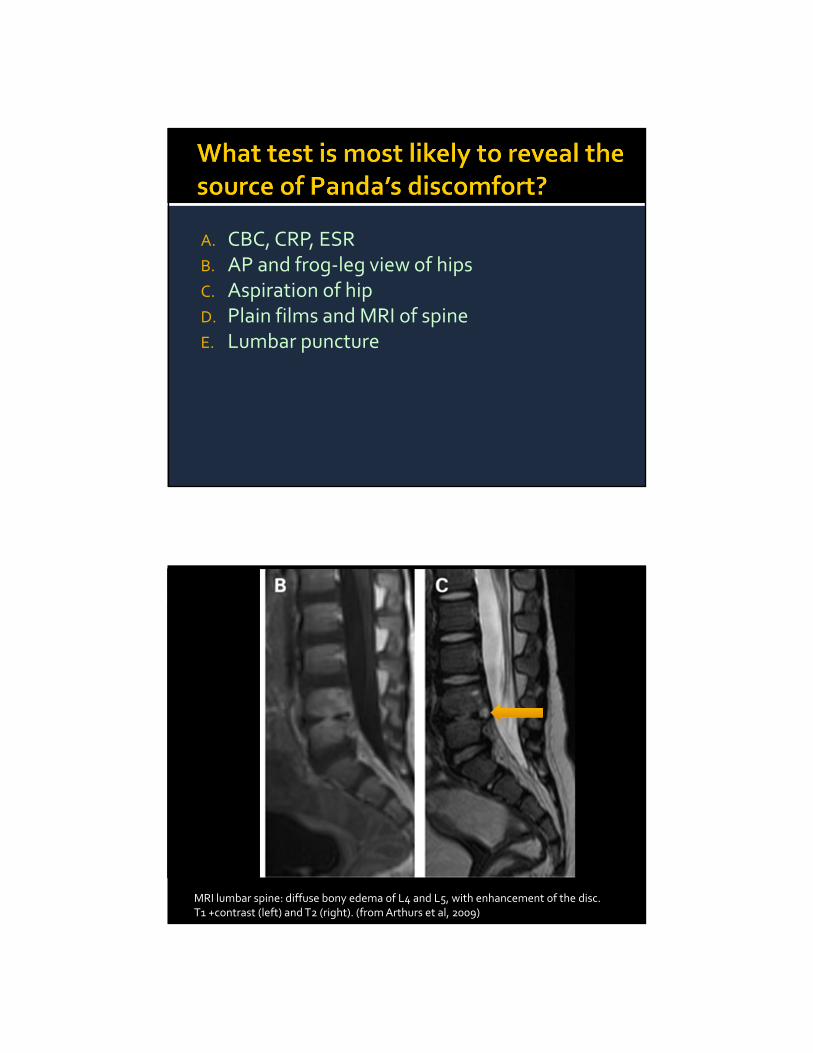
MRI lumbar spine: diffuse bony edema of L4 and L5, with enhancement of the disc.T1 +contrast (left) and T2 (right). (from Arthurs et al, 2009)
Inflammatory/infectious etiology
Diagnosis commonly delayed
Refusal to walk/sit/limp/crying > back pain
Recent 18 year series (Fernandez, 2000)
Mean age: 2.8 years
Only 28% febrile
Mean days of symptoms = 22
Fernandez, Pediatrics, 2000
Diagnostic pearls: Inflammatory markers poor predictors (may be normal)
MRI best sensitivity/specificity▪ 76% seen on plain film (narrowing of disc @ 2‐4 wks)
Consider scintigraphy – sensitive but non‐specific Management Blood cultures rarely positive
Parenteral antibiotics (vanco, clinda) recommended▪ In some series, patients did well without antibiotics
Follow ESR/CRP

Kodiak is a 4 week old boy brought in for “crying nonstop”
Seen by PCP yesterday, told it was colic Not feeding well, and seems to cry morewith the 5 S’s….
PMH: ex‐ 32 weeker, got “a few days” of antibiotics after birth, no other illnesses
T= 36.0, P 190, R 50, BP 90/50 Very fussy, inconsolable Flat fontanelle, well‐perfused, no rash Slight erythema/warmth/swelling of left calf
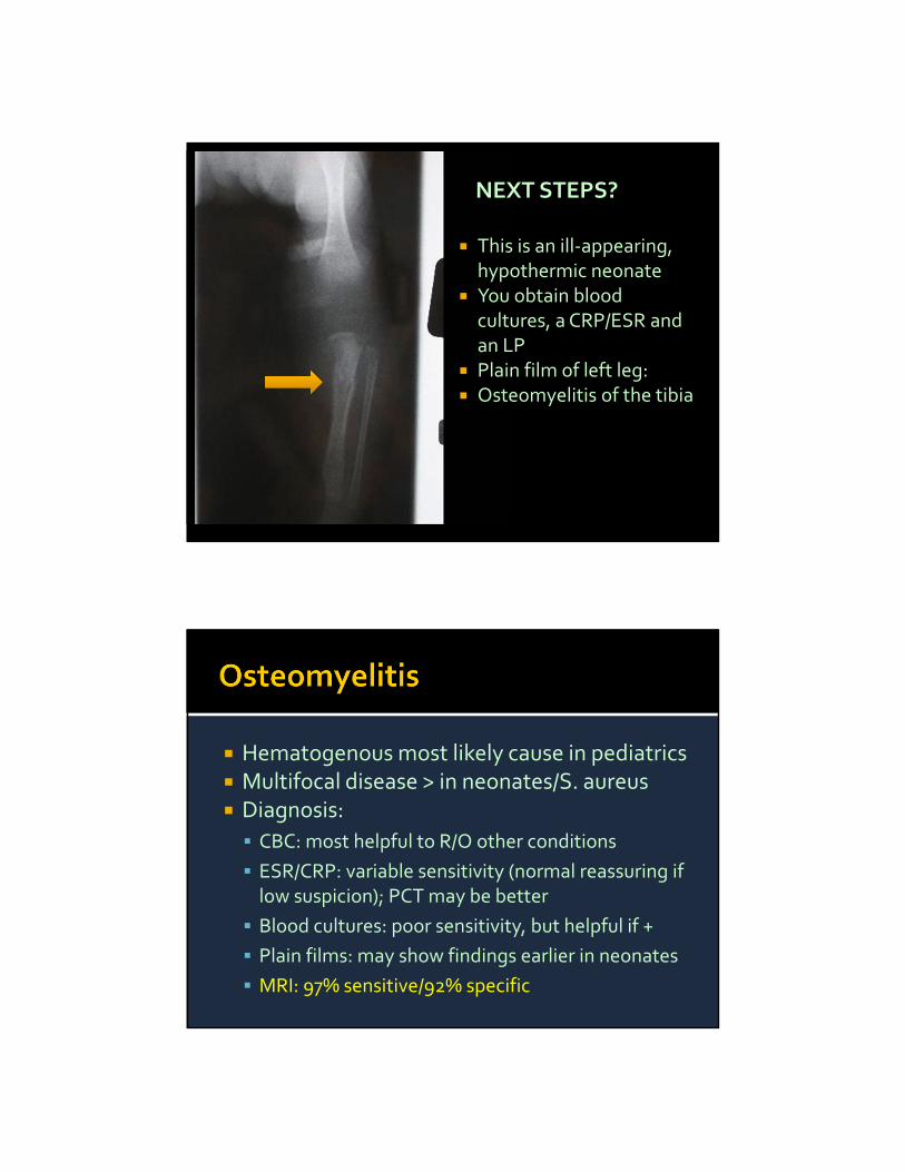
NEXT STEPS?
This is an ill‐appearing, hypothermic neonate
You obtain blood cultures, a CRP/ESR and an LP
Plain film of left leg: Osteomyelitis of the tibia
Hematogenous most likely cause in pediatrics Multifocal disease > in neonates/S. aureus Diagnosis:
CBC: most helpful to R/O other conditions
ESR/CRP: variable sensitivity (normal reassuring if low suspicion); PCT may be better
Blood cultures: poor sensitivity, but helpful if +
Plain films: may show findings earlier in neonates
MRI: 97% sensitive/92% specific

Neonatal:
S. aureus (MRSA), E, Coli, GBS (late‐onset)
Vancomycin and cefotaxime
Infants/kids:
S. aureus (MRSA), GAS: vancomycin
Kingella? Add cefazolin
Sickle cell? Add ceftriaxone
Gobi is a 6 mo old girl, brought in for “crying nonstop”
Usually consolable when held, but now it seems to make her cry more
Dad notes that she seems to be breathing fast, but otherwise has been afebrile, eating well, and no other symptoms
No PCP identified, but has been “healthy”

T 37.3, P 130, R 45, O2 sat 99% Well‐nourished, comfortably tachypneic, no rashes/bruises, smiles and coos when sitting in dad’s lap
Screams when you pick her up, and will not lie on her back
You are able to range all of the limbs without difficulty, the rest of the exam is normal

Virtually pathognomonic for abuse
Can be missed on plain films
Let radiologist know what you are looking for…
Thoracic cage, sternum, scapula, spine Metaphyseal corner lesions (MCL)/bucket handle fractures
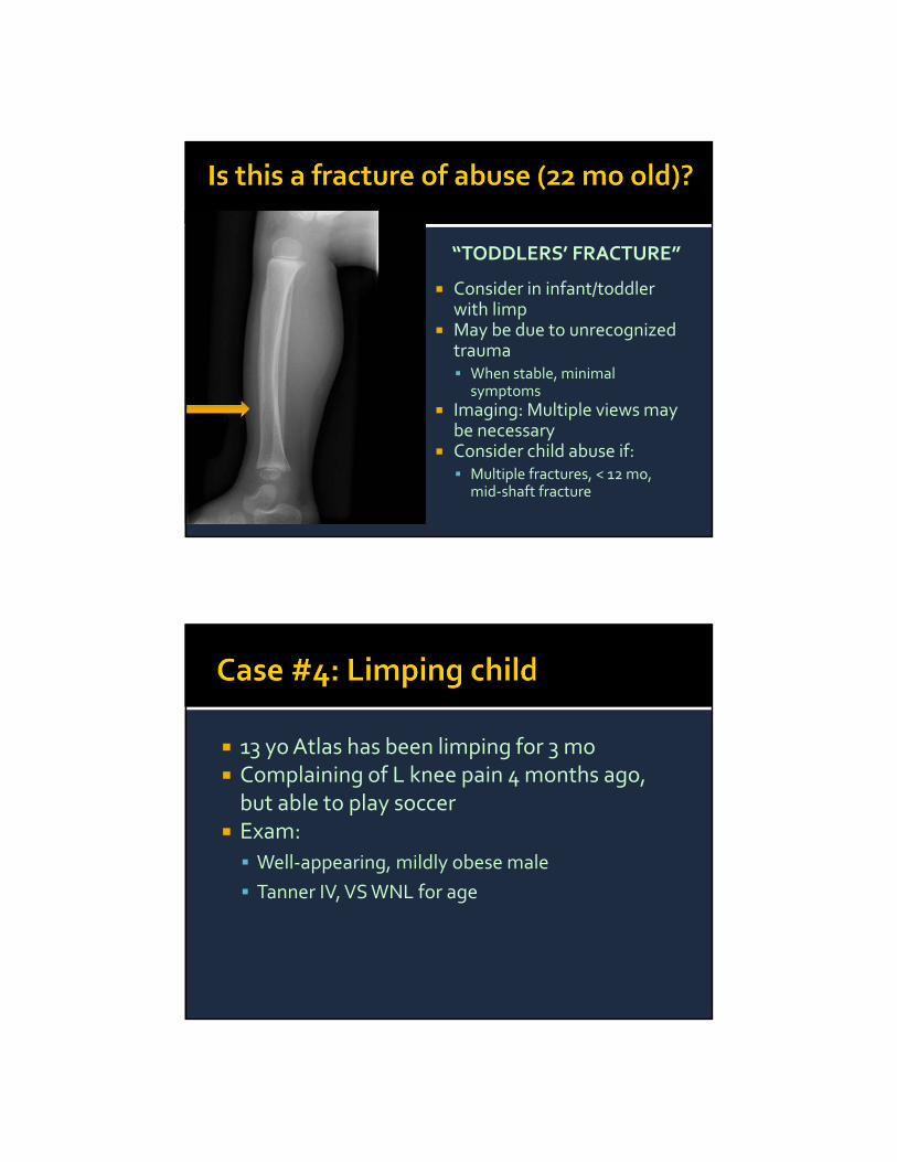
“TODDLERS’ FRACTURE”
Consider in infant/toddler with limp
May be due to unrecognized trauma When stable, minimal
symptoms Imaging: Multiple views may
be necessary Consider child abuse if: Multiple fractures, < 12 mo,
mid‐shaft fracture
13 yoAtlas has been limping for 3 mo Complaining of L knee pain 4 months ago, but able to play soccer
Exam:
Well‐appearing, mildly obese male
Tanner IV, VS WNL for age

Lies with L leg flexed and externally rotated
Obligate external rotation on flexion of L hip
Internal rotation of L hip severely limited
Knee exam normal

Normal Abnormal
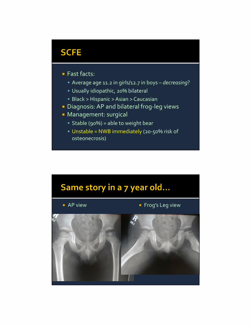
Fast facts:
Average age 11.2 in girls/12.7 in boys – decreasing?
Usually idiopathic, 20% bilateral
Black > Hispanic > Asian > Caucasian
Diagnosis: AP and bilateral frog‐leg views Management: surgical
Stable (90%) = able to weight bear
Unstable = NWB immediately (20‐50% risk of osteonecrosis)
AP view Frog’s Leg view
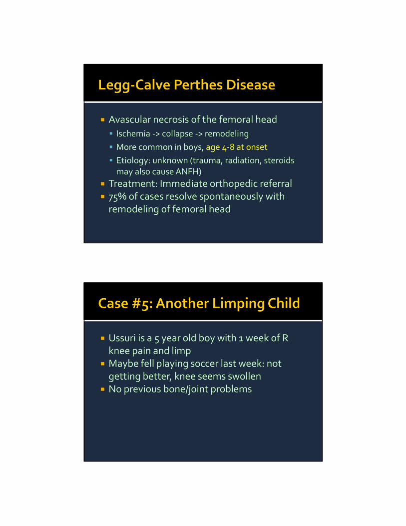
Avascular necrosis of the femoral head
Ischemia ‐> collapse ‐> remodeling
More common in boys, age 4‐8 at onset
Etiology: unknown (trauma, radiation, steroids may also cause ANFH)
Treatment: Immediate orthopedic referral 75% of cases resolve spontaneously with remodeling of femoral head
Ussuri is a 5 year old boy with 1 week of R knee pain and limp
Maybe fell playing soccer last week: not getting better, knee seems swollen
No previous bone/joint problems
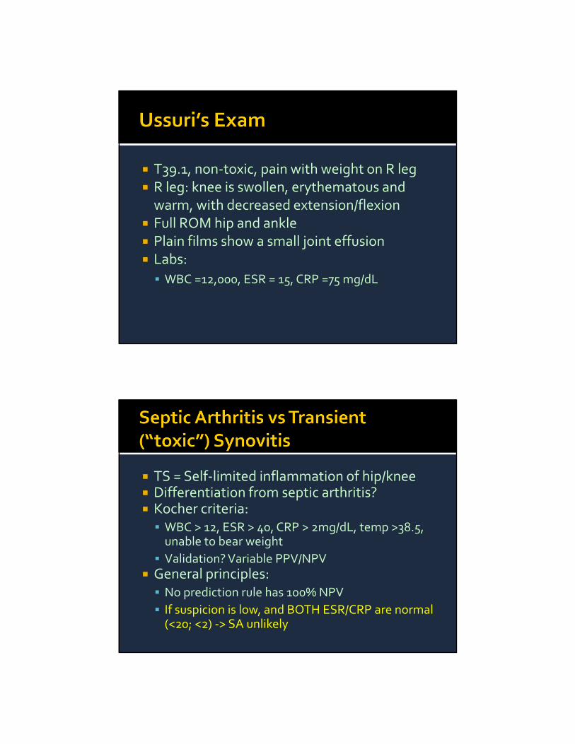
T39.1, non‐toxic, pain with weight on R leg R leg: knee is swollen, erythematous and warm, with decreased extension/flexion
Full ROM hip and ankle Plain films show a small joint effusion Labs:
WBC =12,000, ESR = 15, CRP =75 mg/dL
TS = Self‐limited inflammation of hip/knee Differentiation from septic arthritis? Kocher criteria: WBC > 12, ESR > 40, CRP > 2mg/dL, temp >38.5, unable to bear weight
Validation? Variable PPV/NPV General principles: No prediction rule has 100% NPV
If suspicion is low, and BOTH ESR/CRP are normal (<20; <2) ‐> SA unlikely
Most common cause of hip pain in kids 3‐10 years of age
Etiology unknown Management: NSAID’s, rest 1‐2% develop LCP
Joint aspiration reveals 40,000 WBC What is your next step?
Steroids prior to abx:
Reduce duration of sx, treatment and hospitalization and improve long‐term outs
Antibiotic choice
Vancomycin for good MRSA/GAS coverage
+ cefotaxime in neonate (E Coli)
+ consider cefazolin for Kingella in kids < 3
+ ceftriaxone in teens/sickle cell
Harel 2011; Odio 2003
Nandi is a 15 yo girl complaining of R knee pain for 2 months
Pain is intermittent, improves at night Told she has “growing pains” No specific trauma, but has been unable to play basketball
Exam: tender mass distal R thigh, otherwise normal
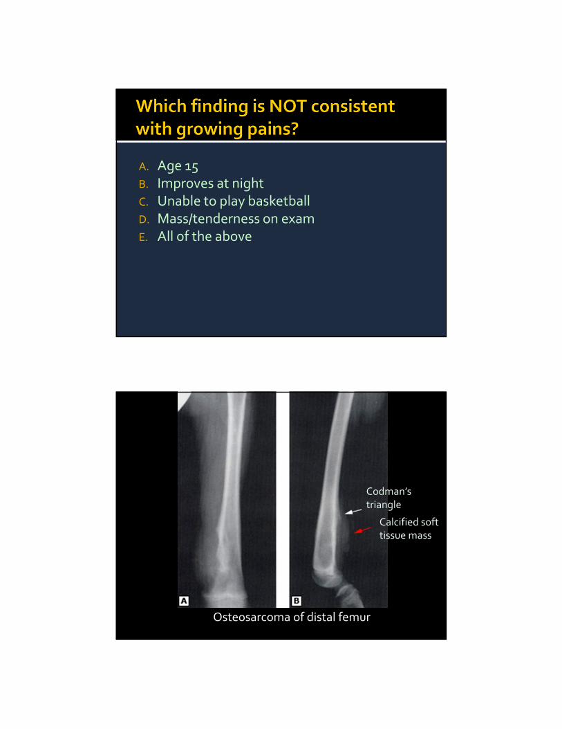
A. Age 15B. Improves at nightC. Unable to play basketballD. Mass/tenderness on examE. All of the above
Osteosarcoma of distal femur
Calcified soft tissue mass
Codman’s triangle

Osteosarcoma > Ewing’s Peak age: 13‐16, boys:girls = 1.5:1 Delay in dx common: average 2‐3 mo Clinical:
Intermittent pain, improves at night
Mass in 30‐40%
Long bones most frequently involved
Constitutional symptoms are rare
Ewing’s Sarcoma: Onion‐skinningPelvis> long bones
Osteosarcoma: sunburst reaction

Pedi orthopedic emergencies may present as crying, limp or refusal to walk
Consider in neonates/infants with unexplained fever Diagnosis/Treatment: MRI = study of choice for discitis, OM
Plain films sufficient to diagnose SCFE, LCP and bone malignancies (but get the right views!)
CRP/ESR: if normal = reassuring against septic arthritis (but get fluid if concern is high)
Steroids before antibiotics in SA enhances recovery
Andrea Marmor, MD UCSF High Risk Emergency Medicine May, 2014
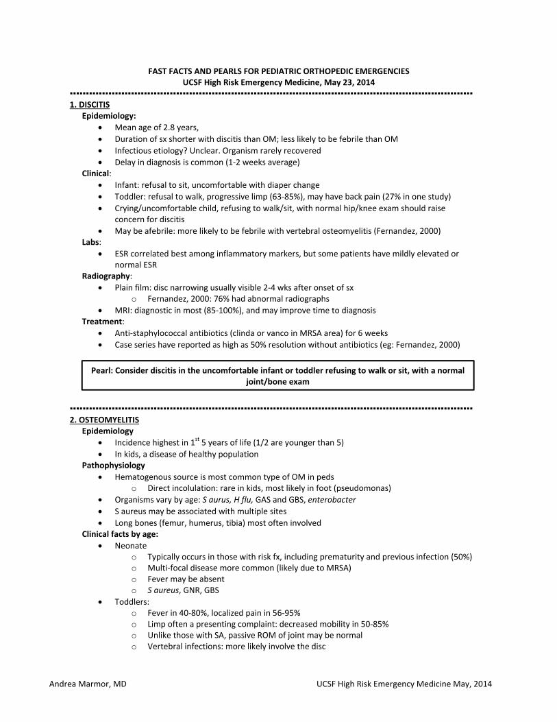
FAST FACTS AND PEARLS FOR PEDIATRIC ORTHOPEDIC EMERGENCIES UCSF High Risk Emergency Medicine, May 23, 2014
▪▪▪▪▪▪▪▪▪▪▪▪▪▪▪▪▪▪▪▪▪▪▪▪▪▪▪▪▪▪▪▪▪▪▪▪▪▪▪▪▪▪▪▪▪▪▪▪▪▪▪▪▪▪▪▪▪▪▪▪▪▪▪▪▪▪▪▪▪▪▪▪▪▪▪▪▪▪▪▪▪▪▪▪▪▪▪▪▪▪▪▪▪▪▪▪▪▪▪▪▪▪▪▪▪▪▪▪▪▪▪▪▪▪▪▪▪▪▪▪▪▪▪▪▪ 1. DISCITIS
Epidemiology:
Mean age of 2.8 years,
Duration of sx shorter with discitis than OM; less likely to be febrile than OM
Infectious etiology? Unclear. Organism rarely recovered
Delay in diagnosis is common (1‐2 weeks average) Clinical:
Infant: refusal to sit, uncomfortable with diaper change
Toddler: refusal to walk, progressive limp (63‐85%), may have back pain (27% in one study)
Crying/uncomfortable child, refusing to walk/sit, with normal hip/knee exam should raise concern for discitis
May be afebrile: more likely to be febrile with vertebral osteomyelitis (Fernandez, 2000) Labs:
ESR correlated best among inflammatory markers, but some patients have mildly elevated or normal ESR
Radiography:
Plain film: disc narrowing usually visible 2‐4 wks after onset of sx o Fernandez, 2000: 76% had abnormal radiographs
MRI: diagnostic in most (85‐100%), and may improve time to diagnosis Treatment:
Anti‐staphylococcal antibiotics (clinda or vanco in MRSA area) for 6 weeks
Case series have reported as high as 50% resolution without antibiotics (eg: Fernandez, 2000)
▪▪▪▪▪▪▪▪▪▪▪▪▪▪▪▪▪▪▪▪▪▪▪▪▪▪▪▪▪▪▪▪▪▪▪▪▪▪▪▪▪▪▪▪▪▪▪▪▪▪▪▪▪▪▪▪▪▪▪▪▪▪▪▪▪▪▪▪▪▪▪▪▪▪▪▪▪▪▪▪▪▪▪▪▪▪▪▪▪▪▪▪▪▪▪▪▪▪▪▪▪▪▪▪▪▪▪▪▪▪▪▪▪▪▪▪▪▪▪▪▪▪▪▪▪ 2. OSTEOMYELITIS
Epidemiology
Incidence highest in 1st 5 years of life (1/2 are younger than 5)
In kids, a disease of healthy population Pathophysiology
Hematogenous source is most common type of OM in peds o Direct incolulation: rare in kids, most likely in foot (pseudomonas)
Organisms vary by age: S aurus, H flu, GAS and GBS, enterobacter
S aureus may be associated with multiple sites
Long bones (femur, humerus, tibia) most often involved Clinical facts by age:
Neonate o Typically occurs in those with risk fx, including prematurity and previous infection (50%) o Multi‐focal disease more common (likely due to MRSA) o Fever may be absent o S aureus, GNR, GBS
Toddlers: o Fever in 40‐80%, localized pain in 56‐95% o Limp often a presenting complaint: decreased mobility in 50‐85% o Unlike those with SA, passive ROM of joint may be normal o Vertebral infections: more likely involve the disc
Pearl: Consider discitis in the uncomfortable infant or toddler refusing to walk or sit, with a normal joint/bone exam

Andrea Marmor, MD UCSF High Risk Emergency Medicine May, 2014
o S aureus and GAS
Older Kids: o More likely to have localized pain o Brodie’s abscess: occurs most commonly in teens
Differential dx:
Neonate/Infant: occult fx, other infection, malignancy (rare) NM disorder,
Toddler/kid: septic arthritis, cellulitis, malignancy, bone infarction, Caffey’s disease (infantile cortical hyperostosis)/fibrodysplasia ossificans progressive
Labs
Labs should be interpreted in light of clinical suspicion
ESR: o 70‐100% sensitive; lower sens in puncture‐related OM (Harris, 2011)
CRP: o Prospective trial (Unkila‐Kallio): CRP performed as well as ESR o Jaakkola and Kehl: only 47% sensitive
PCT better predictor of OM than other bone/joint infections, but sensitivity still poor o Butbul‐Aviel, 2005: PCT value was elevated in 7 patients (58.3%) with osteomyelitis, only
3 children (27.2%) with septic arthritis and NO children with other (benign) diagnoses
CBC o Generally lacks sens/spec, but may identify other conditions (eg: leukemia) o Lower sensitivity (12‐58%)
Blood cultures o Poor sensitivity (< 50%) – but may be helpful in isolating organism (eg: Kingella –
fastidious org, longer to grow) o Only 40‐60% of bone cultures are +
Imaging:
Plain film: evidence by 10‐21 days (in neonates, may be apparent by 7‐10 days)
Scintigraphy may be useful when attempting to localize infection (80‐100% sens/70‐96% spec)
MRI: best imaging modality 97% sens/92% spec Treatment
Neonate: cefotaxime + vanco
Older infants/kids: vancomycin (+nafcillin if known MSSA) o Kingella; susc to cephalosporins but resistant to vanco/clinda: consider adding cefazolin
if suspected
Sickle cell; add ceftriaxone for salmonella/H. flu
▪▪▪▪▪▪▪▪▪▪▪▪▪▪▪▪▪▪▪▪▪▪▪▪▪▪▪▪▪▪▪▪▪▪▪▪▪▪▪▪▪▪▪▪▪▪▪▪▪▪▪▪▪▪▪▪▪▪▪▪▪▪▪▪▪▪▪▪▪▪▪▪▪▪▪▪▪▪▪▪▪▪▪▪▪▪▪▪▪▪▪▪▪▪▪▪▪▪▪▪▪▪▪▪▪▪▪▪▪▪▪▪▪▪▪▪▪▪▪▪▪▪▪▪▪ 3. TODDLERS’ FRACTURE:
Definition: a spiral fracture of the distal tibia, typically associated with the accidental twisting of the distal leg that occurs when a toddler catches their foot while running/walking
Toddlers’ fracture may be a subset of CAST fractures (childhood accidental spiral tibial fractures) Accidental fractures typically occur in distal ½ of tibia, without displacement
Age: In one series, 1/3 occur in kids < 3, NONE in kids < 12 mo (Mellick, 1999) Overall, the majority of fractures of abuse occur in kids < 12 mo:
Pearl: Consider osteomyelitis in neonates with FWS, and cover empirically for GBS, enterococcus and S. aureus
Pearl: Osteomyelitis is most often hematogenous in the pediatric population

Andrea Marmor, MD UCSF High Risk Emergency Medicine May, 2014
Accidental fx are RARE in kids < 12 mo Diagnosis:
Initial radiographs may be normal in 43% of cases Internal oblique view best for visualizing the spiral fracture
▪▪▪▪▪▪▪▪▪▪▪▪▪▪▪▪▪▪▪▪▪▪▪▪▪▪▪▪▪▪▪▪▪▪▪▪▪▪▪▪▪▪▪▪▪▪▪▪▪▪▪▪▪▪▪▪▪▪▪▪▪▪▪▪▪▪▪▪▪▪▪▪▪▪▪▪▪▪▪▪▪▪▪▪▪▪▪▪▪▪▪▪▪▪▪▪▪▪▪▪▪▪▪▪▪▪▪▪▪▪▪▪▪▪▪▪▪▪▪▪▪▪▪▪▪ 4. SCFE
Epidemiology:
Average age: 12.7 years for boys and 11.2 years for girls, o Near the end of linear growth, prior to menarche/ Tanner IV o Age decreasing over time: earlier puberty?
Lehmann (2006): o Rates almost 4 x higher in blacks, 2.5 x higher in Hispanics and 1.62x higher in
Asian/Pacific Islanders compared to white children
Obesity is recognized as a strongly associated factor of SCFE. o Increased BMI increases the shear stress across the physis, thus weakening it and
causing a slip. The varying surge and level of hormonal activity associated with adolescent growth spurt may also contribute to the cause of SCFE
Commonly bilateral (~20%)
Rarely the result of an endocrine or metabolic disorder
Delay in diagnosis worsens prognosis Clinical Factors:
History o Typically present with knee, hip, groin, thigh pain or all o May be trivial trauma or discomfort (painless limp also common) o Acute major trauma is rarely involved; gradual onset of symptoms and deformity
(external rotation) is more common
Exam: o Limited internal rotation is UNIVERSAL o External rotation of extremity o Obligatory external rotation with passive flexion of 90 degrees
Diagnosis:
Radiology: plain films diagnostic in most, although may be negative with early/posterior slip o AP and frog‐leg lateral (frog‐leg view more sensitive) o Important to visualize both hips o Klein’s line (AP view): line drawn along femoral neck should intersect the lateral portion
of the femoral head – if not, suspect SCFE Management:
Treatment is surgical, with stabilization across the physis by in‐situ pinning
Urgency based on stability: o Stable = able to bear weight (>90%)
Manage surgically as soon as possible May progress to a more severe or unstable slip
o Unstable = unable to bear weight (even with support) Make non‐weight bearing immediately and admit Risk of osteonecrosis 20‐50%)
Pearl: A spiral tibial fracture in a child < 12 months should prompt concern for child abuse
Pearl: Always get bilateral hip views in suspected SCFE: 20% are bilateral

Andrea Marmor, MD UCSF High Risk Emergency Medicine May, 2014
▪▪▪▪▪▪▪▪▪▪▪▪▪▪▪▪▪▪▪▪▪▪▪▪▪▪▪▪▪▪▪▪▪▪▪▪▪▪▪▪▪▪▪▪▪▪▪▪▪▪▪▪▪▪▪▪▪▪▪▪▪▪▪▪▪▪▪▪▪▪▪▪▪▪▪▪▪▪▪▪▪▪▪▪▪▪▪▪▪▪▪▪▪▪▪▪▪▪▪▪▪▪▪▪▪▪▪▪▪▪▪▪▪▪▪▪▪▪▪▪▪▪▪▪▪ 5. LEGG‐CALVE‐PERTHES DISEASE
Idiopathic avascular necrosis of hip
Epidemiology/Presentation o Clinical: Insidious onset of limp, with pain often referred to thigh or knee o Peak incidence between 5 and 7 (seen between ages 3 and 12) o 10% of cases are familial o Male: female ratio = 4:1
Exam: Limited internal rotation of hip, may result in atrophy of thigh/buttocks o Galeazzi test (leg length discrepancy) and Trendelenberg test (for unilateral gluteal
muscle weakness) may be positive o Trendelenburg test also abnormal in SCFE, DDH ‐ suggests hip pathology
Diagnosis: o Generally visible on plain film, although initial radiographs may be normal o Obtain AP and lateral films, and views of both hips. o Repeat if symptoms persistent
Management: make NON‐WT‐BEARING immediately, and obtain orthopedic consultation
▪▪▪▪▪▪▪▪▪▪▪▪▪▪▪▪▪▪▪▪▪▪▪▪▪▪▪▪▪▪▪▪▪▪▪▪▪▪▪▪▪▪▪▪▪▪▪▪▪▪▪▪▪▪▪▪▪▪▪▪▪▪▪▪▪▪▪▪▪▪▪▪▪▪▪▪▪▪▪▪▪▪▪▪▪▪▪▪▪▪▪▪▪▪▪▪▪▪▪▪▪▪▪▪▪▪▪▪▪▪▪▪▪▪▪▪▪▪▪▪▪▪▪▪▪ 6. SKELETAL MALIGNANCIES: OSTEOSARCOMA VS EWING’S SARCOMA
Osteosarcoma Ewing’s Sarcoma “Growing pains”
Origin Primitive bone mesenchyme Poorly differentiatiated (?mesenchyme?)
Unknown (NOT caused by growth!)
Gender Boys > girls (1.5:1) Boys > girls (1.5:1) Girls> boys
Age Peak 13‐16 (growth spurt)Adults > 65
Peak 13‐16Can be seen into 40’s
2‐12 years
Race Black> Caucasian Caucasian> Black/Asian None
Frequency Rare; most common bone malignancy
Rare; 2nd most common bone malignancy
Common (10‐20%)
Location Metaphyses of long bones (distal femur> proximal tibia, proximal humerus)
Pelvis > metaphysis/diaphysis of LE long bones > spine
More common in lower extremities
Clinical Signs/ Symptoms
Intermittent local pain/ tenderness
Rarely at night +/‐ mass (~30‐40%)
Average 2‐3 months duration
Constitutional symptoms rare
Intermittent local pain/ tenderness
Rarely at night +/‐ mass (~30‐40%)
Average 3‐4 mo duration
Constitutional symptoms: 10‐20%
Nightly bilateral, deep pain in thigh/calf
Absent during day No physical findings Chronic, episodic pattern Otherwise normal activity
Radiographic appearance
Lytic/sclerotic mass
Calcified soft tissue mass
“Sunburst” periosteal reaction
“Moth‐eaten” lytic/sclerotic mass
“Onion‐skinning” of periosteum
None
Pearl: Findings suggestive of a SCFE in younger child (5‐7 years of age) should prompt concern for Legg‐Calve‐Perthes Disease
Pearl: “Growing pains” should never cause pain during the day, or interfere with activity

Andrea Marmor, MD UCSF High Risk Emergency Medicine May, 2014
▪▪▪▪▪▪▪▪▪▪▪▪▪▪▪▪▪▪▪▪▪▪▪▪▪▪▪▪▪▪▪▪▪▪▪▪▪▪▪▪▪▪▪▪▪▪▪▪▪▪▪▪▪▪▪▪▪▪▪▪▪▪▪▪▪▪▪▪▪▪▪▪▪▪▪▪▪▪▪▪▪▪▪▪▪▪▪▪▪▪▪▪▪▪▪▪▪▪▪▪▪▪▪▪▪▪▪▪▪▪▪▪▪▪▪▪▪▪▪▪▪▪▪▪▪ 7. SEPTIC ARTHRITIS
Epidemiology/Pathophysiology
80% lower extremity (hip and knee most common)
10 % more than one joint Causes
Neonates/infants = GBS, N. gonorrhea, E.coli, S. aureus
Infants/Toddlers: S. aureus (including MRSA) + Kingella kingae o Kingella = gram negative coccobacillus, an emerging pediatric pathogen o Nationwide study of Kingella (Dubnov‐Raz, 2010):
96% of children with Kingella were <3 53% of infections were skeletal infections 43% of infections were bacteremia
Diagnosis
Also consider: Toxic Synovitis, JIA, post‐strep arthritis, serum sickness, trauma, SCFE, LCP
Labs: o ESR and CRP better negative than positive predictors o Eg: CRP < 1 mg/dL has NPV of 87% (Levine 2003) o CRP peaks 36‐50hrs after onset of infection o PCT a poor predictor: only 27% sensitivity in one study (Butbul‐Aviel, 2005)
Radiography o Plain radiographs:
May demonstrate joint effusion, but not sensitive Frog leg view of hips: may increase sensitivity for joint effusion Better to R/O other bone abnormalities
o Ultrasound: May identify and quantify joint effusion, high NPV for hip arthritis
o Bone scan: Generally not indicated, unless searching for a source of fever, or osteo
suspected o MRI:
May help distinguish b/w SA and TS/ Can evaluate comcominant osteo or abscess
Fluid: o Best diagnostic test, but studies inconsistent due to varying gold standards o Higher WBC associated with higher likelihood of SA o WBC > 50 K with > 90% neuts suggests SA, but is not 100% sensitive or specific
Treatment
Antibiotics o Neonate (< 3 mo):
Bugs: S. aureus, GBS, E. Coli Drugs: vanco + ceftriaxone or cefotaxime Consider gonorrhea if risk factors present
o Infants/kids (> 3 mo): Bugs: S. aureus, GAS (Kingella in kids < 3 yrs) Drugs: vancomycin or clindamycin Consider adding cefazolin for Kingella in kids < 3 years Consider adding CTX in teens (N. gonorrhea) or sickle cell (Salmonella spp)
Corticosteroids: o Dexamethasone before antibiotics speeds recovery and improves long‐term outcomes
o Harel 2011: Shortened duration of fever, inflammation, parenteral therapy and hospital stay
Andrea Marmor, MD UCSF High Risk Emergency Medicine May, 2014
o Odio 2003: Shortened hospital stay and reduction in residual dysfunction at 6 and 12 months
o Recs: Dexamethasone 0.15mg/kg IV before antibiotics, and q6h for 4 days
▪▪▪▪▪▪▪▪▪▪▪▪▪▪▪▪▪▪▪▪▪▪▪▪▪▪▪▪▪▪▪▪▪▪▪▪▪▪▪▪▪▪▪▪▪▪▪▪▪▪▪▪▪▪▪▪▪▪▪▪▪▪▪▪▪▪▪▪▪▪▪▪▪▪▪▪▪▪▪▪▪▪▪▪▪▪▪▪▪▪▪▪▪▪▪▪▪▪▪▪▪▪▪▪▪▪▪▪▪▪▪▪▪▪▪▪▪▪▪▪▪▪▪▪▪ 8. SEPTIC ARTHRITIS (SA) VS TRANSIENT SYNOVITIS (TS)
Both present with similar symptoms, in similar joints, and in similar patient populations Multiple studies have attempted to develop a clinical prediction rule that can identify children at
low risk of SA Kocher Criteria
Kocher (1999) found that 5 findings were associated with septic arthritis (99.7% positive predictive value, AUC of .96) 1. Fever ≥38.5º C (101ºF) 2. Inability to bear weight 3. White blood cell count >12,000/mm3 4. Erythrocyte sedimentation rate >40 mm per hour 5. C‐reactive protein > 2.0 mg/dL (20 mg/L)
Subsequent Validation of Kocher criteria: PPV varies from 59‐93% in retrospective and prospective studies (Kocher 2004, Luhmann 2004,
Caird 2006), NPV only 83% (Caird)
Other predictive models:
Luhmann, 2004 (retrospective) o 3 variables had PPV of 71% for septic arthritis:
1. History of fever 2. WBC of >12K 3. Previous health‐care visit
Singhal, 2007 (retrospective) o A CRP > 20 mg/l had OR of 81.9 o 2 determinants
1. Weight‐bearing status 2. CRP > 20 mg/l
o Absence of both: < 1% had septic arthritis o Presence of both: 74% had septic arthritis
Pakkonen, 2010 (prospective) o Best sensitivity (98%) for SA with combined ESR (>20) and CRP (>20 mg/L)
Caird 2006 (prospective) o C‐reactive protein level of >2.0 mg/dL (>20 mg/L) was a strong independent risk factor and
a valuable tool for assessing and diagnosing children suspected of having septic arthritis of the hip.
Pearl: In differentiation SA and TS, take into account the entire clinical picture If suspicion is high, obtain hip ultrasound and arthrocentesis even if labs are normal If suspicion is low, and CRP/ESR are normal, unlikely to have SA

Andrea Marmor, MD UCSF High Risk Emergency Medicine May, 2014
▪▪▪▪▪▪▪▪▪▪▪▪▪▪▪▪▪▪▪▪▪▪▪▪▪▪▪▪▪▪▪▪▪▪▪▪▪▪▪▪▪▪▪▪▪▪▪▪▪▪▪▪▪▪▪▪▪▪▪▪▪▪▪▪▪▪▪▪▪▪▪▪▪▪▪▪▪▪▪▪▪▪▪▪▪▪▪▪▪▪▪▪▪▪▪▪▪▪▪▪▪▪▪▪▪▪▪▪▪▪▪▪▪▪▪▪▪▪▪▪▪▪▪▪▪▪▪▪ REFERENCES:
1. Arthurs OJ, et al. The toddler refusing to weight‐bear: a revised imaging guide from a case series. Emerg Med J 2009;26:797‐801
2. Bhatia NN, Pirpiris M, Otsuka NY. Body mass index in patients with slipped capital femoral epiphysis. J Pediatr Orthop. 2006 Mar‐Apr;26(2):197‐9.
3. Brown R, Hussain M, McHugh K, et al. Discitis in young children.J Joint Bone Surgery (Br)2001;83‐B:106–11.
4. Butbul‐Aviel Y, et al. Procalcitonin as a diagnostic aid in osteomyelitis and septic arthritis. Pediatr Emerg Care. 2005 Dec;21(12):828‐32.
5. Caird MS, et al. Factors distinguishing septic arthritis from transient synovitis of the hip in children. A prospective study. J Bone Joint Surg Am. 2006 Jun;88(6):1251‐7.
6. Dodwell ER. Osteomyelitis and septic arthritis in children: current concepts. Curr Opin Pediatr 2013; 25(1): 58‐63
7. Dubnov‐Raz G, et al. Invasive pediatric Kingella kingae infections: a nationwide collaborative study. Pediatr Infect Dis 2010; 29(7): 639‐43
8. Fernandez M, Carrol CL, Baker CJ Discitis and vertebral osteomyelitis in children: an 18‐year review. Pediatrics. 2000;105(6):1299
9. Gholve PA, Cameron DB, Millis MB. Slipped capital femoral epiphysis update. Curr Opin Pediatr. 2009 Feb;21(1):39‐45.
10. Harris JC, et al. How useful are laboratory investigations in the emergency department evaluation of possible osteomyelitis? Emerg Med Australas. 2011 Jun;23(3):317‐30
11. Harel L, et al. Dexamethasone therapy for septic arthritis in children: results of a randomized controlled study. J Pediatr Orthop 2011; 31(2): 211‐5
12. Karabouta Z, et al. Discitis in toddlers: a case series and review. Acta Paediatrica 2005; 94(10): 1516‐1518 13. Kocher MS et al. Validation of a clinical prediction rule for the differentiation between septic arthritis and
transient synovitis of the hip in children. J Bone Joint Surg Am 2004; 86‐A(8): 1629‐35 14. Lehmann C, et al. The epidemiology of slipped capital femoral epiphysis: an update. J Pediatr Orth 2006;
26(3): 286‐290 15. Loder RT, Starnes T, Dikos G, Aronsson DD. Demographic predictors of severity of stable slipped capital
femoral epiphyses. J Bone Joint Surg Am 2006; 88:97–105. 16. Luhmann SJ, et al. Differentiation between septic arthritis and transient synovitis of the hip in children
with clinical prediction algorithms. J Bone Joint Surg Am 2004; 86‐A(5): 956‐62 17. McCarville MB. The child with bone pain: malignancies and mimickers. Cancer Imaging. 2009 Oct 2;9 Spec
No A:S115‐21. 18. Mellick LB, et al. Childhood accidental spiral tibial (CAST) fractures. Pediatr Emerg Care 1999; 15(5): 307‐9 19. Odio CM, et al. Double blind, randomized, placebo‐controlled study of dexamethasone therapy for
hematogenous septic arthritis in children. Pediatr Infect Dis J 2003; 22(10): 883‐889 20. Pääkkönen M, Kallio MJ, Kallio PE, Peltola H. Sensitivity of erythrocyte sedimentation rate and C‐reactive
protein in childhood bone and joint infections. Clin Orthop Relat Res. 2010 Mar;468(3):861‐6. Epub 2009 Jun 17.
21. Singhal R, Perry DC, et al. The use of CRP within a clinical prediction algorithm for the differentiation of septic arthritis and transient synovitis in children. J Bone Joint Surg Br. 2011 Nov;93(11):1556‐61.
22. Sultan J,Hughes PJ. Septic arthritis or transient synovitis of the hip in children: the value of clinical prediction algorithms. J Bone Joint Surg Br 2010; 92(9): 1289‐93
23. Widhe B, Widhe T. Initial symptoms and clinical features in osteosarcoma and Ewing sarcoma. J Bone Joint Surg 2000; 82(5): 667‐