marques stoco steindel vallejo grisard 2015 no.pdf
TRANSCRIPT

International Journal for Parasitology xxx (2015) xxx–xxx
Contents lists available at ScienceDirect
International Journal for Parasitology
journal homepage: www.elsevier .com/locate / i jpara
Trypanosoma rangeli displays a clonal population structure, revealing asubdivision of KP1(�) strains and the ancestry of the Amazonian group q
http://dx.doi.org/10.1016/j.ijpara.2014.11.0040020-7519/� 2015 The Authors. Published by Elsevier Ltd. on behalf of Australian Society for Parasitology Inc.This is an open access article under the CC BY-NC-SA license (http://creativecommons.org/licenses/by-nc-sa/4.0/).
q Sequence data reported in this paper are available in GenBank under accessionnumbers KJ525754–KJ525771.⇑ Corresponding author at: Universidade Federal de Santa Catarina, Cx. Postal
476, Florianópolis, SC 88.040-970, Brazil. Tel.: +55 (48) 3721 2955.E-mail addresses: [email protected] (T.C.M. Sincero), edmundo.grisard@ufsc.
br (E.C. Grisard).
Please cite this article in press as: Sincero, T.C.M., et al. Trypanosoma rangeli displays a clonal population structure, revealing a subdivision of KP1(�)and the ancestry of the Amazonian group. Int. J. Parasitol. (2015), http://dx.doi.org/10.1016/j.ijpara.2014.11.004
Thaís Cristine Marques Sincero a,⇑, Patricia Hermes Stoco b, Mário Steindel b, Gustavo Adolfo Vallejo c,Edmundo Carlos Grisard b,⇑a Universidade Federal de Santa Catarina (UFSC), Centro de Ciências da Saúde (CCS), Departamento de Análises Clínicas (ACL), Setor E, Bloco K, Florianópolis, SC 88.040-970, Brazilb Universidade Federal de Santa Catarina (UFSC), Centro de Ciências Biológicas (CCB), Departamento de Microbiologia, Imunologia e Parasitologia (MIP), Setor F, Bloco A,Florianópolis, SC 88.040-970, Brazilc Laboratorio de Investigaciones en Parasitología Tropical, Universidad del Tolima, Altos de Santa Helena, A.A. 546, Ibagué, Colombia
a r t i c l e i n f o a b s t r a c t
Article history:Received 3 July 2014Received in revised form 12 November 2014Accepted 24 November 2014Available online xxxx
Keywords:Genetic polymorphismVector–parasite coevolutionParasite ancestryIntra-specific variabilityMicrosatellite and SNP analysis
Assessment of the genetic variability and population structure of Trypanosoma rangeli, a non-pathogenicAmerican trypanosome, was carried out through microsatellite and single-nucleotide polymorphismanalyses. Two approaches were used for microsatellite typing: data mining in expressed sequence tag/open reading frame expressed sequence tags libraries and PCR-based Isolation of Microsatellite Arraysfrom genomic libraries. All microsatellites found were evaluated for their abundance, frequency and use-fulness as markers. Genotyping of T. rangeli strains and clones was performed for 18 loci amplified by PCRfrom expressed sequence tag/open reading frame expressed sequence tags libraries. The presence of sin-gle-nucleotide polymorphisms in the nuclear, multi-copy, spliced leader gene was assessed in 18 T. ran-geli strains, and the results show that T. rangeli has a predominantly clonal population structure, allowinga robust phylogenetic analysis. Microsatellite typing revealed a subdivision of the KP1(�) genetic group,which may be influenced by geographical location and/or by the co-evolution of parasite and vectorsoccurring within the same geographical areas. The hypothesis of parasite–vector co-evolution was cor-roborated by single-nucleotide polymorphism analysis of the spliced leader gene. Taken together, theresults suggest three T. rangeli groups: (i) the T. rangeli Amazonian group; (ii) the T. rangeli KP1(�) group;and (iii) the T. rangeli KP1(+) group. The latter two groups possibly evolved from the Amazonian group toproduce KP1(+) and KP1(�) strains.� 2015 The Authors. Published by Elsevier Ltd. on behalf of Australian Society for Parasitology Inc. This isan open access article under the CC BY-NC-SA license (http://creativecommons.org/licenses/by-nc-sa/4.0/).
1. Introduction
Trypanosoma rangeli and Trypanosoma cruzi are kinetoplastidprotozoan parasites infecting humans and a variety of wild anddomestic mammals in South and Central America. Due to thenon-pathogenic nature of T. rangeli to its mammalian hosts, thisparasite is a unique and interesting biological model for addressinghost–parasite interactions, and the genome has been recentlyunveiled (Snoeijer et al., 2004; Grisard et al., 2010; Stoco et al.,2014). Furthermore, considering the wide and superimposedgeographical distribution and sharing of reservoirs, vectors and
soluble antigens with T. cruzi, the etiological agent of Chagas dis-ease, T. rangeli is of epidemiological and medical importance, oftenleading to a misdiagnosis of South American trypanosomiasis andresulting in social and economic losses (Stevens et al., 2001;Guhl and Vallejo, 2003; Grisard et al., 2010).
Polymorphism among the T. rangeli strains isolated from mam-mals and triatomines from different geographical regions has beendemonstrated by molecular and biochemical methods. Two maingenetic lineages based on kinetoplast DNA (kDNA) mini-circle typ-ing have been described: KP1(+) and KP1(�) (Vallejo et al., 2002,2003, 2009). Comparative sequence analysis of the ssrDNA, inter-nal transcribed spacer 1 (ITS-1) and spliced leader intergenicregions (SL-IRs) noted increased variability within this taxon andresulted in the proposal of the existence of five lineages (A–E)(Maia da Silva et al., 2004, 2007, 2009).
The distinct molecular markers and methods used to assessDNA polymorphisms must consider (i) the expected level ofvariability, (ii) the rate of mutation, (iii) the mode of inheritance
strains
2 T.C.M. Sincero et al. / International Journal for Parasitology xxx (2015) xxx–xxx
(segregation during cell division), and (iv) the cost, time and needfor specialised equipment and practices. Based on the type of infor-mation generated by various loci, we may group these methodsinto three categories as follows: dominant bi-allelic (e.g., randomamplified polymorphic DNA (RAPD), amplified fragment lengthpolymorphism (AFLP), bi-allelic co-dominant (e.g., single-nucleo-tide polymorphism (SNP), restriction fragment length polymor-phism (RFLP), and multi-allelic co-dominant (e.g., microsatellites(MSs) (Vignal et al., 2002).
Despite the description of several MS loci for T. cruzi (Oliveiraet al., 1998, 1999; Macedo et al., 2001; Koffi et al., 2007; Njiruet al., 2007; Lewis et al., 2009; Llewellyn et al., 2009; Venegaset al., 2009; Simo et al., 2010), Trypanosoma brucei (Kanmogneet al., 1997; Jamonneau et al., 2002, 2004; Truc et al., 2002; Koffiet al., 2007; Cabrine-Santos et al., 2010; McInnes et al., 2012),and Trypanosoma evansi (Botero et al., 2010; McInnes et al.,2012), the number of these markers described for trypanosomatidsis still small. Similarly, the number of SNP markers in kinetoplast-ida has revealed a few loci, but only data for T. cruzi are available atdbSNP (http://www.ncbi.nlm.nih.gov/snp/) and TcSNP (http://snps.tcruzi.org/) (Ackermann et al., 2009). Because the number ofboth MS and SNP markers is relatively small for T. rangeli(Grisard et al., 1999; Urrea et al., 2011), we searched for and usedMS and SNP markers to assess the genetic variability and popula-tion structure of T. rangeli strains isolated from distinct geograph-ical regions, hosts and vectors.
2. Materials and methods
2.1. Parasites and DNA extraction
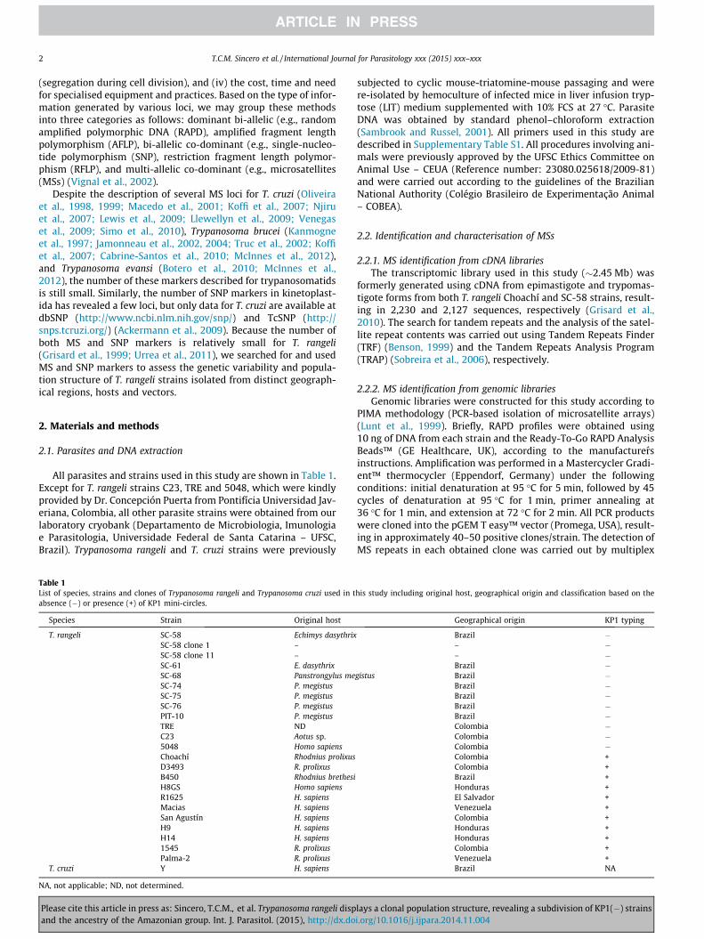
All parasites and strains used in this study are shown in Table 1.Except for T. rangeli strains C23, TRE and 5048, which were kindlyprovided by Dr. Concepción Puerta from Pontifícia Universidad Jav-eriana, Colombia, all other parasite strains were obtained from ourlaboratory cryobank (Departamento de Microbiologia, Imunologiae Parasitologia, Universidade Federal de Santa Catarina – UFSC,Brazil). Trypanosoma rangeli and T. cruzi strains were previously
Table 1List of species, strains and clones of Trypanosoma rangeli and Trypanosoma cruzi used in tabsence (�) or presence (+) of KP1 mini-circles.
Species Strain Original host
T. rangeli SC-58 Echimys dasythrixSC-58 clone 1 –SC-58 clone 11 –SC-61 E. dasythrixSC-68 Panstrongylus meSC-74 P. megistusSC-75 P. megistusSC-76 P. megistusPIT-10 P. megistusTRE NDC23 Aotus sp.5048 Homo sapiensChoachí Rhodnius prolixusD3493 R. prolixusB450 Rhodnius brethesiH8GS Homo sapiensR1625 H. sapiensMacias H. sapiensSan Agustín H. sapiensH9 H. sapiensH14 H. sapiens1545 R. prolixusPalma-2 R. prolixus
T. cruzi Y H. sapiens
NA, not applicable; ND, not determined.
Please cite this article in press as: Sincero, T.C.M., et al. Trypanosoma rangeli dispand the ancestry of the Amazonian group. Int. J. Parasitol. (2015), http://dx.do
subjected to cyclic mouse-triatomine-mouse passaging and werere-isolated by hemoculture of infected mice in liver infusion tryp-tose (LIT) medium supplemented with 10% FCS at 27 �C. ParasiteDNA was obtained by standard phenol–chloroform extraction(Sambrook and Russel, 2001). All primers used in this study aredescribed in Supplementary Table S1. All procedures involving ani-mals were previously approved by the UFSC Ethics Committee onAnimal Use – CEUA (Reference number: 23080.025618/2009-81)and were carried out according to the guidelines of the BrazilianNational Authority (Colégio Brasileiro de Experimentação Animal– COBEA).
2.2. Identification and characterisation of MSs
2.2.1. MS identification from cDNA librariesThe transcriptomic library used in this study (�2.45 Mb) was
formerly generated using cDNA from epimastigote and trypomas-tigote forms from both T. rangeli Choachí and SC-58 strains, result-ing in 2,230 and 2,127 sequences, respectively (Grisard et al.,2010). The search for tandem repeats and the analysis of the satel-lite repeat contents was carried out using Tandem Repeats Finder(TRF) (Benson, 1999) and the Tandem Repeats Analysis Program(TRAP) (Sobreira et al., 2006), respectively.
2.2.2. MS identification from genomic librariesGenomic libraries were constructed for this study according to
PIMA methodology (PCR-based isolation of microsatellite arrays)(Lunt et al., 1999). Briefly, RAPD profiles were obtained using10 ng of DNA from each strain and the Ready-To-Go RAPD AnalysisBeads™ (GE Healthcare, UK), according to the manufacturersinstructions. Amplification was performed in a Mastercycler Gradi-ent™ thermocycler (Eppendorf, Germany) under the followingconditions: initial denaturation at 95 �C for 5 min, followed by 45cycles of denaturation at 95 �C for 1 min, primer annealing at36 �C for 1 min, and extension at 72 �C for 2 min. All PCR productswere cloned into the pGEM T easy™ vector (Promega, USA), result-ing in approximately 40–50 positive clones/strain. The detection ofMS repeats in each obtained clone was carried out by multiplex
his study including original host, geographical origin and classification based on the
Geographical origin KP1 typing
Brazil �– �– �Brazil �
gistus Brazil �Brazil �Brazil �Brazil �Brazil �Colombia �Colombia �Colombia �Colombia +Colombia +Brazil +Honduras +El Salvador +Venezuela +Colombia +Honduras +Honduras +Colombia +Venezuela +Brazil NA
lays a clonal population structure, revealing a subdivision of KP1(�) strainsi.org/10.1016/j.ijpara.2014.11.004

T.C.M. Sincero et al. / International Journal for Parasitology xxx (2015) xxx–xxx 3
PCR using primers specific to the cloning vector (EXCEL-R andPGEM-F, Supplementary Table S1) and an additional primer direc-ted to CA motif repeats, as described for T. cruzi (Oliveira et al.,1998). The 10 lL reactions comprised 1 U of Taq DNA polymerase(LGC Biotechnology, Brazil), 0.2 mM of each dNTP (GE Healthcare),10 pmol of each primer in a buffer containing 10 mM Tris–HCl (pH8.5), 50 mM KCl and 1.5 mM MgCl2. The amplification was per-formed in a Mastercycler Gradient™ Thermocycler with an initialdenaturation at 94 �C for 3 min, followed by 35 cycles at 94 �Cfor 45 s, primer annealing at 55 �C for 30 s, and extension at72 �C for 1 min followed by final extension at 72 �C for 10 min.DNA sequencing of both strands from all positive clones was car-ried out using the DYEnamic™ ET Dye Terminator kit (GE Health-care) on a MegaBACE 1000™ DNA Analysis System (GEHealthcare). Briefly, each sequencing reaction used 5 pmol of eacholigonucleotide, pGEM-F or EXCEL-R, and 800 ng of plasmid DNAunder the following thermal conditions: 95 �C for 25 s, 35 cyclesof 95 �C for 15 s, 50 �C for 20 s, and 60 �C for 90 s. After the labellingreaction, the products were precipitated in 70% isopropanol,injected at 2 kV for 100 s, and electrophoresed for 140 min at8 kV. The resulting contigs were then analysed using the TRAPand TRF software, as described in Section 2.4.2.
2.2.3. Selection and genotyping of MS loci and phylogenetic inferencesFollowing size (P24 bp) and quality assessment (Phred >20)
and the TRF and TRAP analyses, high-quality sequences wereselected according to the criteria described by Subirana andMesseguer (2008), whereby a given sequence of 24 bp has a verylow chance of occurring randomly (10�5) and is unlikely to gener-ate a coding sequence (Subirana and Messeguer, 2008). Primers foreach selected MS locus were designed based on the obtainedsequences, and the sense oligonucleotide of each primer pair was50-labelled with fluorescein (FAM) (Supplementary Table S1).
These defined MS loci were then used in genotyping assays ofall T. rangeli strains and the control T. cruzi strain (Table 1). PCRwas performed using the GoTaq™ Green Master Mix (Promega)in a final volume of 12.5 lL containing GoTaq™ DNA polymerase,1 lM of each primer, 50 ng of template DNA in a buffer containing200 lM dNTPs and 1.5 mM MgCl2 (pH 8.5). Amplification was per-formed in a Mastercycler Gradient™ Thermocycler with an initialdenaturation at 94 �C for 3 min, followed by 35 cycles at 94 �Cfor 45 s, primer annealing at 55 �C for 30 s, extension at 72 �C for1 min, and a final extension at 72 �C for 10 min. The ampliconswere precipitated in 70% isopropanol and eluted in 10 lL of ultra-pure water. The genotyping reaction was set up using 7.75 lL of0.1% Tween, 0.25 lL of ET-ROX™ 400 or 900 (GE Healthcare), and2 lL of diluted PCR product (1:5–1:15 in water), as instructed bythe manufacturer. The samples were gently homogenised, dena-tured at 95 �C for 1 min, and kept on ice until genotyping using aMegaBace 1000™ DNA Analysis System (GE Healthcare). The sam-ples were injected for 80 s at 3 kV and electrophoresed for 80 minat 10 kV. Determination of the allele size and quality, as well as thegenotypes of the strains for each marker, was performed by analy-sis of the obtained electropherograms using the Fragment Profilersoftware (GE Healthcare).
Phylogenetic inferences from the obtained MS data were basedon the Stepwise Mutation Model (SMM) (Kimura and Ohta, 1978).The MS multilocus genotypes were analysed using the Factor, Mixand Seqboot software of the Phylip package (v. 3.69) (Felsenstein,2005). Briefly, binary characters (0/1) generated by Factor wereloaded into Mix and analysed by the Wagner Parsimony Method,allowing a Seqboot bootstrap (1,000 bootstraps) estimation of theevolutionary relationships among the strains expressed as a Wag-ner network tree.
Please cite this article in press as: Sincero, T.C.M., et al. Trypanosoma rangeli dispand the ancestry of the Amazonian group. Int. J. Parasitol. (2015), http://dx.do
2.3. Population analysis
A number of metrics were used to test for population geneticstructure such as MS allelic frequencies, Nei’s unbiased measuresof genetic distance, Wright’s fixation index (FST) and heterozygos-ity were calculated with GENEPOP (V. 3.4) (http://genepop.curtin.edu.au). An Analysis of Molecular Variance (AMOVA) and the prob-ability of occurrence of subpopulations or genetic subdivisionswere carried out using ARLEQUIN (v. 3.1) (Excoffier et al., 2005)using the original haplotype definition with the following settings:Deletion Weight = 1, Transition Weight = 1, TransversionWeight = 1, Epsilon Value = 1e-07, Significant digits for output = 5and Allowed Level of Missing Data = 0.05.
2.4. SNP analysis of the SL gene
2.4.1. PCR, cloning and sequencingAmplification of the SL gene for all strains was performed using
primers ME-L (50-TTC GAA CCC ATA ACT TGT TTG GT-30) and ME-R(50-AGC CAT TGT TTC AAA GTA CTC AAT CAG AAA CTG-30), asdescribed by Fernandes et al. (1997), generating an amplicon ofapproximately 900 bp. The reaction was performed using the Qia-gen HotStar HiFidelity™ system, with HiFi buffer (Tris–HCl, pH8.7), 300 lM dNTP ultrapure, BSA, Triton X-100™, and 1.5 mMMgSO4), 1 lM of each primer, 50 ng of DNA and 1 U of HotStarHiFidelity DNA polymerase (Qiagen, USA) in a total volume of25 lL. The amplification was performed in a Mastercycler GradientThermocycler™ (Eppendorf) under the following thermal condi-tions: denaturation at 95 �C for 5 min, followed by five cycles at95 �C for 20 s, primer annealing at 60 �C for 30 s, and extensionat 72 �C for 30 s, 30 cycles of 95 �C for 20 s, annealing at 55 �C for30 s, and extension at 72 �C for 30 s, and a final extension step of72 �C for 5 min.
The expected bands were purified using GFX PCR DNA and a GelBand Purification Kit™ (GE Healthcare), cloned into the pGEM™ TEasy vector (Promega), and transformed by electroporation in Esch-erichia coli DH5a cells. After transformation, recombinant cloneswere obtained by selective growth on Luria–Bertani (LB) mediumsupplemented with 100 g/mL of ampicillin and checked for thepresence of the inserts with PCR using primers pGEM-F (50-ACGCCA AGC TAT TTA GGT GAC ACT ATA-30) and EXCEL-R (50-GTTGTA AAA CGA CGG CCA GTG AAT-30).
For sequencing, five positive clones of each strain were grown inLB medium supplemented with 100 lg/mL of ampicillin for 20 h at37 �C with shaking. Plasmid DNA was recovered by alkaline lysisaccording to standard protocols (Sambrook and Russel, 2001). BothDNA strands were sequenced for all clones as described in Section2.2.2.
2.4.2. SL gene sequence analysisAfter quality analysis using the Phred/Phrap/Consed package
(http://www.phrap.org) (Ewing and Green, 1998; Ewing et al.,1998), high quality sequences (Phred P30) were checked for theiridentity using a BLAST search (www.ncbi.nlm.nih.gov/BLAST) andthen aligned using ClustalW (Thompson et al., 1994). Phylogeneticreconstructions using the SL gene sequences were obtained for allT. rangeli strains using the bootstrapped Maximum Parsimony(1,000 replicates) and Kimura 2 parameter (K2P) models in MEGAsoftware (v. 5) (Tamura et al., 2011).
2.4.3. Network analysisA reconstruction of the phylogenetic relationship among T. ran-
geli strains was carried out as formerly described by Herrera et al.(2013) for T. cruzi strains. For that, the intergenic region of 63 gene
lays a clonal population structure, revealing a subdivision of KP1(�) strainsi.org/10.1016/j.ijpara.2014.11.004

4 T.C.M. Sincero et al. / International Journal for Parasitology xxx (2015) xxx–xxx
sequences from the T. rangeli SL-IR, corresponding to 18 newsequences obtained in this study and 45 non-redundant sequencesretrieved from GenBank, were comparatively analysed (Supple-mentary Table S2, Supplementary Fig. S1). The detailed character-istics of these strains, including their genetic lineages(KP1+/KP1� or A–E groups), are presented in SupplementaryTable S3 (Vallejo et al., 2002; Maia Da Silva et al., 2007, 2009;Urrea et al., 2011).
Sequences ranging from 230 to 311 bp were aligned and editedmanually using BioEdit (v. 7.0.90) (http://www.mbio.ncsu.edu/bio-edit/bioedit.html) and MEGA (v. 5) (Tamura et al., 2011); the 5.8SrRNA gene was removed, generating two distinct regions of SL-IRthat were separately analysed as proposed by Herrera et al.(2013). An analysis of the first region, named the SNP region, wasperformed by deleting the beginning of the sequence alignment,the entire MS region, and one site where two sequences presenteda position annotated ‘‘S’’ (C/G). After trimming, alignment of the173 bp-long SNP-enriched fraction revealed 24 polymorphic sites,of which 14 was parsimony informative. The analysis of the secondregion, named the MS region, was carried out by selecting the sitescorresponding to the MS region (CA)n (TA)n (TG)n (TATG)n (TA)nin the alignment. Because the T. rangeli SL gene is far more con-served than that in T. cruzi, gaps were retained to allow analysisusing the Single Mutation Model.
Haplotype reconstruction was performed using the algorithmPHASE (Stephens et al., 2001, 2003) of DnaSP (v. 5) (Librado andRozas, 2009) using default parameters. The phylogenetic relation-ship was built using the median-joining (MJ) network method inNetwork version 4.5.1.6 (Bandelt et al., 1999) (http://www.flux-us-engineering.com). As recommended by Herrera et al. (2013),the network approach is preferable to conventional phylogeneticmethods for intraspecific phylogeny. The network was obtainedafter haplotype reconstruction using DnaSP. After star contraction(pre-processing), with default parameters and epsilon 10, post-processing was used to remove unnecessary median vectors andlinks (cleaning procedure). Network diagrams of haplotypes weremanually arranged considering geographical and host origin, cycleand subgroup classification (Supplementary Table S4).
Table 2Characteristics of microsatellite markers identified and selected from Trypanosoma rangeli
Marker Motif Size range alleles (bp) No. of allele
TR_Di-01 (CA)21 174–247 11TR_Di-03 (CT)10(GT)10 313–321 4TR_Di-04 (AC)14AG(AC)6 195–241 9TR_Di-05 (AC)13 366–374 3TR_Di-06 (AC)17 165–183 3TR_Di-07 (GT)15 266–288 7TR_Di-09 (CT)13 109–129 7TR_Tri-01 (GCC)10 221–242 3TR_Tri-01b (GCC)10 316–364 4TR_Tetra-02b (TTTG)8 108–230 5TR_Hexa-01 (TGTGCG)4 379–396 3TR_Hexa-01b (TGTGCG)4 334–347 3TR_Hexa-02 (CTCCTT)4 229–260 3TR_Hexa-03 (TGTGCG)4 307–388 7TR_Hexa-04 (ATCCGC)4 358–376 4TR_Hexa-05 (CCTTTT)4 359–366 2TR_Hexa-06 (ATAAAT)4 205–235 3
MCLE-03 (CT)4(GT)2CTAT(GT)15 408–418 5
Ho, observed heterozygosity; He, expected heterozygosity.a Sequence origin as genome (g) and/or transcriptome (t).b Trypanosoma cruzi strain.
Please cite this article in press as: Sincero, T.C.M., et al. Trypanosoma rangeli dispand the ancestry of the Amazonian group. Int. J. Parasitol. (2015), http://dx.do
3. Results
3.1. Search for MS repeats in T. rangeli genomic and cDNA libraries
The search for MS repeats on T. rangeli genomic and transcrip-tomic libraries by TRF revealed seven and 2,138 potential MSmarkers, respectively, which after analysis by TRAP resulted intwo genome and 36 transcriptome-derived sequences. Amongthese, a single genomic sequence (Tr_Di-05) and 18 cDNAsequences (Table 2) were in accordance with formerly establishedcriteria (Subirana and Messeguer, 2008) and submitted to experi-mental validation. PCR amplification of these markers wasachieved for 16 out of the 18 pre-selected repeats (88.9%), includ-ing MCLE-03 previously described for T. cruzi (Oliveira et al., 1998).
A total of 2,127 contigs clustered from 2.45 Mb of sequencefrom the T. rangeli transcriptome (Grisard et al., 2010) were usedfor MS mining. The TRF analysis revealed 2,132 simple-sequencerepeats (SSRs) distributed in 418 non-redundant classes at a fre-quency of one repeat every 1.24 Kb. The ratio of various classesof MSs (dimers to hexamers) is not equally distributed throughoutthe sequences. Hexameric repeats were the most abundant class ofMSs, with an average of 37.2% of all loci, followed by trimers(18.5%), pentamers (11.1%), dimers (10.4%), and tetramers (7.7%).
The TRAP analysis revealed that a majority of the 2,132 contigsrevealed by TRF were not suitable for MS typing due to either (i)reduced size of the regions flanking the repeat motifs did not allowthe design of PCR primers or (ii) the MS size was less than 24 bp inlength, which could have an increased chance of occurring ran-domly and thus did not represent a robust locus. The selectedmarkers found in both the SC-58 and Choachí strains are shownin Table 2 and reveal simple and composed repeats, perfect orimperfect, formed by two to six nucleotides. The efficiency of thetranscriptome mining approach is indicated by the successfulamplification of 18 of the 23 loci tested.
All the T. rangeli MS loci described in the present study wererevealed to be specific because attempts to amplify these loci fromT. cruzi (Y strain), Leishmania infantum and Leishmania braziliensiswere unsuccessful (data not shown). Furthermore, the stability of
genomic and/or transcriptomic sequences from Choachí and SC-58 strains.
s Ho He Strain Sequence
Origina Reference
0.5556 0.8222 Choachí t CHEG204007B06.b0.0000 0.7100 Choachí t CHEG202001E11.b0.8500 0.8282 Choachí t CHEG203001A03.b0.0000 0.6694 Choachí g/t CHEG205001C03.b0.0000 0.6205 Choachí t CHEG205003A01.b0.8500 0.8462 Choachí t CHEG205005A10.b0.6842 0.7838 SC-58 t SCEG216003B10.b0.0000 0.2495 SC-58 t SCEG216009G07.b0.0000 0.3256 SC-58 t SCEG216009G07.b0.4118 0.7522 Choachí t CHEG004010A02.b0.0000 0.5947 SC-58 t SCEG212007E09.b0.0000 0.6032 SC-58 t SCEG212007E09.b0.0500 0.5141 Choachí t CHEG201009E03.b0.1500 0.7179 SC-58 t SCEG212007E09.b0.6000 0.6564 Choachí t CHEG204015E05.b0.0667 0.0667 Choachí t CHEG203003A12.b0.2308 0.4400 Choachí t CHEG204001A07.b
0.2000 0.5244 CL Brenerb g Oliveira et al. (1998)
lays a clonal population structure, revealing a subdivision of KP1(�) strainsi.org/10.1016/j.ijpara.2014.11.004

T.C.M. Sincero et al. / International Journal for Parasitology xxx (2015) xxx–xxx 5
these loci as MS markers was assessed by the PCR amplification ofeach MS repeat after 70 passages of the studied strains in axenicculture, revealing no size or sequence changes (data not shown).
3.2. Genotyping of T. rangeli strains
The 18 polymorphic MS markers were successfully amplifiedfrom 22 of the 23 T. rangeli strains (except strain 5048) (Supple-mentary Table S5). It is noteworthy to mention that the amplifica-tion of a single allele for all markers in both SC-58 strain clones (1and 11) could be the result of monosomic chromosomes. Trypano-soma rangeli is considered a diploid parasite as is also assumed forother trypanosomatids such as T. cruzi, T. brucei, Leishmania major,Leishmania amazonensis and L. braziliensis (Stoco et al., 2014).Although the T. rangeli population structure was analysed whileassuming such a premise, we cannot rule out the possibility ofaneuploidy since this phenomenon has been described in thesephylogenetically related parasites, especially for the genus Leish-mania (Gaunt et al., 2003; Minning et al., 2011; Rogers et al.,2011; Mannaert et al., 2012). Complete data of the MS genotypingare shown in Supplementary Table S5.
The number of alleles per studied locus is shown in Table 3. Allloci showed a variable number of alleles, ranging from two to 11.Table 2 also shows the observed (Ho) and expected (He) heterozy-gosity obtained for each locus, confirming the expected deficiencyin heterozygosity for organisms lacking sexual reproduction, suchas T. rangeli.
An analysis of the unrooted Wagner network based on the MSgenotyping of 18 T. rangeli polymorphic loci (Fig. 1) using theSMM (Kimura and Ohta, 1978) supports the clustering of threepopulations: (i) KP1(+) strains, (ii) Colombian KP1(�) strains, and(iii) Brazilian KP1(�) strains.
3.3. Population structure of T. rangeli
The level of population subdivision was estimated using FST
(Wright’s fixation index) between population pairs. The likelihoodof this subdivision was evaluated using STRUCTURE software with100,000 interactions of the Markov chain algorithm and consider-
Table 3Distribution of Trypanosoma rangeli strains according to genotypes identified by microsatellineage classification and the frequency of repeats per genotype.
T. rangeli strains
Legeri, 4176AM80, AAA, AAB, Saimiri, PreguiciChoachí, B450, D3493, H8GS, H14, H9, Macias, Palma2, San_Agustin, R1625, 1545, SC
Duran, 444, 3123, Rp500, Rp528, D99, VE00, Lobita, AEI, ROma01, P21, VE9, VE3, RROR85
Tra643R.col, P53, 44, 43, 201271555LDGRGBSC76, PIT10G5401, 1141TREPG5048C23GAL 47, SO 29, SO 48Peru1, Peru1A, Peru2
a Imperfect repeats.
Please cite this article in press as: Sincero, T.C.M., et al. Trypanosoma rangeli dispand the ancestry of the Amazonian group. Int. J. Parasitol. (2015), http://dx.do
ing a model of mixed ancestry (admixture model) and correlatedallelic frequencies. Assessment of the population structure of allT. rangeli strains (Table 1) revealed an FST value of 0.30380(P = 0.00293), indicating a moderate subdivision of this taxon(Porter, 1990). A separate analysis of the genetic diversity of theKP1(�) and KP1(+) strains revealed FST values of 0.4718 (±0.2652) and 0.2572 (± 0.1981), respectively, indicating an effectiveisolation for KP1(�) strains and a moderate subdivision for KP1(+)strains (Porter, 1990; Hey and Pinho, 2012). As expected for asex-ual and clonal organisms, the Ho values were lower than the He
(Table 2).
3.4. SNP analysis of the T. rangeli SL gene
Cloning and high quality sequencing (Phred quality P90) of theSL gene was achieved for 18 of the 23 T. rangeli strains (except forSC-74, SC-61, SC-68 and both SC-58 strain clones). A discrete sizepolymorphism was observed among the strains, as formerlyreported (Grisard et al., 1999), varying from 904 bp for strain 5048to 1,022 bp for the PIT10 strain. All obtained sequences are availablein GenBank under accession numbers KJ525754–KJ525771. All par-simonious sites are shown in Supplementary Table S6.
A phylogram resulting from the Maximum Parsimony analysisof the SL gene from the 18 T. rangeli strains is shown in Fig. 2. Usingalmost the same strains, the clustering obtained by this methodcorroborates the genotyping results shown in Fig. 1 and reinforcesthe classical KP1(+)/KP1(�) mini-circle classification (Vallejo et al.,1994, 2002, 2003, 2009).
A further assessment of these differences was carried out byanalysing the MS region of the SL-IR (Supplementary Fig. S1) ofthe 18 T. rangeli strains cited above, aggregating 23 non-redundantsequences described by Urrea et al. (2011) and 22 sequencesdescribed by Maia da Silva et al. (2007, 2009) which were retrievedfrom GenBank. The haplotype analysis of all 63 non-redundantsequences is shown in Fig. 3. Despite the lack of resolution forsome branches, especially among KP1(+) strains, the tree topologycorroborates the results shown in Figs. 1 and 2, emphasising withhigh bootstrap support the existence of a third group formedexclusively by T. rangeli strains isolated from the Brazilian Amazonregion.
lite (MS) region analysis of the spliced leader intergenic region (SL-IR), their KP1(+/�)
MSGenotypes
No. of sequencesattributed
Repeats frequency
KP1(+) KP1(�) CA TA TG TATG TA
G1 2 0 1 0 0 0 0G2 6 0 2 0 0 0 0
75, SC58, P19,OR20, ROR62,
G3 29 0 3 0 1 0 2
G4 0 1 6 6 1 1 2G5 0 5 6 9 9 3 0G6 0 1 6 9 9 3 0G7 0 1 6 10 9 3 0G8 0 1 7 4 1 5 2G9 0 1 7 8 1 3 2G10 0 2 7 14a 1 8a 1a
G11 0 1 8 5 1 4 2G12 0 2 9 4 1 3 2G13 0 1 9 5 1 4 2G14 0 1 9 13 1 6 2G15 0 1 10 4 1 3 2G16 0 1 10 4 1 5 2G17 0 3 11 7 1 1 2G18 0 3 12 9 2 2 2
lays a clonal population structure, revealing a subdivision of KP1(�) strainsi.org/10.1016/j.ijpara.2014.11.004

6 T.C.M. Sincero et al. / International Journal for Parasitology xxx (2015) xxx–xxx
4. Discussion
The search for SSRs in T. rangeli expressed sequence tag (EST)/open reading frame expressed sequence tags (ORESTES) librariesproved to be an efficient method for selecting MS markers, alsoallowing the assessment of repetitive content within the codingregions of the T. rangeli genome. Although less polymorphic thanthe MSs found within non-coding regions (Li et al., 2004), therepeats found in coding regions are abundant, easily detectableby computational screening, with no bias concerning the types ofrepetition (except A/T monomers), and allow the direct mappingof genes representing the transcribed part of the genome.
The assessment of the MS polymorphism in T. rangeli, as definedby the number of alleles per locus, revealed that 18 out of the 20loci tested (Table 3) showed polymorphism that is comparablewith other MS genotyping of phylogenetically related organismssuch as T. cruzi and Leishmania spp. (Oliveira et al., 1999; Fakharet al., 2008), and a consequent reduction in Ho of the studiedstrains.
Our studies of T. rangeli population structure were based on MSgenotyping. In general, the Ho values were lower than the He values(Table 2), a result that is consistent with other MS-based studies ofL. infantum (Ferreira et al., 2012) and T. cruzi populations (Oliveiraet al., 1998, 1999).
Assessment of a given population structure, which variesaccording to the species studied, may be influenced by the num-bers and types of markers used. For T. rangeli, as few as threehypervariable MS loci (TR_Di-04, TR_Di-07, TR_Di-09) were suffi-cient to distinguish the populations according to the KP1(+) andKP1(�) genetic groups.
FST is calculated as the variance that distinguishes a populationover the total variation present. Porter (1990) suggested the fol-lowing: FST < 0.2 indicates a negligible population subdivision;0.2 < FST < 0.3 indicates a moderate subdivision; FST values >0.3are indicative of effective isolation. More recently, Hey and Pinho(2012) suggested an FST value >0.35 to identify a distinct or isolatedpopulation. With regard to the question of whether FST tends toreflect mostly gene flow or the time since population separation,the studies compiled by Hey and Pinho (2012) suggest a fairly evenbalance between the two.
Considering all T. rangeli strains in a single analysis, a moderatesubdivision is shown (FST = 0.30380 ± 0.00203, P = 0.00293), inaccordance with the KP1(�) and KP1(+) lineages. However, consid-ering strains from each group in a separate analysis, the KP1(�)strains show a genetic diversity almost two times higher(0.4718 ± 0.2652) than the KP1(+) strains (0.2572 ± 0.1981), indi-cating a further subdivision of the KP1(�) strains (FST > 0.35). Thesedata are consistent with those observed in the phylogenetic trees(Figs. 1 and 2) and are in accordance with the geographical distri-bution of the strains, whereby the KP1(�) strains are grouped intotwo branches: the strains isolated in Colombia and the strains iso-lated in Brazil. The KP1(+) population appears to have some degreeof subdivision but at a more subtle level (0.2 < FST > 0.3).
These results reinforce the classification of T. rangeli strains intodistinct genetic groups according to the presence/absence of theKP1 kDNA mini-circle as well as the vector–parasite co-evolutiontheory that proposes the isolation of vectors in nature, thus pre-venting gene flow between the KP1(+) and KP1(�) strains(Steindel et al., 1994; Vallejo et al., 2007; Urrea et al., 2011).
The phylogenetic analyses (Fig. 1) suggest that the populationstructure of T. rangeli can be partly explained by classificationbased on the presence or absence of the KP1 mini-circle. However,two highly significant branches were observed among the KP1(�)strains, one comprising strains from southern Brazil (PIT10, SC-58, SC-61, SC-68, SC-74, SC-75, and SC-76) and another with strainsfrom Colombia (C23, 5048, and TRE), corroborating the results of a
Please cite this article in press as: Sincero, T.C.M., et al. Trypanosoma rangeli dispand the ancestry of the Amazonian group. Int. J. Parasitol. (2015), http://dx.do
possible population subdivision among KP1(�) strains as evi-denced by FST discussed above.
Analyses of SNPs are accurate but require high qualitysequencing from well-characterised biological material. For thisstudy, we initially subjected all T. rangeli strains to a cyclicalmouse-triatomine-mouse passage to assure the biological charac-teristics of T. rangeli (anterior transmission). After amplification ofthe SL gene using a high fidelity proof-reading Taq DNA polymer-ase, all high quality reads (Phred P30) were considered for clus-tering, which resulted in a majority of sequences with Phredquality P90 (1 error/109 bases). MEGA 4.0 analysis revealed atotal of 77 parsimony informative sites based on the alignmentof the 18 sequences of the T. rangeli SL gene by ClustalW(Supplementary Table S6).
A phylogram based on the SNPs found within the SL geneallowed interspecific clustering, clearly separating T. rangeli fromT. cruzi and T. vivax (Fig. 2). Even with no resolution for intraspe-cific clustering, the T. rangeli KP1(�) strains isolated in Brazilremained clustered closer to the Colombian strains and outsidethe main KP1(+) clade of strains, as was also observed in formerstudies using the same strains but distinct markers such as the His-tone H2A intergenic region (Puerta et al., 2009) and PTP2 gene(phosphotyrosine phosphatase) (Prestes et al., 2012).
In contrast to the SNP analysis of the SL-IR gene, the analysis ofMS repeats allowed a clear assembly of T. rangeli strains accordingto the KP1(+) and KP1(�) genetic groups, with high bootstrap sup-port (Fig. 1), as previously reported by other groups (Cuervo et al.,2006; Suarez et al., 2007). These results indicate a recent diver-gence between the strains due to the high mutation rate of MSmarkers (Wilson and Balding, 1998), in contrast to SNPs. The sep-arate clustering of the Brazilian and Colombian KP1(�) strains alsoreinforces the vector–parasite co-evolution theory and indicatesthat the T. rangeli population structure should be more complexthan the classification according to the type of kDNA mini-circle,as proposed by Maia da Silva et al. (2007, 2009). These authors sug-gest the existence of at least five genetic groups for T. rangeli (A–E).Group A is composed of isolates from Colombia, Venezuela, Hondu-ras and Brazil (Rondônia and Marajó Island), group B of Brazilianisolates from Acre, Amazonas and Pará States, and group C ofstrains isolated in Panama, El Salvador and Colombia. Group D iscomposed exclusively of strain SC-58 isolated from southern Braziland group E of isolates from bats (Platyrrhinus lineatus) from cen-tral Brazil.
Using RAPD and sequencing analyses of the SL-IR from 25 T.rangeli strains, Urrea et al. (2011) recently reported results thatsupport a strict association of T. rangeli genotypes and Rhodniusspp., suggesting a possible co-evolutionary association betweenparasites and their vectors. The authors suggested associationsbetween T. rangeli KP1(+) strains and Rhodnius prolixus, Rhodniusrobustus and Rhodnius neglectus, and between T. rangeli KP1(�)strains and Rhodnius pallescens, Rhodnius colombiensis and Rhodniusecuadoriensis.
Considering this vector–parasite clustering and aiming to assessthe evolutionary history of T. rangeli, we performed a comparativeanalysis including the 18 sequences of the SL-IR generated in thepresent study and formerly described sequences (Maia Da Silvaet al., 2007, 2009; Urrea et al., 2011) (Fig. 3). It is worth mentioningthat despite the lack of resolution for some branches, the clusteringof the strains was maintained and corroborated the analysis usingcomplete sequences (Fig. 2), showing the robustness of thisapproach and reinforcing the KP1(+/�) classification, whichappears to be more general than proposed by Maia da Silva et al.(2007, 2009).
This analysis also revealed a distinct and interesting group com-posed of T. rangeli strains isolated in the Brazilian Amazon region(related to Rhodnius brethesi), which clustered separately from all
lays a clonal population structure, revealing a subdivision of KP1(�) strainsi.org/10.1016/j.ijpara.2014.11.004

Fig. 1. Unrooted Wagner network of Trypanosoma rangeli strains based on the genotyping of 18 polymorphic loci. Each microsatellite allele was considered one state from amultistate character; the genetic distance between any two strains was estimated as the number of mutational steps necessary to transform one into the other. Numbers onthe nodes represent the bootstrap values based on 1,000 replications. Bootstrap values not shown were all below 600 and are not given individually. Bar indicates KP1 (+/�)classification.
Fig. 2. Phylogram resulting from maximum parsimony analysis (1,000 replicates) (Kimura 2 parameter model using MEGA software) of the spliced leader gene fromTrypanosoma rangeli strains. The tree was rooted using Trypanosoma cruzi (Tc CL, U57984.1) and Trypanosoma vivax (Tv Desowitz, AJ250749.1) sequences. Bootstrap values areshown in each tree node.
T.C.M. Sincero et al. / International Journal for Parasitology xxx (2015) xxx–xxx 7
other KP1(+) and KP1(�) strains, with a high bootstrap value(Fig. 3). Because the Amazon region is considered the origin ofRhodnius spp. (Hiraki et al., 1978) and the fact that T. rangeli evolvedtogether with its invertebrate hosts (Kimura and Ohta, 1978; Urreaet al., 2011), it would be reasonable to speculate that this group rep-resents the ancestral genotype of T. rangeli strains. This hypothesis
Please cite this article in press as: Sincero, T.C.M., et al. Trypanosoma rangeli dispand the ancestry of the Amazonian group. Int. J. Parasitol. (2015), http://dx.do
was further addressed through a network analysis for intraspecificvariability using two regions of SL-IR (SNP and MS regions), asdescribed in Section 2.4.3 (Table 3, Fig. 4, Supplementary Table S4).
The analysis of the MS region of SL-IR (Table 3, SupplementaryTable S4) shows a progressive increase in repeat number,especially for the CA motif, starting with one and two for the
lays a clonal population structure, revealing a subdivision of KP1(�) strainsi.org/10.1016/j.ijpara.2014.11.004

Fig. 3. Phylogram obtained with spliced leader intergenic region sequences from Trypanosoma rangeli strains described in this study (denoted by Tr prefix) and from all non-redundant sequences obtained in GenBank (Maia da Silva et al., 2007, 2009; Urrea et al., 2011). The two main branches separated the Robustus genotype from Pallescens II,Colombiensis and Ecuadoriensis II genotypes. KP1 mini-circle classifications are also indicated. The bootstrap values obtained by maximum likelihood analysis are shown ontree nodes (1,000 replicates).
8 T.C.M. Sincero et al. / International Journal for Parasitology xxx (2015) xxx–xxx
Please cite this article in press as: Sincero, T.C.M., et al. Trypanosoma rangeli displays a clonal population structure, revealing a subdivision of KP1(�) strainsand the ancestry of the Amazonian group. Int. J. Parasitol. (2015), http://dx.doi.org/10.1016/j.ijpara.2014.11.004

Colombiensis genotype
Ecuadoriensis II genotype
Pallescens II genotype
Robustus genotype
Amazonian strains
Santa Catarina strains
N1
N2N3 G10
G04
G05, 06, 07
G18
G01 e 02
KP1?
G14, 16
G08, 11, 12, 13, 15
G17
G09
G17
KP1(-) G03
KP1(+)
Fig. 4. Distribution of Trypanosoma rangeli strains according to the single nucleotide polymorphism region analysis of the spliced-leader intergenic region. Haplotype (H_1–H_23) median network trees are resolved from 63 sequences of T. rangeli. A network program was used for the reconstruction of the trees, applying the star contraction optionbefore construction with the median-joining option. A cleaning procedure was done with the post-processing option. Black circles are median vectors that are hypothesisedsequences not found in the sample and required to connect the existing haplotypes. The other circles are nodes corresponding to one haplotype or contracted haplotypesfound in the current data set; their size is proportional to the number of haplotypes that form each node. Sco is the result of a star contraction of haplotypes 3 and 4. The torsoof analysis is represented by N1, N2 and N3, and partitioned haplotypes into three main branches. The different colours represent triatomine genotypes described by Urreaet al. (2011). Localisation of genotypes obtained from microsatellite region analysis (Table 3, G01–G18) are also indicated.
T.C.M. Sincero et al. / International Journal for Parasitology xxx (2015) xxx–xxx 9
Amazonian group (G1, G2) and up to 12 for the Peruvian strains(G18). The motifs TA and TATG were absent in the first three geno-types, with the TG and final TA dinucleotides starting to appear inthe KP1(+) strains (G3). The most heterogeneous group was formedby KP1(�) strains (G4–G18), revealing a variable number ofrepeats. The SMM used to analyse all the data indicated that theMS regions showing lower numbers of repeats could be consideredancestral markers, thus indicating that T. rangeli strains evolvedfrom the Amazonian group to the KP1(+) and KP1(�) strains.
Fig. 4 shows the analysis of the SNP region (Haplotypes 1–23)median network trees resolved from 63 sequences of T. rangeliSL-IR, reinforcing the observations discussed above. The trunk ofthe analysis is represented by N1, N2 and N3, partitioning haplo-types into three main branches: the Amazonian group, which ismore distant from the other strains; the KP1(+) strains isolatedfrom Robustus group triatomines, which are more similar to eachother; and the more diverse KP1(�) strains isolated from Colombi-ensis, Pallescens and Ecuadoriensis group triatomines. The dataalso reveal no apparent geographical structure except for the Ama-zonian group. As expected there was no specific association of theT. rangeli haplotypes with a given vertebrate host but a more visi-ble association with the triatomine vectors (Urrea et al., 2011).While the KP1(+) group is exclusively associated with the Robustusgenotype, the KP1(�) group is associated with the Pallescens, Ecua-doriensis and Colombiensis groups of triatomines (Fig. 4, Supple-mentary Tables S3, S4). The Amazonian haplotype (KP1?) wasnot clustered along the established triatomine groups.
In conclusion, our results characterised MS markers foundwithin coding sequences of the T. rangeli genome, allowing anunderstanding of the population structure of this parasite and sup-
Please cite this article in press as: Sincero, T.C.M., et al. Trypanosoma rangeli dispand the ancestry of the Amazonian group. Int. J. Parasitol. (2015), http://dx.do
porting the clonal structure of this species. Additionally, suchmarkers reinforce the robustness of the two major groups circulat-ing in central/South America, KP1(+) and KP1(�) and their intrinsicrelationship with triatomine groups, highlighting variability withinthe KP1(�) group composed of strains from southern Brazil and thepossibility of the ancestry of this taxon based on Amazonianstrains.
Taken together, the analysis of MS genotyping and SL sequencessuggests the following three T. rangeli groups: (i) the Amazoniangroup, which is related to R. brethesi and some vertebrates; (ii)the T. rangeli KP1(�) group, which is related to R. pallescens, R.colombiensis, R. ecuadoriensis, Panstrongylus megistus and some ver-tebrates (although strains from Santa Catarina remain betweenother KP1(�) and KP1(+) strains); and (iii) the T. rangeli KP1(+)group, which is related to R. prolixus, R. robustus, R. neglectus andsome vertebrates (Vallejo et al., 2003; Urrea et al., 2011).
The analysis of the T. rangeli population structure reinforces itsuse as a model organism to study the genomic organisation andevolution of trypanosomatids. Also, the present study points outthe influence of vector species on the clonality of these parasitestrains, which is not well understood for most of the vector-bornehuman or animal trypanosomatid-related diseases.
Acknowledgments
We are grateful to Dr. LDP Rona for critical reading of the man-uscript. TCMS and PHS were recipients of Coordenação de Aper-feiçoamento de Pessoal de Nível Superior (CAPES, Brazil)scholarships. This study was supported by grants from ConselhoNacional de Desenvolvimento Científico e Tecnológico (CNPq,
lays a clonal population structure, revealing a subdivision of KP1(�) strainsi.org/10.1016/j.ijpara.2014.11.004
10 T.C.M. Sincero et al. / International Journal for Parasitology xxx (2015) xxx–xxx
Brazil), CAPES and Financiadora de Estudos e Projetos (FINEP, Bra-zil). The funders had no role in the study design, data generationand analysis, decision to publish, or preparation of the manuscript.GE Healthcare Brazil is acknowledged for technical support.
Appendix A. Supplementary data
Supplementary data associated with this article can be found, inthe online version, at http://dx.doi.org/10.1016/j.ijpara.2014.11.004.
References
Ackermann, A.A., Carmona, S.J., Aguero, F., 2009. TcSNP: a database of geneticvariation in Trypanosoma cruzi. Nucleic Acids Res. 37, D544–D549.
Bandelt, H.J., Forster, P., Rohl, A., 1999. Median-joining networks for inferringintraspecific phylogenies. Mol. Biol. Evol. 16, 37–48.
Benson, G., 1999. Tandem repeats finder: a program to analyze DNA sequences.Nucleic Acids Res. 27, 573–580.
Botero, A., Ortiz, S., Munoz, S., Triana, O., Solari, A., 2010. Differentiation ofTrypanosoma cruzi and Trypanosoma rangeli of Colombia using minicirclehybridization tests. Diagn. Microbiol. Infect. Dis. 68, 265–270.
Cabrine-Santos, M., Ramirez, L.E., Lages-Silva, E., de Souza, B.F., Pedrosa, A.L., 2010.Sequencing and analysis of chromosomal extremities of Trypanosoma rangeli incomparison with Trypanosoma cruzi lineages. Parasitol. Res. 108, 459–466.
Cuervo, C., Lopez, M.C., Puerta, C., 2006. The Trypanosoma rangeli histone H2A genesequence serves as a differential marker for KP1 strains. Infect. Genet. Evol. 6,401–409.
Ewing, B., Green, P., 1998. Base-calling of automated sequencer traces using phred.II. Error probabilities. Genome Res. 8, 186–194.
Ewing, B., Hillier, L., Wendl, M.C., Green, P., 1998. Base-calling of automatedsequencer traces using phred. I. Accuracy assessment. Genome Res. 8, 175–185.
Excoffier, L., Laval, G., Schneider, S., 2005. Arlequin (version 3.0): an integratedsoftware package for population genetics data analysis. Evol. Bioinform. Online1, 47–50.
Fakhar, M., Motazedian, M.H., Daly, D., Lowe, C.D., Kemp, S.J., Noyes, H.A., 2008. Anintegrated pipeline for the development of novel panels of mappedmicrosatellite markers for Leishmania donovani complex, Leishmaniabraziliensis and Leishmania major. Parasitology 135, 567–574.
Fernandes, O., Teixeira, M.M., Sturm, N.R., Sousa, M.A., Camargo, E.P., Degrave, W.M.,Campbell, D.A., 1997. Mini-exon gene sequences define six groups within thegenus Crithidia. J. Eukaryot. Microbiol. 44, 535–539.
Felsenstein, J., 2005. PHYLIP (Phylogeny Inference Package) version 3.6. Distributedby the author. Department of Genome Sciences, University of Washington,Seattle.
Ferreira, G.E., dos Santos, B.N., Dorval, M.E., Ramos, T.P., Porrozzi, R., Peixoto, A.A.,Cupolillo, E., 2012. The genetic structure of Leishmania infantum populations inBrazil and its possible association with the transmission cycle of visceralleishmaniasis. PLoS One 7, e36242.
Gaunt, M.W., Yeo, M., Frame, I.A., Stothard, J.R., Carrasco, H.J., Taylor, M.C., Mena,S.S., Veazey, P., Miles, G.A., Acosta, N., de Arias, A.R., Miles, M.A., 2003.Mechanism of genetic exchange in American trypanosomes. Nature 421, 936–939.
Grisard, E.C., Campbell, D.A., Romanha, A.J., 1999. Mini-exon gene sequencepolymorphism among Trypanosoma rangeli strains isolated from distinctgeographical regions. Parasitology 118 (Pt 4), 375–382.
Grisard, E.C., Stoco, P.H., Wagner, G., Sincero, T.C., Rotava, G., Rodrigues, J.B.,Snoeijer, C.Q., Koerich, L.B., Sperandio, M.M., Bayer-Santos, E., Fragoso, S.P.,Goldenberg, S., Triana, O., Vallejo, G.A., Tyler, K.M., Davila, A.M., Steindel, M.,2010. Transcriptomic analyses of the avirulent protozoan parasite Trypanosomarangeli. Mol. Biochem. Parasitol. 174, 18–25.
Guhl, F., Vallejo, G.A., 2003. Trypanosoma (Herpetosoma) rangeli Tejera, 1920: anupdated review. Mem. Inst. Oswaldo Cruz 98, 435–442.
Herrera, C.P., Barnabe, C., Breniere, S.F., 2013. Complex evolutionary pathways ofthe intergenic region of the mini-exon gene in Trypanosoma cruzi TcI: a possibleancient origin in the Gran Chaco and lack of strict genetic structuration. Infect.Genet. Evol. 16, 27–37.
Hey, J., Pinho, C., 2012. Population genetics and objectivity in species diagnosis.Evolution 66, 1413–1429.
Hiraki, S., Miyoshi, I., Nakamura, K., Ohta, T., Ikeda, H., Tsubota, T., Uno, J., Tanaka, T.,Kimura, I., 1978. Heterotransplantation of a human leukemic T-cell line inhamsters (author’s transl). Rinsho Ketsueki 19, 1519.
Jamonneau, V., Garcia, A., Ravel, S., Cuny, G., Oury, B., Solano, P., N’Guessan, P., N’Dri,L., Sanon, R., Frezil, J.L., Truc, P., 2002. Genetic characterization of Trypanosomabrucei gambiense and clinical evolution of human African trypanosomiasis inCote d’Ivoire. Trop. Med. Int. Health 7, 610–621.
Jamonneau, V., Ravel, S., Garcia, A., Koffi, M., Truc, P., Laveissiere, C., Herder, S.,Grebaut, P., Cuny, G., Solano, P., 2004. Characterization of Trypanosoma bruceis.l. infecting asymptomatic sleeping-sickness patients in Cote d’Ivoire: a newgenetic group? Ann. Trop. Med. Parasitol. 98, 329–337.
Kanmogne, G.D., Bailey, M., Gibson, W.C., 1997. Wide variation in DNA contentamong isolates of Trypanosoma brucei spp. Acta Trop. 63, 75–87.
Please cite this article in press as: Sincero, T.C.M., et al. Trypanosoma rangeli dispand the ancestry of the Amazonian group. Int. J. Parasitol. (2015), http://dx.do
Kimura, M., Ohta, T., 1978. Stepwise mutation model and distribution of allelicfrequencies in a finite population. Proc. Natl. Acad. Sci. USA 75, 2868–2872.
Koffi, M., Solano, P., Barnabe, C., de Meeus, T., Bucheton, B., Cuny, G., Jamonneau, V.,2007. Genetic characterisation of Trypanosoma brucei s.l. using microsatellitetyping: new perspectives for the molecular epidemiology of human Africantrypanosomiasis. Infect. Genet. Evol. 7, 675–684.
Lewis, M.D., Llewellyn, M.S., Gaunt, M.W., Yeo, M., Carrasco, H.J., Miles, M.A., 2009.Flow cytometric analysis and microsatellite genotyping reveal extensive DNAcontent variation in Trypanosoma cruzi populations and expose contrastsbetween natural and experimental hybrids. Int. J. Parasitol. 39, 1305–1317.
Li, B., Xia, Q., Lu, C., Zhou, Z., Xiang, Z., 2004. Analysis on frequency and density ofmicrosatellites in coding sequences of several eukaryotic genomes. GenomicsProteomics Bioinformatics 2, 24–31.
Librado, P., Rozas, J., 2009. DnaSP v5: a software for comprehensive analysis of DNApolymorphism data. Bioinformatics 25, 1451–1452.
Llewellyn, M.S., Lewis, M.D., Acosta, N., Yeo, M., Carrasco, H.J., Segovia, M., Vargas, J.,Torrico, F., Miles, M.A., Gaunt, M.W., 2009. Trypanosoma cruzi IIc: phylogeneticand phylogeographic insights from sequence and microsatellite analysis andpotential impact on emergent Chagas disease. PLoS Negl. Trop. Dis. 3, e510.
Lunt, D.H., Hutchinson, W.F., Carvalho, G.R., 1999. An efficient method for PCR-based isolation of microsatellite arrays (PIMA). Mol. Ecol. 8, 891–893.
Macedo, A.M., Pimenta, J.R., Aguiar, R.S., Melo, A.I., Chiari, E., Zingales, B., Pena, S.D.,Oliveira, R.P., 2001. Usefulness of microsatellite typing in population geneticstudies of Trypanosoma cruzi. Mem. Inst. Oswaldo Cruz 96, 407–413.
Maia da Silva, F., Rodrigues, A.C., Campaner, M., Takata, C.S., Brigido, M.C., Junqueira,A.C., Coura, J.R., Takeda, G.F., Shaw, J.J., Teixeira, M.M., 2004. Randomlyamplified polymorphic DNA analysis of Trypanosoma rangeli and allied speciesfrom human, monkeys and other sylvatic mammals of the Brazilian Amazondisclosed a new group and a species-specific marker. Parasitology 128, 283–294.
Maia Da Silva, F., Junqueira, A.C., Campaner, M., Rodrigues, A.C., Crisante, G.,Ramirez, L.E., Caballero, Z.C., Monteiro, F.A., Coura, J.R., Anez, N., Teixeira, M.M.,2007. Comparative phylogeography of Trypanosoma rangeli and Rhodnius(Hemiptera: Reduviidae) supports a long coexistence of parasite lineages andtheir sympatric vectors. Mol. Ecol. 16, 3361–3373.
Maia da Silva, F., Marcili, A., Lima, L., Cavazzana Jr., M., Ortiz, P.A., Campaner, M.,Takeda, G.F., Paiva, F., Nunes, V.L., Camargo, E.P., Teixeira, M.M., 2009.Trypanosoma rangeli isolates of bats from Central Brazil: genotyping andphylogenetic analysis enable description of a new lineage using spliced-leadergene sequences. Acta Trop. 109, 199–207.
Mannaert, A., Downing, T., Imamura, H., Dujardin, J.C., 2012. Adaptive mechanismsin pathogens: universal aneuploidy in Leishmania. Trends Parasitol. 28, 370–376.
McInnes, L.M., Dargantes, A.P., Ryan, U.M., Reid, S.A., 2012. Microsatellite typing andpopulation structuring of Trypanosoma evansi in Mindanao, Philippines. Vet.Parasitol. 187, 129–139.
Minning, T.A., Weatherly, D.B., Flibotte, S., Tarleton, R.L., 2011. Widespread, focalcopy number variations (CNV) and whole chromosome aneuploidies inTrypanosoma cruzi strains revealed by array comparative genomichybridization. BMC Genomics 12, 139.
Njiru, Z.K., Constantine, C.C., Gitonga, P.K., Thompson, R.C., Reid, S.A., 2007. Geneticvariability of Trypanosoma evansi isolates detected by inter-simple sequencerepeat anchored-PCR and microsatellite. Vet. Parasitol. 147, 51–60.
Oliveira, R.P., Broude, N.E., Macedo, A.M., Cantor, C.R., Smith, C.L., Pena, S.D., 1998.Probing the genetic population structure of Trypanosoma cruzi withpolymorphic microsatellites. Proc. Natl. Acad. Sci. USA 95, 3776–3780.
Oliveira, R.P., Melo, A.I., Macedo, A.M., Chiari, E., Pena, S.D., 1999. The populationstructure of Trypanosoma cruzi: expanded analysis of 54 strains using eightpolymorphic CA-repeat microsatellites. Mem. Inst. Oswaldo Cruz 94 (Suppl 1),65–70.
Porter, A.H., 1990. Testing nominal species boundaries using gene flow statistics –the taxonomy of 2 hybridizing admiral butterflies (Limenitis, Nymphalidae).Syst. Zool. 39, 131–147.
Prestes, E.B., Bayer-Santos, E., Hermes Stoco, P., Sincero, T.C., Wagner, G., Umaki, A.,Fragoso, S.P., Bordignon, J., Steindel, M., Grisard, E.C., 2012. Trypanosoma rangeliprotein tyrosine phosphatase is associated with the parasite’s flagellum. Mem.Inst. Oswaldo Cruz 107, 713–719.
Puerta, C.J., Sincero, T.C., Stoco, P.H., Cuervo, C., Grisard, E.C., 2009. Comparativeanalysis of Trypanosoma rangeli histone H2A gene intergenic region withdistinct intraspecific lineage markers. Vector Borne Zoonotic Dis. 9, 449–456.
Rogers, M.B., Hilley, J.D., Dickens, N.J., Wilkes, J., Bates, P.A., Depledge, D.P., Harris,D., Her, Y., Herzyk, P., Imamura, H., Otto, T.D., Sanders, M., Seeger, K., Dujardin,J.C., Berriman, M., Smith, D.F., Hertz-Fowler, C., Mottram, J.C., 2011.Chromosome and gene copy number variation allow major structural changebetween species and strains of Leishmania. Genome Res. 21, 2129–2142.
Sambrook, J., Russel, D., 2001. Molecular Cloning: A Laboratory Manual, 2nd ed.Cold Spring Harbor Laboratory Press, New York.
Simo, G., Njiokou, F., Tume, C., Lueong, S., De Meeus, T., Cuny, G., Asonganyi, T., 2010.Population genetic structure of Central African Trypanosoma brucei gambienseisolates using microsatellite DNA markers. Infect. Genet. Evol. 10, 68–76.
Snoeijer, C.Q., Picchi, G.F., Dambros, B.P., Steindel, M., Goldenberg, S., Fragoso, S.P.,Lorenzini, D.M., Grisard, E.C., 2004. Trypanosoma rangeli Transcriptome Project:Generation and analysis of expressed sequence tags. Kinetoplastid Biol. Dis. 3, 1.
Sobreira, T.J., Durham, A.M., Gruber, A., 2006. TRAP: automated classification,quantification and annotation of tandemly repeated sequences. Bioinformatics22, 361–362.
lays a clonal population structure, revealing a subdivision of KP1(�) strainsi.org/10.1016/j.ijpara.2014.11.004

T.C.M. Sincero et al. / International Journal for Parasitology xxx (2015) xxx–xxx 11
Steindel, M., Dias Neto, E., Pinto, C.J., Grisard, E.C., Menezes, C.L., Murta, S.M.,Simpson, A.J., Romanha, A.J., 1994. Randomly amplified polymorphic DNA(RAPD) and isoenzyme analysis of Trypanosoma rangeli strains. J. Eukaryot.Microbiol. 41, 261–267.
Stephens, M., Donnelly, P., 2003. A comparison of bayesian methods for haplotypereconstruction from population genotype data. Am. J. Hum. Genet. 73, 1162–1169.
Stephens, M., Smith, N.J., Donnelly, P., 2001. A new statistical method for haplotypereconstruction from population data. Am. J. Hum. Genet. 68, 978–989.
Stevens, J.R., Noyes, H.A., Schofield, C.J., Gibson, W., 2001. The molecular evolutionof trypanosomatidae. Adv. Parasitol. 48, 1–56.
Stoco, P.H., Wagner, G., Talavera-Lopez, C., Gerber, A., Zaha, A., Thompson, C.E.,Bartholomeu, D.C., Luckemeyer, D.D., Bahia, D., Loreto, E., Prestes, E.B., Lima,F.M., Rodrigues-Luiz, G., Vallejo, G.A., Filho, J.F., Schenkman, S., Monteiro, K.M.,Tyler, K.M., Almeida, L.G., Ortiz, M.F., Chiurillo, M.A., Moraes, M.H., Cunha Ode,L., Mendonca-Neto, R., Silva, R., Teixeira, S.M., Murta, S.M., Sincero, T.C., Mendes,T.A., Urmenyi, T.P., Silva, V.G., DaRocha, W.D., Andersson, B., Romanha, A.J.,Steindel, M., Vasconcelos, A.T., Grisard, E.C., 2014. Genome of the avirulenthuman-infective trypanosome-Trypanosoma rangeli. PLoS Negl. Trop. Dis. 8,e3176.
Suarez, B.A., Cuervo, C.L., Puerta, C.J., 2007. The intergenic region of the histone H2agene supports two major lineages of Trypanosoma rangeli. Biomedica 27, 410–418.
Subirana, J.A., Messeguer, X., 2008. Structural families of genomic microsatellites.Gene 408, 124–132.
Tamura, K., Peterson, D., Peterson, N., Stecher, G., Nei, M., Kumar, S., 2011. MEGA5:molecular evolutionary genetics analysis using maximum likelihood,evolutionary distance, and maximum parsimony methods. Mol. Biol. Evol. 28,2731–2739.
Thompson, J.D., Higgins, D.G., Gibson, T.J., 1994. CLUSTAL W: improving thesensitivity of progressive multiple sequence alignment through sequenceweighting, position-specific gap penalties and weight matrix choice. NucleicAcids Res. 22, 4673–4680.
Truc, P., Ravel, S., Jamonneau, V., N’Guessan, P., Cuny, G., 2002. Genetic variabilitywithin Trypanosoma brucei gambiense: evidence for the circulation of different
Please cite this article in press as: Sincero, T.C.M., et al. Trypanosoma rangeli dispand the ancestry of the Amazonian group. Int. J. Parasitol. (2015), http://dx.do
genotypes in human African trypanosomiasis patients in Cote d’Ivoire. Trans. R.Soc. Trop. Med. Hyg. 96, 52–55.
Urrea, D.A., Guhl, F., Herrera, C.P., Falla, A., Carranza, J.C., Cuba-Cuba, C., Triana-Chavez, O., Grisard, E.C., Vallejo, G.A., 2011. Sequence analysis of the spliced-leader intergenic region (SL-IR) and random amplified polymorphic DNA(RAPD) of Trypanosoma rangeli strains isolated from Rhodnius ecuadoriensis, R.colombiensis, R. pallescens and R. prolixus suggests a degree of co-evolutionbetween parasites and vectors. Acta Trop. 120, 59–66.
Vallejo, G.A., Macedo, A.M., Chiari, E., Pena, S.D., 1994. Kinetoplast DNA fromTrypanosoma rangeli contains two distinct classes of minicircles with differentsize and molecular organization. Mol. Biochem. Parasitol. 67, 245–253.
Vallejo, G.A., Guhl, F., Carranza, J.C., Lozano, L.E., Sanchez, J.L., Jaramillo, J.C.,Gualtero, D., Castaneda, N., Silva, J.C., Steindel, M., 2002. KDNA markers definetwo major Trypanosoma rangeli lineages in Latin-America. Acta Trop. 81, 77–82.
Vallejo, G.A., Guhl, F., Carranza, J.C., Moreno, J., Triana, O., Grisard, E.C., 2003. Paritybetween kinetoplast DNA and mini-exon gene sequences supports either clonalevolution or speciation in Trypanosoma rangeli strains isolated from Rhodniuscolombiensis, R. pallescens and R. prolixus in Colombia. Infect. Genet. Evol. 3, 39–45.
Vallejo, G.A., Guhl, F., Carranza, J.C., Triana, O., Perez, G., Ortiz, P.A., Marin, D.H., Villa,L.M., Suarez, J., Sanchez, I.P., Pulido, X., Rodriguez, I.B., Lozano, L.E., Urrea, D.A.,Rivera, F.A., Cuba-Cuba, C., Clavijo, J.A., 2007. Trypanosoma rangeli parasite–vector-vertebrate interactions and their relationship to the systematics andepidemiology of American trypanosomiasis. Biomedica 27, 110–118.
Vallejo, G.A., Guhl, F., Schaub, G.A., 2009. Triatominae-Trypanosoma cruzi/T. rangeli:vector–parasite interactions. Acta Trop. 110, 137–147.
Venegas, J., Conoepan, W., Pichuantes, S., Miranda, S., Jercic, M.I., Gajardo, M.,Sanchez, G., 2009. Phylogenetic analysis of microsatellite markers furthersupports the two hybridization events hypothesis as the origin of theTrypanosoma cruzi lineages. Parasitol. Res. 105, 191–199.
Vignal, A., Milan, D., SanCristobal, M., Eggen, A., 2002. A review on SNP and othertypes of molecular markers and their use in animal genetics. Genet. Sel. Evol. 34,275–305.
Wilson, I.J., Balding, D.J., 1998. Genealogical inference from microsatellite data.Genetics 150, 499–510.
lays a clonal population structure, revealing a subdivision of KP1(�) strainsi.org/10.1016/j.ijpara.2014.11.004