mastermind mediates chromatin-specific transcription and
TRANSCRIPT

Mastermind mediates chromatin-specifictranscription and turnover of the Notchenhancer complexChristy J. Fryer,1 Elise Lamar,2 Ivana Turbachova,1 Chris Kintner,2 and Katherine A. Jones1,3
1Regulatory Biology Laboratory and 2Molecular Neurobiology Laboratory, The Salk Institute for Biological Studies,La Jolla, California 92037, USA
Signaling through the Notch pathway activates the proteolytic release of the Notch intracellular domain(ICD), a dedicated transcriptional coactivator of CSL enhancer-binding proteins. Here we show thatchromatin-dependent transactivation by the recombinant Notch ICD–CBF1 enhancer complex in vitrorequires an additional coactivator, Mastermind (MAM). MAM provides two activation domains necessary forNotch signaling in mammalian cells and in Xenopus embryos. We show that the central MAM activationdomain (TAD1) recruits CBP/p300 to promote nucleosome acetylation at Notch enhancers and activatetranscription in vitro. We also find that MAM expression induces phosphorylation and relocalization ofendogenous CBP/p300 proteins to nuclear foci in vivo. Moreover, we show that coexpression with MAM andCBF1 strongly enhances phosphorylation and proteolytic turnover of the Notch ICD in vivo. Enhancedphosphorylation of the ICD and p300 requires a glutamine-rich region of MAM (TAD2) that is essential forNotch transcription in vivo. Thus MAM may function as a timer to couple transcription activation withdisassembly of the Notch enhancer complex on chromatin.
[Key Words: Notch; Mastermind; transcription activation; chromatin; phosphorylation; proteolysis]
Received March 15, 2002; revised version accepted April 22, 2002.
The Notch signaling pathway regulates important cellfate decisions, including neurogenesis and hematopoi-esis (for reviews, see Greenwald 1998; Artavanis-Tsako-nas et al. 1999; Mumm and Kopan 2000; Anderson et al.2001). The transmembrane Notch receptors characteris-tically possess EGF- and Notch/Lin12-type repeats in theextracellular domain, as well as multiple ankyrin (ANK)repeats, a nuclear localization motif, and PEST controlsequences in the intracellular domain. In the presence ofcells expressing Delta or Jagged/Serrate ligands, theNotch receptor undergoes sequential proteolytic cleav-age steps that ultimately release the intracellular do-main (ICD) from the membrane. The untethered ICDenters the nucleus and targets members of the CSL(CBF1, Suppressor of Hairless, LAG-1) family of en-hancer-binding proteins, providing an activation domainthat enables the CSL proteins to activate transcription ofNotch target genes. Identified targets of Notch signalinginclude basic helix–loop–helix proteins that antagonizecell differentiation, such as those encoded by the Dro-sophila Enhancer of split or mammalian Hairy enhancerof split genes.
The Notch transactivation mechanism generally re-sembles that characterized for other inducible enhanc-ers. Thus, Notch target genes are actively repressed inthe absence of signaling by the association of CSL pro-teins with histone deacetylases and CtBP, SMRT, or CIRcorepressors (Dou et al. 1994; Kao et al. 1998; Hsieh et al.1999; Morel et al. 2001). CBF1 may also repress basaltranscription through binding to TFIIA (Olave et al.1998). Several other important components of the Notchenhancer complex have been identified genetically andbiochemically, including Mastermind (MAM; Smoller etal. 1990; Xu et al. 1990), SKIP (Zhou et al. 2000),NRARP/5D9 (Krebs et al. 2001; Lamar et al. 2001), andDeltex (Yamamoto et al. 2001). MAM is a glutamine-richnuclear protein essential for Notch signaling that inter-acts stoichiometrically with the ICD–CBF1 complex andstabilizes its binding to DNA in vitro (Helms et al. 1999;Wu et al. 2000; Petcherski and Kimble 2000a,b; Kitagawaet al. 2001). Similarly, SKIP can associate with the ICD–CBF1 complex and is thought to displace Notch-associ-ated corepressors (Zhou et al. 2000). NRARP is an an-kyrin repeat protein that binds to MAM–ICD–CBF1complexes and promotes Notch proteolysis in Xenopusembryos (Krebs et al. 2001; Lamar et al. 2001). AlthoughDeltex may also function as a transcriptional coactivator(Yamamoto et al. 2001), the presence of WWE and RINGfinger domains suggests a possible role for this protein in
3Corresponding author.E-MAIL [email protected]; FAX (858) 535-8194.Article and publication are at http://www.genesdev.org/cgi/doi/10.1101/gad.991602.
GENES & DEVELOPMENT 16:1397–1411 © 2002 by Cold Spring Harbor Laboratory Press ISSN 0890-9369/02 $5.00; www.genesdev.org 1397
Cold Spring Harbor Laboratory Press on March 20, 2022 - Published by genesdev.cshlp.orgDownloaded from

the ubiquitination of the Notch receptor as well. Bindingof the Notch ICD to CBF1 is mediated by the ANK re-peats and N-terminal RAM domain, and a proximal C-terminal region carries the transactivation domain. Sev-eral transcriptional coactivators have been proposed tobind the Notch ICD directly, including the histone acet-yltransferases PCAF/GCN-5 (Kurooka and Honjo 2000)and CBP/p300 (Oswald et al. 2001), and in Caenorhab-ditis elegans, the homolog of the Spt6 transcription elon-gation factor (Hubbard et al. 1996).
The Notch ICD is expressed at very low levels in sig-naling cells, suggesting that it may undergo rapid proteo-lytic turnover in the nucleus. The hyperphosphorylatednuclear ICD is recognized through a C-terminal PESTmotif by Sel-10, an F-box protein component of a nuclearE3 ubiquitin ligase complex, which marks the ICD forproteolytic degradation (Hubbard et al. 1997; Gupta-Rossi et al. 2001; Oberg et al. 2001; Wu et al. 2001). Theankyrin repeat protein NRARP can promote turnover ofthe Notch ICD in embryos; however, its action is inde-pendent of the PEST domain (Lamar et al. 2001; E. Lamarand C. Kinter, unpubl.). Similarly, ITCH is a cytoplasmicubiquitin ligase that promotes ubiquitination of Notchin a PEST-independent manner (Qiu et al. 2000). Thus,ubiquitin-dependent proteolytic events may controlNotch signaling at multiple stages (Lai 2002).
Although the events that regulate the turnover of theICD in the nucleus are not yet fully defined, emergingstudies indicate that strong transcriptional activators arefrequently marked for destruction by the transcriptionmachinery (for review, see Tansey 2001). For example,the yeast GCN4 activator is phosphorylated by Srb10, acomponent of the RNA polymerase II-associated Media-tor complex, which signals its subsequent destruction byubiquitin-mediated proteolysis (Chi et al. 2001). Simi-larly, phosphorylation by the TFIIH CycH–CDK7 com-plex promotes the turnover of the E2F activator (Vandeland Kouzarides 1999). Recruitment of CBP/p300 appearsto connect transcription activation to ubiquitin-medi-ated proteolysis of the p53 transactivator (Grossman etal. 1998; Zhu et al. 2001). Importantly, ubiquitination ofenhancer factors can also be required for transcriptionactivation, as shown recently for the VP16 activationdomain (Salghetti et al. 2001), or for exchange of tran-scriptional coactivator and corepressor complexes (Os-tendorff et al. 2002). Moreover, both E3 ubiquitin ligases(Kamura et al. 1998) as well as the 19S regulatory pro-teasomal complex (Ferdous et al. 2001) have been impli-cated as functioning directly in the control of RNAPIItranscription elongation.
To further characterize the mechanism of transcrip-tional activation by Notch, we sought to model the ac-tivity of the Notch enhancer complex in a chromatin-based, cell-free transcription system. In contrast withcurrent models of Notch function, we find that the ac-tivation domain of the ICD is not capable of supportingtranscription on its own, and that MAM is an essentialcomponent of the minimal functional Notch enhancer.We show that MAM interacts strongly with CBP/p300and is required for p300-dependent acetylation of nucleo-
somes at a minimal Notch enhancer in vitro. Expressionof MAM promotes phosphorylation and accumulation ofendogenous CBP/p300 proteins in nuclear foci. More-over, MAM expression strongly enhances the modifica-tion and proteolytic degradation of the Notch ICD in thenucleus. The ability of MAM to promote phosphoryla-tion and turnover of the ICD and CBP/p300 dependsupon a glutamine-rich C-terminal domain that is re-quired for Notch transcription in vivo. Therefore, MAMmay link transcriptional activation with modification,disassembly, and turnover of the Notch enhancer com-plex in vivo.
Results
The Notch ICD–CBF1 complex is not sufficientto direct transcription on chromatin in vitro
To reconstitute the Notch enhancer complex on chro-matin templates, recombinant human CBF-1 and the hu-man (TAN-1) and Xenopus Notch ICD proteins were ex-pressed in bacteria as fusion proteins with a glutathione-S-transferase (GST) tag and purified by affinitychromatography. The purified proteins were incubatedwith pNRE (Notch regulatory element), a plasmid con-taining multiple CBF-binding sites located upstream of aTATA-containing minimal core promoter, in the pres-ence of a Drosophila embryo (S-190) nucleosome-assem-bly extract and purified Drosophila core histones (Bulgerand Kadonaga 1994). Micrococcal nuclease digestionanalysis of the chromatin confirmed the presence of aregularly spaced nucleosome array (data not shown) thatwas capable of repressing core pNRE promoter activity(Fig. 1A, lane 1). In these experiments, transcription wascarried out by incubating the chromatin template with aHeLa nuclear extract, and pNRE RNA was detected witha template-specific primer. Transcription from a non-chromatin plasmid containing the �-globin promoter (�-glo) was included as a control for RNA recovery in thereactions.
As expected, neither CBF1 nor the ICD alone had anyeffect on pNRE transcription in vitro (Fig. 1A, lanes 2,3).Surprisingly, however, the CBF1–ICD complex was alsotranscriptionally inactive in this system (Fig. 1A, lane 6).The absence of transcriptional activity was not owing toan inability of the ICD to bind CBF1, because the pro-teins readily formed a stable ternary complex with DNAin EMSA experiments (Fig. 1B), and the ICD significantlyenhanced binding of CBF1 to the nucleosomal pNREtemplate in DNase I footprint experiments (data notshown). These findings suggest that the Notch ICD, atleast in its unmodified state, does not contain an inde-pendent activation domain sufficient to initiate tran-scription on chromatin.
Human MAM provides an activation domainnecessary for Notch-regulated transcription in vitro
Several Notch-specific coactivators have been shown tointeract directly with the ICD and may function as an
Fryer et al.
1398 GENES & DEVELOPMENT
Cold Spring Harbor Laboratory Press on March 20, 2022 - Published by genesdev.cshlp.orgDownloaded from

integral part of the Notch enhancer complex in vivo.Foremost among these is MAM, which has been showngenetically to be essential for Notch function in manyorganisms (Xu et al. 1990; Helms et al. 1999; Petcherskiand Kimble 2000a,b.; Wu et al. 2000). Consequently wepurified the full-length MAM protein using both recom-binant baculovirus (MAM1016) and bacterial expression
systems (1–1016MM), and we also expressed a C-termi-nal truncated form of the protein (1–301MM). Interest-ingly, both the full-length and truncated MAM proteinsactivated pNRE transcription strongly when incorpo-rated into a complex with the ICD and CBF1 (Fig. 1A,lane 7; see also Fig. 2A). MAM could not activate pNREtranscription on its own (Fig. 1A, lane 4), nor did it enhance
Figure 1. MAM interacts with and is required for chromatin-dependent transcriptional activation by the Notch (CBF1:ICD) enhancercomplex in vitro. (A) Primer-extension analysis of the transcriptional activity of purified recombinant Notch complexes on pNREchromatin in vitro. Where indicated, the reactions either lacked enhancer factors (lanes 1,8), or contained CBF1 (120 nM, lanes2,5–7,9–14), the wild-type Xenopus Notch ICD (120 nM, lanes 3,6,7,9), or different mutant ICD proteins (lanes 10–14), and human1–301MM (120 nM, lanes 4,5,7,9–14). Arrows indicate transcripts originating from the pNRE or control (nonchromatin) �-globin (�-glo)templates. The Notch enhancer complex is assembled through binding of the ICD ankyrin repeat (ANK) domain to the central regionof CBF1 (amino acids 179–361) and to an N-terminal domain of MAM, as indicated schematically at the bottom of the figure. (B) EMSAanalysis of the ability of wild-type and mutant ICD proteins (lanes 3–18, as indicated above each lane) to support a stable ternarycomplex with CBF1 (lanes 2–18) and MAM (1–301MM; lanes 3,5,7,9,11,13,15) on DNA in vitro. Arrows indicate the various CBF1,ICD–CBF1, and MAM:ICD–CBF1 complexes on DNA. The different mutant ICD proteins tested are depicted to the right of the figure.
Mastermind controls Notch activation and turnover
GENES & DEVELOPMENT 1399
Cold Spring Harbor Laboratory Press on March 20, 2022 - Published by genesdev.cshlp.orgDownloaded from

transcription by CBF1 in the absence of the ICD (Fig. 1A,lane 5). Purified MAM bound avidly to the ICD–CBF1–DNA complex in EMSA experiments (Fig. 1B), but couldnot recognize the CBF1–DNA complex in the absenceof the ICD (data not shown). MAM did not bind tothe pNRE enhancer on its own but modestly en-hanced binding of the ICD–CBF1 complex to chro-matin in DNase I footprint experiments (data notshown). We conclude that the minimal functional Notchenhancer is a heterotrimer composed of MAM, ICD, andCBF1.
Previous studies have shown that the ANK and RAMregions of the Notch ICD mediate binding to CBF1,whereas the central region contains the transactivation(TAD) and nuclear localization (NLS) domains (Kurookaand Honjo 2000; Oswald et al. 2001). Consequently, weanalyzed a series of mutant ICD proteins (Fig. 1A, rightpanel) to determine which regions of the ICD contributeto Notch transcription in vitro. An ICD mutant lackingthe ANK repeats (ICD31) was transcriptionally inactive
(Fig. 1A, lane 14) and was able to bind CBF1 but unableto form a ternary complex with MAM (Fig. 1B). The Cterminus of the ICD was dispensable for pNRE transcrip-tion (ICD22; Fig. 1A, lane 10), whereas the TAD/NLSregion was required (ICD23; Fig. 1A, lane 11). The ICD23protein, which lacks the TAD/NLS, was able to bindCBF1 and form a ternary complex with MAM on DNA(Fig. 1B), indicating that its failure to activate Notchtranscription was most likely caused by the loss of theactivation domain. Removal of the RAM domain(ICD32) disrupted binding to CBF1; however, this mu-tant retained the ability to form a three-way complexwith MAM (Fig. 1B) and was able to support Notch tran-scription in vitro (Fig. 1A, lane 13). A protein containingonly the ANK repeats (ICDANK) failed to bind CBF1(Fig. 1B) but could weakly support a three-way complexwith MAM (Fig. 1B). The ICDANK protein was tran-scriptionally inactive (Fig. 1A, lane 12), but the transcrip-tional defect of this mutant as well as that of the ICD23mutant could be overcome by using higher levels of the
Figure 2. MAM activates pNRE transcriptionon chromatin in vitro through an N-terminal ac-tivation domain (TAD1). (A, left panel) Analysisof the ability of various MAM proteins to acti-vate pNRE transcription in the presence of CBF1and ICD in vitro. The different MAM truncationor deletion mutants tested are indicated aboveeach lane and are shown schematically at thebottom of the figure. Transcription conditionsare as described for Figure 1A. (Right panel) Theisolated MAM TAD1 fragment (amino acids 75–301) selectively blocks Notch transcription invitro. Reactions either lacked the Notch en-hancer complex (lane 9) or contained the com-plex (120nM MAM:ICD–CBF1; lanes 10–14) inthe absence (lane 10) or presence of 75–301MM(480 nM, lane 11; 960 nM, lane 12), a C-terminalfragment of MAM (amino acids 301–1016 frag-ment, 480 nM; lane 13), or the �-catenin CTARMtransactivation domain (960 nM, lane 14). (B, leftpanel) EMSA analysis of Notch complexes con-taining wild-type or mutant MAM proteins, asindicated above each lane. Reactions eitherlacked enhancer factors (lane 1) or containedCBF1 (lanes 2–9), the ICD (lanes 3–9), and variousMAM proteins (lanes 4–9), as indicated aboveeach lane. (Right panel) The MAM TAD1 frag-ment (amino acids 75–301) does not disrupt as-sembly of the Notch enhancer complex on DNA.The binding reactions either lacked enhancerfactors (lane 10) or contained 100 nM each ofCBF1 (lanes 11–15), the ICD (lanes 12–15), and1–1016MM (lanes 14,15), either in the absence(lanes 12,14) or presence (lanes 13,15) of a fivefoldexcess of 75–301MM. Arrows indicate the differ-ent Notch enhancer factor:DNA complexes.
Fryer et al.
1400 GENES & DEVELOPMENT
Cold Spring Harbor Laboratory Press on March 20, 2022 - Published by genesdev.cshlp.orgDownloaded from

protein (data not shown). From these data, we concludethat the primary role of the ICD is to tether MAM toCBF1 through the ANK repeats, but that the TAD/NLSregion of the ICD also contributes to Notch transcriptionin a MAM-dependent manner.
The central MAM transactivation domain (TAD1)interacts with CBP/p300
These findings suggest that MAM provides an activationdomain necessary for initiation of Notch transcriptionon chromatin in vitro. We found that a C-terminal trun-cated form of MAM (1–301MM) activated pNRE tran-scription even more strongly than wild-type MAM (1–1016MM; Fig. 2A, cf. lanes 3 and 4), suggesting that theactivation domain is contained within the first third ofthe MAM protein. Further analysis indicated that twoMAM mutants lacking the ICD-binding domain at the Nterminus were transcriptionally inactive (Fig. 2B, lanes 6and 7), and unable to bind to the ICD in EMSA experi-ments (Fig. 2B, lanes 8 and 9), confirming that MAMmust interact with the ICD to activate Notch transcrip-tion in vitro. An N-terminal fragment of MAM that wasable to bind the ICD (Fig. 2B, lane 7) was also found to betranscriptionally inactive in vitro (1–74MM; Fig. 2A,lane 5), indicating that the MAM activation domain(TAD1) lies between amino acids 75 and 301. Consistentwith this possibility, a full-length MAM protein lackingTAD1 (1–74/301–1016MM) was significantly impairedin its ability to activate pNRE transcription (Fig. 2B, lane8), even though it could assemble into the Notch en-hancer complex (Fig. 2B, lane 5). Moreover, the isolatedTAD1 fragment (75–301MM) selectively blocked pNREtranscription in vitro when incubated at levels four- toeightfold higher than that of the ICD (Fig. 2A, cf. lane 10with lanes 11 and 12). In contrast, pNRE transcriptionwas unaffected by equivalent levels of either a C-termi-nal fragment of MAM (amino acids 301–1016; Fig. 2A,lane 13) or the CT-ARM fragment of the �-catenin (�-cat)transcription factor (Fig. 2A, lane 14; Tutter et al. 2001).The inhibition by 75–301MM was specific because it didnot affect transcription in vitro by the unrelated LEF-1–�-catenin enhancer complex (data not shown), nor did itdisrupt the assembly of the Notch enhancer complex inEMSA experiments (Fig. 2B, lanes 14,15).
To determine whether the 75–301MM fragment wasacting to sequester Notch coactivators, we examined theHeLa nuclear proteins that interact with TAD1 using aGST protein affinity-selection (pull-down) approach. Thebead-coupled 75–301MM protein was incubated with acrude HeLa nuclear extract, and the associated proteinsthat remained following stringent washing were elutedby boiling and visualized by SDS-PAGE and silver-stain-ing. As shown in Figure 3A, the 75–301MM proteinbound two prominent HeLa nuclear proteins of 300 kDand 100 kD (Fig. 3A, lane 3). Neither of these proteinswas present in eluates from 75–301MM beads that hadnot been incubated with the HeLa nuclear extract (Fig.3A, lane 2). The 300-kD protein was subjected to analy-sis by MALDI-TOF mass spectrometry and identified as
the histone acetyltransferase, CBP/p300. The identifica-tion was confirmed by Western blot analysis, which re-vealed that p300 interacts strongly with full-length1–1016MM as well as 75–301MM proteins (Fig. 3B, lanes6 and 7, respectively), but not with 1–74MM or GSTalone (Fig. 3B, lanes 2 and 8, respectively). Removal ofthe 75–301aa domain greatly reduced binding to p300(Fig. 3B, cf. lanes 5 and 6), indicating that MAM does notcontain other high-affinity interaction sites for p300. Al-though it has been reported that the Notch ICD interactsdirectly with p300 (Oswald et al. 2001), we failed to de-tect any interaction between p300 and GST–ICD byWestern blot under the stringent conditions used here(data not shown), and conclude that MAM, not the ICD,is the preferred target for p300 in the Notch enhancer.
To assess whether p300 functions as a positive cofac-tor for Notch in vitro, CBF1 and the ICD were incubatedwith the template during nucleosome assembly, andMAM and recombinant p300 were added at a later step,following chromatin assembly but prior to transcription.Transcription of the pNRE template by the Notch en-hancer complex was relatively inefficient under theseconditions (Fig. 3C, lane 6), but was strongly activated bypurified p300 (Fig. 3C, lane 7). As expected, p300 wastranscriptionally inactive in the absence of the Notchenhancer factors (Fig. 3C, lanes 2–4). Activation of theNotch enhancer complex by p300 was blocked by Lys-CoA, a selective inhibitor of the histone acetyltransfer-ase activity of p300 (Lau et al. 2000; data not shown),indicating that acetylation is important for p300 func-tion in this system.
Although p300 can readily acetylate free histones invitro, acetylation of assembled nucleosomal arrays hasbeen shown to require an activator-mediated targetingstep (Ito et al. 2000). Consequently, we asked whetherthe Notch enhancer factors could recruit p300 to acety-late the nucleosomal pNRE template. For this experi-ment, the enhancer proteins were allowed to bind thetemplate during nucleosome assembly, and the chroma-tin template was purified prior to incubation with re-combinant p300. Acetylation of pNRE nucleosomal his-tones was measured by incorporation of 3H-acetyl CoA.As shown in Figure 3D, p300 was unable to acetylatepNRE chromatin templates that had been assembled ei-ther in the absence of enhancer factors (Fig. 3D, lane 1) orin the presence of the ICD–CBF1 complex (Fig. 3D, lane2). However, p300 was able to access the pNRE templatein the presence of Notch enhancer complexes containingeither full-length MAM (Fig. 3D, lane 3) or 1–301MM(Fig. 3D, lane 4). As expected, p300-mediated acetylationof the template also required CBF1 and the Notch ICD(Fig. 3D, lane 5). Importantly, the TAD1 region of MAMwas required for p300-directed histone acetylation (Fig.3D, lane 6), and acetylation could be competed in transby the 75–301MM fragment (data not shown). Theshorter MAM protein 1–301MM was more efficient thanwild-type MAM at supporting acetylation by p300, con-sistent with its higher transcriptional activity in vitro.Taken together, these data indicate that MAM recruitsp300 to acetylate pNRE nucleosomal histones, and that
Mastermind controls Notch activation and turnover
GENES & DEVELOPMENT 1401
Cold Spring Harbor Laboratory Press on March 20, 2022 - Published by genesdev.cshlp.orgDownloaded from

TAD1 likely blocks Notch transcription by disruptingthe binding of p300 to MAM.
Overall these findings were surprising, because aMAM truncation mutant similar to 1–301MM has beenshown previously to block Notch signaling in vivo (Wuet al. 2000). We interpreted these results to suggest thatMAM may contain two distinct transcription activationdomains: (1) a previously unrecognized central activa-tion domain (TAD1; amino acids 75–301); and (2) a C-terminal activation domain (TAD2) that is essential forNotch activity in vivo but does not contribute to Notchtranscription in vitro under the conditions tested here.To investigate this possibility further, we analyzedwhether both TAD1 and TAD2 are required for Notchsignaling in vivo, and whether either domain influencesthe subnuclear localization of MAM, in the experimentsdescribed below.
A mutant MAM unable to bind CBP/p300 functionsas a dominant-negative inhibitor of Notch signalingin Xenopus embryos
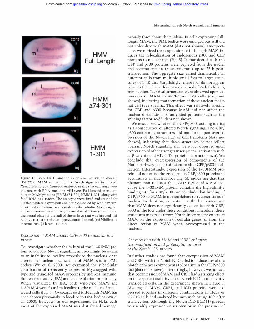
To determine whether the p300 interaction domain(TAD1; amino acids 75–301) is required for MAM func-tion in vivo, Xenopus embryos were injected with RNAencoding either the wild-type or mutant MAM proteins,and analyzed for Notch function by scoring the number
of primary neurons that form in the neural plate (Lamaret al. 2001). In this assay, increased Notch signaling re-duces the number of differentiated cells (primary neu-rons) that form, as visualized by staining for a neural-specific form of tubulin, whereas disabled Notch signal-ing results in an increased number of neurons. Injectionof �74–301MM RNA markedly increased the number ofneurons formed relative to the uninjected control half ofthe embryo (Fig. 4), indicating that Notch signaling isblocked in vivo by a MAM mutant protein that lacksTAD1. Consistent with a previous report (Wu et al.2000), we also found that 1–301MM is a dominant-nega-tive inhibitor of Notch signaling in Xenopus embryos(Fig. 4). Both �74–301MM and 1–301MM also blockedthe induction by the ICD of two primary Notch targetgenes, ESR1 and ESR7, in animal cap experiments (La-mar et al. 2001; data not shown), confirming that thedefect in Notch signaling was transcriptional. Moreover,neither of the MAM mutant proteins was able to en-hance ICD-dependent Notch transcription in transientexpression experiments (data not shown). We concludethat both TAD1 (amino acids 75–301) and the C-termi-nal activation domain (TAD2; amino acids 301–1016) arerequired for MAM-dependent activation of Notch targetand reporter genes in vivo, whereas the initiation ofNotch transcription on chromatin in vitro requires theMAM TAD1, but not TAD2, domain.
Figure 3. MAM interacts specifically withnuclear CBP/p300 and promotes p300-medi-ated nucleosome acetylation in vitro. (A)SDS-PAGE analysis of HeLa nuclear proteinsthat interact with the MAM TAD1 fragment.A crude HeLa nuclear extract was incubatedwith either glutathione beads (lane 1) or GST–MAMTAD1 beads (lane 3), and the associatedproteins were eluted by boiling and analyzedby SDS-PAGE and silver staining. The arrowindicates a band identified as CBP/p300through tryptic digestion and MALDI-TOFmass-spectrometry analysis. (B) HeLa nuclearextract was incubated with either GST (lane8) or various GST–MAM protein-coupledbeads (lanes 2–7) as indicated above each lane,and analyzed by immunoblotting for associ-ated nuclear p300 protein. The input nuclearp300 protein is shown in lane 1. (C) Purifiedrecombinant p300 enhances Notch transcrip-tion in vitro. Transcription reactions con-tained the pNRE template incubated in theabsence of enhancer factors (lane 1,5) or withthe Notch enhancer complex (MAM–ICD–CBF1; lanes 6,7), which was added followingchromatin assembly. Where indicated, re-combinant p300 was incubated with the chro-matin template for 30 min prior to analysis oftranscription by primer extension (lanes2–4,7). (D) Purified p300 promotes acetylationof pNRE nucleosomal histones in a MAM-dependent manner in vitro. The pNRE chromatin assembly reactions either lacked enhancerfactors (lane 1), or contained CBF1–ICD (lane 2–4,6) and wild-type or mutant MAM protein (lanes 3–6), as indicated above the lanes.The pNRE template was purified following chromatin assembly and incubated with recombinant p300 and 14C-acetyl CoA, and thelabeled nucleosomal histones were identified by SDS-PAGE and fluorography.
Fryer et al.
1402 GENES & DEVELOPMENT
Cold Spring Harbor Laboratory Press on March 20, 2022 - Published by genesdev.cshlp.orgDownloaded from
Expression of MAM directs CBP/p300 to nuclear fociin vivo
To investigate whether the failure of the 1–301MM pro-tein to support Notch signaling in vivo might be owingto an inability to localize properly to the nucleus, or toaltered subnuclear localization of MAM within PMLbodies (Wu et al. 2000), we examined the subcellulardistribution of transiently expressed Myc-tagged wild-type and truncated MAM proteins by indirect immuno-fluorescence assay (IFA) and deconvolution microscopy.When visualized by IFA, both wild-type MAM and1–301MM were found to localize to the nucleus of trans-fected cells (Fig. 5). Overexpressed full-length MAM hasbeen shown previously to localize to PML bodies (Wu etal. 2000); however, in our experiments in HeLa cellsmost of the expressed MAM was distributed homoge-
neously throughout the nucleus. In cells expressing full-length MAM, the PML bodies were enlarged but still didnot colocalize with MAM (data not shown). Unexpect-edly, we noticed that expression of full-length MAM in-duces the relocalization of endogenous p300 and CBPproteins to nuclear foci (Fig. 5). In transfected cells theCBP and p300 proteins were depleted from the nucleiand accumulated in these structures up to 72 h post-transfection. The aggregate size varied dramatically indifferent cells from multiple small foci to larger struc-tures of 1–10 µm. Surprisingly, these foci do not appeartoxic to the cells, at least over a period of 72 h followingtransfection. Identical structures were observed upon ex-pression of MAM in MCF7 and 293 cells (data notshown), indicating that formation of these nuclear foci isnot cell-type-specific. This effect was relatively specificfor CBP and p300 because MAM did not affect thenuclear distribution of unrelated proteins such as thesplicing factor sc-35 (data not shown).
We next asked whether the CBP/p300 foci might ariseas a consequence of altered Notch signaling. The CBP/p300-containing structures did not form upon overex-pression of the Notch ICD or CBF1 proteins (data notshown), indicating that these structures do not reflectaberrant Notch signaling, nor were foci observed uponexpression of other strong transcriptional activators suchas �-catenin and HIV-1 Tat protein (data not shown). Weconclude that overexpression of components of theNotch pathway is not sufficient to alter CBP/p300 local-ization. Interestingly, expression of the 1–301MM pro-tein did not cause the endogenous CBP/p300 proteins toaccumulate in nuclear foci (Fig. 5), indicating that thisphenomenon requires the TAD2 region of MAM. Be-cause the 1–301MM protein contains the high-affinitybinding site for CBP/p300, we conclude that binding ofCBP/p300 to MAM is not sufficient to redirect its sub-nuclear localization, consistent with the observationthat MAM does not significantly colocalize with CBP/p300 in the foci under these conditions. Therefore, thesestructures may result from Notch-independent effects ofMAM on the expression of cellular genes, or from thedirect action of MAM when overexpressed in thenucleus.
Coexpression with MAM and CBF1 enhancesthe modification and proteolytic turnoverof the Notch ICD in vivo
In further studies, we found that coexpression of MAMand CBF1 with the Notch ICD failed to induce any of theNotch enhancer components to localize in the CBP/p300foci (data not shown). Interestingly, however, we noticedthat coexpression of MAM and CBF1 had a striking effecton the apparent stability of the Notch ICD in transientlytransfected cells. In the experiment shown in Figure 6,Myc-tagged MAM, CBF1, and ICD proteins were ex-pressed together in different combinations in HeLa orC2C12 cells and analyzed by immunoblotting 48 h aftertransfection. Although the Notch ICD (ICD11) proteinwas readily expressed on its own or in the presence of
Figure 4. Both TAD1 and the C-terminal activation domain(TAD2) of MAM are required for Notch signaling in injectedXenopus embryos. Xenopus embryos at the two-cell stage wereinjected with RNA encoding wild-type (Full-length) or mutanthuman MAM proteins (HMM�74–301; HMM1–301) along withlacZ RNA as a tracer. The embryos were fixed and stained for�-galactosidase expression and double-labeled by whole-mountin situ hybridization for a neural-specific tubulin. Notch signal-ing was assessed by counting the number of primary neurons onthe neural plate for the half of the embryo that was injected (inj)relative to that for the uninjected control (cont). (m) Midline; (i)interneuron; (l) lateral neuron.
Mastermind controls Notch activation and turnover
GENES & DEVELOPMENT 1403
Cold Spring Harbor Laboratory Press on March 20, 2022 - Published by genesdev.cshlp.orgDownloaded from

CBF1, only very low levels of the ICD accumulatedwhen coexpressed with MAM and CBF1 (Fig. 6A, cf.lanes 1 and 3). In contrast, a mutant form of the ICDlacking the PEST domain (ICD22) was stable in the pres-ence of MAM and CBF1. The apparent decreased stabil-ity of the ICD was most pronounced when all threeNotch enhancer proteins were expressed. Thus, the ICDwas stable when expressed with MAM (Fig. 6B, cf. lanes2 and 4), but not with MAM and CBF1 (Fig. 6B, cf. lanes6 and 8). Although the Notch cofactor NRARP influ-ences the stability of Notch in Xenopus embryos (Lamaret al. 2001), coexpression of NRARP (N) had no effect onthe apparent stability of any of the Notch enhancer com-ponents in these experiments (Fig. 6A,B). Interestingly,the Notch ICD was stable when coexpressed with CBF1and the C-terminally truncated MAM (1–301MM) pro-tein, which lacks the TAD2 domain (Fig. 6C, cf. lanes 2and 5).
In experiments where low levels of the Notch ICD
could be detected in the presence of MAM and CBF1, theICD was found to migrate on SDS-PAGE gels as a modi-fied form that resembles the hyperphosphorylated form(Fig. 6C, cf. lanes 9 and 10). In contrast, no evidenceof hyperphosphorylation was observed when the ICDwas coexpressed with CBF1 and the shorter MAM(1–301MM) protein (Fig. 6C, cf. lanes 9–11). Furtheranalysis revealed that modification of the ICD by MAMwas reversed upon treatment with protein phosphatase(Fig. 7A, cf. lanes 4 and 5), but not with Vanadate-inac-tivated phosphatase (Fig. 7A, lane 6). We conclude thatMAM promotes the hyperphosphorylation and proteo-lytic turnover of the Notch ICD in the presence of CBF1.Interestingly, Western blot analyses also revealed achange in the migration of endogenous CBP in cells ex-pressing MAM (Fig. 7B, cf. lanes 2 and 3). This alteredmigration was reversed by treatment of the cells withprotein phosphatase in a manner that could be inacti-vated with Vanadate (Fig. 7B, cf. lanes 7 and 8), indicat-
Figure 5. Human MAM directs endogeneous CBP/p300 to nuclear foci in vivo in a TAD2-dependent manner. Transiently expressedMyc-tagged wild-type (1–1016MM) or mutant MAM proteins (1–301MM) and endogenous p300 and CBP proteins were visualized byindirect immunofluorescence and deconvolution microscopy in HeLa cells. (A) HeLa cells transiently transfected with 1016MM-Mycwere imaged for Myc immunofluorescence (red) and endogenous p300 (upper panel) or CBP (lower panel) immunofluorescence (green).Cells were counterstained with DAPI (blue) to show the position of the nucleus for all the cells in the field. (B) HeLa cells weretransiently transfected with 1–301MM-Myc and analyzed as in A.
Fryer et al.
1404 GENES & DEVELOPMENT
Cold Spring Harbor Laboratory Press on March 20, 2022 - Published by genesdev.cshlp.orgDownloaded from

ing that MAM also promotes phosphorylation of CBP invivo. This change in CBP phosphorylation was not ob-served upon expression with the C-terminal truncatedMAM protein (1–301MM), indicating that TAD2 is re-quired for MAM to promote phosphorylation of endog-enous CBP in vivo. Taken together, these findings sug-gest that assembly of MAM into the Notch enhancercomplex promotes the phosphorylation of CBP as well asthe phosphorylation and turnover of the associatedNotch ICD. These findings are summarized in the modelshown in Figure 8, and discussed further below.
Discussion
A key role for MAM in the Notch enhancer complex
A common feature of developmental signaling pathwayssuch as Notch, Wnt/Wg, Hedgehog, and TGF-� is theligand-dependent mobilization of dedicated coactivatorsthat target specific classes of enhancer factors. Like theseother induced coactivators, the intracellular domain ofthe Notch receptor carries a potent transactivation do-
main that is otherwise lacking in its DNA-binding part-ner, CBF1 (Artavanis-Tsakonas et al. 1999; Mumm andKopan 2000). Through reconstituting the activity of theNotch enhancer complex on chromatin, we find that theNotch ICD functions primarily to connect CBF1 to asecond non-DNA-binding coactivator, MAM. In addi-tion, we find that the activation domain of the ICD func-tions in a MAM-dependent manner in vitro. These ob-servations strongly suggest that the minimal functionalNotch enhancer complex is a heterotrimer composed ofMAM, the Notch ICD, and CBF1.
In this respect, the Notch transactivation mechanismdiffers from that used by the Wnt/Wg signaling pathway,because the Wnt-responsive �-catenin–LEF-1 enhancercomplex, unlike the ICD–CBF1 complex, is sufficient toinitiate transcription on chromatin templates in vitro(Tutter et al. 2001). A key role for Notch in the assemblyof multisubunit signaling complexes might serve tomodulate the response of different cells to Notch ligand,depending on the level of available MAM in the nucleus.Moreover, the ICD may assemble multiple types of com-plexes with different coactivators such as SKIP (Zhou et
Figure 6. The MAM TAD2 region promotes the modification and turnover of Notch ICD in vivo. (A) MAM and CBF1 enhance thePEST-dependent proteolytic turnover of the Notch ICD in vivo. The expression of Myc-tagged Notch enhancer proteins in C2C12 cellswas detected 48 h after transfection by SDS-PAGE of cell extracts and immunoblotting with an anti-Myc antibody. The expressedproteins included CBF1 (lanes 1–6), full-length ICD (ICD11; lanes 1–3), a C-terminal truncated ICD lacking the PEST domain (ICD22,lanes 4–6), full-length MAM (1–1016MM; lanes 3–6), and the Notch-specific factor NRARP (N, lanes 2,5). (B) MAM-dependentturnover of the Notch ICD requires CBF1 in vivo. Full-length Notch ICD (ICD; lanes 2–9), MAM (MM; lanes 4,5,8,9,11), CBF1 (lanes6–10), and NRARP (lanes 3,5,7,9) were expressed in HeLa cells and analyzed for protein expression 48 h after transfection in vivo. (C)HeLa cells (left panel) or C2C12 cells (right panel) were transfected with CBF1 (lanes 1,2,5,9–11), ICD (lanes 1,2,4,5,8–11), and eitherfull-length (FL; lanes 2–4,10) or 1–301MM (1–301; lanes 5,6,11), and protein levels were examined as in A. The MAM-dependentmodification of the ICD is evidenced by its altered mobility by SDS-PAGE (lane 10 ).
Mastermind controls Notch activation and turnover
GENES & DEVELOPMENT 1405
Cold Spring Harbor Laboratory Press on March 20, 2022 - Published by genesdev.cshlp.orgDownloaded from

al. 2000), which might functionally substitute for MAMin some cells. Previous studies have shown that bindingof the Notch ICD to yet a different potential coactivator,Deltex, excludes binding to CBF1 (Yamamoto et al.2001). Thus, an ICD–Deltex complex might target otherenhancer-binding proteins and activate a distinct set ofgenes in response to Notch signaling. Although the com-position of individual Notch signaling complexes hasnot yet been well defined, our data indicate that displace-ment of CBF-associated corepressors by a heterodimericICD–CBF1 complex would not be sufficient to activate aNotch target gene in chromatin (Fig. 8A).
Previous studies have shown that MAM enters theNotch enhancer complex through binding to the ICD, ina step that also requires CBF1 (Fig. 8B; Petcherski andKimble 2000b). Our findings suggest that MAM carriestwo distinct transactivation domains (TAD1, TAD2)that act in concert with the ICD activation domain. Invitro, MAM acts through TAD1 to recruit CBP/p300 tothe Notch enhancer complex (Fig. 8B) and direct acety-lation of the nucleosomal pNRE template. Although arecent report indicates that the ICD can bind directly top300 (Oswald et al. 2001), we show that MAM is essen-tial for the ICD–CBF1 complex to recruit p300 to thetemplate in vitro (Fig. 3D). One possibility is that theICD–TAD may help stabilize the association of p300with the enhancer complex after it has been recruited byMAM. It is interesting to note that the MAM C-terminalactivation domain (TAD2) does not contribute to tran-scription in the system used here, which monitors onlythe steps leading to initiation of transcription. Conse-quently, it will be important to assess whether the MAMTAD2 controls a postinitiation step, such as promoter
clearance or elongation, or whether the action of TAD2requires additional coactivators in the complex, or post-translational modification of the Notch enhancer pro-teins.
MAM expression induces phosphorylationand accumulation of CBP/p300 in nuclear foci
Unexpectedly, we find that expression of MAM inducesendogenous CBP/p300 proteins to accumulate in mul-tiple nuclear foci in vivo (Fig. 5). We also show that thesestructures do not form upon expression of a mutantMAM protein lacking the C-terminal TAD2 region (1–301MM). Thus, binding of MAM to CBP/p300, which ismediated through TAD1, is not sufficient to cause CBP/p300 to accumulate in these structures. Expression ofother Notch components (ICD, CBF1) did not affect thesubnuclear localization of CBP/p300, indicating thatthese foci are not a consequence of high levels of Notchsignaling in the nucleus. One possibility is that MAMmay regulate the expression or modification of CBP/p300 independently of Notch signaling. Indeed, we findthat the MAM-induced foci are accompanied by in-creased phosphorylation of CBP, and this phosphoryla-tion requires the C-terminal TAD2 domain of MAM (Fig.7). Consequently, overexpression of MAM in the nucleusmay promote widespread phosphorylation of CBP, whichmay cause the CBP/p300 proteins to concentrate in thesestructures. Changes in CBP/p300 phosphorylation havebeen shown to alter its activity and differentially affectits interactions with other transcription factors (Ait-Si-Ali et al. 1998). It will therefore be important to assesswhether MAM promotes CBP/p300 phosphorylation
Figure 7. Transient expression of MAM alters the phosphorylation of the Notch ICD and endogenous CBP proteins. (A) C2C12 cellswere transfected with myc-tagged forms of ICD and CBF1 in the absence (lanes 1–3) or presence (lanes 4–6) of MAM, and cell extractsprepared 48 h after transfection were immunoprecipitated with an anti-myc antibody (9E10). The isolated proteins were resuspendedin phosphatase buffer (lanes 1,4), phosphatase buffer containing 0.2 U of acid phosphatase (lanes 2,5), or phosphatase buffer containing0.2 U of acid phosphatase plus 1 mM sodium orthovanadate (Van; lanes 3,6). Immunoprecipitated complexes were detected by Westernblotting with an anti-myc antibody. (B) 293T cells were transfected with pCDNA empty vector (lane 1), 1–301MM (lane 2), or1–1016MM (lanes 3–6), and cell extracts prepared 48 h after transfection were immunoprecipitated with an anti-CBP antibody. Theisolated complexes were untreated (lanes 1–3) or resuspended in phosphatase buffer (lane 4), phosphatase buffer containing 0.2 U of acidphosphatase (lane 5), or phosphatase buffer containing 0.2 U of acid phosphatase plus 1 mM sodium orthovanadate (Van; lane 6). Theimmunoprecipitated complexes were detected by Western blotting with an anti-CBP antibody. Lanes 7 and 8 show a longer exposureof the reactions shown in lanes 5 and 6.
Fryer et al.
1406 GENES & DEVELOPMENT
Cold Spring Harbor Laboratory Press on March 20, 2022 - Published by genesdev.cshlp.orgDownloaded from

within the Notch enhancer complex (Fig. 8B), andwhether phosphorylation of CBP/p300 is important fortranscriptional activation by Notch.
The MAM C-terminal activation domain couplestranscription activation with turnover of the ICD
The timing of Notch signaling is tightly controlled indevelopmental processes such as somite formation, dur-
ing which Notch target genes such as cHairy1 andmHES1 undergo periodic cycles of expression at the di-rection of a molecular oscillator, or vertebrate segmen-tation clock (for review, see Pourquie 1999). This clockmay be established through the intrinsic timing ofNotch signaling as well as the half-life of Notch-inducedtranscriptional repressors. Previous studies have estab-lished that the Notch ICD is subject to proteolytic deg-radation in the nucleus through the action of the ubiq-uitin ligases such as Sel-10 (Hubbard et al. 1997; Gupta-Rossi et al. 2001; Oberg et al. 2001; Wu et al. 2001). Rapidturnover of the ICD may be required to allow genes torespond rapidly to subsequent cycles of Notch signaling.We find that coexpression with MAM and CBF1 pro-motes the phosphorylation and proteolytic turnover ofthe ICD in vivo (Fig. 6), indicating that MAM couplestranscription activation with degradation of the ICD. Inthis respect, MAM may act as a timer to control thelength of time that the Notch complex remains associ-ated with the enhancer. By extension, MAM might con-tribute to the periodic expression of Notch target genesduring somitogenesis through its potential effects on thedisassembly of the Notch enhancer complex.
Our data indicate that CBF1 acts in concert withMAM to control the proteolytic turnover of the ICD invivo (Fig. 6B). Importantly, both MAM and CBF1 appearto be stable upon coexpression with the ICD, and thus itappears that the ICD can be destabilized independentlyof its interacting partners. The requirement for CBF1may reflect its ability to enhance binding of MAM to theICD (Petcherski and Kimble 2000b), or alternativelyCBF1 might be needed to target the Notch enhancercomplex to DNA. We show that the stability of a mutantICD protein lacking the PEST domain is unaffected bycoexpression with MAM and CBF1 (Fig. 6A), and thatturnover is accompanied by increased phosphorylationof the ICD (Fig. 7). Importantly, we find that the MAMTAD2 domain is necessary for both enhanced phos-phorylation and turnover of the ICD. Because p300 hasbeen shown to be critical for the regulated turnover ofthe p53 transactivator by MDM2 (Grossman et al. 1998;Zhu et al. 2001), it will be important to assess whetherrecruitment of p300 by MAM may similarly be requiredfor proteolytic degradation of the ICD. Nevertheless, it isclear that recruitment of CBP/p300 through the MAMTAD1 region is not sufficient to couple activation withturnover of the Notch ICD under the conditions exam-ined here.
Thus the TAD2 region is required for MAM to pro-mote the phosphorylation of its two associated factors,CBP/p300 and the Notch ICD. Because MAM does notpossess intrinsic ICD protein kinase activity (data notshown), it is attractive to consider that the Notch ICDand CBP/p300 may instead be targeted for phosphoryla-tion by cyclin-dependent kinases that associate with thetranscription complex (Price 2000; Orphanides and Rein-berg 2002) and are recruited to the promoter by MAM(Fig. 8B). Phosphorylation events mediated by CDK7 andSrb10 (the CDK8 homolog in yeast) have been implicatedin the proteolytic destruction of other enhancer factors
Figure 8. Model for the mechanism of Notch transcription. (A)Notch target genes are repressed through CBF1 complexes thatcontain histone deacetylases (HDAC) and other corepressors(Dou et al. 1994; Kao et al. 1998; Hsieh et al. 1999; Morel et al.2001). Although previous studies have shown that binding ofthe Notch ICD to CBF1 can displace corepressor complexes(Kao et al. 1998; Zhou et al. 2000), the data presented here in-dicate that a CBF1–ICD complex would be insufficient to acti-vate Notch transcription in the absence of MAM. (B) A three-way complex containing CBF1, ICD, and MAM is required forNotch transcription on chromatin templates in vitro. CBP/p300is recruited through the MAM TAD1 region to promote tran-scription initiation and acetylate nearby nucleosomal histones.The ICD activation domain also contributes to transcription atthis step. We find that MAM induces the phosphorylation ofboth CBP/p300 and the Notch ICD in a step that requires theMAM TAD2 region. Phosphorylation of the Notch ICD byMAM also requires CBF1, which may stabilize binding of MAMto the ICD (Petcherski and Kimble 2000b). Widespread phos-phorylation of CBP/p300 proteins by MAM may contribute tothe accumulation of these proteins in nuclear foci. It remains tobe determined whether the MAM-induced phosphorylationevents might be carried out by cyclin-dependent kinases asso-ciated with the RNA polymerase II (RNAPII) complex (Price2000; Orphanides and Reinberg 2002) or other protein kinasesassociated with MAM. (C) The phosphorylated ICD may be tar-geted for ubiquitination by ubiquitin ligase complexes, leadingto disassembly of the enhancer complex and proteolytic degra-dation of the ICD.
Mastermind controls Notch activation and turnover
GENES & DEVELOPMENT 1407
Cold Spring Harbor Laboratory Press on March 20, 2022 - Published by genesdev.cshlp.orgDownloaded from

(Vandel and Kouzarides 1999; Chi et al. 2001). The CDK9subunit of the positive transcription elongation factor,P-TEFb, also associates with RNAPII (Price 2000),whereas CDK8 interacts with RNAPII as a component ofhuman and yeast mediator complexes that have beenvariously implicated in activation and repression of tran-scription (Orphanides and Reinberg 2002). Another pos-sibility is that the ICD is phosphorylated by a proteinkinase that associates with MAM directly. It remains tobe determined whether the MAM-induced phosphoryla-tion is accompanied by increased ubiquitination of theICD, and whether the degradation of the ICD we observeis caused by ubiquitin-dependent proteolysis such asthat described for the nuclear Sel-10 ubiquitin ligase(Gupta-Rossi et al. 2001; Oberg et al. 2001; Wu et al.2001). It will also be important to learn whether modi-fication of the ICD regulates its transcriptional activity,as has been observed for other transcription factors (Hirstet al. 1999; Salghetti et al. 2001; Vincent et al. 2001;Ostendorff et al. 2002), and whether these steps may ul-timately be coupled to disassembly of the Notch en-hancer complex and turnover of the Notch ICD (Fig. 8C).
In summary, we show that MAM is an essential com-ponent of the Notch enhancer complex in vitro as well asin vivo. The human MAM protein recruits p300/CBP tothe Notch enhancer complex and controls the stabilityof the Notch ICD through the action of its unique C-terminal activation domain. Further studies will beneeded to evaluate whether these properties are sharedamong the various MAM proteins in different species,and to learn how MAM-induced phosphorylation of theICD and CBP/p300 proteins is coordinated with the regu-lation of Notch transcription.
Materials and methods
Plasmids and expression of recombinant proteins
Full-length human Mastermind (cDNA clone KIAA0200) andmutants were subcloned into pGEX-KG, a modified form of thePGEX-2T vector (Pharmacia) by standard PCR methods to yieldconstructs encoding in-frame fusions with glutathione-S-trans-ferase (GST). Full-length MAM and 1–301MM mutants weresubcloned into the CS2-MT vector by standard PCR methods togenerate a Mastermind construct in frame with a C-terminalmyc tag. GST–CBF1 was a generous gift of Lynne Vales(UMDNJ) and was expressed as described (Olave et al. 1998).The generation of the GST–ICD constructs was described pre-viously (Wettstein et al. 1997). Protein expression was inducedby addition of IPTG to a final concentration of 0.2 mM andincubation at 37°C for an additional 4 h. GST fusion proteinswere purified as described (Tutter et al. 2001). Full-length hu-man histidine-tagged p300 was expressed and purified frombaculovirus-infected Sf9 cells as described previously (Kraus etal. 1999).
DNA-binding experiments
EMSA were carried out with the high-affinity CBF1 binding sitefrom the HES1 promoter (CTAGGTTACTGTGGGAAAGAAAGTCC) in a final reaction volume of 15 µL containing 20 mMHEPES at pH 8.0, 50 mM KCl, 2.5 mM EDTA, 8 mM MgCl2, 5
mM Spermidine, 30–35 µg/mL poly(dIdC), 250 µg/mL BSA,0.025% NP-40, and 15% glycerol.
GST protein affinity-selection experiments
First, 15 µL of glutathione-sepharose 4B (Pharmacia) was equili-brated in GST-pulldown buffer (50 mM Tris at pH 7.9, 120 mMKCl, 0.5% NP-40, 2 mM DTT, 0.1 mM PMSF, 2 mg/mL benza-midine, 1 µg/mL Pepstatin A, 4 µg/mL Leupeptin, 10 µg/mLAprotinin, 20 µg/mL Soybean Trypsin Inhibitor) and combinedwith 100 µg of purified recombinant GST alone or the indicatedGST-tagged recombinant protein in a final volume of 300 µL ofGST-pulldown buffer (4°C for 1 h). Excess unbound GST orGST-fusion protein was removed by briefly washing the beadswith 300 µL of GST-pulldown buffer. In a separate reaction, 150µL of HeLa nuclear extract (8–10 mg/mL) was combined with550 µL of GST-pulldown buffer and precleared once over 15 µLof glutathione sepharose 4B (4°C for 1.5 h), and once over 20 µLof the aforementioned GST-bound glutathione sepharose 4B(4°C for 1.5 h). The precleared nuclear extract was then incu-bated with the aforementioned GST-fusion-bound glutathionesepharose at 4°C for 4 h with constant mixing. The depletedsupernatant was discarded, and the beads were briefly washed 3times with 300 µL of HEGN0.3M (20 mM HEPES at pH 8.0, 0.2mM EDTA, 10% gycerol, 0.3 M KCl, 0.1%NP-40) and once with300 µL of HEG0.1M (20 mM HEPES at pH 8.0, 0.2 mM EDTA,10% glycerol, 0.1 M KCl). The beads were then combined with20 µL of SDS-PAGE loading buffer, boiled, and electrophoresedthrough an 8% acrylamide SDS gel. Proteins were visualized bystaining with silver. Association of p300 with GST–Mastermindconstructs was detected by Western blotting with p300 (C20)antibody (Santa Cruz). Following the GST-pulldown reactionand Coomassie staining, proteins bands were excised, destainedin acetonitrile and ammonium bicarbonate buffer, and sub-jected to in gel tryptic digest. A portion of the concentratedtryptic fragments was analyzed by MALDI-TOF with Reflec-tron, and proteins were identified by database comparison(Scripps Proteomics/Mass Spec Facility, La Jolla, CA).
In vitro chromatin assembly and transcription
Chromatin assembly was performed essentially as described(Bulger and Kadonaga 1994). pNRE contains eight copies of aconsensus CBF1-binding site upstream of HIV-1 sequences (−79to +80, relative to the RNA start site) in a luciferase vector. Forchromatin reconstitution of pNRE, 1.25 µg of supercoiled plas-mid DNA was used in each 250-µL chromatin assembly reac-tion. Unless indicated, the GST–CBF1, GST–ICD, and GST–MAM were incubated with the pNRE template during chroma-tin assembly. Following assembly, 20-µL aliquots of thechromatin reaction were incubated with HeLa cell nuclear ex-tract and 25 ng of nonchromatin �-globin DNA at 30°C for 30min. Transcription and DNase I footprint reactions on chroma-tin were carried out as described previously (Tutter et al. 2001).
Chromatin HAT assay
Chromatin was assembled in the presence of GST–CBF1, GST–ICD, and GST–MAM as indicated. After assembly, 100 µL ofchromatin was purified on a Sepharose CL4B gel filtration col-umn as described (Mizuguchi and Wu 1999). Then 20 µL ofpurified chromatin was incubated +/− p300 in the presence of 5µM 14C-acetyl CoA (Amersham) at 30°C for 1 h. Reactions werestopped by boiling in SDS-PAGE loading buffer and analyzed ona 15% SDS-PAGE gel. The gel was fixed and treated with fluo-
Fryer et al.
1408 GENES & DEVELOPMENT
Cold Spring Harbor Laboratory Press on March 20, 2022 - Published by genesdev.cshlp.orgDownloaded from

rography enhancing solution (Amersham) prior to drying andexposure to autoradiography
Xenopus embryo RNA injection and in situ hybridization
Embryos were obtained from Xenopus laevis adult frogs by hor-mone-induced egg-laying and in vitro fertilization using stan-dard methods. Synthetic RNAs for injection into embryos weregenerated for full-length or deleted forms of human MAM byinserting the appropriate open reading frame from the cDNAclone KIAA0200 into the CS2+MT vector. Templates for Notch-ICD, XSu(H)DBM, and a nuclear-localized form of �-galactosi-dase (nlacZ) were described previously (Wettstein et al. 1997).Albino embryos at the two-cell stage were injected into oneanimal blastomere with 0.2–1 ng of test RNAs, along withnlacZ RNA (500 pg) as a tracer. At early neurulae stages, in-jected embryos were fixed, stained for �-galactosidase activitywith 5-bromo-4-chloro-3-indolyl-�-galactopyranoside (X-Gal),and then processed for whole-mount in situ hybridization forN-tubulin expression as described previously using digoxigenin-labeled antisense riboprobes. (Lamar et al. 2001). At least 30injected embryos were analyzed for each test RNA, in two in-dependent experiments, and the phenotypes reported occurredin >80% of the embryos. Embryos injected with just nLacZserved as a negative control.
Indirect immunofluorescence and deconvolution microscopy
All manipulations were done at room temperature. HeLa cellswere grown on glass coverslips and transfected with 1 µg ofCS2-Mastermind DNA using Effectene (QIAGEN) according tothe manufacturer’s protocol. Cells were fixed for immunofluo-rescence 24–72 h after transfection with 3% paraformaldehyde,0.05% glutaraldehyde in PBS for 20 min, followed by 3 min ofincubation with 0.1 M glycine/PBS. Fixed cells were permeabi-lized with 0.2% Triton X-100 in PBS for 20 min and blocked in2% BSA, 0.2% Triton X-100 for 20 min. The cells were incu-bated for 1 h with primary antibodies, followed by 40 min ofincubation with appropriate secondary antibodies. The mono-clonal antibodies 9E10 against the myc-epitope were purchasedfrom Covance/Babco and used at a 1:2000 dilution. Rabbit anti-CBP (A22) and anti-p300 (C20; Santa Cruz Biotechnology) wereused at dilutions of 1:200 and 1:500, respectively. The primaryantibodies were detected by secondary antibodies conjugated toAlexa 594 (goat anti-mouse IgG conjugate) or Alexa 488 (goatanti-rabbit IgG conjugate; Molecular Probes) at dilutions of1:1000. Before mounting the cells onto slides using Vectashieldmounting media (Vector Laboratories), the cell nucleus wascounterstained with DAPI. Immunofluorescence images werecollected as a Z-series with Olympus IX70 Microscope and de-convolved with Deltavision Software for image acquisition andprocessing (deconvolution). Adobe Photoshop (Adobe System)was used for image presentation, each image representing asingle Z-section after deconvolution.
Cell culture and Western analysis
HeLa or C2C12 cells were grown in DMEM containing 10%FBS, L-glutamine, penicillin, and streptomycin. For transfec-tion, 60,000 cells were plated in each well of 24-well dishes andtransfected the next day in serum-free medium with 2 µg ofDNA per well using the PEI reagent (Fluka) according to pub-lished protocols (HeLa cells) or with 500 ng of DNA using lipo-fectamine (GIBCO BRL; C2C12 cells). In general, each effectorconstituted ∼20% of the total DNA transfected, which was nor-malized to a constant amount with carrier CS2 vector. Cells
were washed in PBS and lysed in 400 µL of ice-cold lysis buffer(25 mM Tris at pH 7.5, 300 mM NaCl, 1% Triton X-100, 1 mMPMSF, 1 µM pepstatin, 1 µM leupeptin, 1 mM sodium vanadate,and 50 mM NaF) 48 h after transfection. Cell lysates werecleared by centrifugation at 4°C for 15 min at 15,000 rpm. Celllysates were analyzed for the Myc-tagged proteins by Westernblotting using the 9E10 myc mouse monoclonal antibody.
Phosphatase treatment of 293T and C2C12 extracts
The 293T cells were transiently transfected with pCDNAempty vector, 1–301MM-myc, or 1016MM-myc, using calciumphosphate precipitation. Cell extracts were prepared 48 h aftertransfection and immunoprecipitated with anti-CBP antibody(Santa Cruz). Immunoprecipitated complexes were untreated orresuspended in phosphatase buffer and treated with 0.2 U of acidphosphatase or 0.2 U of acid phosphatase plus 1 mM sodiumvanadate. After these reactions were incubated at 37°C for 1 h,cell lysis buffer was added, and the immune complexes werepelleted, resuspended in SDS-PAGE sample buffer, and loadedon a 6% gel. The immunoprecipitated complexes were detectedby Western blotting with an anti-CBP antibody. C2C12 cellswere transfected with myc-tagged forms of ICD, CBFI, andMAM as indicated, and cell extracts prepared 48 h after trans-fection were immunoprecipitated with an anti-myc antibody.The isolated proteins were treated with phosphatase as for 293Tcells. The immunoprecipitated complexes were detected byWestern blotting with an anti-myc antibody (9E10).
Acknowledgments
We thank Lynne Vales for the GST-CBF1 plasmid, and LeeKraus and James Kadonaga for the p300 baculovirus expressionvector. These studies were funded by grants from the NIH toK.A.J. and C.K. C.J.F. is supported by the Cancer Research fundof the Damon Runyan–Walter Winchell Foundation Fellowship(DRG-1610), and I.T. is funded by the Mac S. Rau Foundation.
The publication costs of this article were defrayed in part bypayment of page charges. This article must therefore be herebymarked “advertisement” in accordance with 18 USC section1734 solely to indicate this fact.
References
Ait-Si-Ali, S., Ramirez, S., Barre, F.X., Dkhissi, F., Magnaghi-Jaulin, L., Girault, J.A., Robin, P., Knibiehler, M., Pritchard,L.L., Ducommun, B., et al. 1998. Histone acetyltransferaseactivity of CBP is controlled by cycle-dependent kinases andoncoprotein E1A. Nature 396: 184–186.
Anderson, A., Robey, E., and Huang, Y. 2001. Notch signaling inlymphocyte development. Curr. Op. Genet. Dev. 11: 554–560.
Artavanis-Tsakonas, S., Rand, M.D., and Lake, R.J. 1999. Notchsignaling: Cell fate control and signal integration in devel-opment. Science 284: 770–776.
Bulger, M. and Kadonaga, J.T. 1994. Biochemical reconstitutionof chromatin with physiological nucleosome spacing. Meth-ods Mol. Genet. 5: 242–262.
Chi, Y., Huddleston, M., Zhang, X., Young, R., Annan, R., Carr,S., and Deshaies, R.J. 2001. Negative regulation of Gcn4 andMsn2 transcription factors by Srb10 cyclin-dependent ki-nase. Genes & Dev. 15: 1078–1092.
Dou, S., Zeng, X., Cortes, P., Erdjument-Bromage, H., Tempst,P., Honjo, T., and Vales, L.D. 1994. The recombination sig-nal sequence-binding protein RBP-2N functions as a tran-
Mastermind controls Notch activation and turnover
GENES & DEVELOPMENT 1409
Cold Spring Harbor Laboratory Press on March 20, 2022 - Published by genesdev.cshlp.orgDownloaded from

scriptional repressor. Mol. Cell. Biol. 14: 3310–3319.Ferdous, A., Gonzalez, F., Sun, L., Kodadek, T., and Johnston, S.
2001. The 19S regulatory particle of the proteasome is re-quired for efficient transcription elongation by RNA poly-merase II. Mol. Cell 7: 981–991.
Greenwald, I. 1998. LIN-12/Notch signaling: Lessons fromworms and flies. Genes & Dev. 12: 1751–1762.
Grossman, S., Perez, M., Kung, A., Joseph, M., Mansur, C., Xiao,Z., Kumar, S., Howley, P., and Livingston, D. 1998. p300/MDM2 complexes participate in MDM2-mediated p53 deg-radation. Mol. Cell 2: 405–415.
Gupta-Rossi, N., Le Bail, O., Gonen, H., Brou, C., Logeat, F., Six,E., Ciechanover, A., and Israel, A. 2001. Functional interac-tion between SEL-10, an F-box protein, and the nuclear formof activated Notch1 receptor. J. Biol. Chem. 276: 34371–34378.
Helms, W., Lee, H., Ammerman, M., Parks, A.L., Muskavitch,M.A., and Yedvobnick, B. 1999. Engineered truncations inthe Drosophila mastermind protein disrupt Notch pathwayfunction. Dev. Biol. 215: 358–374.
Hirst, M., Kobor, M.S., Kuriakose, N., Greenblatt, J., and Sad-owski, I. 1999. GAL4 is regulated by the RNA polymerase IIholoenzyme-associated cyclin-dependent protein kinaseSRB10/CDK8. Mol. Cell 3: 673–678.
Hsieh, J., Zhou, S., Chen, L., Young, D., and Hayward, S. 1999.CIR, a corepressor linking the DNA binding factor CBF1 tothe histone deacetylase complex. Proc. Natl. Acad. Sci.96: 23–28.
Hubbard, E., Dong, Q., and Greenwald, I. 1996. Evidence forphysical and functional association between EMB-5 andLIN-12 in Caenorhabditis elegans. Science 273: 112–115.
Hubbard, E., Wu, G., Kitajewski, J., and Greenwald, I. 1997.sel-10, a negative regulator of lin-12 activity in Caenorhab-ditis elegans, encodes a member of the CDC4 family of pro-teins. Genes & Dev. 11: 3182–3193.
Ito, T., Ikehara, T., Nakagawa, T., Kraus, W., and Muramatsu,M. 2000. p300-mediated acetylation facilitates the transferof histone H2A–H2B dimers from nucleosomes to a histonechaperone. Genes & Dev. 14: 1899–1907.
Kamura, T., Sato, S., Haque, D., Liu, L., Kaelin, W.G., Jr., Con-away, R.C., and Conaway, J.W. 1998. The elongin BC com-plex interacts with the conserved SOCS-box motif present inmembers of the SOCS, ras, WD-40 repeat, and ankyrin repeatfamilies. Genes & Dev. 12: 3872–3881.
Kao, H.Y., Ordentlich, P., Koyano-Nakagawa, N., Tang, Z.,Downes, M., Kintner, C.R., Evans, R.M., and Kadesch, T.1998. A histone deacetylase corepressor complex regulatesthe Notch signal transduction pathway. Genes & Dev.12: 2269–2277.
Kitagawa, M., Oyama, T., Kawashima, T., Yedvobnick, B., Ku-mar, A., Matsuno, K., and Harigaya, K. 2001. A human pro-tein with sequence similarity to Drosophila mastermind co-ordinates the nuclear form of notch and a CSL protein tobuild a transcriptional activator complex on target promot-ers. Mol. Cell. Biol. 21: 4337–4346.
Kraus, W., Manning, E., and Kadonaga, J. 1999. Biochemicalanalysis of distinct activation functions in p300 that en-hance transcription initiation with chromatin templates.Mol. Cell. Biol. 19: 8123–8135.
Krebs, L.T., Deftos, M.L., Bevan, M.J., and Gridley, T. 2001. TheNrarp gene encodes an ankyrin-repeat protein that is tran-scriptionally regulated by the Notch signaling pathway.Dev. Biol. 238: 110–119.
Kurooka, H. and Honjo, T. 2000. Functional interaction be-tween the mouse notch1 intracellular region and histoneacetyltransferases PCAF and GCN5. J. Biol. Chem.
275: 17211–17220.Lai, E.C. 2002. Protein degradation: Four e3s for the notch path-
way. Curr. Biol. 12: R74–R78.Lamar, E., Deblandre, G., Wettstein, D., Gawantka, V., Pollet,
N., Niehrs, C., and Kintner, C. 2001. Nrarp is a novel intra-cellular component of the Notch signaling pathway. Genes& Dev. 15: 1885–1899.
Lau, O.D., Kundu, T.K., Soccio, R.E., Ait-Si-Ali, S., Khalil, E.M.,Vassilev, A., Wolffe, A.P., Nakatani, Y., Roeder, R.G., andCole, P.A. 2000. HATs off: Selective synthetic inhibitors ofthe histone acetyltransferases p300 and PCAF. Mol. Cell5: 589–595.
Mizuguchi, G. and Wu, C. 1999. Nucleosome remodeling factorNURF and in vitro transcription of chromatin. Methods Mol.Biol. 119: 333–342.
Morel, V., Lecourtois, M., Massiani, O., Maier, D., Preiss, A.,and Schweisguth, F. 2001. Transcriptional repression bySuppressor of Hairless involves the binding of a Hairless–dCtBP complex in Drosophila. Curr. Biol. 11: 789–792.
Mumm, J.S. and Kopan, R. 2000. Notch signaling: From theoutside in. Dev. Biol. 228: 151–165.
Oberg, C., Li, J., Pauley, A., Wolf, E., Gurney, M., and Lendahl,U. 2001. The notch intracellular domain is ubiquitinatedand negatively regulated by the mammalian sel-10 homolog.J. Biol. Chem. 276: 35847–35853.
Olave, I., Reinberg, D., and Vales, L.D. 1998. The mammaliantranscriptional repressor RBP (CBF1) targets TFIID andTFIIA to prevent activated transcription. Genes & Dev.12: 1621–1637.
Orphanides, G. and Reinberg, D. 2002. A unified theory of geneexpression. Cell 108: 439–451.
Ostendorff, H.P., Pelrano, R.I., Peters, M.A., Schluter, A.,Bossenz, M., Scheffner, M., and Bach, I. 2002. Ubiquitina-tion-dependent cofactor exchange on LIM homeodomaintranscription factors. Nature 416: 99–103.
Oswald, F., Tauber, B., Dobner, T., Bourteele, S., Kostezka, U.,Adler, G., Liptay, S., and Schmid, R.M. 2001. p300 acts as atranscriptional coactivator for mammalian notch-1. Mol.Cell. Biol. 21: 7761–7774.
Petcherski, A.G. and Kimble, J. 2000a. LAG-3 is a putative tran-scriptional activator in the C. elegans Notch pathway. Na-ture 405: 364–368.
———. 2000b. Mastermind is a putative activator for Notch.Curr. Biol. 10: R471–R473.
Pourquie, O. 1999. Notch around the clock. Curr. Op. Genet.Dev. 9: 559–565.
Price, D.H. 2000. P-TEFb, a cyclin-dependent kinase controllingelongation by RNA polymerase II. Mol. Cell. Biol. 20: 2629–2634.
Qiu, L., Joazeiro, C., Fang, N., Wang, H.Y., Elly, C., Altman, Y.,Fang, D., Hunter, T., and Liu, Y.C. 2000. Recognition andubiquitination of Notch by Itch, a hect-type E3 ubiquitinligase. J. Biol. Chem. 275: 35734–35737.
Salghetti, S., Caudy, A., Chenoweth, J., and Tansey, W. 2001.Regulation of transcriptional activation domain function byubiquitin. Science 293: 1651–1653.
Smoller, D., Friedel, C., Schmid, A., Bettler, D., Lam, L., andYedvobnick, B. 1990. The Drosophila neurogenic locus mas-termind encodes a nuclear protein unusually rich in aminoacid homopolymers. Genes & Dev. 4: 1688–1700.
Tansey, W. 2001. Transcription activation: Risky business.Genes & Dev. 15: 1045–1050.
Tutter, A., Fryer, C., and Jones, K. 2001. Chromatin-specificregulation of LEF-1-�-catenin transcription activation andinhibition in vitro. Genes & Dev. 15: 3342–3354.
Vandel, L. and Kouzarides, T. 1999. Residues phosphorylated by
Fryer et al.
1410 GENES & DEVELOPMENT
Cold Spring Harbor Laboratory Press on March 20, 2022 - Published by genesdev.cshlp.orgDownloaded from

TFIIH are required for E2F-1 degradation during S-phase.EMBO J. 18: 4280–4291.
Vincent, O., Kuchin, S., Hong, S.P., Townley, R., Vyas, V.K., andCarlson, M. 2001. Interaction of the Srb10 kinase with Sip4,a transcriptional activator of gluconeogenic genes in Saccha-romyces cerevisiae. Mol. Cell. Biol. 21: 5790–5796.
Wettstein, D.A., Turner, D.L., and Kintner, C. 1997. The Xeno-pus homolog of Drosophila Suppressor of Hairless mediatesNotch signaling during primary neurogenesis. Development124: 693–702.
Wu, G., Lyapina, S., Das, I., Li, J., Gurney, M., Pauley, A., Chui,I., Deshaies, R., and Kitajewski, J. 2001. SEL-10 is an inhibi-tor of notch signaling that targets notch for ubiquitin-medi-ated protein degradation. Mol. Cell. Biol. 21: 7403–7415.
Wu, L., Aster, J.C., Blacklow, S.C., Lake, R., Artavanis-Tsako-nas, S., and Griffin, J.D. 2000. MAML1, a human homologueof Drosophila mastermind, is a transcriptional co-activatorfor Notch receptors. Nat. Genet. 26: 484–489.
Xu, T., Rebay, I., Fleming, R.J., Scottgale, T.N., and Artavanis-Tsakonas, S. 1990. The Notch locus and the genetic circuitryinvolved in early Drosophila neurogenesis. Genes & Dev.4: 464–475.
Yamamoto, N., Yamamoto, S., Inagaki, F., Kawaichi, M., Fuka-mizu, A., Kishi, N., Matsuno, K., Nakamura, K., Weinmas-ter, G., Okano, H., et al. 2001. Role of Deltex-1 as a tran-scriptional regulator downstream of the Notch receptor. J.Biol. Chem. 276: 45031–45040.
Zhou, S., Fujimuro, M., Hsieh, J.J., Chen, L., Miyamoto, A.,Weinmaster, G., and Hayward, S. 2000. SKIP, a CBF1-asso-ciated protein, interacts with the ankyrin repeat domain ofNotchIC to facilitate NotchIC function. Mol. Cell. Biol.20: 2400–2410.
Zhu, Q., Yao, J., Wani, G., Wani, M., and Wani, A. 2001. Mdm2mutant defective in binding p300 promotes ubiquitinationbut not degradation of p53. J. Biol. Chem. 276: 29695–29701.
Mastermind controls Notch activation and turnover
GENES & DEVELOPMENT 1411
Cold Spring Harbor Laboratory Press on March 20, 2022 - Published by genesdev.cshlp.orgDownloaded from

10.1101/gad.991602Access the most recent version at doi: 16:2002, Genes Dev.
Christy J. Fryer, Elise Lamar, Ivana Turbachova, et al. the Notch enhancer complexMastermind mediates chromatin-specific transcription and turnover of
References
http://genesdev.cshlp.org/content/16/11/1397.full.html#ref-list-1
This article cites 51 articles, 31 of which can be accessed free at:
License
ServiceEmail Alerting
click here.right corner of the article or
Receive free email alerts when new articles cite this article - sign up in the box at the top
Cold Spring Harbor Laboratory Press
Cold Spring Harbor Laboratory Press on March 20, 2022 - Published by genesdev.cshlp.orgDownloaded from