maxillofacial prosthesis
TRANSCRIPT

Maxillofacial Prosthetics
Assistant Professor
Ibrahim Hamad Al-Fahdawi
Department of Prosthodontics
College of Dentistry
University of Anbar
,


It is the God given right of every human being to appear human
“the art and science of anatomic, functional,or cosmetic reconstruction by means of nonliving substitutes of those regions in the maxilla, mandible, and face that are missingor defective because of surgical intervention, trauma, pathology, ordevelopmental or congenital malformations”
Maxillofacial prosthodontic
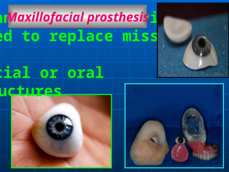
is an artificial device used to replace missing facial or oral structures.
Maxillofacial prosthesis
Reconstruct of missing parts in maxilla, mandible and face with prosthesis.To achieve:1- Preservation of residual structures.2- Reconstruction of function.3- Improvement in esthetic.
The Aim of Maxillofacial Prosthetic:
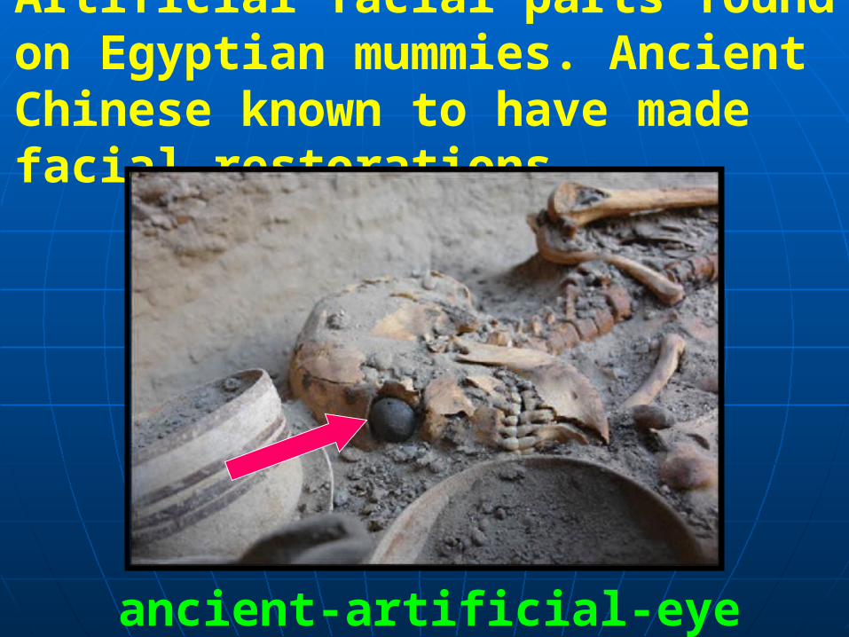
Artificial facial parts found on Egyptian mummies. Ancient Chinese known to have made facial restorations.
ancient-artificial-eye

1-Congental2-Traumatic3-Pathological with radical surgery
1- Intra Oral (Maxilla and Mandible).2- Extra Oral (eye, nose, ear).
Causes of Facial and oral Tissues loss
These factors result to 2 types of defects either:
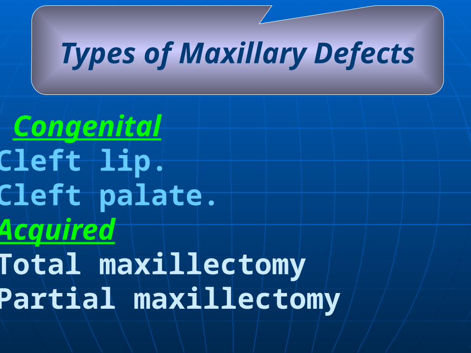
- Congenital.Cleft lip..Cleft palate.-Acquired.Total maxillectomy.Partial maxillectomy
Types of Maxillary Defects
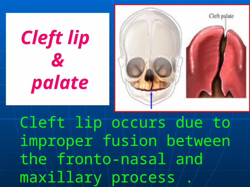
Cleft lip occurs due to improper fusion between the fronto-nasal and maxillary process .
Cleft lip &
palate
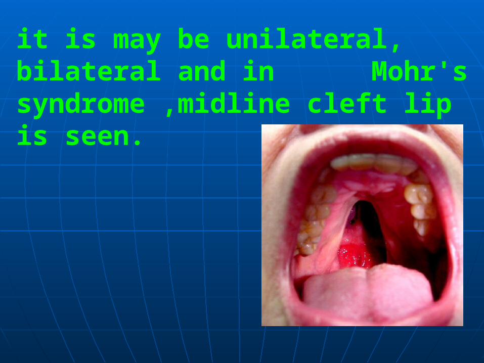
it is may be unilateral, bilateral and in Mohr's syndrome ,midline cleft lip is seen.
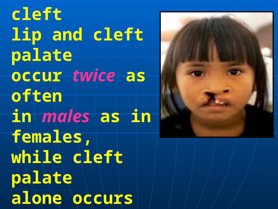
Cleft lip and the combination of cleftlip and cleft palate occur twice as often in males as in females,while cleft palate alone occurs more often in females.
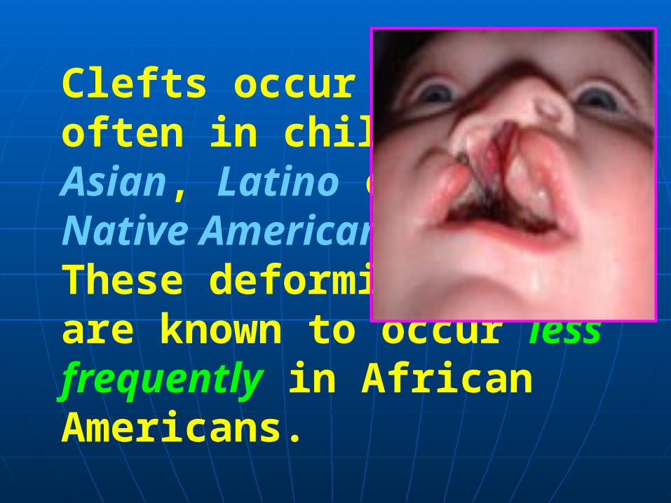
Clefts occur mostoften in children ofAsian, Latino orNative AmericanThese deformities are known to occur less frequently in African Americans.
As a result of the abnormalities in the upper arch of the mouth, teeth may not erupt properly or may be missing completely. In such cases, artificial teeth and orthodontics (braces) are usually required. Routine oral hygiene, tooth brushing and flossing are still required to maintain healthy teeth and gums and prevent gum disease (periodontitis) and tooth decay.
Dental Problems

Treatments for Cleft Lip and Cleft Palate
Children with cleft lip and/or cleft palate are treated over the course of 18 or more years. Treatment can involve a team of professionals beginning shortly after birth and continuing throughout adolescence .

The treatment team includes medical, dental and other healthcare specialists who work together to address the many different and complicated needs specific to the individual.
Most acquired defect occur due to surgical resection of tumors or trauma .
Acquired Maxillary Defect
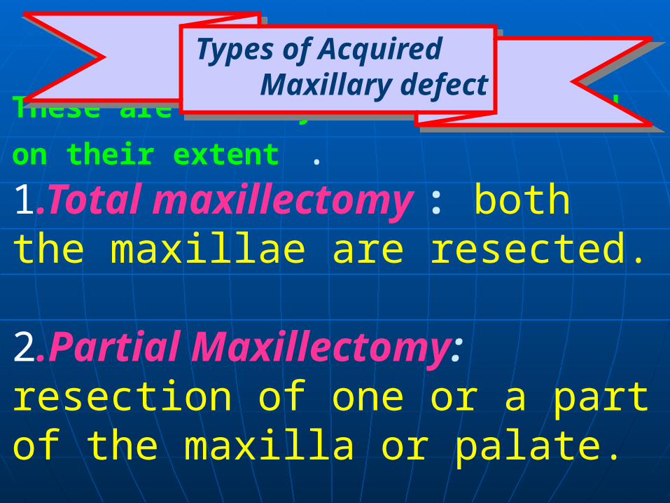
These are usually classified based on their extent .1.Total maxillectomy : both the maxillae are resected. 2.Partial Maxillectomy: resection of one or a part of the maxilla or palate.
Types of Acquired Maxillary defect
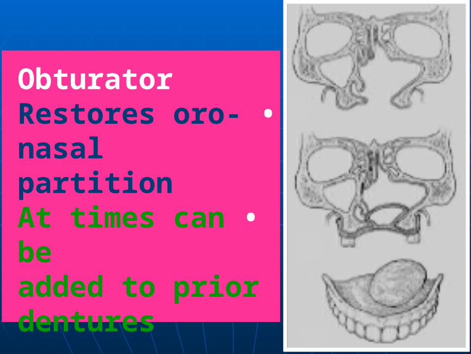
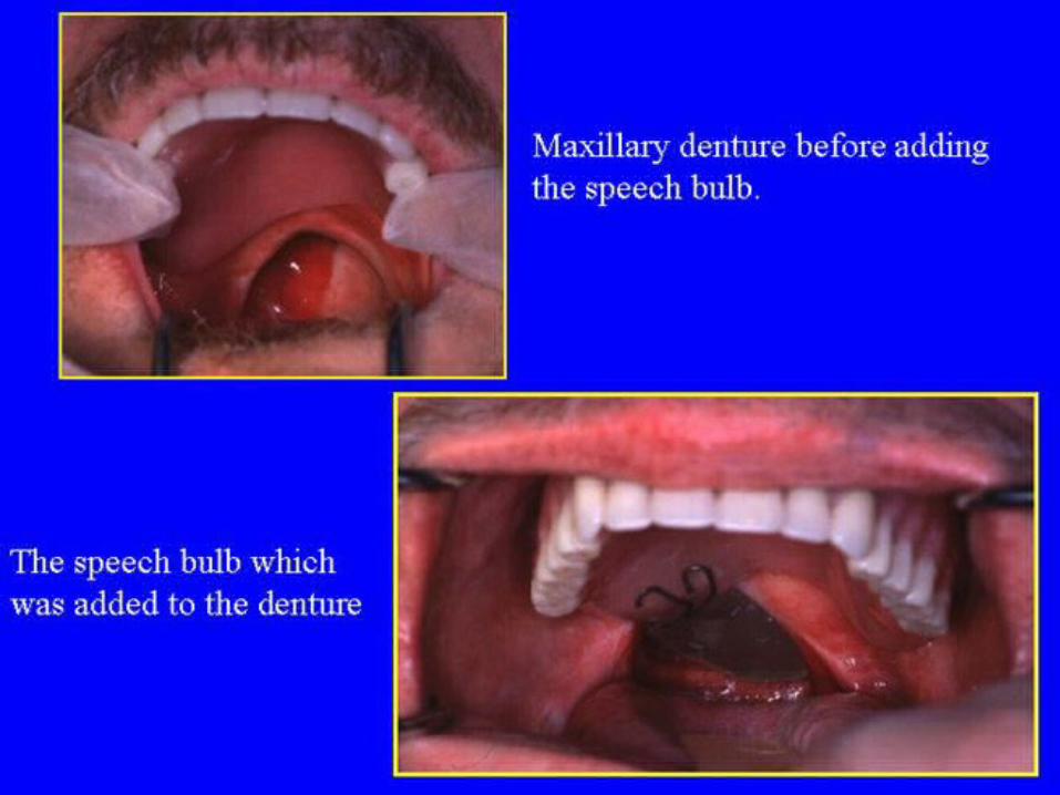
Obturator •Restores oro-nasal
partition •At times can be
added to priordentures
The three types of prostheses are constructed for both edentulous and dentulous patients
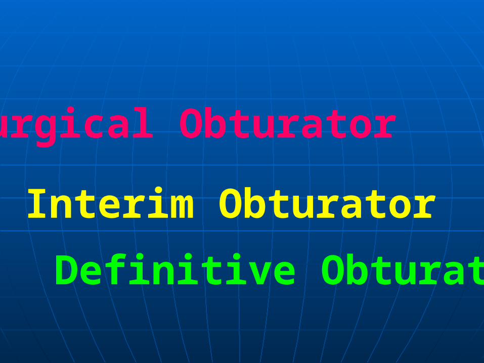
Surgical Obturator
Interim Obturator
Definitive Obturator
surgical obturator is constructed before the surgery and is inserted in the operating room

advantagesrestoration of normal speech and eating habits. Preventing the collapse of the soft tissues. Facial symmetry will be preserved, and retention of the interim and definitive prostheses will be facilitated. Above all, the mental well-being of the patient is improved considerably.

When the surgical dressing is removed (7 to 10 days after the operation), the immediate presurgical prosthesis can be relined with a provisional denture liner.
Interim Obturator
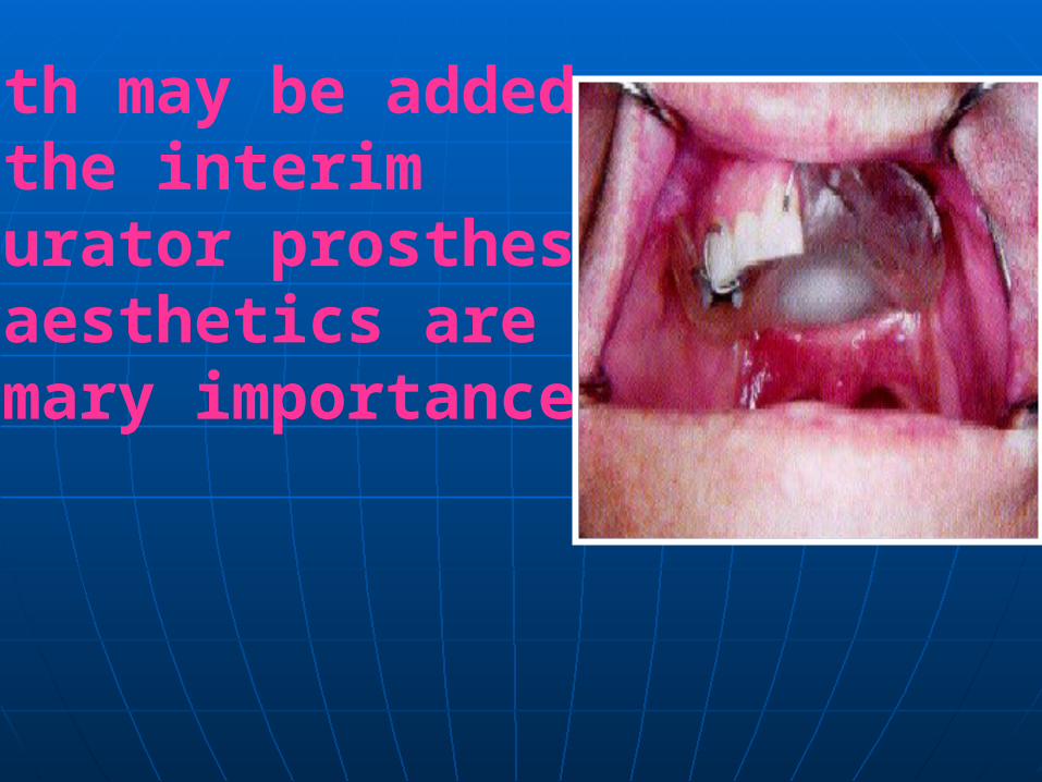
Teeth may be addedto the interim obturator prosthesisif aesthetics are of primary importance.

However, it is advantageous to omit the placement of teeth to prevent occlusal loading in the region of resection during the early stage of healing. This delay reduces the chances of irritation that could affect healing of the surgical site.
The interim prosthesis may be inserted 1 to 3 weeks after maxillary
resection.
Most prostheses require relining or refitting within the first 6 months to 1 year because of slow and continuous tissue changes about the surgical defect and normal alveolar bone changes.

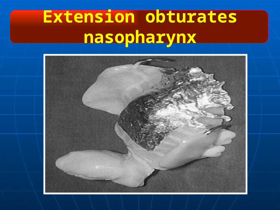
Extension obturates nasopharynx
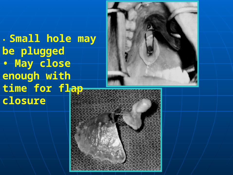
• Small hole maybe plugged• May closeenough withtime for flapclosure
Neoplastic resection is one of the most common causes for an acquired mandibular defect ( carcinoma of the tongue , floor of the mouth ).
Acquired Defect Of The Mandible

Resection of the mandible may often lead to speech and swallowing dysfunction , which are difficult to manage
A Large Maxillofacial Prosthesis for Total Mandibular Defect
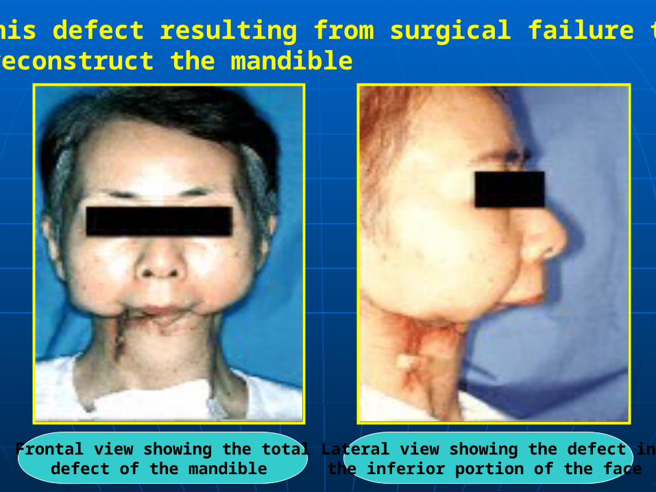
Frontal view showing the total defect of the mandible
Lateral view showing the defect in the inferior portion of the face
This defect resulting from surgical failure to reconstruct the mandible.
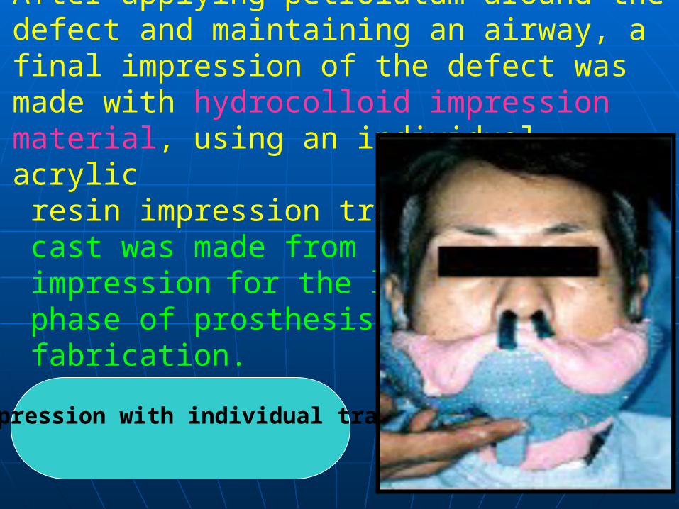
After applying petrolatum around the defect and maintaining an airway, a final impression of the defect was made with hydrocolloid impression material, using an individual acrylic resin impression tray. A stone cast was made from the impression for the laboratory phase of prosthesis fabrication.
Impression with individual tray

The wax contours of the facial prosthesis were formed with the aid of a presurgical photograph of the patient. The wax prosthesis was evaluated on the patient for esthetics and marginal adaptation. However, the soft tissue around the defect lacked sufficient anatomicundercuts to retain the prosthesisand the remaining bony structures were inadequate for dental implants.
Wax prosthesis positioned on the defect
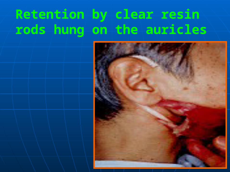
Retention by clear resin rods hung on the auricles
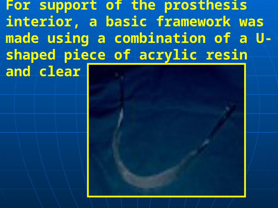
For support of the prosthesis interior, a basic framework was made using a combination of a U-shaped piece of acrylic resin and clear resin rods .
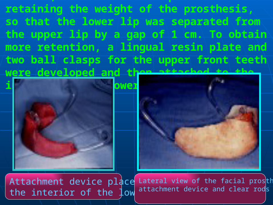
However, this was not adequate for retaining the weight of the prosthesis, so that the lower lip was separated from the upper lip by a gap of 1 cm. To obtain more retention, a lingual resin plate and two ball clasps for the upper front teeth were developed and then attached to the interior of the lower lip.
Attachment device placed onthe interior of the lower lip.
Lateral view of the facial prosthesis with an attachment device and clear rods for hanging.
These devices enabled the patient to retain the prosthesis adequately without the use of adhesives or implants. Finally, the wax facial prosthesis was invested and cast with silicone, which was suitably colored with a base pigment to match the patient's skin.

Extraoral DefectsThese defects occur due to trauma , neoplasm or congenital malformation .
Extraoral congenital malformations that require maxillofacial prostheses include:
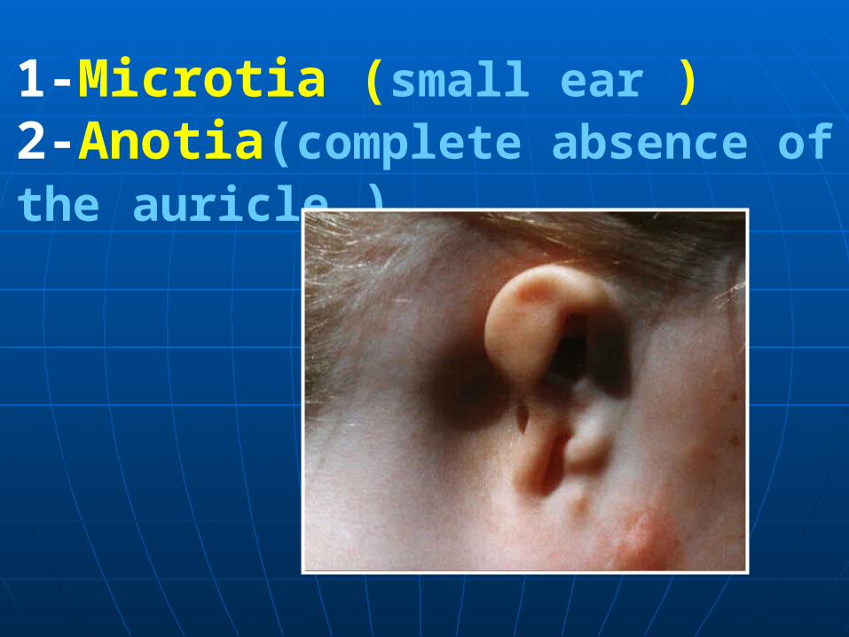
1-Microtia (small ear ) 2-Anotia(complete absence of the auricle )
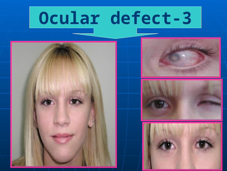
3-Ocular defect
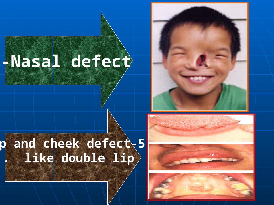
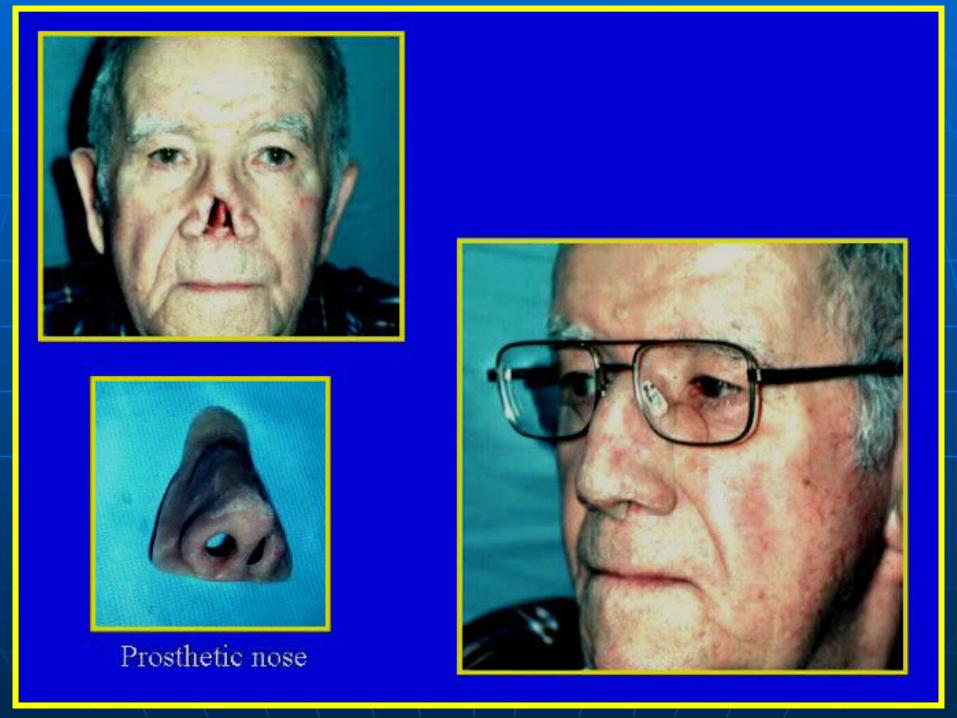
4-Nasal defect
5-Lip and cheek defect like double lip .
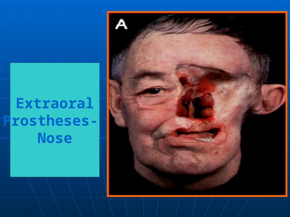
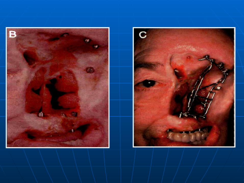
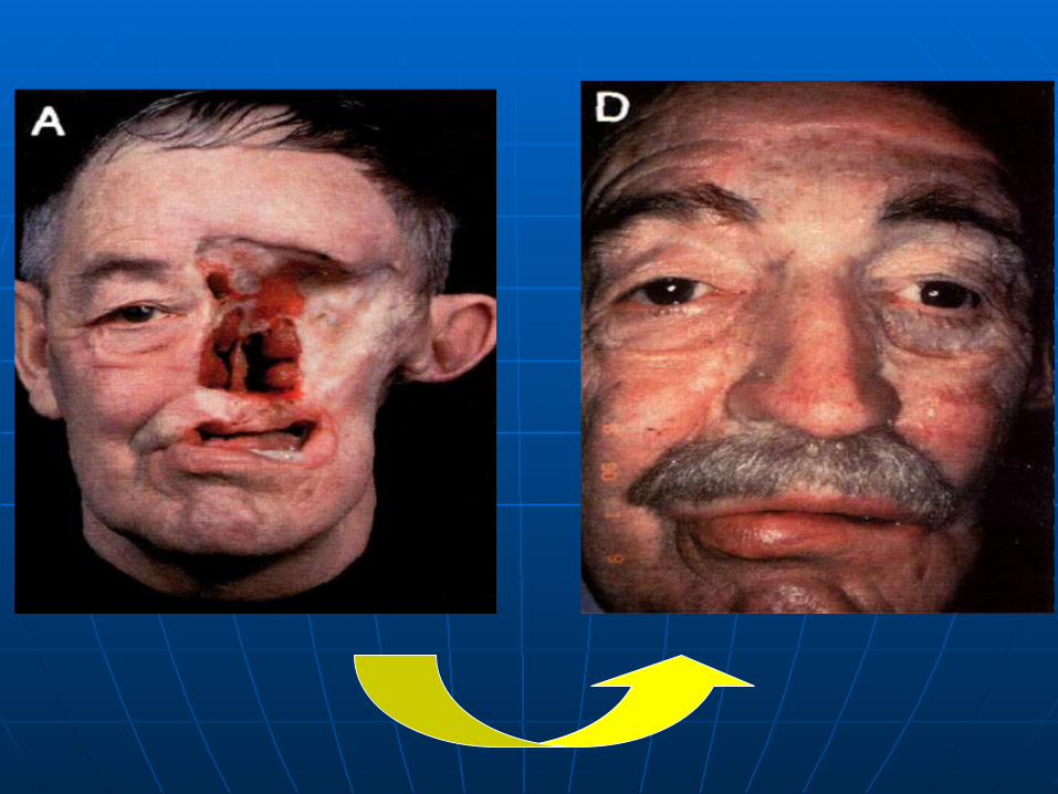
ExtraoralProstheses-
Nose
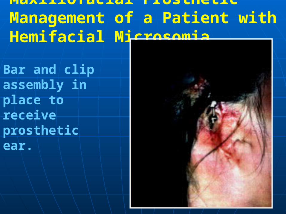
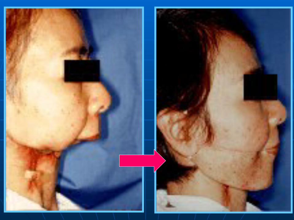
Maxillofacial Prosthetic Management of a Patient with Hemifacial Microsomia
Bar and clip assembly in place to receive prostheticear.
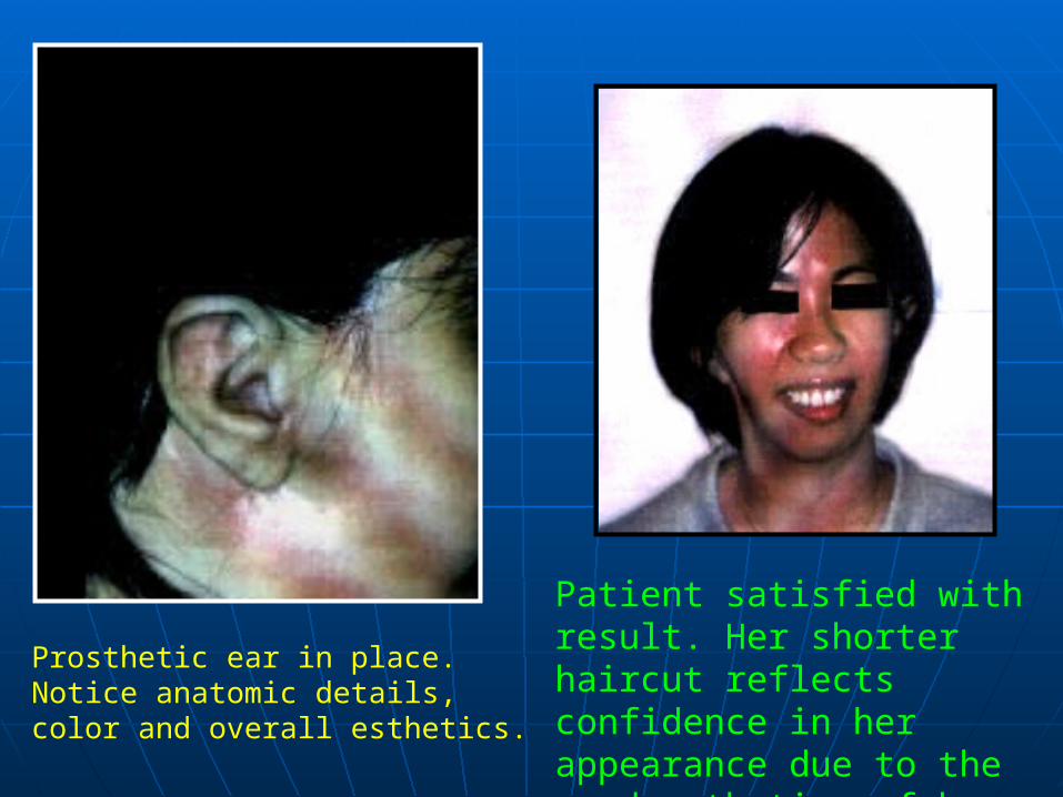
Patient satisfied with result. Her shorter haircut reflects confidence in her appearance due to the good esthetics of her new prosthetic ear
Prosthetic ear in place. Notice anatomic details, color and overall esthetics.
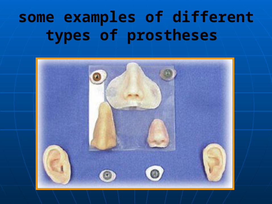
some examples of different types of prostheses

A prosthetic ear maybe retained withosseointegrated implants.
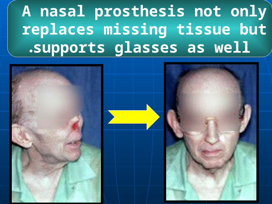
A nasal prosthesis not only replaces missing tissue but
supports glasses as well.
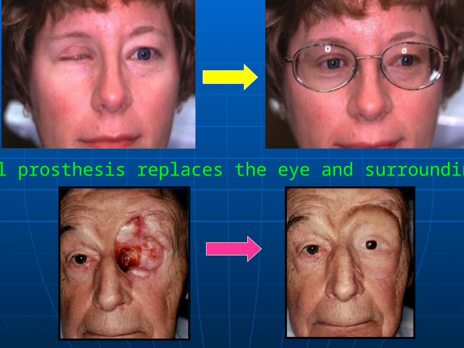
oculofacial prosthesis replaces the eye and surrounding tissues.

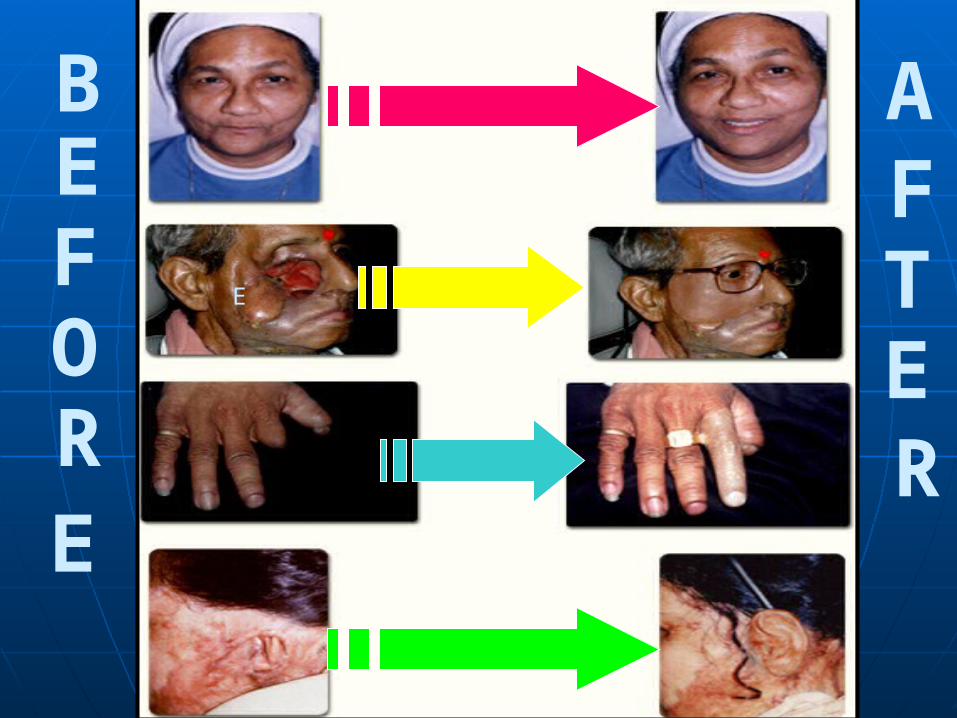
B
EFO
R
AFTE
R
E
E
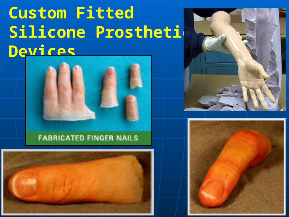
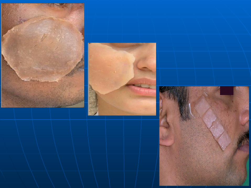
Custom Fitted Silicone Prosthetic Devices

implants in radiated patients experienced a very high success rate. The benefits gained by the use of implants are great. This makes it highly recommended to use dental implants in radiated patients whenever it is possible.
Materials Used in Maxillofacial Reconstruction
was once commonly used for maxillo- facial prostheses, and is still used occasionally to make artificial facial parts. When properly pigmented, these prostheses can look quite realistic.
POLY(METHYL METHACRYLATE)

these are plasticized methyl methacrylate polymer , which show elastic property .These are not commonly used because they get tacky lead to collection of dust and stain , have poor edge strength and degrade under sun light.
Acrylic copolymers
it is a hard , clear , tasteless and odourless resin ,extensively used in the beginning but its used decreased due to shrinkage and long processing time , discoloration and hardening of the margin .
Polyvinyl Chloride And Copolymers
these materials have excellent properties like elasticity without compromised edge strength ( this help to thin material at the margin ) . They can be used to restore defect with mobile tissue beds .The disadvantages include the moisture sensitivity during processing and poor color stability .
Polyurethane Elastomers

it is the most commonly used material for facial restoration but poor tear strength and life- less appearance have limited them from universal acceptance .

The process of crosslinking the silicone is known as vulcanizing. Vaulcanizing can occur with or without heat accordingly silicones are available in two forms.
1-HTV-Silicone : it requires heat for vulcanization . It is highly viscous , white , opaque and has better physical properties .
2-RTV-Silicones : they are room temperature polymerizing silicones . It is esear to process and allow intrinsic colouration .
Metal : metal implants are used to obtain bone anchorage for a prosthesis . Implant metals used are Titanium alloys , base metal alloys are used for denture base fabrication
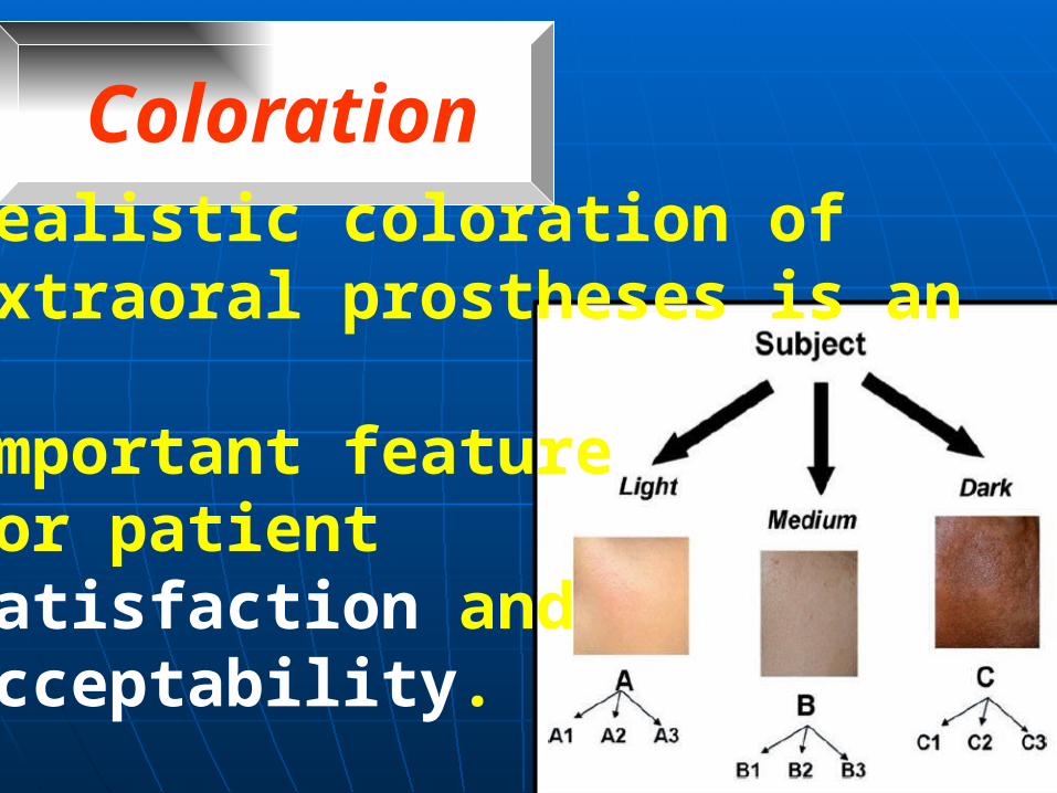
Realistic coloration of extraoral prostheses is an important feature for patient satisfaction and acceptability.
Coloration
Cosmetic realism involves the correct application of colorant formulations within the base material before polymerization (intrinsic) and after
polymerization (extrinsic).
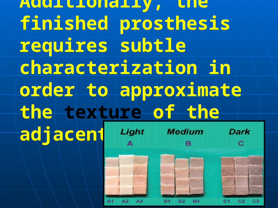
Additionally, the finished prosthesis requires subtle characterization in order to approximate the texture of the adjacent tissue
The spectral values in natural skin must be matched by corresponding pigments to accommodate environmental changes, seasonal changes, and varying light conditions.

The ultimate in realistic cosmetic matching depends on the combination of intrinsic and extrinsic colorations.
is the first step in incorporating indepth coloration reflected internally by discrete pigment particles spectrally equivalent or approximating those of the physiologic colorant and color centers, namely arterial red, venous red-purple, carotenoid yellow, melanoid brown, and opaque dispersed cellular lipids.
Intrinsic Coloration
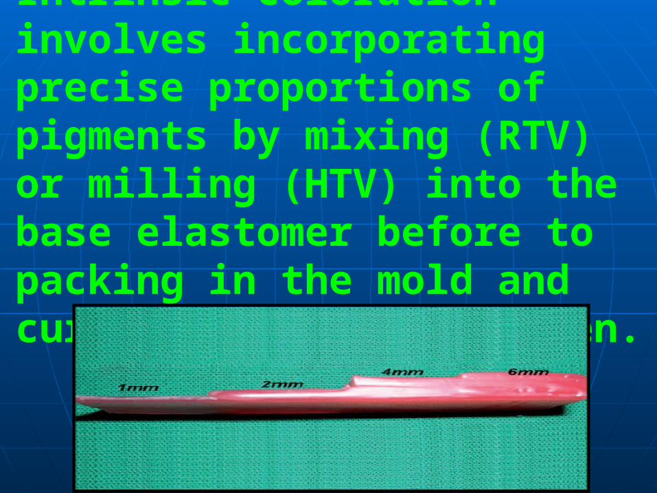
Intrinsic coloration involves incorporating precise proportions of pigments by mixing (RTV) or milling (HTV) into the base elastomer before to packing in the mold and curing in a dry heat oven.
In general, the extrinsic coloration uses a medical-grade adhesive combined with xylene and earth pigments, which are applied to the external surface of the prosthesis. The prosthesis is then postcured in a dry heat oven to evaporate the xylene.
Extrinsic coloration

Fabrication Of the Prostheses

The method for fabricating a prosthesis is similar for most materials.An impression is made of the affected area with alginate. A master cast ispoured, duplicating the defect on the patient.
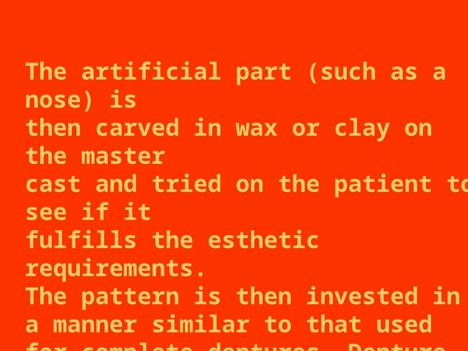
The artificial part (such as a nose) isthen carved in wax or clay on the master cast and tried on the patient to see if it fulfills the esthetic requirements.The pattern is then invested in a manner similar to that used for complete dentures. Denture flasks are often used for this purpose.

When the prosthesis is quite complex (such as an eye and orbit), three- or four-part molds are made. With some materials, metal molds are required because of high processing temperatures. After the pattern is invested, it is removed from the mold by use of a boiling water bath.
The mold is now ready to make the prosthesis. The patient should be present so pigments may be added to the elastomer to give a realistic appearance and match the patient's skin color.Generally, dry mineral earth pigments or artist's oil-based pigments are used.
Color matching is done by mixing small amounts of the pigments into the elastomer. Some clinicians use color tabs and predetermined pigment formulations to match skin color.
When a color match is achieved, the elastomer is compression molded and processed according to the manufacturer's instructions.After processing, the prosthesis is removed from the mold and the excess flash is removed.
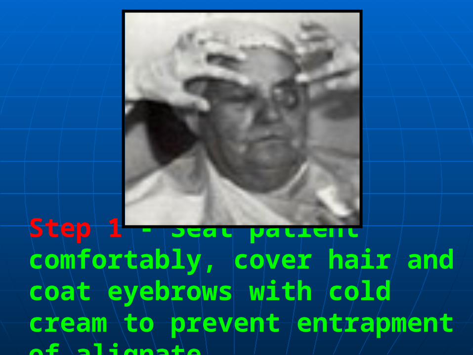
Step 1 - Seat patient comfortably, cover hair and coat eyebrows with cold cream to prevent entrapment of alignate.
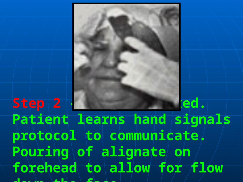
Step 2 - Alignate mixed. Patient learns hand signals protocol to communicate. Pouring of alignate on forehead to allow for flow down the face.
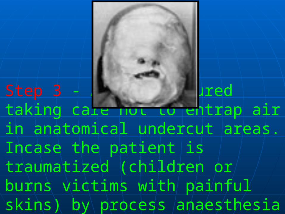
Step 3 - Alignate poured taking care not to entrap air in anatomical undercut areas. Incase the patient is traumatized (children or burns victims with painful skins) by process anaesthesia is needed.
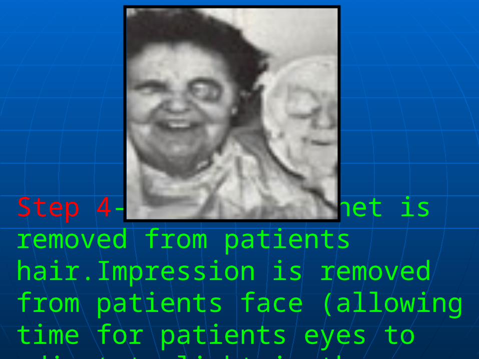
Step 4- Guaze stockinet is removed from patients hair.Impression is removed from patients face (allowing time for patients eyes to adjust to light in the room
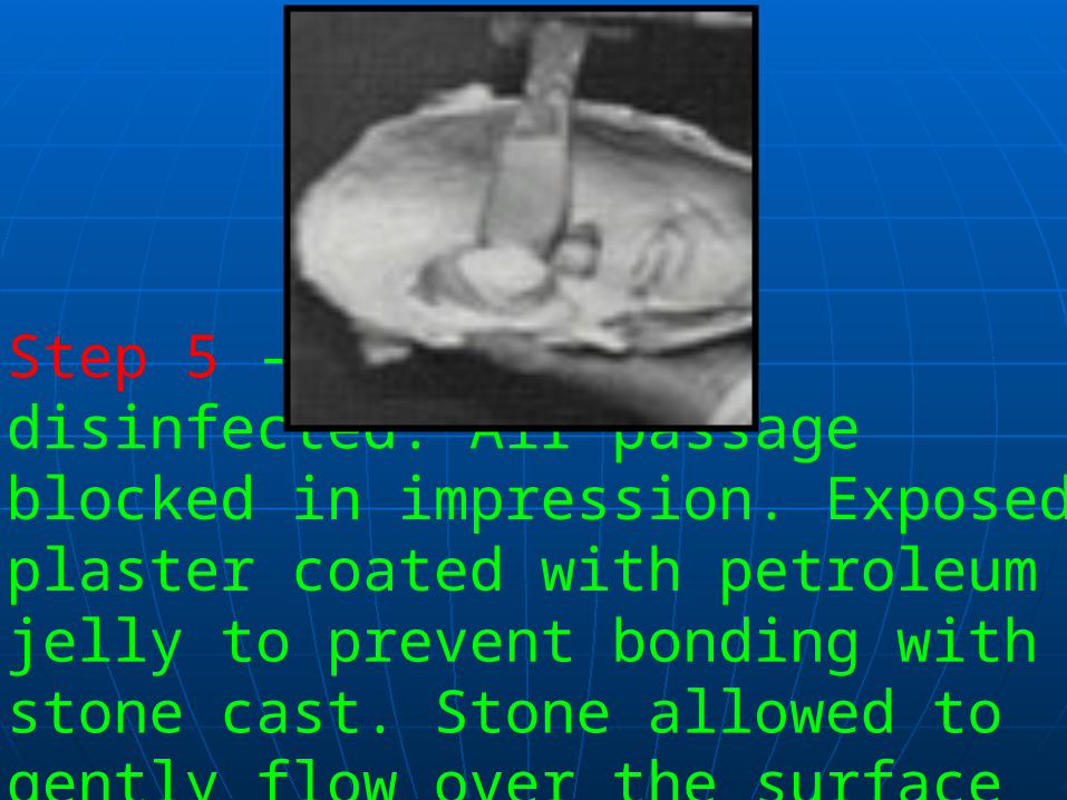
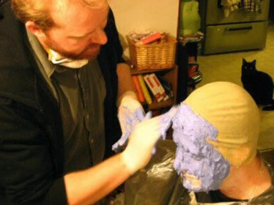
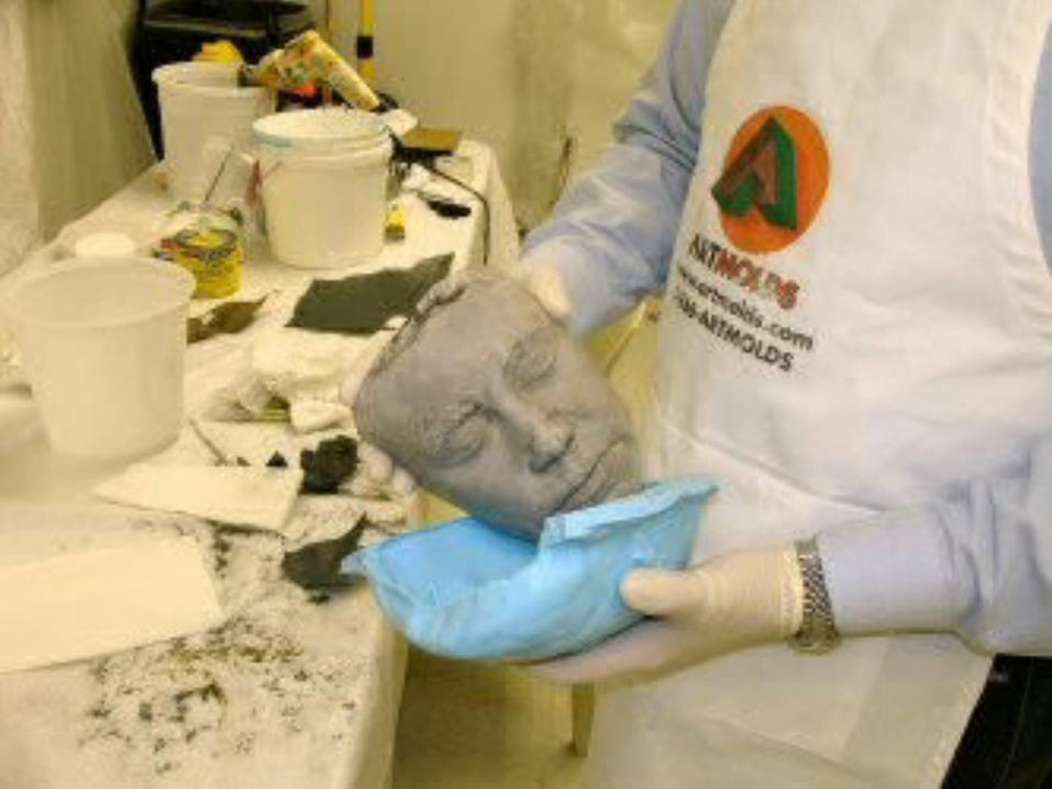
Step 5 - Impression disinfected. Air passage blocked in impression. Exposed plaster coated with petroleum jelly to prevent bonding with stone cast. Stone allowed to gently flow over the surface of the alignate.
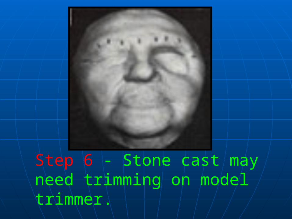
Step 6 - Stone cast may need trimming on model trimmer.


Fabrication of a Maxillofacial Prosthesis Using a Computer-Aided Design and Manufacturing System
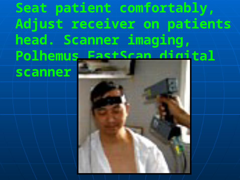
Seat patient comfortably, Adjust receiver on patients head. Scanner imaging, Polhemus FastScan digital scanner
Maxillofacial prostheses are usually fabricated on the basis of impressions made with dental-impression material.The extent to which the prosthesis reproduces normal facial morphology depends on the clinical judgment of the individual fabricating the prosthesis.

This new technique describes a computer-aided design and manufacturing system (CAD/CAM) for the fabrication of maxillofacial prostheses. This system will provide a more consistently accurate reproduction of facial morphology.
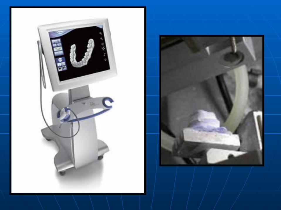
Facial measurements were taken using a non-contact three-dimensional laser morphological measurement system.
The measurements were sent to a computer numerical controlled (CNC) milling machine to generatea cast of the patient's face for thefabrication of prosthesis.


Facial contours were measured using a laser. This method minimizes patient discomfort and avoids soft tissue distortion by impression material. Moreover, the digital data obtained is easy to store and transmit, and mirror-images can be readily generated by computer processing.
Results
This method offers an objective, quantified approach for fabricating maxillofacial prostheses.
Conclusion
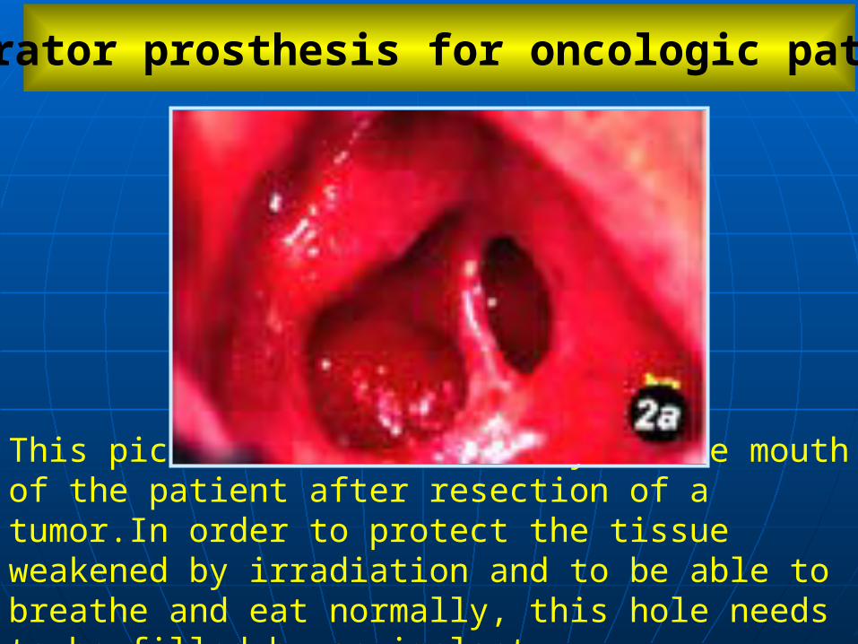
This picture shows the cavity in the mouth of the patient after resection of a tumor.In order to protect the tissue weakened by irradiation and to be able to breathe and eat normally, this hole needs to be filled by an implant.
Obturator prosthesis for oncologic patients
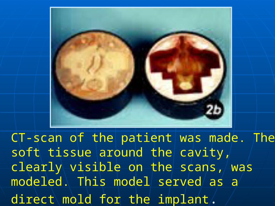
CT-scan of the patient was made. The soft tissue around the cavity, clearly visible on the scans, was modeled. This model served as a direct mold for the implant.

The implant, called obturator prosthesis, was cast from the mold in a bio-compatible silicone
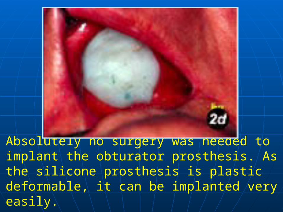
Absolutely no surgery was needed to implant the obturator prosthesis. As the silicone prosthesis is plastic deformable, it can be implanted very easily.
CONCLUSION: The prosthesis fits the cavity much better than ever could have been achieved by using impression techniques. These traditional techniques produce a master of the obturator prosthesis by making an impression of the cavity in a plastic deformable material.

The prostheses cast from such masters are always less accurate because of the presence of undercuts (the impression technique is not sensitive to local internal broadening of the cavity) and can severely damage the sensitive and vulnerable surrounding tissue.
The soft prosthesis is fixed by means of magnets on a hard dental implant. This makes it possible to take it out for inspection and to replace it afterwards.
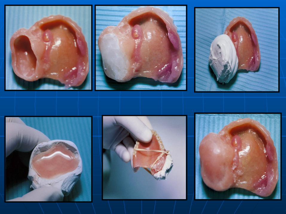
fabrication of a meatus obturator prosthesis made with visible light-cured (VLC) resin. The fabrication technique is relatively easy and saves time by eliminating some laboratory procedures for both the patient and the practitioner.
An alternative approach to fabricatinga meatus obturator prosthesis
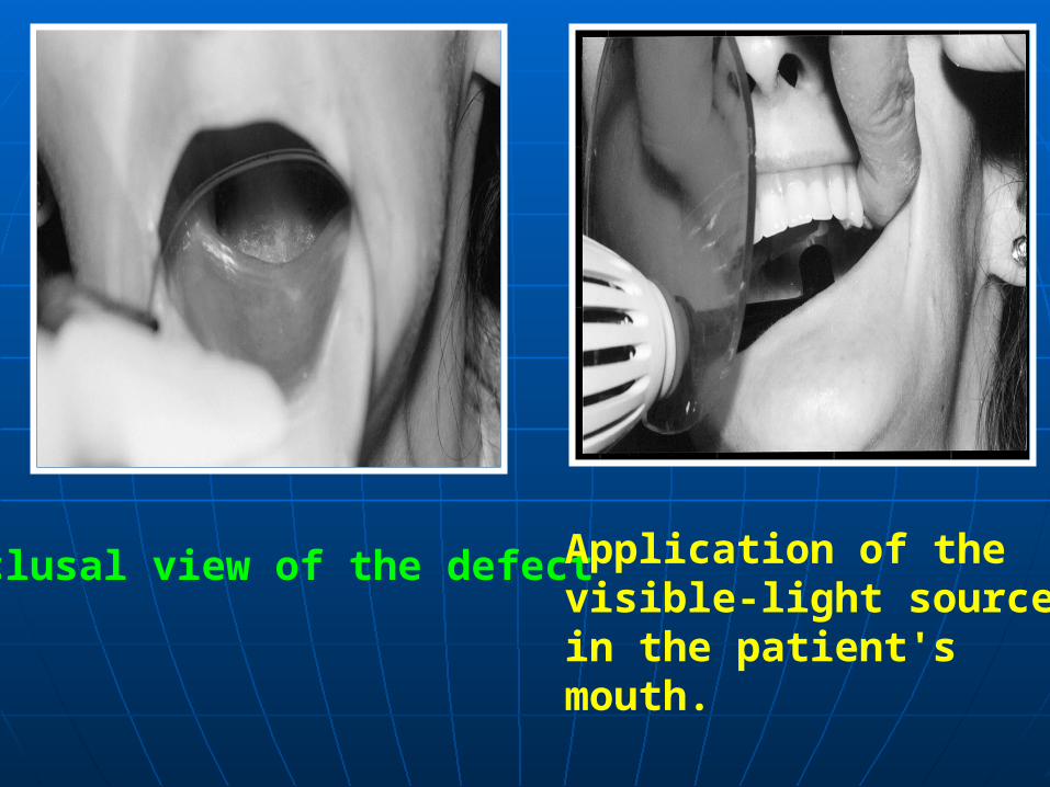
Occlusal view of the defect Application of the visible-light source in the patient'smouth.
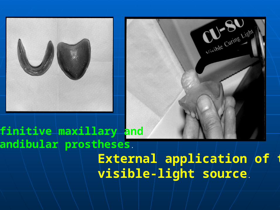
Definitive maxillary and mandibular prostheses.
External application of the visible-light source.
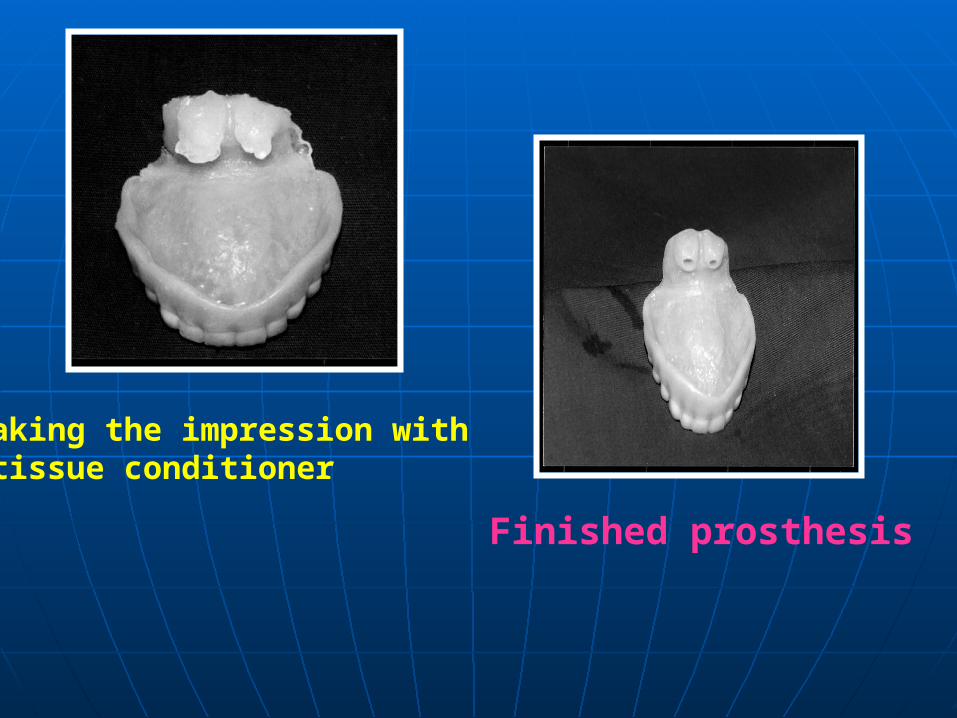
Making the impression with tissue conditioner.
Finished prosthesis

Retention of maxillofacial prostheses
Retention of facial prostheses has been primarily by way of medical adhesives. An ideal adhesive should be one that provides firm functional retention under flexure or extension during speech, facial expressions, and moisture or perspiration contact.
Adhesives for extraoral maxillofacial prostheses require a substantial amount of supportive ingredients properly formulated to provide lasting viscoelasticity with a high degree of retention. Numerous brand names of adhesives have been introduced over the years in maxillofacial prosthetics.
Other methods of retention include engagement of anatomic tissue undercuts, thereby minimizing dependence on adhesives. The potential for tissue irritation exists with this technique, and therefore it must be used prudently. Areas that have been irradiated contraindicate the use of this technique.
Finally, with the increaed use of
osseointegrated implants, dependence on adhesive and anatomic methods of retention has diminished.
Magnets can be used to minimize force transfer to the implant and supporting bone. The resultant decrease in dependence on chemical (adhesives) and anatomic (tissue undercuts) sources of retention is beneficial to both the patient and the prosthetic rehabilitation.
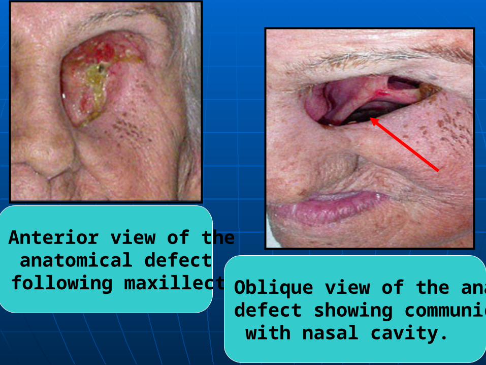
Anterior view of the anatomical defect
following maxillectomy Oblique view of the anatomicaldefect showing communication with nasal cavity.
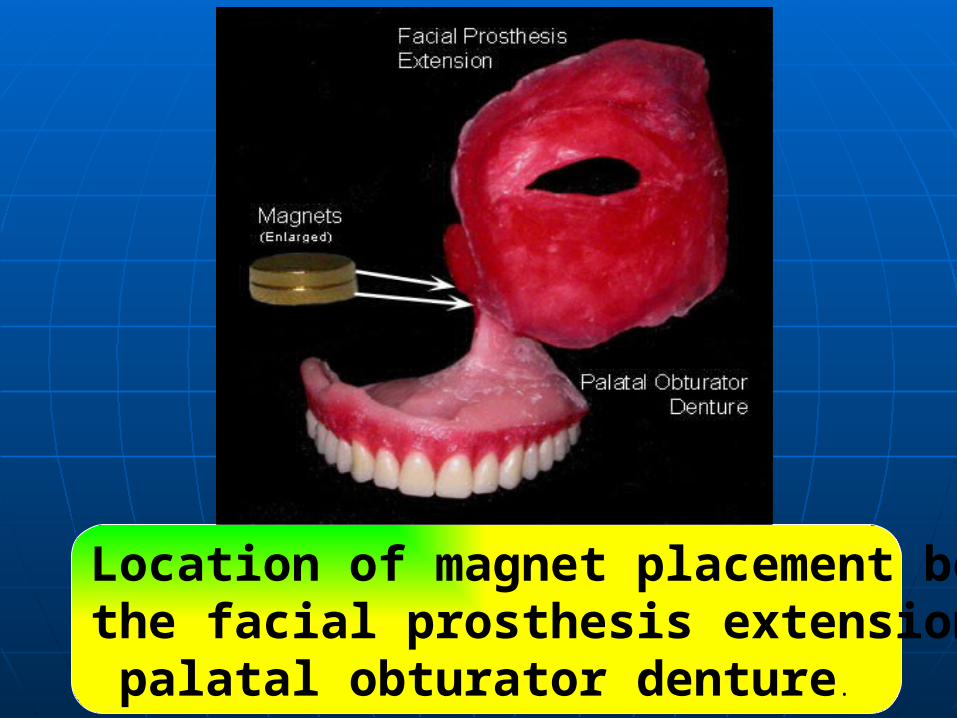
Location of magnet placement between the facial prosthesis extension and the palatal obturator denture.

Positioning of the retention magnetsA.Highligher paste on thesuperior aspect of the palatal obturator prosthesis. B. Transfer of the paste tothe facial prosthesis extension to demarcate thelocation for the secondmagnet.
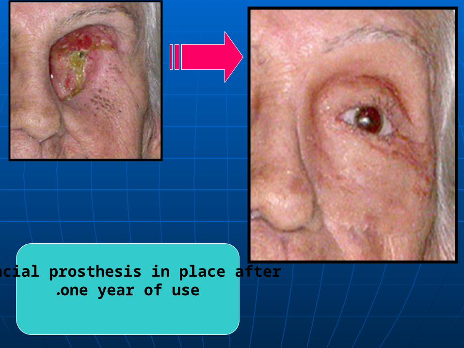
Facial prosthesis in place after one year of use.
