mbbs 3 cerebrovascular disease seminar intimalintimal ... cerebral cortex normal (left) acute...
TRANSCRIPT
CEREBRALVASCULAR DISEASE
Dr Rodney Itaki
Lecturer
Anatomical Pathology Discipline
Division of Pathology
University of Papua New GuineaSchool of Medicine & Health SciencesDivision of Pathology
Cerebral vascular Disease�Definition of term:
�The term cerebrovascular disease designates any abnormality of the brain resulting from a pathologic process of the blood vessels.
�Sudden loss of neurological function is the hallmark of cerebrovascular disease.
�3rd most common cause of death in affluent societies�Its incidence increases with age and is somewhat higher in
men than in women.
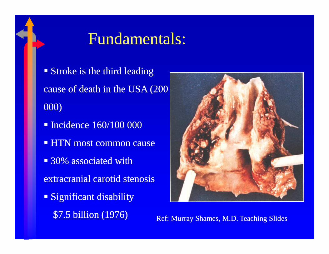
FundamentalsFundamentals:
� Stroke is the third leading Stroke is the third leading
cause of death in the USA (200 cause of death in the USA (200
000)000)
�� Incidence 160/100 000Incidence 160/100 000
�� HTN most common causeHTN most common cause
�� 30% associated with 30% associated with
extracranialextracranial carotid carotid stenosisstenosis
�� Significant disabilitySignificant disability
$7.5 billion (1976)$7.5 billion (1976) Ref: Murray Shames, M.D. Teaching SlidesRef: Murray Shames, M.D. Teaching Slides
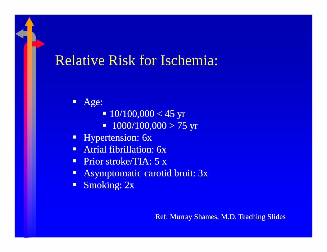
Relative Risk for Ischemia:
�� Age:Age:�� 10/100,000 < 45 yr10/100,000 < 45 yr�� 1000/100,000 > 75 yr1000/100,000 > 75 yr
�� Hypertension: 6xHypertension: 6x�� AtrialAtrial fibrillation: 6xfibrillation: 6x�� Prior stroke/TIA: 5 xPrior stroke/TIA: 5 x�� Asymptomatic carotid bruit: 3xAsymptomatic carotid bruit: 3x�� Smoking: 2xSmoking: 2x
Ref: Murray Shames, M.D. Teaching SlidesRef: Murray Shames, M.D. Teaching Slides
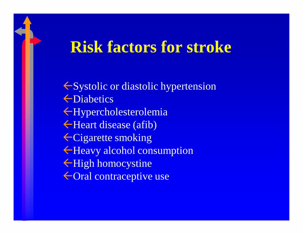
Risk factors for stroke
�Systolic or diastolic hypertension�Diabetics�Hypercholesterolemia�Heart disease (afib)�Cigarette smoking �Heavy alcohol consumption�High homocystine�Oral contraceptive use
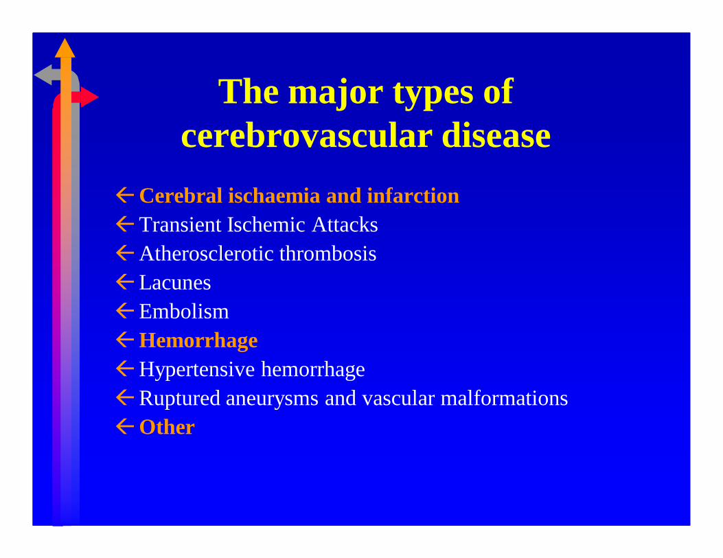
The major types of cerebrovascular disease
�Cerebral ischaemia and infarction�Transient Ischemic Attacks�Atherosclerotic thrombosis�Lacunes�Embolism�Hemorrhage�Hypertensive hemorrhage�Ruptured aneurysms and vascular malformations�Other

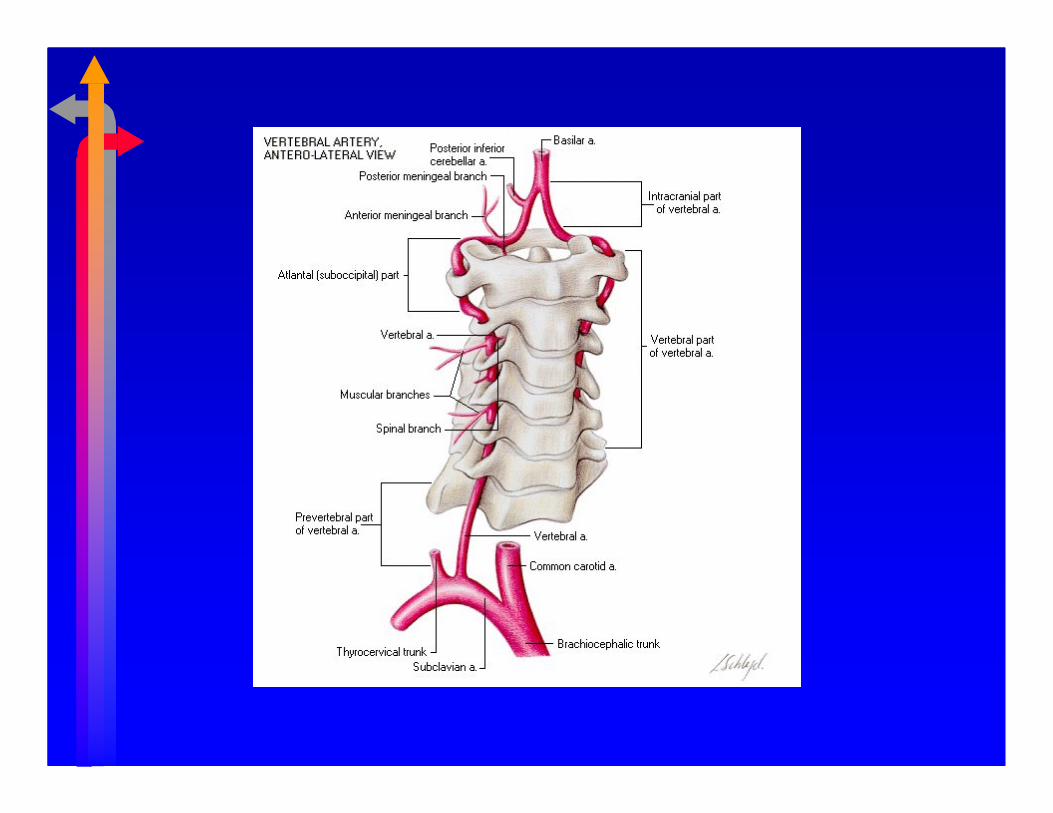
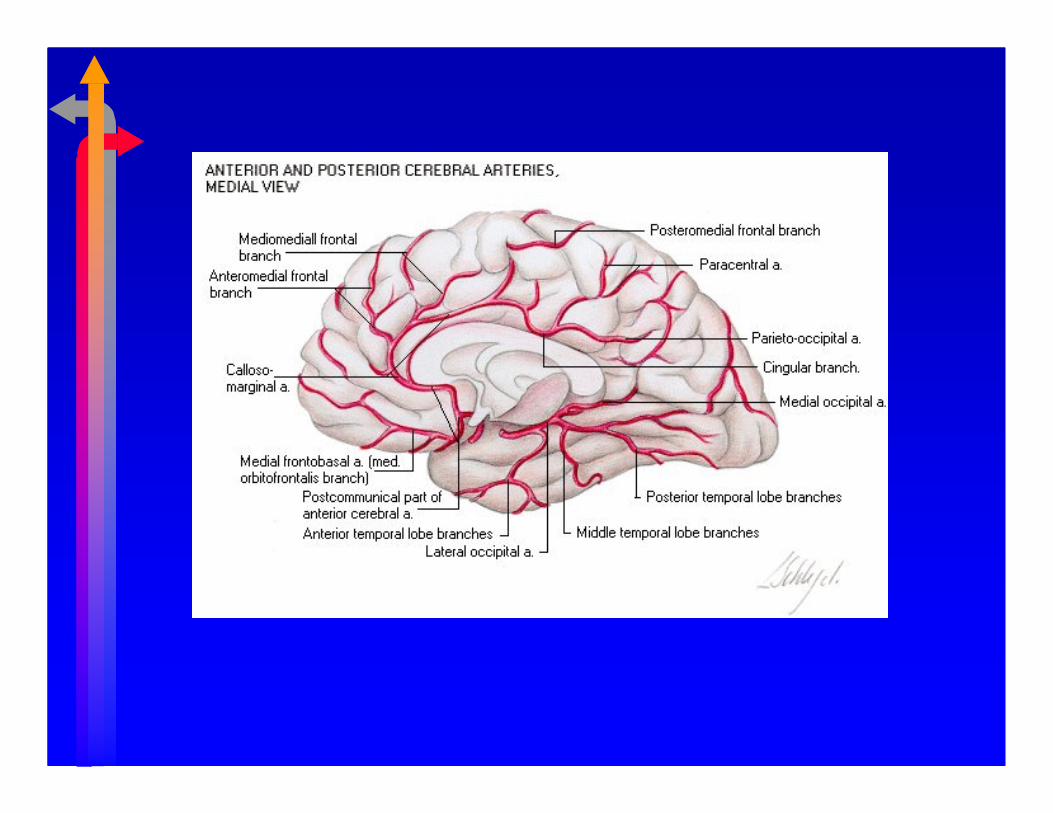
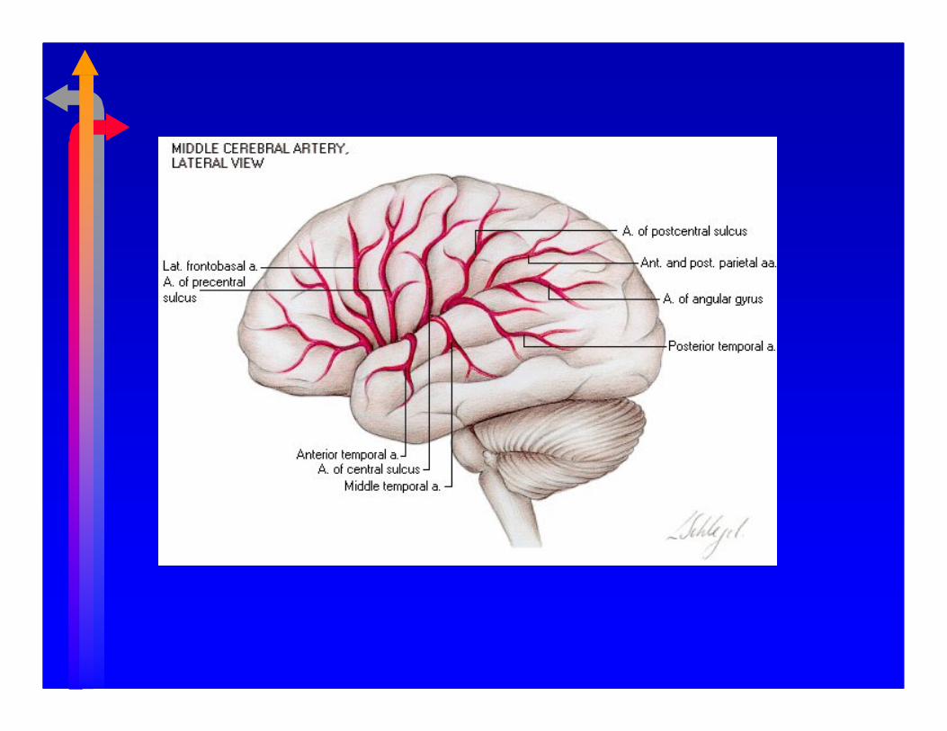
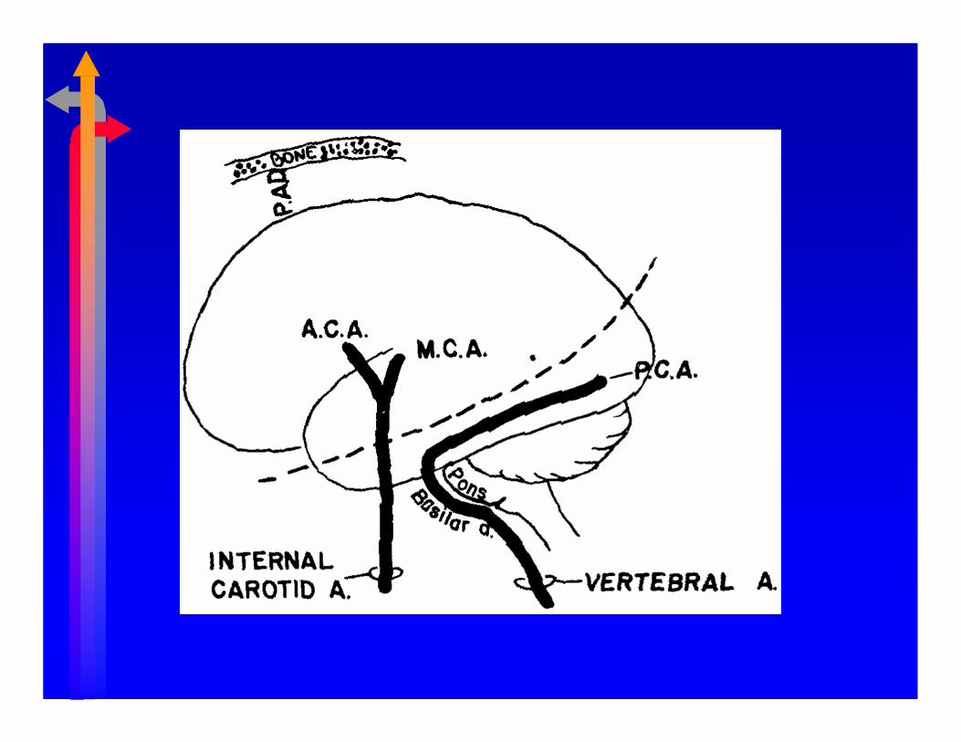
I、、、、Cerebral ischaemia and infarction�Anatomy and pathology
�The principal pathological process is the occlusion of arteries supplying the brain.
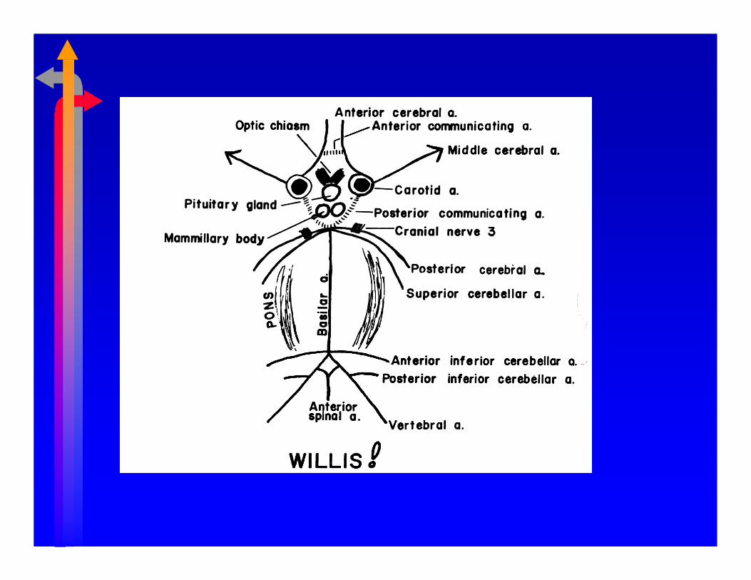
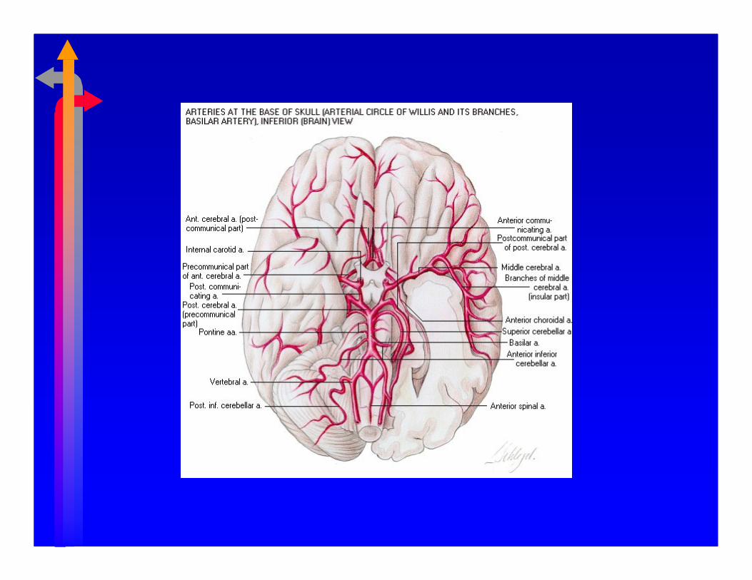
�The two internal carotid arteries and the basilar artery form the Circle of Willis at the base of the brain.
�Efficient anatomotic device in the event of occlusion of arteries proximal to it.
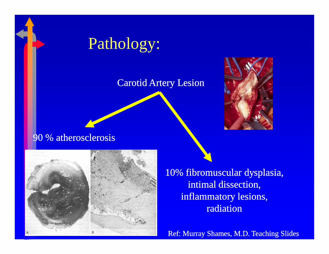
Pathology:
Carotid Artery LesionCarotid Artery Lesion
10% fibromuscular dysplasia, 10% fibromuscular dysplasia, intimal dissection, intimal dissection,
inflammatory lesions, inflammatory lesions, radiationradiation
90 % atherosclerosis90 % atherosclerosis
Ref: Murray Shames, M.D. Teaching SlidesRef: Murray Shames, M.D. Teaching Slides
�Anatomy and pathology
�Occlusion leads to sudden severe ischaemia in the area of brain tissue supplied by the occluded artery, and recovery depends upon rapid lysis or fragmentation of the occluding material:Reversal of neurological function within minutes or hours gives rise to the clinical picture of a transient ischaemic attack.
�Anatomy and pathology
�When the neurological deficit lasts longer than 24 hours, it may be called a reversible ischaemic neurological deficit ( RIND ) if it recovers completely in a few days,or a completed stroke if there is a persistent deficit.Sometimes recovery is very slow and incomplete.
Neurological symptoms and signs
�The loss of function that the patient notices, and which may be apparent on examination, entirely depends on the area of brain tissue involved in the ischaemic process.

1. Transient Ischemic Attacks(TIA)
�Definition of term
�TIAs are brief, reversible episodes of focal, nonconvulsive ischaemic neurologic disturbance, Duration should be less than 24 h.
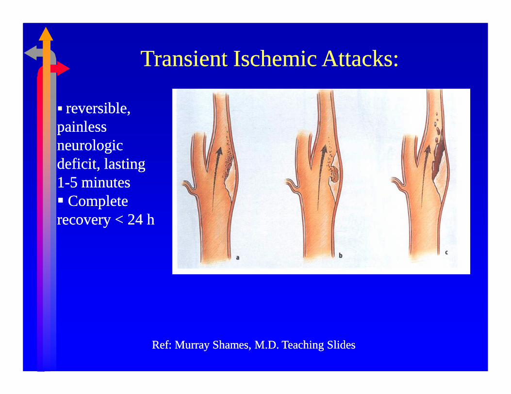
�� reversible, reversible, painless painless neurologic neurologic deficit, lasting deficit, lasting 11--5 minutes5 minutes�� Complete Complete recovery < 24 hrecovery < 24 h
Transient Ischemic Attacks:Transient Ischemic Attacks:
Ref: Murray Shames, M.D. Teaching SlidesRef: Murray Shames, M.D. Teaching Slides
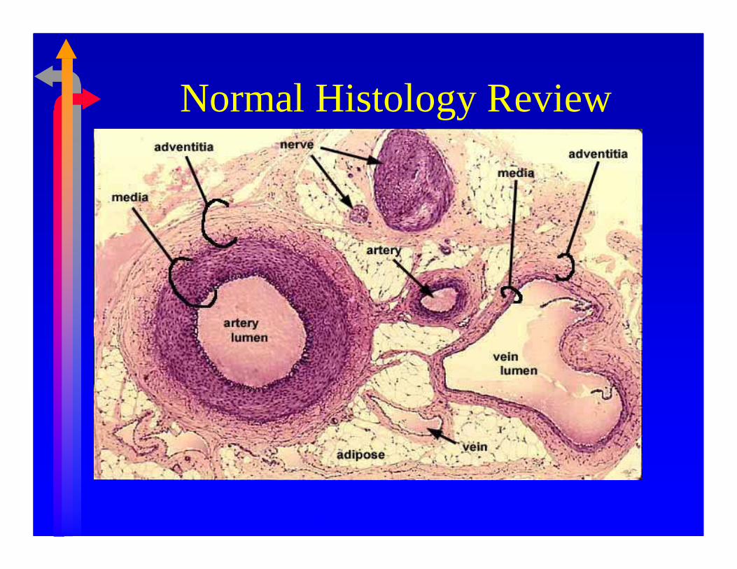
Normal Histology Review
Ref: Wikipedia
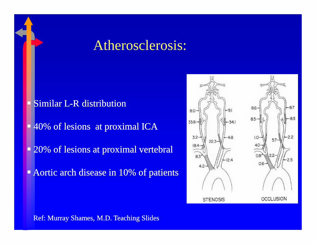
Atherosclerosis:
�� Similar LSimilar L--R distributionR distribution
�� 40% of lesions at proximal ICA40% of lesions at proximal ICA
�� 20% of lesions at proximal vertebral20% of lesions at proximal vertebral
��Aortic arch disease in 10% of patientsAortic arch disease in 10% of patients
Ref: Murray Shames, M.D. Teaching SlidesRef: Murray Shames, M.D. Teaching Slides
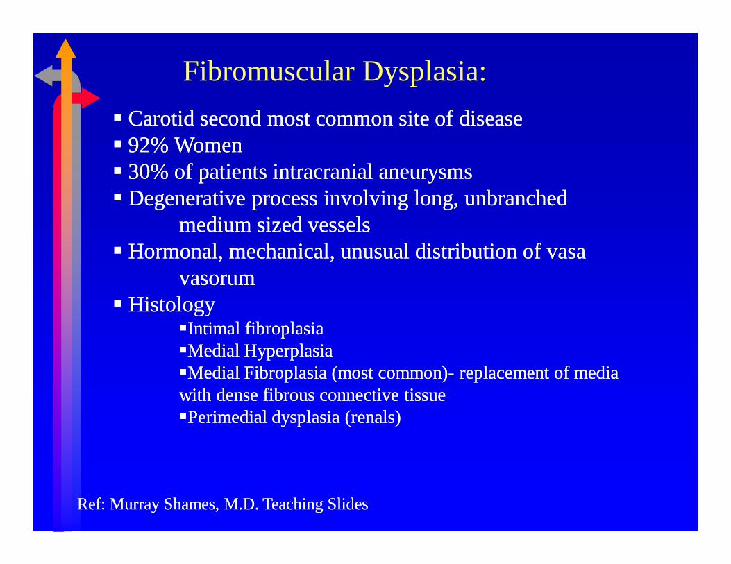
Fibromuscular Dysplasia:
�� Carotid second most common site of diseaseCarotid second most common site of disease�� 92% Women92% Women�� 30% of patients intracranial aneurysms30% of patients intracranial aneurysms�� Degenerative process involving long, Degenerative process involving long, unbranchedunbranched
medium sized vesselsmedium sized vessels�� Hormonal, mechanical, unusual distribution of Hormonal, mechanical, unusual distribution of vasavasa
vasorumvasorum�� HistologyHistology
��IntimalIntimal fibroplasiafibroplasia��Medial HyperplasiaMedial Hyperplasia��Medial Medial FibroplasiaFibroplasia (most common)(most common)-- replacement of media replacement of media with dense fibrous connective tissuewith dense fibrous connective tissue��PerimedialPerimedial dysplasia (dysplasia (renalsrenals))
Ref: Murray Shames, M.D. Teaching SlidesRef: Murray Shames, M.D. Teaching Slides
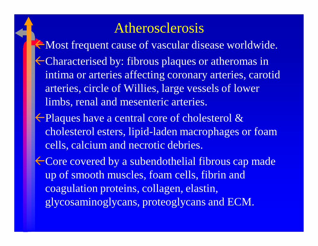
Atherosclerosis�Most frequent cause of vascular disease worldwide.
�Characterised by: fibrous plaques or atheromas in intima or arteries affecting coronary arteries, carotid arteries, circle of Willies, large vessels of lower limbs, renal and mesenteric arteries.
�Plaques have a central core of cholesterol & cholesterol esters, lipid-laden macrophages or foam cells, calcium and necrotic debries.
�Core covered by a subendothelial fibrous cap made up of smooth muscles, foam cells, fibrin and coagulation proteins, collagen, elastin, glycosaminoglycans, proteoglycans and ECM.
Atherosclerosis
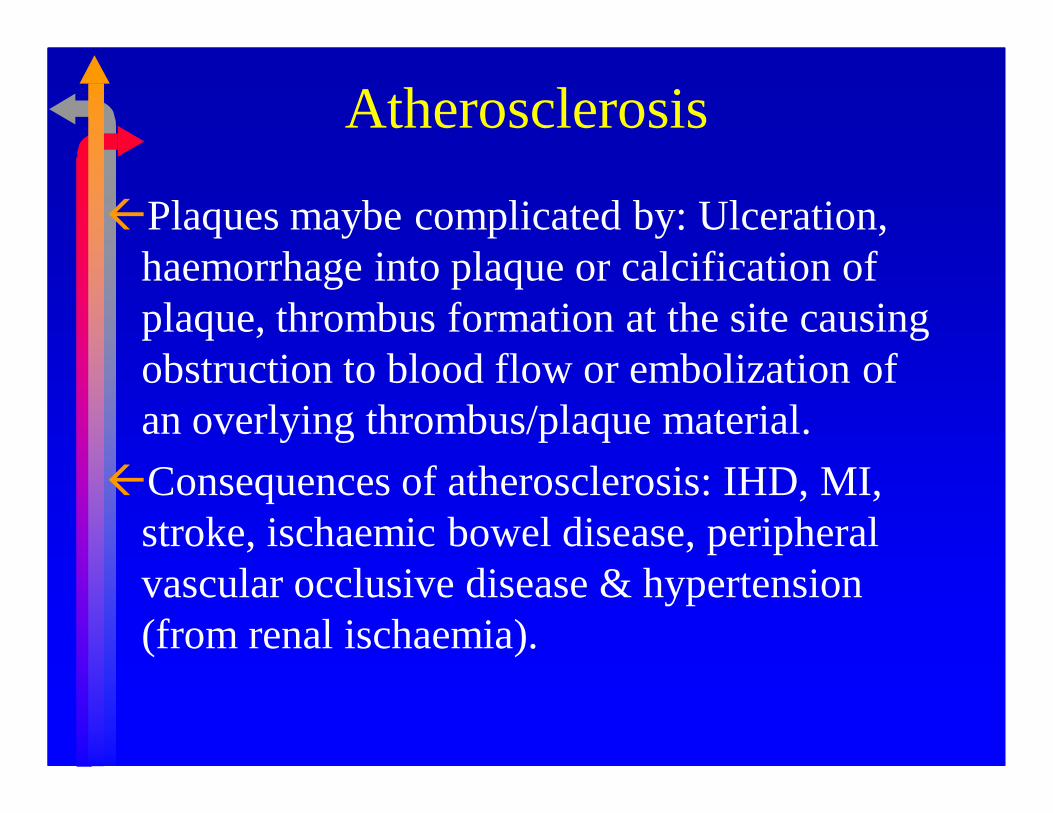
�Plaques maybe complicated by: Ulceration, haemorrhage into plaque or calcification of plaque, thrombus formation at the site causing obstruction to blood flow or embolization of an overlying thrombus/plaque material.
�Consequences of atherosclerosis: IHD, MI, stroke, ischaemic bowel disease, peripheral vascular occlusive disease & hypertension (from renal ischaemia).

Pathogenesis of Atherosclerosis:Pathogenesis of Atherosclerosis:
�� IntimalIntimal injury (injury (hemodynamicshemodynamics))�� Nodular deposition of fat in arterial Nodular deposition of fat in arterial intimaintima��Associated inflammatory response Associated inflammatory response –– fibroblast, fibroblast,
smooth muscle cell proliferationsmooth muscle cell proliferation�� Slow accumulation of lipoproteinsSlow accumulation of lipoproteins�� Calcium precipitation in the primary fatty plaque.Calcium precipitation in the primary fatty plaque.
Ref: Murray Shames, M.D. Teaching SlidesRef: Murray Shames, M.D. Teaching Slides

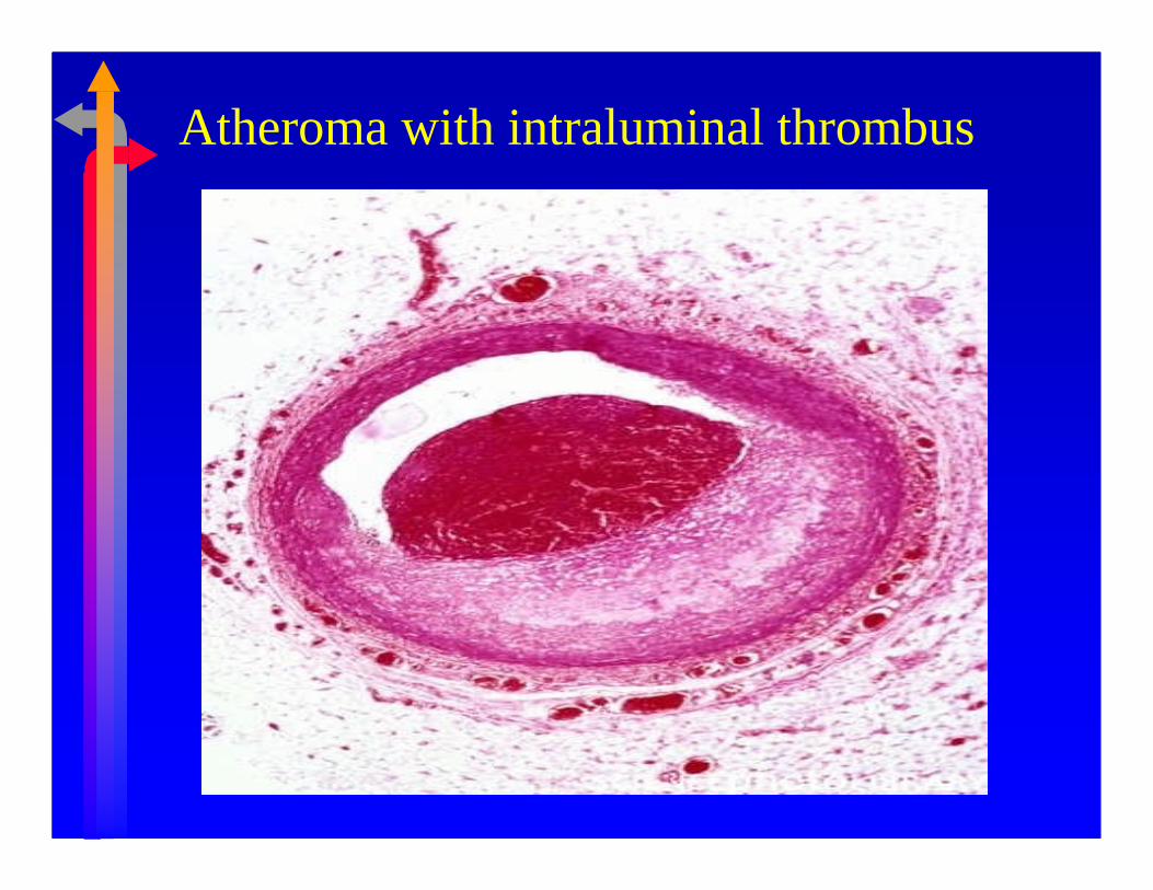
Atheroma with intraluminal thrombus
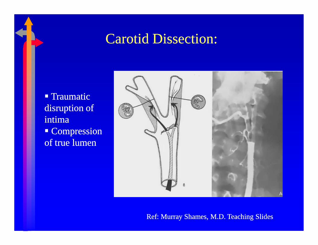
Carotid Dissection:Carotid Dissection:
�� Traumatic Traumatic disruption of disruption of intimaintima�� Compression Compression of true lumenof true lumen
Ref: Murray Shames, M.D. Teaching SlidesRef: Murray Shames, M.D. Teaching Slides
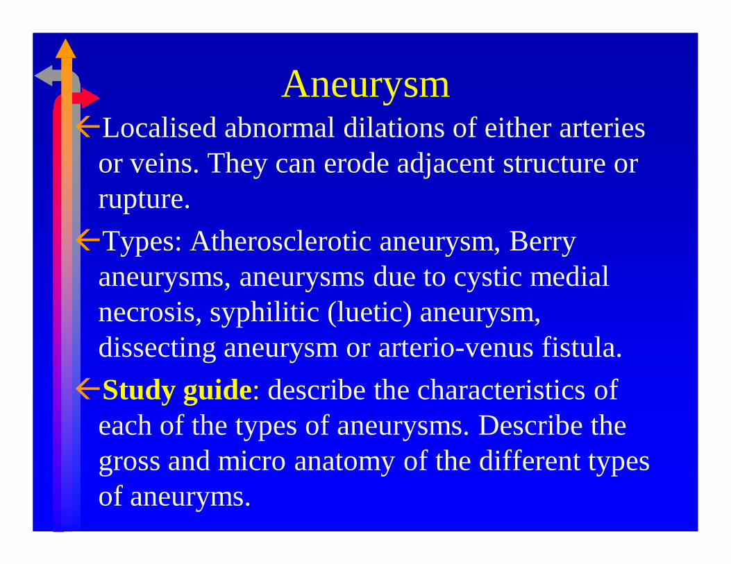
Aneurysm�Localised abnormal dilations of either arteries
or veins. They can erode adjacent structure or rupture.
�Types: Atherosclerotic aneurysm, Berry aneurysms, aneurysms due to cystic medial necrosis, syphilitic (luetic) aneurysm, dissecting aneurysm or arterio-venus fistula.
�Study guide: describe the characteristics of each of the types of aneurysms. Describe the gross and micro anatomy of the different types of aneuryms.
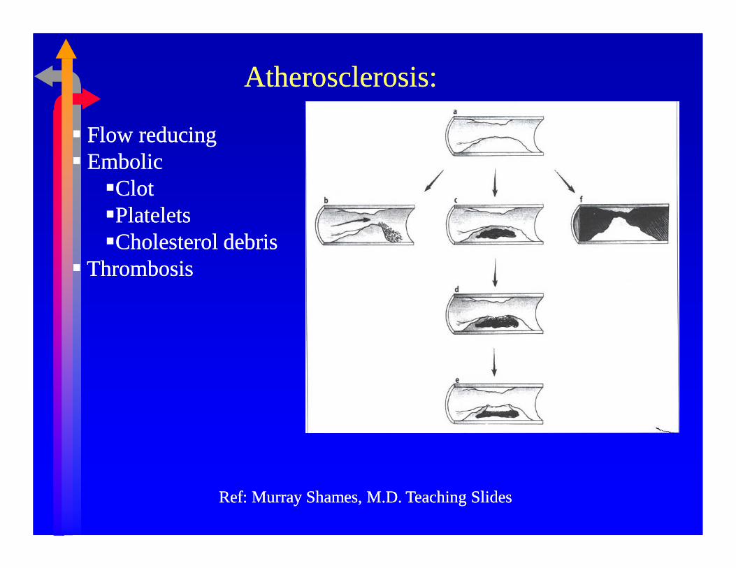
Atherosclerosis:Atherosclerosis:
�� Flow reducingFlow reducing�� EmbolicEmbolic
��ClotClot��PlateletsPlatelets��Cholesterol debrisCholesterol debris
�� ThrombosisThrombosis
Ref: Murray Shames, M.D. Teaching SlidesRef: Murray Shames, M.D. Teaching Slides
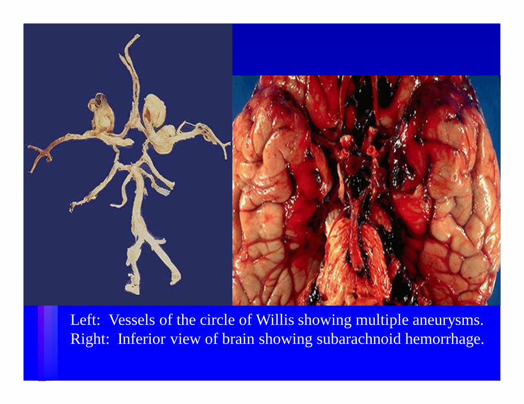
Left: Vessels of the circle of Willis showing multiple aneurysms.Right: Inferior view of brain showing subarachnoid hemorrhage.
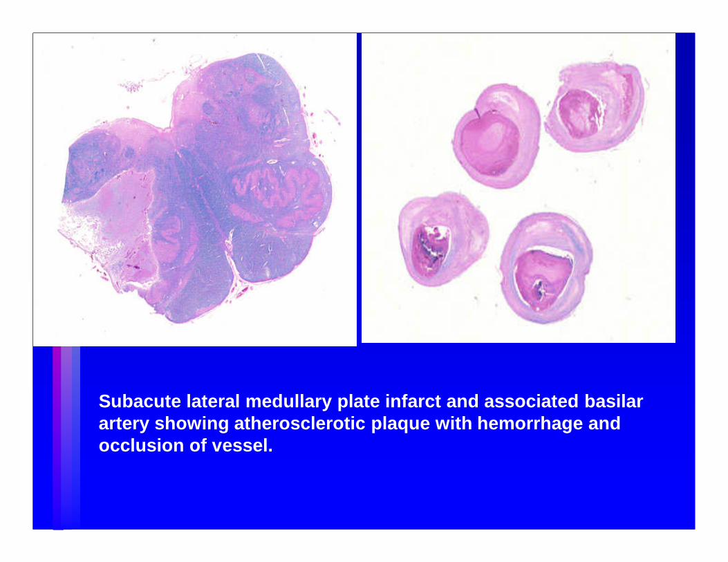
Subacute lateral medullary plate infarct and associated basilarartery showing atherosclerotic plaque with hemorrhage and occlusion of vessel.
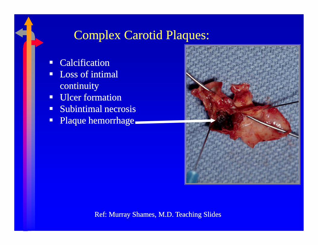
Complex Carotid Plaques:Complex Carotid Plaques:
�� CalcificationCalcification�� Loss of Loss of intimalintimal
continuitycontinuity�� Ulcer formationUlcer formation�� SubintimalSubintimal necrosisnecrosis�� Plaque hemorrhagePlaque hemorrhage
Ref: Murray Shames, M.D. Teaching SlidesRef: Murray Shames, M.D. Teaching Slides

�� a harbinger of stroke (30a harbinger of stroke (30--40% of patients with 40% of patients with surgically accessible carotid surgically accessible carotid stenosisstenosis))
�� No loss of consciousness No loss of consciousness -- syncopesyncope�� AmaurosisAmaurosis fugaxfugax: embolus to : embolus to ipsilateralipsilateral retinal arteryretinal artery�� AphasiaAphasia�� ContraContra--lateral lateral paralysis, paresis, paralysis, paresis, paresthesiasparesthesias�� Stroke rate at 1 ,3 , 5 years 23%, 27%, 45%Stroke rate at 1 ,3 , 5 years 23%, 27%, 45%�� Crescendo TIA’s/ Stroke in evolutionCrescendo TIA’s/ Stroke in evolution
Relationship between TIA & StrokeRelationship between TIA & Stroke
Ref: Murray Shames, M.D. Teaching SlidesRef: Murray Shames, M.D. Teaching Slides
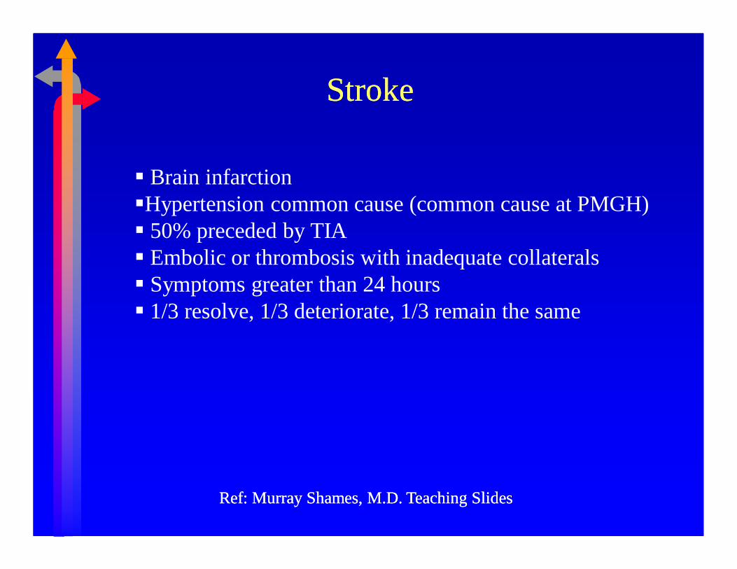
StrokeStroke
� Brain infarction�Hypertension common cause (common cause at PMGH)� 50% preceded by TIA� Embolic or thrombosis with inadequate collaterals� Symptoms greater than 24 hours� 1/3 resolve, 1/3 deteriorate, 1/3 remain the same
Ref: Murray Shames, M.D. Teaching SlidesRef: Murray Shames, M.D. Teaching Slides
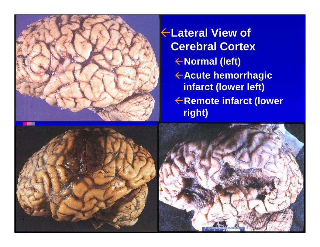
�Lateral View of Cerebral Cortex�Normal (left)�Acute hemorrhagic
infarct (lower left)�Remote infarct (lower
right)
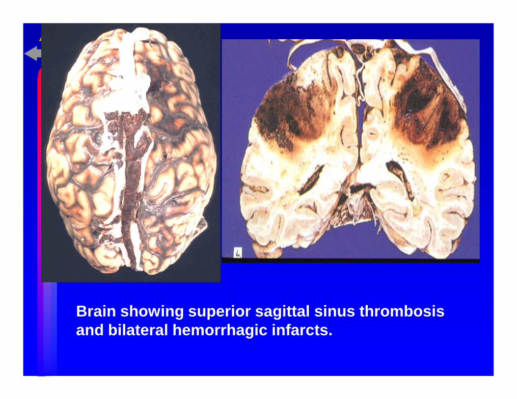
Brain showing superior sagittal sinus thrombosis and bilateral hemorrhagic infarcts.
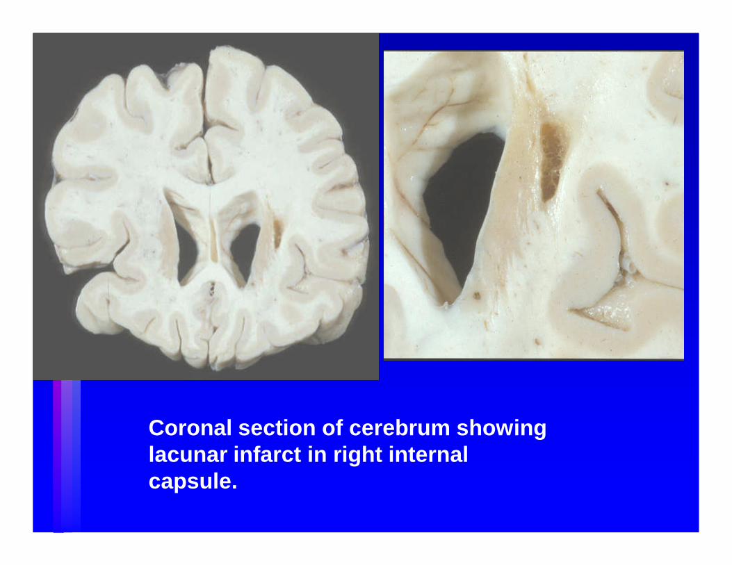
Coronal section of cerebrum showing lacunar infarct in right internal capsule.
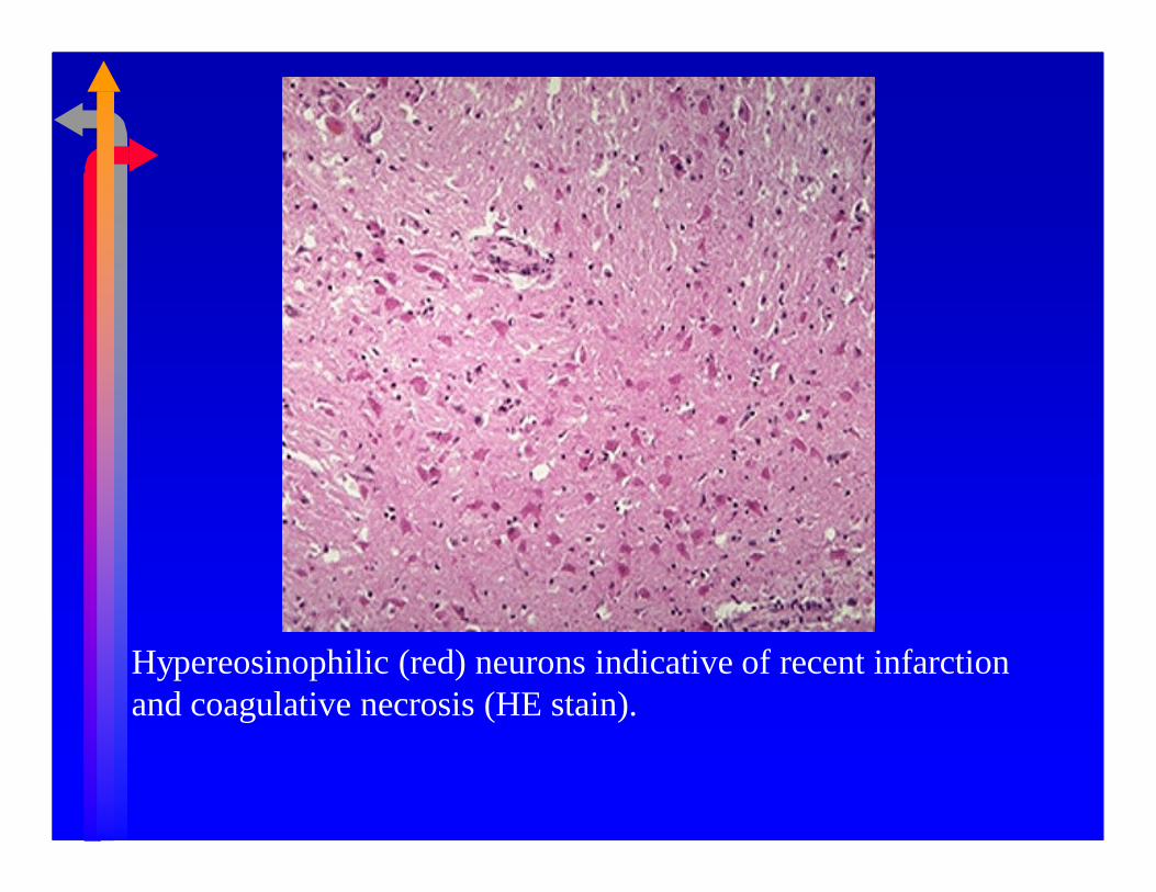
Hypereosinophilic (red) neurons indicative of recent infarction and coagulative necrosis (HE stain).
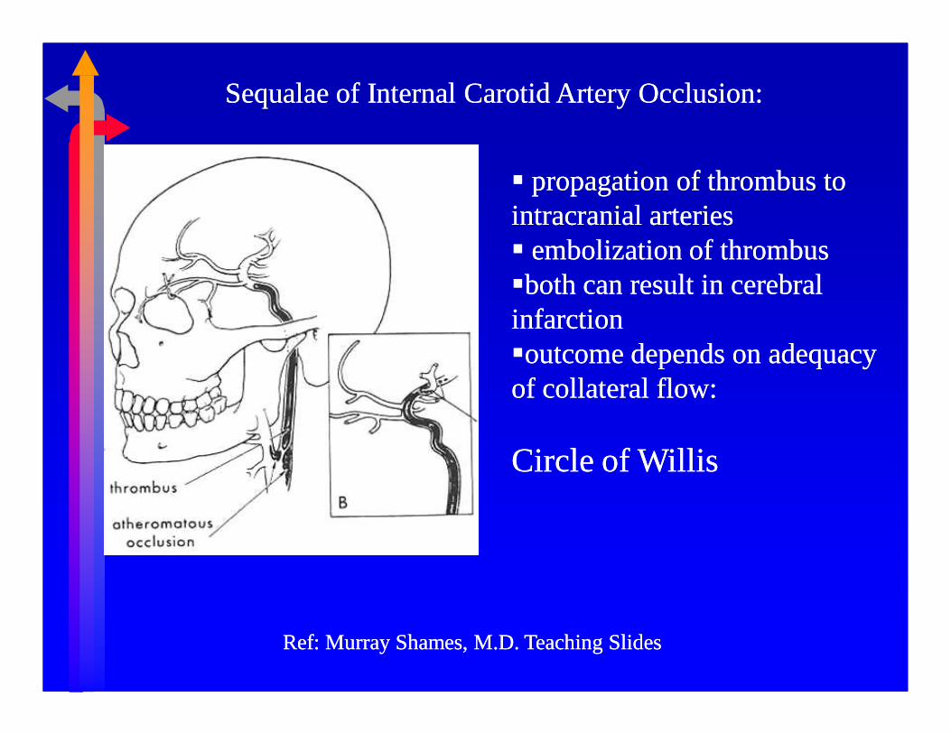
SequalaeSequalae of Internal Carotid Artery Occlusion:of Internal Carotid Artery Occlusion:
�� propagation of thrombus to propagation of thrombus to intracranial arteriesintracranial arteries�� embolizationembolization of thrombusof thrombus��both can result in cerebral both can result in cerebral infarctioninfarction��outcome depends on adequacy outcome depends on adequacy of collateral flow:of collateral flow:
Circle of WillisCircle of Willis
Ref: Murray Shames, M.D. Teaching SlidesRef: Murray Shames, M.D. Teaching Slides
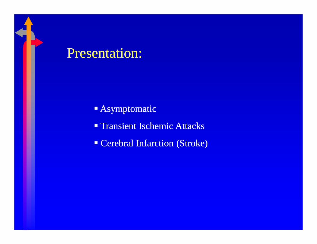
Presentation:Presentation:
��AsymptomaticAsymptomatic
�� Transient Ischemic AttacksTransient Ischemic Attacks
�� Cerebral Cerebral Infarction (Stroke)Infarction (Stroke)
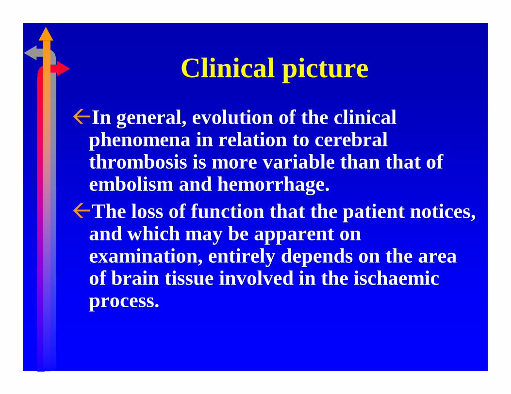
Clinical picture
�In general, evolution of the clinical phenomena in relation to cerebral thrombosis is more variable than that of embolism and hemorrhage. �The loss of function that the patient notices,
and which may be apparent on examination, entirely depends on the area of brain tissue involved in the ischaemicprocess.
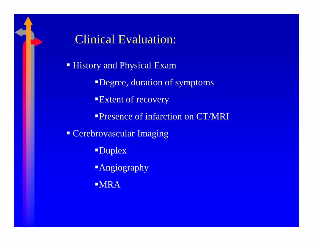
Clinical Evaluation:
� History and Physical Exam
�Degree, duration of symptoms
�Extent of recovery
�Presence of infarction on CT/MRI
� Cerebrovascular Imaging
�Duplex
�Angiography
�MRA
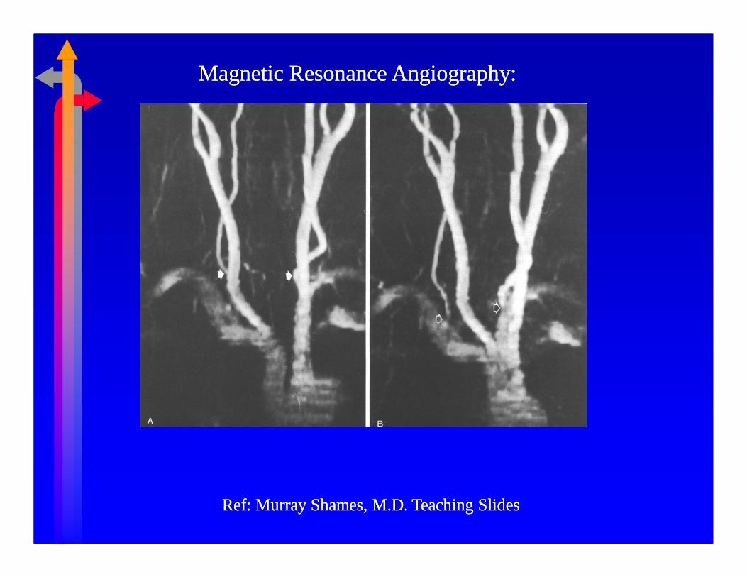
Magnetic Resonance Angiography:Magnetic Resonance Angiography:
Ref: Murray Shames, M.D. Teaching SlidesRef: Murray Shames, M.D. Teaching Slides
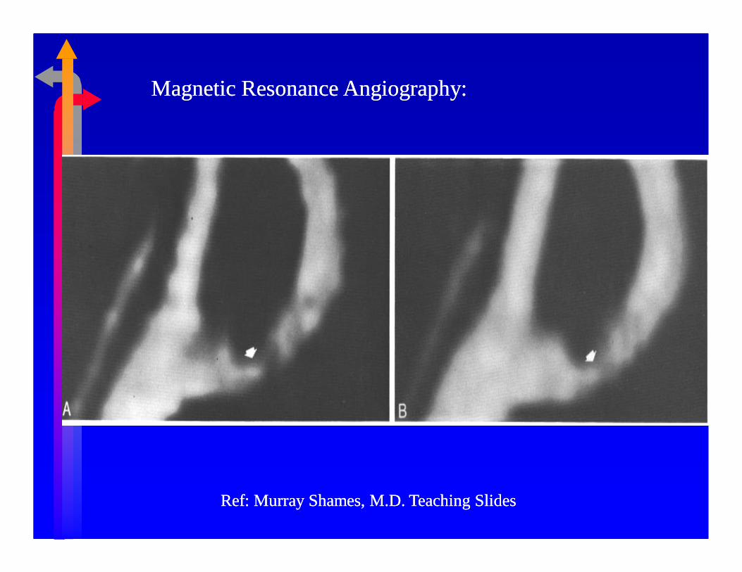
Magnetic Resonance Angiography:Magnetic Resonance Angiography:
Ref: Murray Shames, M.D. Teaching SlidesRef: Murray Shames, M.D. Teaching Slides
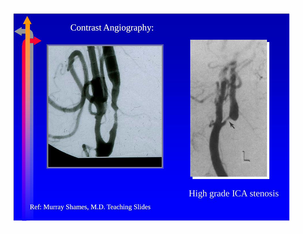
Contrast Angiography:Contrast Angiography:
High grade ICA stenosisRef: Murray Shames, M.D. Teaching SlidesRef: Murray Shames, M.D. Teaching Slides
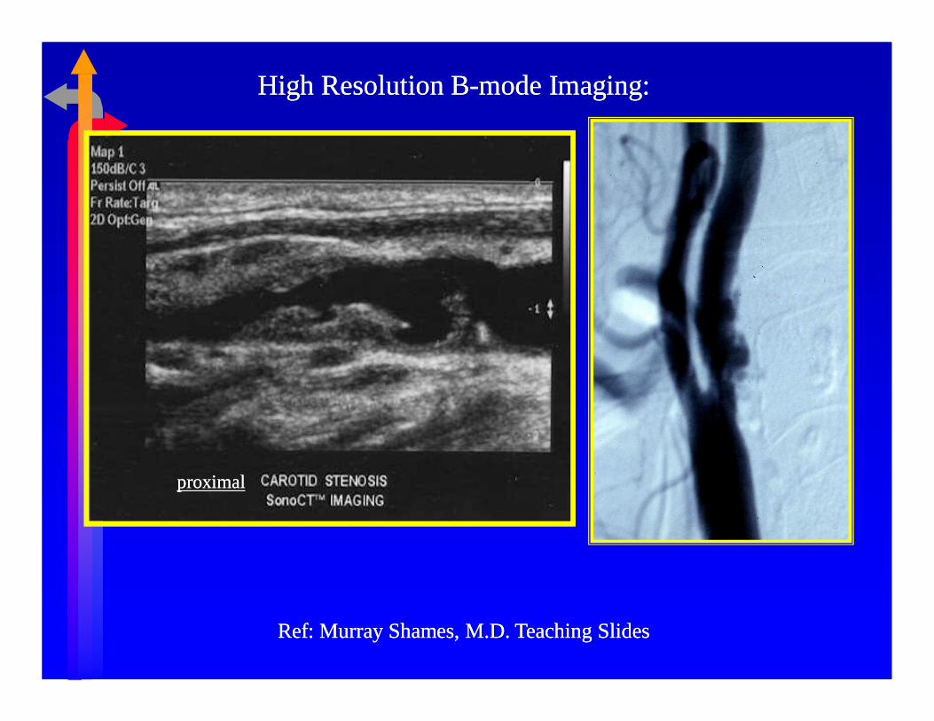
High Resolution BHigh Resolution B--mode Imaging:mode Imaging:
proximalproximal
Ref: Murray Shames, M.D. Teaching SlidesRef: Murray Shames, M.D. Teaching Slides
θ
v
The DopplerPrinciple
skin
fo
f
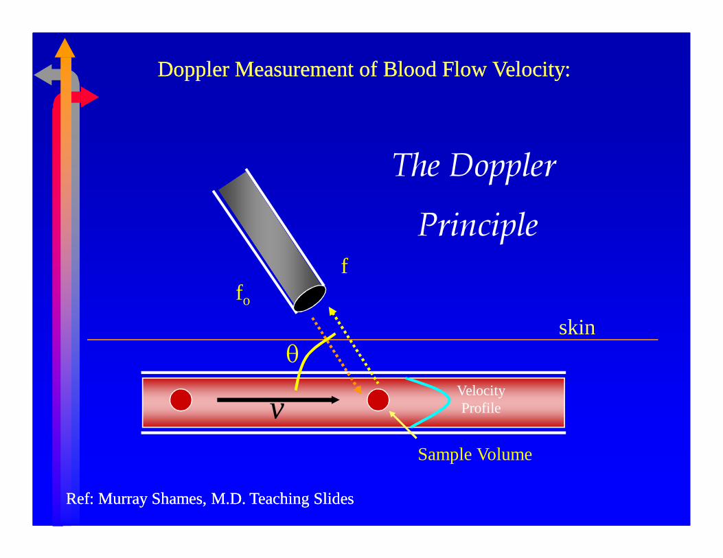
Doppler Measurement of Blood Flow Velocity:Doppler Measurement of Blood Flow Velocity:
Sample Volume
VelocityProfile
Ref: Murray Shames, M.D. Teaching SlidesRef: Murray Shames, M.D. Teaching Slides
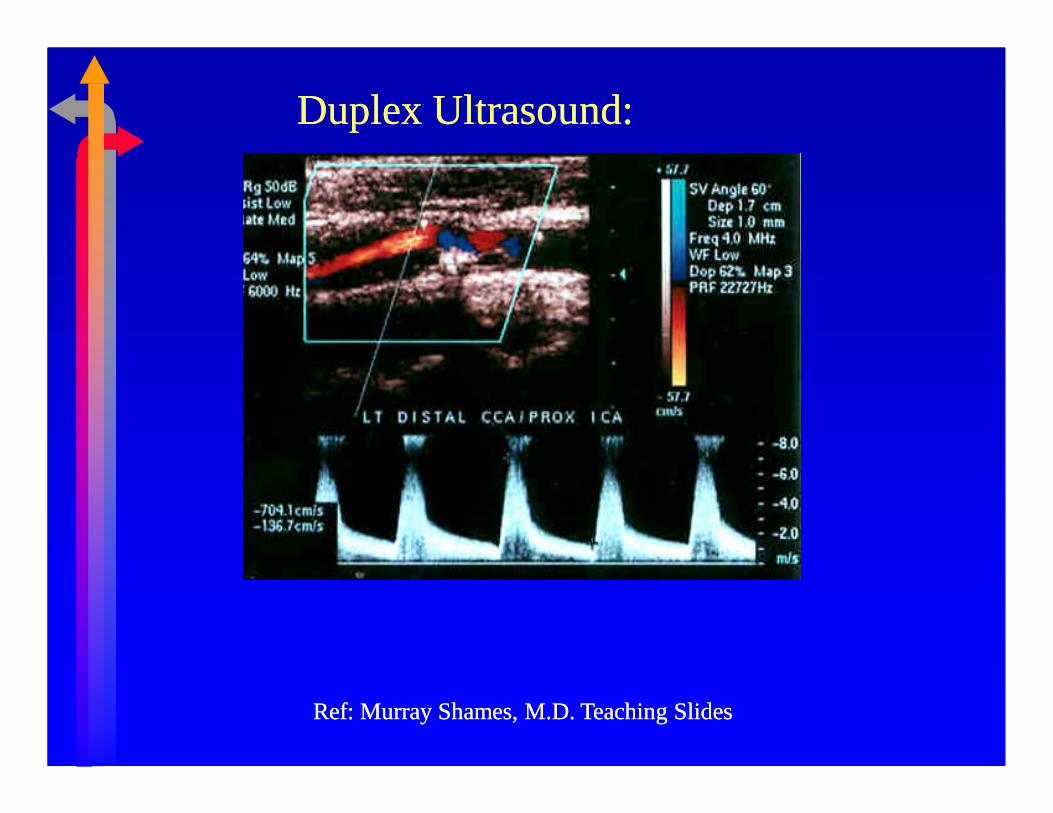
Duplex Ultrasound:Duplex Ultrasound:
Ref: Murray Shames, M.D. Teaching SlidesRef: Murray Shames, M.D. Teaching Slides
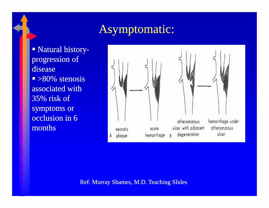
Asymptomatic:Asymptomatic:
�� Natural historyNatural history--progression of progression of diseasedisease�� >80% >80% stenosisstenosisassociated with associated with 35% risk of 35% risk of symptoms or symptoms or occlusion in 6 occlusion in 6 monthsmonths
Ref: Murray Shames, M.D. Teaching SlidesRef: Murray Shames, M.D. Teaching Slides
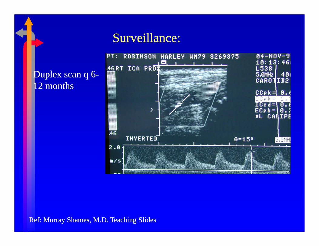
Surveillance:Surveillance:
Duplex scan q 6Duplex scan q 6--12 months12 months
Ref: Murray Shames, M.D. Teaching SlidesRef: Murray Shames, M.D. Teaching Slides
The ultimate goal of vascular testing is to identify The ultimate goal of vascular testing is to identify clinically clinically significantsignificant carotid disease, so that treatment can be applied and carotid disease, so that treatment can be applied and risk of stroke reducedrisk of stroke reduced..
Diagnose hypertension early and treat.Diagnose hypertension early and treat.
PREVENTION IS ALWAYS BETTER THAN CURE!!PREVENTION IS ALWAYS BETTER THAN CURE!!
Summary:Summary:
�� Risk of stroke from extraRisk of stroke from extra--cranial carotid atherosclerosis cranial carotid atherosclerosis related to stenosis severity.related to stenosis severity.
�� Patients with carotid territory TIAs or minor stroke; & Patients with carotid territory TIAs or minor stroke; & >60% ICA stenosis benefit from surgical intervention.>60% ICA stenosis benefit from surgical intervention.
�� HighHigh--grade ICA stenosis (>70%) increases the risk of grade ICA stenosis (>70%) increases the risk of stroke in asymptomatic patients.stroke in asymptomatic patients.
END
Reference: Robins Pathological Basis of Diseases.
Murry Shames, MD, Teaching slides at
http://usfvascularsurgery.com/