m.blog.daum.net sub arachnoid hemorrhage : evaluation and medical...
TRANSCRIPT

SUB ARACHNOID HEMORRHAGE : EVALUATION AND MEDICAL MANAGEMENT
www.brain-aneurysm.com/ba1.html
m.blog.daum.net

INTRODUCTION
¡ SAH refers to extravasation of blood into the subarachnoid space between the pial and arachnoid membranes due to some pathological process
¡ Overall incidence reported between 2-49 cases/1 lakh / yr
¡ 11 cases/ lakh / yr for aneurysmal SAH
¡ Comprises 6-8% of all strokes
¡ Associated with an annual cost of $1.75 billion (USA)

INTRODUCTION
¡ M.C. cause of spontaneous SAH is from a ruptured intracranial aneurysm
¡ An estimated 10-15% of patients die before reaching the hospital
¡ Advances in the management of subarachnoid hemorrhage have resulted in a relative reduction in mortality rate that exceeds 25%. However, more than one third of survivors have major neurological deficits

RELEVENT ANATOMY
¡ "Arachnoid" comes from the Greek "arachne" meaning spiderweb + "eidos" meaning resemblance = spider-web-like
¡ Subarachnoid space is the interval between the arachnoid membrane and pia mater
¡ It is occupied by a spongy tissue consisting of trabeculae and intercommunicating channels in which the CSF is contained
¡ The subarachnoid space extends from the basal cisterns surrounding the brainstem superiorly to S2 level inferiorly. The meninges continue caudal from this point fused as the filum terminale
¡ Subarachnoid space is the location of the interface between the vascular tissue and CSF
¡ www.ganfyd.org

HISTORICAL PERSPECTIVE
¡ Ancient Greek, Egyptian & Arabic literature all have references to intracranial aneurysm & SAH
¡ Dandy performed the first successful clipping of an aneurysm in 1937, using a vascular clip designed by Harvey Cushing
¡ Angiographic vasospasm first described by Ecker & Riemen(1951)
¡ Clinical vasospasm described by Pool, Maspes and Marini et al in 1960s

HISTORICAL PERSPECTIVE
¡ Endovascular therapy for the treatment of intracranial aneurysms was pioneered in the mid 1970s by Serbinenko at the Moscow Institute of Neurosurgery
¡ Kosnik and Hunt (1976) followed by Kassel et al (1982) described effectiveness of induced HTN and intravascular volume expansion in DIND
¡ In 1983 Allen and co-workers published first controlled trial nimodipine in SAH
¡ In 1990 Guglielmi et al, at the University of California Los Angeles (UCLA) Medical Center developed the Guglielmi detachable coil system (GDC- platinum coil)

ETIOLOGY OF SAH
¡ TRAUMA – Most common cause
SPONTANEOUS SAH- ¡ ANEURYSM – 70 to 80% of spontaneous SAH ¡ AVM- 4-5% cases ¡ INFECTIONS ¡ NEOPLASIA ¡ INFLAMMATORY (vasculitis ) ¡ COAGULATION DISORDER ¡ DURAL SINUS THROMBOSIS ¡ PRETRUNCAL NON ANERYSMAL SAH ¡ CEREBRAL ARTERY DISSECTION ¡ RUPTURE OF INFUNDIBULUM ¡ PITUITARY APOPLEXY ¡ SICKLE CELL DISEASE ¡ DRUGS and TOXINS (cocaine abuse, amphetamine, MAO inhibitor,
alcohol, CO, nicotine, quinine, snake venom)
¡ IDIOPATHIC (SAH of unknown etiology, 14-22%)

RISK FACTORS
¡ Smoking and hypertension appears to be a significant risk factor, as does heavy alcohol consumption
¡ The risk of AVM rupture is greater during pregnancy
The following are doubtful risk factors for SAH: ¡ Use of oral contraceptives ¡ Hormone replacement therapy ¡ Hypercholesterolemia ¡ Increasing age

PATHOPHYSIOLOGY
¡ Blood extravasates under pressure into the subarachnoid space and quickly spreads through the CSF around the brain & spinal cord
¡ Blood released under high pressure may directly cause damage to local tissues
¡ Blood extravasation causes a global increase in intracranial pressure
¡ Meningeal irritation occurs
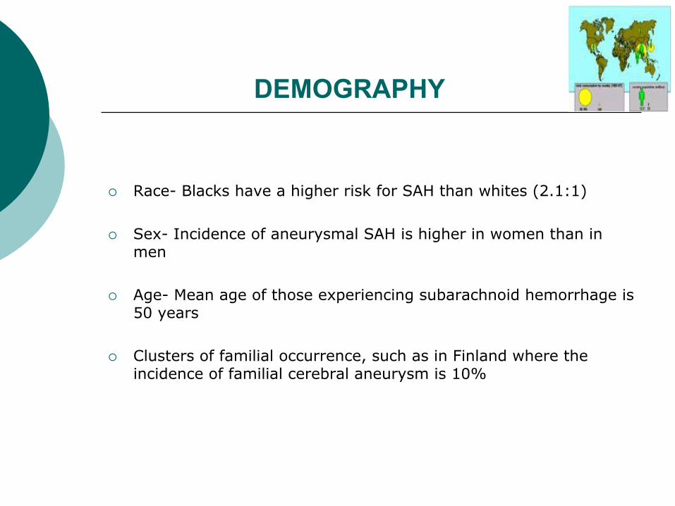
DEMOGRAPHY
¡ Race- Blacks have a higher risk for SAH than whites (2.1:1)
¡ Sex- Incidence of aneurysmal SAH is higher in women than in men
¡ Age- Mean age of those experiencing subarachnoid hemorrhage is 50 years
¡ Clusters of familial occurrence, such as in Finland where the incidence of familial cerebral aneurysm is 10%

CLINICAL PRESENTATION
Headache – ¡ Present in 97% cases ¡ A sudden onset of severe headache, often described as the "worst
headache of my life“
¡ Prodromal (warning) headache(s) from minor blood leakage (sentinel headache) is reported in 30-60% of SAH
¡ Sentinel headaches may occur a few hours to a few months before the rupture, with a reported median of 2 weeks prior to diagnosis of SAH. It may be due to sentinel leaks, mass effect of aneurysm expansion or emboli
¡ Minor leaks are not a feature of AVM

CLINICAL PRESENTATION
¡ Nausea and/or vomiting
¡ Symptoms of meningeal irritation (e.g.- neck stiffness, Kernig’s/ Brudzinski’s sign, low back pain, bilateral leg pain) : >75% of cases of SAH, but many take several (6-24 hrs) hours to develop
¡ Photophobia and visual changes
¡ A sudden LOC occurs at the ictus in 45% of patients, often transient; but in 10% of patients are comatose for several days
¡ Coma in SAH may be d/t raised ICP, ICH, HCP, seizure, low CBF
¡ Seizures during the acute phase of SAH occur in 10-25% of patients

CLINICAL PRESENTATION
¡ Focal neurologic abnormalities (25%) : hemiparesis, aphasia, hemineglect, cranial nerve palsies, memory loss
¡ Ocular hemorrhages (20-40%) : sub hyaloid pre-retinal hemorrhages / intra-retinal hemorrhage surrounding fovea / intra vitreous hemorrhage (Terson syndrome). D/t compression of CRV / retinochoroidal anastomosis by raised, leading to venous disruption
¡ BP elevation is observed in about 50% of patients. BP often becomes labile as ICP increases
¡ Temperature elevation, secondary to chemical meningitis from subarachnoid blood products, is common after the fourth day following bleeding
¡ Tachycardia often is present for several days after SAH

SAH GRADING
Hunt and Hess grading system ¡ Grade 1 - Asymptomatic or mild headache ¡ Grade 2 - Moderate-to-severe headache, nuchal rigidity, and
cranial nerve palsy ¡ Grade 3 - Mild alteration in mental status (confusion, lethargy),
mild focal neurological deficit ¡ Grade 4 - Stupor and/or hemi paresis ¡ Grade 5 - Comatose and/or decerebrate rigidity Modified classification- ¡ Grade 0 – Unruptured aneurysm ¡ Grade 1a – no acute meningeal reaction but with fixed neuro
deficit
* Add one grade for HTN/DM/COPD/Severe Atherosclerosis/ angiographic vasospasm

SAH GRADING
WFNS scale ¡ Grade 0 – Intact aneurysm ¡ Grade 1 - GCS of 15, major focal deficit absent ¡ Grade 2 - GCS of 13-14, major focal deficit absent ¡ Grade 3 - GCS of 13-14, major focal deficit present ¡ Grade 4 - GCS of 7-12, major focal deficit absent or present ¡ Grade 5 - GCS of 3-6, major focal deficit absent or present
* Major focal deficit – aphasia/hemiparesis/hemiplegia

SAH GRADING
BOTTERELL GRADING : ¡ Grade 1 – conscious with or without signs of blood in
subarachnoid space
¡ Grade 2 – drowsy without significant neurological deficit
¡ Grade 3 - drowsy with neurological deficit and probably ICH
¡ Grade 4 - significant neurological deficit, deteriorating because of large ICH or older patients with less severe deficit but pre-existing cerebrovascular disease
¡ Grade 5 – moribund with failing vital centers and extensor rigidity

SAH GRADING
Fischer scale (based on CT scan ) ¡ Group 1 - No blood detected ¡ Group 2 - Diffuse deposition of subarachnoid blood and no layers
of blood greater than 1 mm ¡ Group 3 - Localized clots and/or vertical layers of blood 1 mm or
greater in thickness ¡ Group 4 - Diffuse or no subarachnoid blood, but intracerebral or
intraventricular clots are present

SAH GRADING
¡ The Hunt & Hess and the WFNS grading systems have been shown to correlate with patient outcome
¡ Fischer classification has been used successfully to predict the likelihood of symptomatic cerebral vasospasm
¡ Admission H&H Gr 1 and 2: mortality is 20% & the major cause is rebleed (international cooperative aneurysm study)

WORK UP ¡ CBC count - For evaluation of possible infection or hematologic
abnormality
¡ Prothrombin time (PT) and activated partial thromboplastin time (aPTT) - For evaluation of possible coagulopathy
¡ Serum electrolytes - To establish a baseline for detection of future complications
¡ Blood grouping
¡ Cardiac enzymes - For evaluation of possible myocardial ischemia
¡ Arterial blood gas (ABG) - Assessment is necessary in cases with pulmonary compromise

WORK UP
CT scan: ¡ The diagnosis of SAH usually depends on a high index of clinical
suspicion combined with radiographic confirmation via NCCT. The sensitivity decreases with respect to increased time from ictus. CT scan has a sensitivity of 95% within the first 48 hours of the ictus; 80% at 72 hours and 50% at 1 week

WORK UP (CT SCAN CONTD…)
¡ CT scan findings may be falsely negative in patients with small hemorrhages and in patients with severe anemia
¡ In general, blood localized to the basal cisterns, the sylvian fissure, or the intrahemispheric fissure indicates rupture of a saccular aneurysm. Blood found lying over the convexities or within the superficial parenchyma of the brain often is indicative of AVM or mycotic aneurysm rupture
¡ A.Com aneurysm rupture often a/w ant. interhemispheric fissure bleed, MCA/ PCom aneurysm rupture are a/w sylvian fissure bleed and IVH (3 & 4TH ventricular) usually suggest PICA/ VA aneurysm rupture
¡ CT also helpful to rule out HCP
¡ CECT head may reveal AVM

WORK UP LUMBAR PUNCTURE- ¡ Most sensitive test for SAH (Caution : lowering CSF pressure may
precipitate rebleeding)
¡ CT scan should be performed before LP to exclude significant intracranial ME, obstructive HCP or obvious intracranial hematoma
¡ Findings often are negative within 2 hours of the ictus & is most sensitive 12 hours after the bleed
¡ SAH can be distinguished from traumatic LP
¡ Xanthochromia appear until 2-4 hours after the ictus, is approx 100% present 12 hours after the bleed and remains for approximately 2 weeks. Xanthochromia is present 3 weeks after the bleed in 70% of patients, and it is still detectable at 4 weeks in 40% of patients, better appreciated by spectrophotometry
¡ RBC count usually > 1,00000/c mm, higher protein level d/t blood break down product

WORK UP CEREBRAL ANGIOGRAPHY (DSA)- ¡ Gold standard for evaluation of cerebral aneurysm (80-85%
sensitivity)
¡ Useful in cases of diagnostic uncertainty (after CT and LP)
¡ Can provide surgical information in the setting of SAH: Cerebrovascular anatomy, aneurysm location & source of bleeding, aneurysm size/shape/orientation of dome and neck, relation to the parent/perforating arteries
¡ If cerebral angiography findings are negative (10-20%) a repeat test should be performed 3-4 weeks later
¡ May be useful to evaluate for possible cerebral vasospasm

WORK UP
Magnetic resonance angiography (MRA): ¡ The role of MRA in the detection of SAH currently is under
investigation. Given the current limitations of MRA (e.g., failure to detect PICA and ACom aneurysm in one series) most authors feel that the risk/benefit ratio still favors conventional angiography
¡ Can detect aneurysms >3 mm with 86% sensitivity and is useful for monitoring the status of small, unruptured aneurysms
¡ False +ve in approx. 16%
¡ Can be used to evaluate the degree of intramural thrombus in giant aneurysms
¡ Useful as a screening test in high risk patients including 1st degree relative of patients with IC aneurysm

WORK UP
CTA : ¡ Reported to detect aneurysms larger than 3 mm with a sensitivity
of 95% and 83% specificity. Provide sufficient anatomic detail esp relation to near by bony structure
MRI : ¡ Not sensitive for SAH within the first 48 hours, better after 4-7
days (met-Hb) ¡ Useful tool to diagnose cerebral / spinal AVMs causing SAH
TCD : ¡ TCD useful in the detection and monitoring of arterial vasospasm

WORK UP
ECG : ¡ Changes noted on ECG are attributed to myocardial ischemia
and/or infarction caused by high levels of circulating catecholamines. Myocardial ischemia/infarction is present in 20% of SAH cases
Chest radiograph: ¡ All patients with SAH should have a baseline chest radiograph to
serve as a reference point for evaluation of possible pulmonary complications
Echocardiogram: ¡ Evaluation of ventricular wall motion may be necessary in cases
with suspected myocardial ischemia

MANAGEMENT
INITIAL MANAGEMENT CONCERNS - ¡ Rebleeding ¡ Hydrocephalus ¡ Delayed ischemic neurological deficit (DIND) ¡ Hyponatremia / hypovolemia ¡ DVT / Pulmonary embolism ¡ Seizures ¡ Determining the source of bleeding
MANAGEMENT
¡ Initial management of patients with SAH is stabilization. Assess the level of consciousness, airway, breathing and circulation (ABCs)
¡ Endotracheal intubation should be performed for patients presenting with coma/depressed consciousness/inability to protect airway/ increased ICP
¡ Vascular access should be obtained, including central and arterial lines
¡ Monitoring should include : Cardiac monitoring/Pulse oximetry/ BP monitoring (arterial BP monitoring is indicated in high-grade SAH or when blood pressure is labile)/End-tidal carbon dioxide/Urine output via placement of a Foley catheter

MANAGEMENT
Patients with signs of increased ICP ¡ Intubated and hyperventilated to achieve a PCO2 of 30-35 mm
Hg. Excessive hyperventilation is avoided, which may potentiate vasospasm and ischemia
¡ Osmotic agents (e.g., mannitol), can decrease ICP dramatically (50% after 30 min post administration)
¡ Loop diuretics (e.g., furosemide) also can decrease ICP
¡ The use of IV steroids is controversial

MANAGEMENT
--Intraventricular catheter (ventriculostomy)- possible indication ¡ Acute HCP or significant intraventricular blood. Allows drainage
of CSF as well as ICP monitoring. May increase the risk of rebleed. Symptomatic improvement found in 30%, immediately
¡ H&H grade >3 – some experts feel if there is some improvement with ventriculostomy, prognosis may be favourable
--Serial LP may be tried instead of ventriculostomy

MANAGEMENT STANDARD ADMITTING ORDERS – ¡ Admission in ICU with monitored bed
¡ Vital parameter and neurological status check 1 hrly
¡ Bed rest with head end elevated by30 degree, low external stimulation
¡ Nursing : strict I-O charting / daily weights / DVT pumps at LL / urinary catheterization/ NG tube
¡ IVF : Aggressive fluid therapy to head off cerebral salt wasting. NS + 20 meq KCl/L @ 2 ml/kg/hr
¡ If HCt <40%, 500ml 5% albumin over 4 hrs, on admission
¡ Avoid IM medication/ enema/ NSAID

MANAGEMENT
¡ Prophylactic anticonvulsants : 3% of SAH patients have seizure during acute illness. Use of prophylactic anticonvulsant is controversial, but most authorities recommend for 1 wk post op. Phenytoin is the usual agent used. Barbiturate is usually given for burst suppression in O.R.
¡ Mild sedation (propofol/midazolam/lorazepam)
¡ Analgesic : fentanyl ( lower ICP and doesn’t cause histamine release unlike morphine )
¡ Steroid : effect on brain edema is controversial, usually given prior to craniotomy
¡ Stool softener (bisacodyl) / anti-emetics (ondansetron) / H2 blocker or PPI or sucralfate

MANAGEMENT
ANTI-HYPERTENSIVE ¡ To maintain SBP 120-150 mm Hg for unclipped aneurysm
¡ The current recommendations advocate the use of antihypertensive agents when MAP exceeds 130 mm Hg
¡ Intravenous beta-blockers (labetolol) can be titrated easily and do not increase ICP. Beta-blockers are the agents of choice in patients without contraindications
¡ Hydralazine and calcium channel blockers have a fast onset and lead to relatively less increase in ICP than do nitrates
¡ Angiotensin-converting enzyme inhibitors have a relatively slow onset and are not first-line agents in the setting of acute SAH

MANAGEMENT
Calcium channel blocker : (Nimodipine / nicardipine/ nifedepine) ¡ Nimodipine 60 mg PO/NG q 4 hrs, initiated within 96hrs of SAH
¡ BRANT : British aneurysm nimodipine trial shown 22% vs. 33% incidence of cerebral infarction in compared to placebo
¡ CCB blocks the ‘slow channel’ of calcium influx which reduces the contraction of smooth muscles and may prevent vasospasm
¡ Possible beneficial effects are – improved RBC rheology / prevention of calcium entry in ischemic cells / anti platelet aggregation / dilatation of collateral lepto meningeal arteries
¡ Side effects are – systemic hypotension / renal failure / pulmonary edema

MANAGEMENT
¡ Oxygenation : Non intubated- Oxygen 2 Lit by ventimask Intubated – normocarbia & po2 > 100 mm Hg ¡ Monitoring of CVP q 4 hrs
¡ Following drugs are contraindicated for fear of impaired
coagulation – aspirin / dextran / heparin / repeated administration of hetastarch
¡ For unsecured aneurysm , gentle volume expansion with slight hemodilution and mild elevation of BP may help reducing effects of vasospasm and cerebral salt wasting

MANAGEMENT
LABORATORY INVESTIGATIONS – ¡ ABG / electrolytes / CBC / PT – APTT on admission ¡ ABG / electrolytes / CBC / HCt daily ¡ Serum and urine osmolarity if UO is high/low ¡ C/S of respiratory secretion, urine, blood, CSF if fever or signs of
infection ¡ CXR daily until stable ¡ TCD study

MANAGEMENT OF SPECIFIC PROBLEMS FLUID AND ELCTROLYTE DISTURBANCES : ¡ MC electrolyte disturbances after SAH are hyponatremia (50%) ,
hypernatremia and hypokalemia
¡ Hypokalemia should be corrected fast because it may predispose to cardiac arrhythmia
¡ Mortality a/w hypernatremia is 42%, 15% for hyponatremia and 25% with DI
¡ Most patients of SAH seem to become hypovolemic with low total blood volume even if maintenance fluid requirements are met. Causes may be d/t supine diuresis, pooling in peripheral vascular bed, inadequate erythropoiesis
¡ Incidence of volume depletion increases with decreasing GCS

MANAGEMENT OF SPECIFIC PROBLEMS
HYPONATREMIA FOLLOWING SAH ¡ Hypovolemia and hyponatremia frequently follow SAH as a result
of natriuresis / diuresis
¡ MC cause is SIADH & CSWS ( ANP/BNP/C-peptide )
¡ Hyponatremia may mimic or a/w DIND
¡ Risk factors : h/o DM, CHF, cirrhosis, adrenal insufficiency, use of NSAID/narcotic/thiazide
¡ Treatment : correction of hypovolemia & anemia / salt replacement (oral/IV) fludrocortisone / urea may be tried

MANAGEMENT OF SPECIFIC PROBLEMS
FEATURE SIADH CSWS DI
Ser. Na <135 mEq/L <135 mEq/L Variable,Elevated
PLASMA OSM <280 mOsmol/L <280 mOsmol/L Variable,Elevated
URINE Na >20 mEq/L >20 mEq/L Variable
URINE OSM >Plasma osm >Plasma osm 50-150 mOsm/L
Ser. K Low/normal High/normal Normal
BLOOD VOL Increased Decreased Normal/ Decreased
Na BALANCE Variable Negative High
BODY Wt Increased Decreased Normal/ Decreased
CARDIAC FILLING Pr.
Increased/Normal Decreased Normal/ Decreased
HCT Decreased Increased Normal/ Decreased
BUN : Cr Decreased / Normal Increased Normal/ Decreased
BP Normal Postural hypotension Normal/ Decreased
HR Normal Tachycardia Normal/Tachycardia
OTHER Normal thyroid+adenal+renal function/ no
edema/ no dehydration
S/S of volume depletion UO >3 lit/day UO> 250 ml/hr Ur Sp Gr <1003

MANAGEMENT OF SPECIFIC PROBLEMS
CARDIAC PROBLEMS ¡ SAH may be a/w cardiac arrhythmia and ECG changes in over
60% cases; may be indistinguishable from acute MI
¡ Possible mechanism : hypothalamic ischemia causes increased sympathetic tone with resultant cathecholamine surge producing sub-endocardial ischemia or coronary vasospasm
¡ Stunned myocardium : “reversible post ischemic myocardial dysfunction”. Some patients may develop myocardial hypokinesis following SAH, attributed to defect in Troponin –I. Mimic MI on echo but cardiac enzymes are negative
¡ Hypotension usually does not occur since reduced CO is offset by increased SVR. Reduced CO may impair the ability to tolerate barbiturate and may also impede the use of hyperdynamic therapy for vasospasm

MANAGEMENT OF SPECIFIC PROBLEMS
RESPIRATORY COMPLICATIONS : ¡ Common in poor clinical grade and advanced age
¡ 50% deaths from medical cause
¡ MC respiratory complications are pulmonary edema, pneumonia, atelectasis, aspiration, pneumothorax, asthma and pulmonary embolism
¡ Indication for intubation and controlled respiration include gross abnormality in RR, hypoxia, hypercapnia, depressed consciousness

MANAGEMENT OF SPECIFIC PROBLEMS (RESPIRATORY COMPLICATIONS )
¡ Pulmonary edema may complicate any moment following SAH &
may be cardiogenic/ neurogenic/ 2° to pulmonary insult viz aspiration or shock. Treated by immediate intubation/ ventilation, adequate oxygenation, PEEP, frusemide
¡ Most pneumonia occurs in intubated patients. Prevention include removal of NG & ET tubes as soon as indicated, avoidance of gastric distension, strict hand washing, semi-recumbent positioning, adequate nutrition, use of sucralfate in place of H2 blocker/PPI & proper ventilator care
¡ Use of prophylactic antibiotic is controversial
¡ Tracheostomy and percutaneous feeding gastrostomy

MANAGEMENT OF SPECIFIC PROBLEMS
VENOUS THOMBOEMBOLISM : ¡ 2% of SAH patients develop symptomatic DVT, 50% of them has
PE, which is fatal in half
¡ Asymptomatic DVT occurs in 19-50%
¡ Graduated compression stockings & pneumatic compression devices decreases incidence of DVT
¡ Low dose heparin or LMWH usually started only after securing source viz aneurysm
¡ For diagnosed DVT/PE IVC filters are optimal choice

MANAGEMENT OF SPECIFIC PROBLEMS
GASTRO INTESTINAL COMPLICATION : ¡ Early nutritional support ( oral/NG/PEG/Feeding jejunostomy ) to
counteract hypercatabolic state
¡ Stress ulcer and GI bleed : preventive measures ( sucralfate/H2 blocker/PPI )
¡ Stool softeners are administered routinely to prevent straining/ impaction/abdominal pain. Nimodipine may be a/w pseudo-obstruction & ileus, usually resolves on conservative measure
¡ Up to 24% of SAH patients develop increased hepatic enzymes and 4 % develops severe liver dysfunction. It may be d/t passive liver congestion, SIRS or drug induced

MANAGEMENT OF SPECIFIC PROBLEMS
REBLEEDING : ¡ Maximal frequency is in 1st day (4%), then 1.5% daily for 13
days. 15-20% rebleed within 14 days , 50% within 6 months and then 3% per year
¡ Risk increases with higher H/H grade, pre op ventriculostomy /
lumbar spinal drainage
¡ Risk of rebleeding in SAH of unknown origin / AVM / multiple unruptured aneurysm are similar at approx 1% per year

MANAGEMENT OF SPECIFIC PROBLEMS
Prevention of rebleed : ¡ Early treatment (coiling/surgery). Bed rest do not prevent
rebleeding
¡ Antifibrinolytic therapy – Tranexamic acid reduces the risk of rebleeding. Loading dosage of 1 gm bolus followed by 1 gm q 6 hrs until aneurysm is secured (max 72 hrs)
¡ Epsilon aminocaproic acid (EACA) – competitively blocks activation of plasminogen to plasmin. Incidence of rebleed is reduced but there is more incidence of HCP/DIND on prolonged use. Because of increased risk of cerebral infraction , its use was discouraged

MANAGEMENT OF SPECIFIC PROBLEMS HYDROCEPHALUS FOLLOWING SAH : acute HCP ¡ Optimal range of incidence 15-20% after initial CT following SAH
¡ 30-60% has no impairment of consciousness
¡ 3% develop delayed HCP within 1 week
¡ Possible mechanism : CSF outflow obstruction by blood at aqueduct / 4th ventricular outlet / subarachnoid space and/or at arachnoid granulation
¡ Risk factors : senility, intraventricular blood, diffuse subarachnoid blood, HTN, posterior circulation aneurysm, poor sensorium on admission, use of antifibrinolytics

MANAGEMENT OF SPECIFIC PROBLEMS
Treatment of acute HCP : ¡ Patients in poor grade SAH with large ventricles may be
symptomatic from HCP and considered for ventriculostomy, which causes improvement in 80% cases
¡ ICP recommended to keep in the range of 15-25 mm Hg and to avoid rapid pressure reduction to decrease the risk of ventriculostomy induced aneurysmal rebleed. After securing the aneurysm ICP should be maintained in the range of 5-10 mm Hg

MANAGEMENT OF SPECIFIC PROBLEMS
Chronic HCP : ¡ Reported range is 8-45%
¡ Chronic HCP is due to pia-arachnoid adhesions or permanent impairment of arachnoid granulations
¡ Acute HCP does not inevitably lead to chronic HCP
¡ 50% permanent CSF diversion
¡ Presence of intraventricular blood during acute stage increases the risk of shunt dependency
¡ Controversy: use of ventriculostomy in acute HCP increases or possibly decreases incidence of shunt dependency

MANAGEMENT OF SPECIFIC PROBLEMS
CEREBRAL VASOSPASM : ¡ Usually seen after aneurysmal SAH, but may be seen in IVH from
AVM, head trauma, brain surgery, LP, hypothalamic injury, infection, SAH of any etiology and may be a/w pre-eclampsia

MANAGEMENT OF SPECIFIC PROBLEMS
¡ CLINICAL VASOSPASM –delayed ishchemic neurological deficit (DIND) : a delayed focal ischemic neurologic deficit following SAH, clinically characterized by confusion/deceased level of consciousness with FND (speech/motor)
¡ RADIOGRAPHIC VASOSPASM (angiographic vasospasm) – arterial narrowing demonstrated on cerebral angiography, often with contrast slow filling
¡ SYMPTOMATIC VASOSPASM – when DIND corresponds to a region of vasospasm seen on angiogram
MANAGEMENT OF SPECIFIC PROBLEMS (cerebral vasospasm )
Clinical finding of cerebral vasospasm – usually develop gradually, may progress and fluctuate ¡ Non localizing finding – New or increasing headache Alteration of level of consciousness Disorientation Meningismus ¡ Focal neurological sign – Cranial nerve palsy Focal neurological deficit Anterior cerebral artery syndrome (mc) Middle cerebral artery syndrome

MANAGEMENT OF SPECIFIC PROBLEMS (cerebral vasospasm )
¡ ACA syndrome – frontal lobe findings predominate ( abulia,
grasp/suck reflex, urinary incontinence, drowsiness, slowness, delayed response, confusion ). B/L ACA infract are usually d/t vasospasm after AComA aneurysm rupture
¡ MCA syndrome – hemiparesis, monoparesis, aphasia, ideomotor apraxia

MANAGEMENT OF SPECIFIC PROBLEMS (cerebral vasospasm )
INCIDENCE : ¡ Radiographic vasospasm is found in 30-70% of DSA done around
7th day following SAH ¡ Symptomatic vasospasm occur in 20-30% of patients with SAH ¡ Radiographic cerebral vasospasm (CVS ) may occur in absence
of clinical deficit , and vice-versa

MANAGEMENT OF SPECIFIC PROBLEMS (cerebral vasospasm )
SEVERITY : ¡ CVS is the most significant cause of morbidity and mortality in
patients surviving SAH
¡ Ranges from mild reversible dysfunction to severe permanent deficit secondary to ischemic infarction (7%) and may be fatal (7%)
¡ Earlier onset CVS is a/w greater deficit

MANAGEMENT OF SPECIFIC PROBLEMS (cerebral vasospasm )
TIME COURSE OF VASOSPASM : ¡ Onset is almost never before day 3 post SAH
¡ Max frequency on day 6-8 post SAH
¡ Clinical CVS usually resolves by day 12 post SAH but radiological CVS is demonstrated , it usually resolves slowly over 3-4 weeks time
¡ Onset is usually insidious but in 10% may have abrupt and severe deterioration

MANAGEMENT OF SPECIFIC PROBLEMS (cerebral vasospasm )
CORRELATED FINDINGS : ¡ Blood clots are spasmogenic when in direct contact with proximal 9
cm of ACA and MCA
¡ H & H grade on admission correlate with risk of CVS, grade 1 has 22% incidence and grade 3 and above has > 50% incidence of DIND
¡ Amount of blood in CT correlate with severity of CVS. Fisher group 1 carries 0-2% risk where as group 3 carries up to 23% risk of DIND
¡ Higher incidence found with increasing age, cigarette smoking, preexisting HTN, use antifibrinolytic agents, angiographic dye, hypovolemia
¡ Pial enhancement on CECT approx 3 days after SAH may correlate with higher risk of CVS, indicating increased permeability of BBB

MANAGEMENT OF SPECIFIC PROBLEMS (cerebral vasospasm )
PATHOGENESIS : ¡ Poorly understood ¡ Degraded products from hemolyzed RBC may be the “spasmogens” ¡ Theories of CVS –
STRUCTURAL THEORY VASOCONSTRICTION THEORY
Proliferative vasculopathy Free radical lipid peroxidation
Immune vasculopathy Derangement in eicosanoid production
Vessel wall inflammation Nitric oxide deficit
Extracellular lattice contraction Endothelin excess
-- Neurogenic factors

SAH | V
RELEASE OF VASOCONSTRICTORS | V
OXY HB ENDOTHELIN RELEASE | |
<———————— FREE RADICALS (Fe) RECEPTORS (ETa,ETb) STIMULATION | |
LIPID PEROXIDATION G- POTEIN ACTIVATION | |
ACTIVATION OF PHOSPHOLIPASE (C,D,A2)
| HYDROLYSIS OF PHOSPHOLIPIDS (PG E2, TxA2, LOW PGI2)
| | INCREASED CALCIUM INCREASED DAG
| | CALPAIN ACTIVATION —> PROTEOLYSIS —> PKC ACIVATION
| | PROTEIN DEGADATION (CALPONIN ,OTHERS) PROTEIN PHOSPHORYLATION (CALDESMON ,OTHERS)
| | INHIBITORY EFFECT ON ACTOMYOSIN ATPase LOST
| | V
SUSTAINED VASOCONSTRICTION
CALCIUM INFLUX | INHIBITION OF CA- ATPase

MANAGEMENT OF SPECIFIC PROBLEMS (cerebral vasospasm )
PATHOLOGY : ARTERIAL WALL CHANGES AFTER VASOSPASM
TIME VESSEL LAYER
PATHOLOGIC CHANGE
Day 1-8 Adventitia Increased inflammatory cells (L, PC, MC) & connective tissue
Media Muscle necrosis and corrugation of elastica
Intima Thickening with endothelial swelling and vacuolization, opening of inter-endothelial tight junction
Day 9-60 Intima Proliferation of SMC à progressive intimal thickening

MANAGEMENT OF SPECIFIC PROBLEMS (cerebral vasospasm )
DIAGNOSIS CVS : ¡ Clinical Criteria – Delayed onset of persistent neuro deficit onset 4-20 days post SAH deficit appropriate to involved arteries ¡ R/O other causes of deterioration – Rebleeding HCP Cerebral edema Seizure Metabolic disturbances viz hyponatremia Hypoxia Sepsis

MANAGEMENT OF SPECIFIC PROBLEMS (cerebral vasospasm )
Ancillary tests : Trans Cranial Doppler ( TCD ) ¡ Introduced by Aaslid & colleague ( 1980 ); works on the principle that
as an artery narrows, blood flow velocity within it increases
¡ May precede clinical symptoms by 24-48 hrs
Mean MCA velocity
MCA : ICA (Lindegaard Ratio)
Interpretation
< 120 cm/sec < 3 Normal
120- 200 cm/sec 3-6 Mild vasospasm
> 200 cm/sec > 6 Severe vasospasm

MANAGEMENT OF SPECIFIC PROBLEMS (cerebral vasospasm )
¡ CEREBRAL IADSA : vasospasm appear as concentric narrowing which can be focal/segmental/diffuse. Graded as mild (<25%), moderate (25-50%) or severe (>50%)
¡ MRA / CTA : may be useful, not a practical alternative to DSA
¡ EEG : decline in percent of Alpha activity ( 6-14 Hz ), called relative alpha ( 0.45 – 0.17 ) predicted onset of vasospasm earlier then TCD
¡ MRI : DWI & PWI may detect early ischemia
¡ Xenon CT : may detect large global changes in CBF but insensitive to detect focal blood flow changes
¡ PET / SPECT : non-quantitative and takes longer time

MANAGEMENT OF SPECIFIC PROBLEMS (cerebral vasospasm )
PREVENTION OF CVS : ¡ Vasospasm can often be mitigated by preventing post SAH
hypovolemia & anemia by employing slight over hydration & blood transfusion
¡ Smooth muscle relaxants : - CCB didn’t succeed in counteracting vasospasm but may
provide neuro-protectant effect - Endothelin receptor antagonist , clazosentan ( ETa )
¡ Direct mechanical arterial dilatation : balloon angioplasty ( 60-80% clinical improvement )
¡ Indirect arterial dilatation : utilizing hyperdynamic therapy

MANAGEMENT OF SPECIFIC PROBLEMS (cerebral vasospasm )
¡ Removal of potential vasospasmogenic agents : - Removal of blood clot (mechanical removing during
surgery / subarachnoid irrigation with thrombolytic agents viz rt-PA at surgery or cisternal catheters or intrathecally )
(Findlay JM et al : RCT of intraoperative, intracisternal TPA for prevention of vasospasm)
- CSF drainage via serial LP/EVD/post op. cisternal catheter ¡ Sympatholytics / Cervical sympathectomy ¡ Intra-arterial or intra-thecal papaverine / verapamil ¡ Alpha ICAM 1 inhibition (Ab)

MANAGEMENT OF SPECIFIC PROBLEMS (cerebral vasospasm )
¡ Protection of CNS from ischemic injury : - CCB - NMDA receptor antagonist ( selfotel / eliprodil / cerestat ) - Free radical scavengers ( tirilazad mesylate / nicaraven )
¡ Improvement of rheological properties of intravascular blood to enhance perfusion
- plasma / albumin / LMW dextran / PFC / mannitol - optimal hematocrit is controversial but 30-35% is a good
compromise between lowered viscosity without overlay reducing O2 carrying capacity
¡ Extracranial – intracranial bypass around zone of vasospasm

MANAGEMENT OF SPECIFIC PROBLEMS (cerebral vasospasm )
Promising agents in trial – ¡ Nicardipine prolonged release implants (NPRIs) ¡ Clazosentan ¡ Nizofenone ( neuroprotectant, effective in controlled clinical trial) ¡ High dose methylprednisolone (effective in cohort clinical trial) ¡ calcitonin gene related peptide slow release tab IT (effective in
primate model ) ¡ Hydroxyl radical scavenger – nicaraven (effective in controlled
clinical trial) ¡ combination of inotrope & vasodilator drug milrinone (controlled
dog study) ¡ Vasodilator papaverine slow release pellets (effective IT in dog
model) ¡ Neuroprotectant ebselen/simvastatin (effective in controlled clinical
trial) ¡ Pulse dye LASER delivered by endovascular catheter to reverse
vasospasm ¡ Role of intra cisternal papaverine

MANAGEMENT OF SPECIFIC PROBLEMS (cerebral vasospasm )
STRATEGIES FOR VASOSPASM PREVENTION :
Avoidance of hypovolemia/ hypotension/ antihypertensive/ antifibrinolytics / raised ICP
Prevent ischemia
Subarachnoid clot removal or lysis with fibrinolytic agents Prevent vasospasm
CCB or other potential calcium based neuro-protectant Prevent ischemic damage (?)
21- aminosteroid & other antioxidant, lipid peroxidation inhibitor, free radical scavenger
Prevent ischemic damage (?)
Intra-thecal slow release vasodilator Prevent vasospasm (experimental )
Endothelin inhibitor and antagonist Prevent vasospasm (experimental )

MANAGEMENT OF SPECIFIC PROBLEMS (cerebral vasospasm )
STRATEGIES FOR VASOSPASM REVERSAL :
Hypervolemia, hemodilution and hypertension Reverses ischemia
Intra-arterial papaverine administration Reverses vasospasm
Percutaneous transluminal balloon angioplasty (PTBA) Reverses vasospasm
Intraventricular nitroprusside Reverses vasospasm (experimental)
Intra aortic balloon counterpulsation (for concomitant heart failure )
Reverses vasospasm along With PTBA

Condition Management
No vasospasm ¡ clinically intact ¡ normal TCD
1. Hemodynamics : normotension, keep SBP > 120 mm Hg
2. IVF : NS 200 ml /hr
Subclinical vasospasm ¡ high TCD (> 200 cm/sec) ¡ radiographic vasospasm ¡ clinically intact
1. Monitors : PA catheter, A-line 2. Elderly & pt. with CAD : ECG, cardiac echo, cardiac
enzymes to assess left ventricular function 3. Monitor for S/S of a/e of 3-H therapy (chest pain ,
pulmonary rhonchi, ECG changes ) 4. Hemodynamics : maintain SBP 160-220 mm Hg (may
add DA/NorAd/Dobutamine), maintain PCWP 12-14 mm Hg
5. Hematocrit : to maintain < 33% 6. Fluid : - monitor I/O and ser. Na - IVF = NS + plasma 200-250 ml/hr, ½ NS if
Na>150 meq/L - DDAVP if UO > 200 ml / hr X 4 hrs
Clinical vasospasm ¡ DIND ¡ high TCD (> 200 cm/sec) ¡ radiographic vasospasm
1. Increase SBP 200-220 mm Hg 2. Increase PCWP to 18-21 mm Hg (CXR- to r/o
pulmonary edema) / CVP 8-12 cm H2O 3. Refractory case : angioplasty , IA verapamil
CVS MANAGEMENT PROTOCOL : ( post clipping )

TRIPLE “H” THERAPY
HYPERTENSION
1. Dopamine : start at 2.5 mcg/kg/min and titrate up to 15-20 mcg/kg/min
2. Dobutamine : start at 5 mcg/min titrate to 20 mcg/kg/min
3. Phenylephrine : start at 5 mcg/min titrate up to 10 mcg/kg
4. Norepinephrine : start at 1-2 mcg/min & then increase by 10 mcg/min
HYPERVOLEMIA 1. IVF : NS @ 200 -250 ml / hr
2. Albumin / plasma 3. DDAVP : antidiuretic , 2-4 mcg SQ q D in divided
doses
HEMODILUTION 1. Target hematocrit : < 33%
2. Blood transfusion for Hct < 25%

MANAGEMENT OF SPECIFIC PROBLEMS (cerebral vasospasm )
Complication of hyperdynamic therapy : ¡ Intra cranial – cerebral edema raised ICP hemorrhagic infarction in the area of ischemia
¡ Extra cranial – pulmonary edema (17%) cardiac failure dilutional hyponatremia (3%) MI (2%) ¡ Complication related to catheter – catheter related sepsis (13%) subclavian vein thrombosis (1.3%) pneumothorax (1%) hemothorax

EARLY ANEURYSM REPAIR
DIND
HCT 35%, Hb >10 Hypervolemia (CVP 8-10, PCWP 14-16) Inotropic infusion (DA / Dobutamine)
No clinical &/or BP response in 1 hr
Phenylephrine or NorAd
CLINICAL RESPONSE
H/O CCF or IHD
PA CATHETER
OR DIRECT BALLOON ANGIOPLASTY
BP > 200 mm Hg NO CLINICAL RESPONSE IN 1 HR
CT to r/o Infarction
BALLOON ANGIOPLASTY

MANAGEMENT OF SPECIFIC PROBLEMS
MISCELLANEOUS COMPLICATIONS FOLLOWING SAH : ¡ Fever and SIRS
¡ Genito-urinary : cystitis/ renal failure
¡ Metabolic : hyperglycemia / hypercatabolic state
¡ Hematological : anemia
¡ Poor immunity

SAH IN PREGNANCY
¡ Intracranial hemorrhage of pregnancy (ICHOP) commonly occurs in the setting of eclampsia, MC being intra-parenchymal bleed
¡ ICHOP- related SAH revealed 77% were aneurysmal, 22% from ruptured AVM
¡ Increasing tendency for bleeding with advancing gestational age for both aneurysm & AVM
¡ CT/ DSA scan with shielding of fetus, well hydration of mother to diminish s/e of contrast

SAH IN PREGNANCY
Management modification : ¡ Mannitol should be avoided to prevent fetal dehydration maternal
hypovolemia with uterine hypoperfusion
¡ Seizure prophylaxis – carbamazepine / valproate with lowest dose is the preferred AED
¡ Nitroprusside should not be used
¡ Nimodipine is potentially teratogenic in animals , should be used only when potential benefit justify the risk
¡ C.S. may be used for fetal salvage for moribund mother in 3rd trimester

`