mcb 730: southern blot lab southern, northern & western blotting background overview detailed...
TRANSCRIPT
MCB 730: Southern Blot Lab
• Southern, Northern & Western blotting• Background• Overview• Detailed Protocol

Who is this guy?
Sir Edwin Mellor Southern of course (1938- )
• Inventor of the Southern blot
• http://en.wikipedia.org/wiki/Edwin_Southern
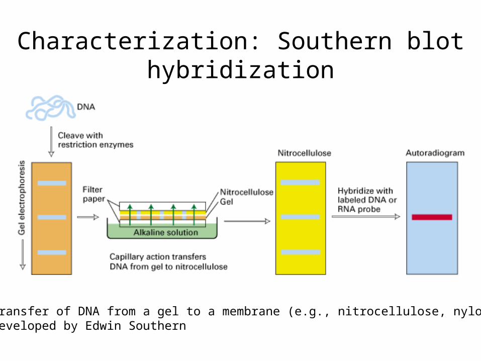
Characterization: Southern blot hybridization
-transfer of DNA from a gel to a membrane (e.g., nitrocellulose, nylon)-developed by Edwin Southern
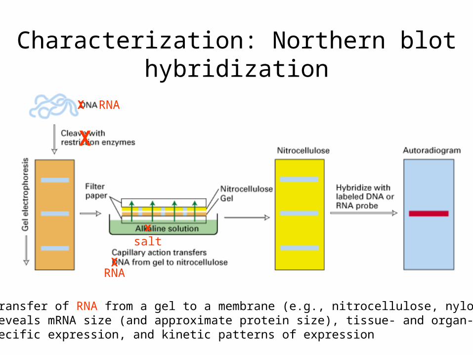
Characterization: Northern blot hybridization
-transfer of RNA from a gel to a membrane (e.g., nitrocellulose, nylon)-reveals mRNA size (and approximate protein size), tissue- and organ-specific expression, and kinetic patterns of expression
X RNA
X
xsalt
XRNA
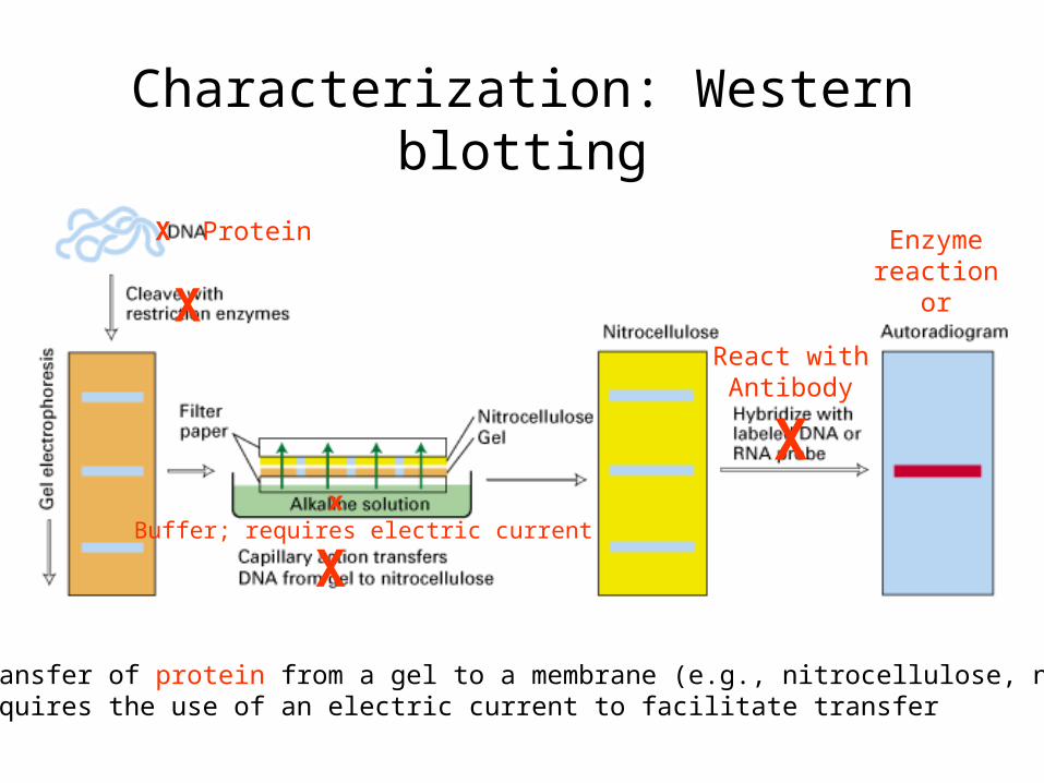
Characterization: Western blotting
-transfer of protein from a gel to a membrane (e.g., nitrocellulose, nylon)-requires the use of an electric current to facilitate transfer
X Protein
X
x Buffer; requires electric current
X
React withAntibody
X
Enzymereaction
or
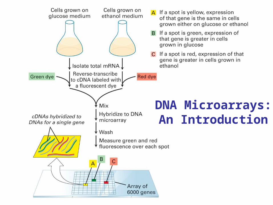
DNA Microarrays:An Introduction
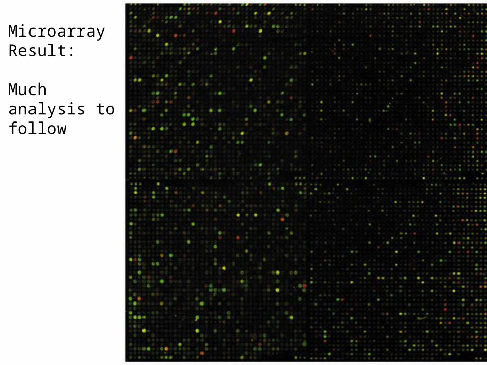
MicroarrayResult:
Much analysis to follow
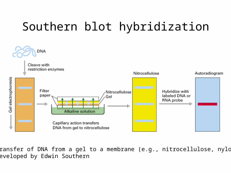
Southern blot hybridization
-transfer of DNA from a gel to a membrane (e.g., nitrocellulose, nylon)-developed by Edwin Southern
Southern blotting of genomic DNA
Steps
1. Extraction of genomic DNA
2. Electrophoresis of genomic DNA (or more commonly restriction enzyme-digested genomic DNA)

3. Before blotting, treat the gel with 0.2N HCl, denaturation and neutralization solution
4. Blotting- Transfer gel to the nitrocellulose membrane by capillary action
A) Sponge method
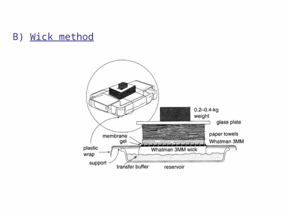
B) Wick method
5. After the transfer, cross-link the DNA to the nitrocellulose membrane
A. UV- cross linking
B. Bake at 120°C for 30 min
C. Bake at 80°C for 2 hrs
6. Making the probe
Label the probe to be hybridized using radioactive or non-radioactive methods
Non-radioactive methods A) Colorimetric
B) Chemiluminescent
* Roche DIG- DNA labeling (Non-radioactive) kits are used for detection
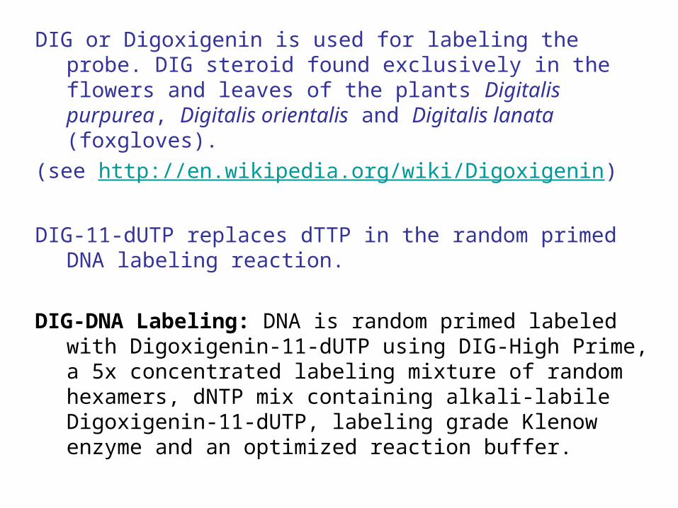
DIG or Digoxigenin is used for labeling the probe. DIG steroid found exclusively in the flowers and leaves of the plants Digitalis purpurea, Digitalis orientalis and Digitalis lanata (foxgloves).
(see http://en.wikipedia.org/wiki/Digoxigenin)
DIG-11-dUTP replaces dTTP in the random primed DNA labeling reaction.
DIG-DNA Labeling: DNA is random primed labeled with Digoxigenin-11-dUTP using DIG-High Prime, a 5x concentrated labeling mixture of random hexamers, dNTP mix containing alkali-labile Digoxigenin-11-dUTP, labeling grade Klenow enzyme and an optimized reaction buffer.
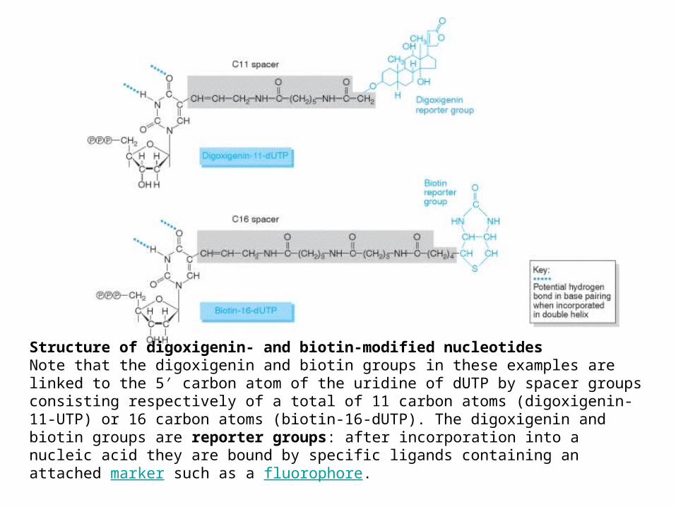
Structure of digoxigenin- and biotin-modified nucleotides Note that the digoxigenin and biotin groups in these examples are linked to the 5′ carbon atom of the uridine of dUTP by spacer groups consisting respectively of a total of 11 carbon atoms (digoxigenin-11-UTP) or 16 carbon atoms (biotin-16-dUTP). The digoxigenin and biotin groups are reporter groups: after incorporation into a nucleic acid they are bound by specific ligands containing an attached marker such as a fluorophore.
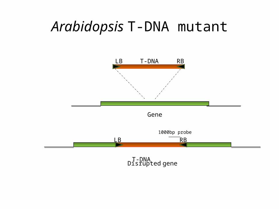
Arabidopsis T-DNA mutant
T-DNA
Gene
RBLB
T-DNA
Disrupted gene
RBLB1000bp probe
7. Prehybridization and hybridization of probe to the membrane at the hybridization temperature.
8. Washes and immunological detection
Detection after stringency washes using anti-digoxigenin antibody conjugated to AP (Alkaline phosphatase)
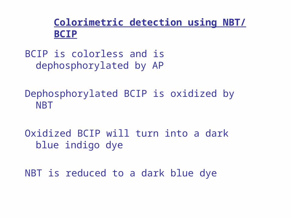
BCIP is colorless and is dephosphorylated by AP
Dephosphorylated BCIP is oxidized by NBT
Oxidized BCIP will turn into a dark blue indigo dye
NBT is reduced to a dark blue dye
Colorimetric detection using NBT/ BCIP
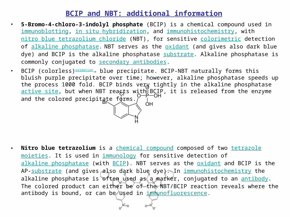
• 5-Bromo-4-chloro-3-indolyl phosphate (BCIP) is a chemical compound used in immunoblotting, in situ hybridization, and immunohistochemistry, with nitro blue tetrazolium chloride (NBT), for sensitive colorimetric detection of alkaline phosphatase. NBT serves as the oxidant (and gives also dark blue dye) and BCIP is the alkaline phosphatase substrate. Alkaline phosphatase is commonly conjugated to secondary antibodies.
• BCIP (colorless)oxidation→ blue precipitate. BCIP-NBT naturally forms this bluish purple precipitate over time; however, alkaline phosphatase speeds up the process 1000 fold. BCIP binds very tightly in the alkaline phosphatase active site, but when NBT reacts with BCIP, it is released from the enzyme and the colored precipitate forms.
• Nitro blue tetrazolium is a chemical compound composed of two tetrazole moieties. It is used in immunology for sensitive detection of alkaline phosphatase (with BCIP). NBT serves as the oxidant and BCIP is the AP-substrate (and gives also dark blue dye). In immunohistochemistry the alkaline phosphatase is often used as a marker, conjugated to an antibody. The colored product can either be of the NBT/BCIP reaction reveals where the antibody is bound, or can be used in immunofluorescence.
BCIP and NBT: additional information
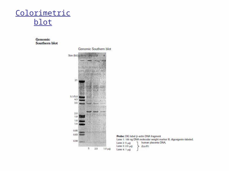
Colorimetric blot
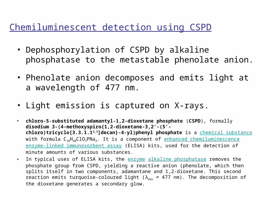
Chemiluminescent detection using CSPD
• Dephosphorylation of CSPD by alkaline phosphatase to the metastable phenolate anion.
• Phenolate anion decomposes and emits light at a wavelength of 477 nm.
• Light emission is captured on X-rays.
• chloro-5-substituted adamantyl-1,2-dioxetane phosphate (CSPD), formally disodium 3-(4-methoxyspiro{1,2-dioxetane-3,2'-(5'-chloro)tricyclo[3.3.1.13,7]decan}-4-yl)phenyl phosphate is a chemical substance with formula C18H20ClO7PNa2. It is a component of enhanced chemiluminescence enzyme-linked immunosorbent assay (ELISA) kits, used for the detection of minute amounts of various substances.
• In typical uses of ELISA kits, the enzyme alkaline phosphatase removes the phosphate group from CSPD, yielding a reactive anion (phenolate, which then splits itself in two components, adamantane and 1,2-dioxetane. This second reaction emits turquoise-coloured light (λmax = 477 nm). The decomposition of the dioxetane generates a secondary glow.