mcb cell signaling lectures 1 and 2 362-1668 lecture...
TRANSCRIPT

MCB Cell Signaling Lectures 1 and 2
Ken Blumer
Dept. of Cell Biology & Physiology
506 McDonnell Sciences
362-1668
Lecture 1
General Concepts of Signal TransductionCell CommunicationTypes of ReceptorsMolecular Signaling
Receptor BindingScatchard AnalysisCompetitive Binding
Second Messengers
G proteins
Signaling throughout Evolution
• Bacteria– Sense nutrients
• Lac operon--bacteria turn on gene expression of 3 genesnecessary to metabolize lactose (Jacob & Monod, Nobel1965)
• Chemotaxis- che proteins that couple nutrient receptorsto flagellar motors
– Quorum sensing• Yeast
– Pheromone signaling for haploid yeast mating• Multicellular Organisms
Many signaling pathways (G proteins, channels, kinases)

Modes of Cell Communication
Lodish, 20-1
• Intracellular ReceptorsLigands need to be
lipophilic– Steroids– Thyroid hormone– Retinoids
• Cell surface receptorsLigands can be either
water soluble orlipophilic--but bind atthe surface
Lodish, 20-2

Four classes of cell-surface receptors
Lodish, 20-3
Transmitting signals from one molecule to another
3 basic modes (may be combined)
1. Allostery
2. Covalent modification
3. Proximity (= regulated recruitment)
P
Shape change, often induced by binding a protein or small moleculeSwitching can be very rapid
Modification itself changes molecule’s shapeMemory device; may be reversible (or not)
Regulated molecule may already be in “signaling mode;” induced proximity to a target promotes transmission of the signal
P P
Signaling speed matches with function
• VERY FAST (milliseconds)Nerve conduction, vision– Ion channels
• FAST (seconds)Vision, metabolism, cardiovascular– G protein-coupled receptors
• SLOW (minutes to hours)Cell division, proliferation, developmental processes– Growth factor receptors– Steroid hormones

Detecting Receptors by Ligand Binding
Saturation Binding studiesCan be performed in intactcells, membranes, or purifiedreceptors1. Add various amounts oflabeled ligand (drug, hormone,growth factor)2. To determine specificbinding, add an excess ofunlabeled ligand to competefor specific binding sites.QU: Why is there non-specificbinding?3. Bind until at equilibrium4. Separate bound fromunbound ligand5. Count labeled ligand
[Adapted from A. Ciechanover et al., 1983, Cell 32:267.]
Receptor: ligand binding must be specific, saturable, and of high affinity
Ligand Binding Reversibility, Affinity & Kinetics
Activity of a signaling machine often depends on its association with another molecule
If the association is reversible, we can talk about . . .
Equilibrium binding
(A) + (B) (AB)k1= association rate
= dissociation rate
At equilibrium, the forward reaction goes at exactly the same rate as the backward reaction
Forward reaction rate = (A)(B)
Backward reaction rate = (AB)
So . . . (A)(B) = (AB)
k2
k1
k2
k1
k2
k1 k2
Kinetics & Affinity
If . . . (A)(B) = (AB) k1 k2
= Kd =(A)(B)(AB)k1
k2
k1
k2=
Define
So . . .
Equilibrium binding is saturable
1.0
0.5
(AB
)
(A)
Kd = conc of A at which half of B binds A
dissociation constantKd =
Bmax
Kd

Kinetics and Half-life
Kd = k1
k2 k1= association rate constant
= dissociation rate constantk2
Units
(M-1)(sec-1)
(sec-1)
k1
k2
usually ~ 108M-1 sec-1 (diffusion-limited)
just a time constant (sec-1)
Thus, knowing the Kd and assuming a “usual” rate of association, you can calculate . . .
k2, and therefore the duration (or half-life*) of the (AB) complex
*Half-life = 0.69 ÷ k2
Half-lives differ greatly
Kd k2
*Half-life = 0.69 ÷ k2
Half-life of (AB)
(sec)(M) (sec-1)
Acetylcholine
Norepinephrine
Insulin
102
100
10-2
0.007
0.7
70
10-6
10-8
10 -10
LIGAND
Scatchard Analysis
Slope = - 1/Kd
X intercept = # rec
(Bound Lig)
(Bound Lig)(Free)
For an excellent discussion of principles of receptor binding, andpractical considerations, see http://www.graphpad.com; also posted on MCB website.

Scatchard Analysis
(Bound Lig)
(Bound Lig)(Free)
Negative cooperativity: binding of ligand to first subunitdecreases affinity of subsequent binding events.
Positive cooperativity:binding of ligand tofirst subunit increasesAffinity of subsequentbinding events.Example: hemoglobinbinding O2
Cooperative binding
The Hill equation accounts for the possibility that not all receptorsites are independent, and states that
Fractional occupancy = Lfn/ (Kd + Lf
n)
n= slope of the Hill plot and also is the avg # of interacting sites
For linear transformation, log [B/(Rt - B)] = n(log Lf) - log Kd
log [B/(Rt - B)]
log Lf
Slope= n
If slope = 1, then singleclass of binding sites
If slope > 1, then positivecooperativity
If slope < 1, then negativecooperativity
Competitive bindingHow many different types of ligands can a receptor bind? Are some ligandsmore avid for a receptor than others?You can use the ability of a compound (could be agonist or antagonist) tocompetitively displace the binding of a fixed amount of a different compound(usually a labeled antagonist).BIG ADVANTAGE: You only need one labeled compound.
Example. Adrenergic agonists: isoproterenol (ISO), epinephrine (EPI)
Adrenergic antagonists: phentolamine (PHEN)
100%
[competitor]
100%
[competitor]
α-adrenergic receptor β-adrenergic receptor
ISO
ISO
PHEN
PHEN

So that’s the theory: How do we know whether or not it is true?
1. Theory is internally consistent (necessary, not sufficient)
2. Binding experiments
Stop binding reaction quickly, measure bound complex, (AB)
Assess k1 = “on-rate”
Assess k2 = “off-rate”
Compare vs. Kd
3. Seeing is believing: Watch behavior of fluorescent-tagged single molecules of ligand bound to receptors
Seeing is believing* . . .
Assess duration of ligand-receptor complexes, during chemotaxis of living Dictyostelium cells
Question: Does signaling differ at front vs. back of the cell?
Experimental system: Dictyostelium discoideum, a soil amoeba
Seeing is believing, Total InternalReflection Fluorescence
http://www.olympusmicro.com/primer/techniques/fluorescence/tirf/tirfintro.html
Question: Does receptor signaling differ at front vs. back of the cell?
Approach: Tag cAMP ligandwith a fluorescent dye
Bound cAMP stays in oneplace on cell surface;unbound tagged cAMPdiffuses rapidly away
Evanescent wave excites onlytagged cAMP near slide

Seeing is believing* . . .
*Ueda et al., Science 294:864,2001
0 5 10 2015 250
400
Time (sec)
Pseudopodk2 = 1.1 and 0.39 s-1
k2 = 0.39 and 0.16 s-1Tail
cAMP-R complexes dissociate ~2.5 x faster at the front than at the back!
True for cells in a ligand gradient and also in a uniform concentration of the ligand
Off & On: cAMP-R complexes (movie: 7 sec total)
Cy3
-cA
MP
b
ound
Cell surface facing the slide
Each point is a separate cAMP/R complex
Other methods of measuring binding
• Surface plasmon resonance (BiaCore)Can measure “on” rates and “off” rates to calculate binding affinities
• Isothermal calorimetryVery accurate, requires lots of protein and expensive equipment
• Equilibrium dialysisUseful for binding of small ligands to large proteins
• Fluorescence anisotropyExcite fluorescent protein with polarized light. Anisotropy refers to the
extent that the emitted light is polarized--the larger theprotein/complex, the slower the tumble rate and the greater theanisotropy
• Co-immunoprecipitation• Yeast two-hybrid
Many receptors regulate cell function byproducing second messengers
• Cyclic nucleotides: cAMP, cGMP• Inositol phosphate (IP)• Diacylglycerol (DAG)• Calcium• Nitric oxide (NO)• Reactive oxygen species (ROS)
Molecular mediators of signal transduction. Cellscarefully, and rapidly, regulate the intracellularconcentrations. Second messengers can be used bymultiple signaling networks (at the same time).

Earl Sutherland1971 Nobel laureate
Rall, et al. JBC 1956
Fischer & Krebs, Nobel 1992
Discovered thatphosphorylase activitywas regulated by thereversible step ofphosphorylation. IdentifiedPKA and some of the firstphosphatases.
cAMP regulates PKA activity
Alberts 15-31,32
Positive cooperativity--binding ofincreases affinity for second cAMP
PKA targets include Phosphorylase kinaseand the transcription regulator, cAMPresponse element binding (CREB) protein

Diacylglycerol and Inositol Phosphates assecond messengers
Alberts, 15-35
Calcium acts as second (third?) messenger
Lodish, 20-39
Calmodulin transduces cytosolic Ca2+ signal
Alberts, 15-40
Calmodulin, found in all eukaryotic cells, and can be up to 1%of total mass. Upon activation by calcium, calmodulin can bindto multiple targets, such as membrane transport proteins,calcium pumps, CaM-kinases

CaM-kinase II regulation
Alberts, 15-41
Frequency of calcium oscillationsinfluences a cell’s response
High frequency Ca2+ oscillationsLow frequency Ca2+ oscillations
CaM
-kin
ase
II ac
tivity
CaM
-kin
ase
II ac
tivity
CaM-kinase uses memory mechanismto decode frequency of calcium spikes.
Requires the ability of the kinase tostay active after calcium drops. This isaccomplished by autophosphorylation.
Alberts 15-39,42
Calcium signaling also occurs in waves
Alberts, 15-37
0 sec 10 sec 20 sec 40 sec
Calcium effects are local, because it diffuses much more slowly thandoes InsP3
Sperm binds
InsP3 receptor is both stimulated and inhibited calcium
[Ca 2+]
Sen
sitiv
ity o
f In
sP3
R to
Ca
2+
InsP3

NO signaling
Lodish, 20-42
NO effects are local, since it has half-life of 5-10 seconds (paracrine).NO activates guanylate cyclase bybinding heme ring (allostericmechanism)
Gases can act as second messengers!
Discovery of NO signaling
Robert F Furchgott showed thatacetylcholine-induced relaxation ofblood vessels was dependent on theendothelium. His "sandwich"experiment set the stage for futurescientific development. He used twodifferent pieces of the aorta; one hadthe endothelial layer intact, in theother it had been removed.
Louis Ignarro reported that EDRF relaxed bloodvessels. He also identified EDRF as a molecule byusing spectral analysis of hemoglobin. Whenhemoglobin was exposed to EDRF, maximumabsorbance moved to a new wave-length; andexposed to NO, exactly the same shift in absorbanceoccurred! EDRF was identical with NO.
Furchgott, Ignarro, Murad, Nobel Prize 1998
http://www.nobel.se/medicine/laureates/1998/illpres/index.html
Reactive Oxygen Species (ROS) Signaling
Finkel & Holbrook, Nature (2000)
ROS important in cell’sadaptation to stress
Many of longevitymutations map to ROSpathways
Mutations in SuperoxideDismutase (SOD)cause amyotrophiclateral sclerosis (ALS,Lou Gehrig’s Disease)
Unfortunately, no greatclinical data showingthat anti-oxidants willhelp us live longer!

ROS activates multiple pathways
Finkel & Holbrook, Nature (2000)
Activation mechanisms ????
Mimic ligand effect for GF receptorsOxidants enhance phosphorylation ofRTKs and augment ERK/Akt signaling
Inactivation of phosphatasesHydrogen peroxide inactivates protein-Yphosphatase 1B
Redox sensorsThioredoxin (Trx) binds and inhibitsASK1, an upstream activator of JNK/p38pathways. ROS dissociates Trx-ASK1complex
HSF1, NF-kB, and ERK activitieschange with age (Pink boxes)
G proteins:switches linking receptors & 2nd messengers
• Discovery and Structure of Heterotrimeric Gproteins
• Signaling pathways of G proteins• Receptors that activate G proteins• Small G proteins-discovery and structure• Activation and inactivation mechanisms• Alliance for Cell Signaling (AfCS)
Discovery of G proteinsMartin Rodbell first proposed the concept of “discriminator-transducer-amplifier” to address the problem: “How canmany hormones (epinephrine, ACTH, TSH, LH, secretin,and glucagon) activate lipolysis and cAMP production inadipocytes through presumably a single cyclase? He calledthis problem “too many angels on a pinhead.” His workidentified GTP as important for the “transducer”.
His work was notinitially receivedwell by thescientificcommunity:
Nobel prize, 1994

Discovery of G proteinsAl Gilman purified the first G proteins. His lab tookadvantage of S49 lymphoma cells that lacked Gsα(although at the time, the cells were thought to lackadenylate cyclase, thus the name cyc-).
Reconstitution experiment rationale: Isolate membranesfrom cyc- cells, then add back fractions from donor wtmembranes that restore adenylate cyclase activity. Nobel prize, 1994
Donor membranes were incubated forincreasing time at 30oC, whichinactivates the adenylate cyclaseactivity (- - - - -). Fortunately, G proteinsare relatively heat stable.
Addition of NaF, Gpp(NH)p, GTP, orGTP and isoproterenol restored activityin the cyc- membranes.
Ross, et al. JBC (1978)
Signal Transduction by G proteins
• Discovery and Structure of Heterotrimeric Gproteins
• Signaling pathways of G proteins• Receptors that activate G proteins• Small G proteins-discovery and structure• Activation and inactivation mechanisms• Alliance for Cell Signaling (AfCS)
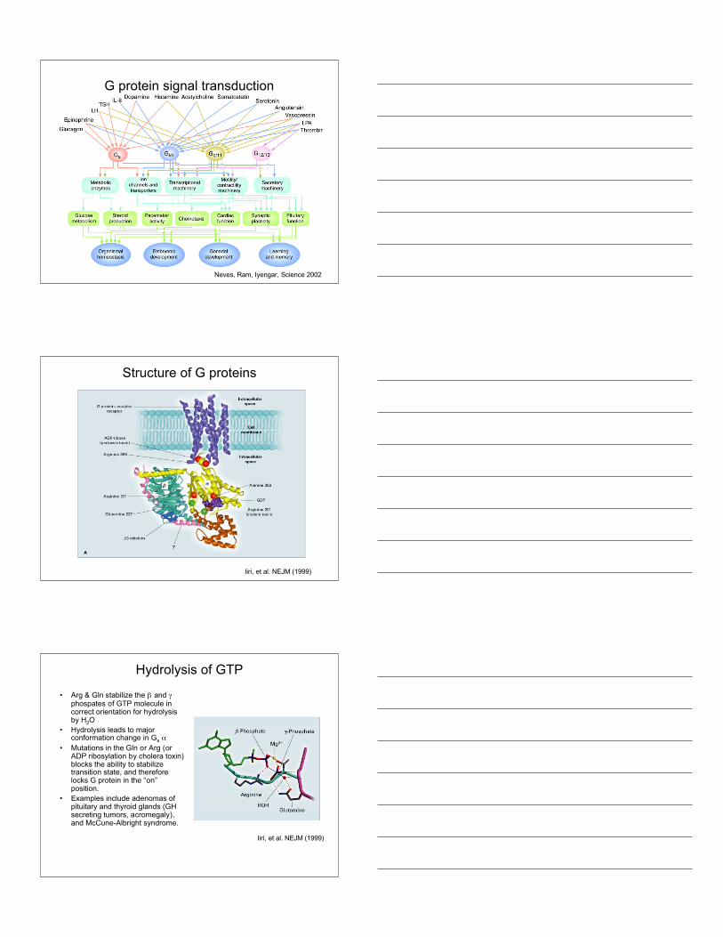
G protein signal transduction
Neves, Ram, Iyengar, Science 2002
Structure of G proteins
Iiri, et al. NEJM (1999)
Hydrolysis of GTP
• Arg & Gln stabilize the β and γphospates of GTP molecule incorrect orientation for hydrolysisby H2O
• Hydrolysis leads to majorconformation change in Gs α
• Mutations in the Gln or Arg (orADP ribosylation by cholera toxin)blocks the ability to stabilizetransition state, and thereforelocks G protein in the “on”position.
• Examples include adenomas ofpituitary and thyroid glands (GHsecreting tumors, acromegaly),and McCune-Albright syndrome.
Iiri, et al. NEJM (1999)

Canonical Gs Signaling PathwayFor interactive pathways atSTKE:
Gs pathwayhttp://stke.sciencemag.org/cgi/cm/CMP_6634
Gi pathwayhttp://stke.sciencemag.org/cgi/cm/CMP_7430
Gq pathwayhttp://stke.sciencemag.org/cgi/cm/CMP_6680
G12 pathwayhttp://stke.sciencemag.org/cgi/cm/CMP_8022
Neves, Ram, Iyengar, Science 2002
Signal Transduction by G proteins
• Discovery and Structure of Heterotrimeric Gproteins
• Signaling pathways of G proteins• Receptors that activate G proteins• Small G proteins-discovery and structure• Activation and inactivation mechanisms• Alliance for Cell Signaling (AfCS)
G protein signaling
• Many ligands• Robust switches• Multiple effectors• Conserved 7 TM
architecture• More than 50% of
drugs targetGPCRs
Bockaert & Pin, EMBO J (1999)

G protein-coupledreceptors
• 5 main families• Conserved 7 TM
architecture
GPCRs in the Human GenomeSteve Foord, GlaxoWelcome
Rhodopsin Secretin Metabotropic
Liganded 163 25 11Orphan 140 34 4Olfactory 350 6Taste 15 3
Identifying Ligands for Orphan GPCRS
Big Pharm approach: set upindividual stable cell linesexpressing each orphan GPCR.Fractionate peptides, tissuefactors, etc. and apply to eachcell line. Example: Orexinreceptors
Cottage industry approach:expression cloning strategy inXenopus oocytes. Use sibselection to identify cDNAs thatencode desired receptor.Example: Thrombin receptor

GPCR desensitization mechanisms
10 seconds is too long! αt-GTP must be inactivated in < 1 sec
Many variations: eg, effectors with RGS activity
eg, phospholipase C β acts on αqE
E*EPi
EFFECT
Regulators of G Signaling (= RGS1-~RGS16; RGS9 in ROS)
GTP
RGSRGS
RGSPi
GDPαt GTP
αt αt
Most RGSs act on αi or αq families
RGSSwi1 Swi2
GTPAccelerate GTPase from < 1/sec to >10 3/sec
GTP GDPαq GTP
αq αq
eg, γ subunit of cGMP PDE enhances effect of retinal RGS on αt
New concepts for GPCR signalingUsing mainly two-hybrid screeningapproaches, many proteins have beenfound to interact with portions of theGPCRs.Non-PDZ scaffolds: AKAPs (A-KinaseAnchoring Proteins, JAK2 (JanusActivated Kinase), homer, β-arrestinsPDZ scaffolds: InaD, PSD-95 (Post-Synaptic Density), NHERF (Na/HExchanger Regulatory Factor).
The arrestins have been found to bindto other signaling proteins and activatedownstream effectors:Examples: src, Raf & ERK, ASK1 &JUNK3
Lefkowitz reviews

Arrestins act as scaffolds for ERK andJNK signaling pathways
Lefkowitz reviews
Signal Transduction by G proteins
• Discovery and Structure of Heterotrimeric Gproteins
• Signaling pathways of G proteins• Receptors that activate G proteins• Small G proteins-discovery and structure• Activation and inactivation mechanisms• Alliance for Cell Signaling (AfCS)
Discovery of Small G proteinsRas genes first identified in‘60’s as transforming genes ofrat sarcoma viruses.
Weinberg, Varmus, Bishop andothers in the early ‘80’s showedthat many cancer cells havemutated versions of ras.
Activated form of ras found in90% of pancreatic carcinomas,50% of colonadenocarcinomas, and 20% ofmalignant melanomas.
Ras-GTP vs. Ras-GDP
Signaling GTPases are Allosteric Switches
γ-phosphate
Ras = classical “monomeric” GTPase
Binding γ-phosphate changes the conformations of two small surface elements, called “switch 1 and 2”
Swi1Swi2

Rho/Rac/Cdc42
In early ‘90’s, Alan Hall discovered that newly characterizedRas homologs (Rho, Rac, Cdc42) induced cytoskeletalchanges.
Hall, Science 1998
Actin Stress fibers Focal adhesions
Lamellipodia Filopodia
Ras superfamily of small G proteins
Takai, et al. Physiological Reviews, 2001

GTPases: How to use reverse genetics to identify their roles in cell regulation
Depends on understanding how the machines work
Epistasis question: Where in a pathway does a specific protein convey its particular message?
A B
C D E
Response
M N Q
Idea: 1. Inhibit activity of the protein of interest
2. Increase activity of the protein of interest
How to do this? Drugs, genetic diseases, mouse KOs, and . . .
Reverse genetics: express one or two mutant versions of the protein of interest
Depends on understanding how the machines work
1. Inhibit activity of the protein with a “dominant-negative” interfering mutant of that protein
2. Increase activity of the protein with a “dominant-positive” or “constitutively active” interfering mutant of the protein
The mutant titrates (binds up) a limiting component to block the normal protein’s signal
The mutant exerts the same effect as the normal protein would, if it were activated in the cell
Reverse genetics: small GTPases as examplesDepends on understanding how the machines work
“Dominant-negative” mutation “Dominant-positive”
mutation
The mutant titrates (binds up) a limiting component to block the normal protein’s signal
The mutant exerts the same effect as the normal protein would, if it were activated
GAP
GTP
Pi
GDPGEF
GEFGDP
Binds GEF but cannot replace GDP by GTP; so GEF not available for activating normal protein
Cannot hydrolyze GTP, so remains always active

Reverse genetics: advantages/pitfalls of using dominant-interfering mutants
Pro:Quick-and-dirty; no biochem
Many different families of signaling proteins amenable . . . once we understand how one of them works
Examples:
RTKs?Other kinases?Adaptors?
Con:Dominant-negatives
Dominant positives
Therefore . . .Still need biochemistry
Hard to apply to complex networks
Over-expression can titrate too many proteins (or the wrong proteins
Not always precise mimics of the normal protein (e.g., may be in the wrong place))
Can induce adaptation, turn-off mechanisms
Hierachy of small G protein activation
Ras
Use of constitutively active or dominant negative mutant small Gproteins revealed that ras and cdc42 can activate rac. Rac, in additionto inducing lamellipodia, also activates Rho.
y
Rho/Rac/Cdc42 signaling in actin assembly
Takai, et al. Physiological Reviews, 2001

Signal Transduction by G proteins
• Discovery and Structure of Heterotrimeric Gproteins
• Signaling pathways of G proteins• Receptors that activate G proteins• Small G proteins-discovery and structure• Activation and inactivation mechanisms
Small G protein “turn on” mechanisms
First mammalian GEF,Dbl, isolated in 1985 asan oncogene in NIH 3T3focus forming assay. Ithad an 180 amino aciddomain with homology toyeast CDC24. Thisdomain, named DH (Dblhomology) is necessaryfor GEF activity.
In 1991, Dbl shown tocatalyze nucleotideexchange on Cdc42.
Schmidt & Hall, Genes & Dev. (2002)Dbl= Diffuse B-cell lymphoma
Small G proteins “turn off” mechanismsRhoGAPs outnumber the small Gproteins Rho/Rac/Cdc42 by nearly5-fold.Why so much redundancy?Luo group did RNAi against 17 ofthe 20 RhoGAPs in fly.
Six caused lethality whenexpressed ubiquitously. Tissuespecific expression of RNAirevealed unique phenotypes.
P190RhoGAP implicated in axonwithdrawal. Increasing amounts ofRNAi caused more axonwithdrawal (panels C-G).
Why so many RhoGAPs?Billuart, et al. Cell (2001)

Identification of RasGAP
McCormick injected Xenopus oocyteswith oncogenic ras (V12) versus wt ras(G12) and monitored germinal vesiclebreakdown (GVB) (top panel)
% G
VB
[ras] (ng)
V12
G12
Time (min)
% R
as-G
TP V12
G12
Then loaded ras with α-32P GTP, injectedinto oocytes, did immppt at increasingtimes and determined if GTP or GDP wasbound (bottom panel)
Purified the factor that promoted GTPaseactivity, cloned and named it GAP (or ras-GAP). Another ras-GAP later identified isNF1 (the gene mutated in neurofibromatosis,i.e., Elephant Man Syndrome).
Rate of GTP hydrolysis is 300-fold fasterin oocytes than in vitro!
Rho/Rac/CDC42 activation ofdownstream effectors
Rho
Effectors: PI 3-Kinase, PLD, Rho Kinase, Rhophilin, and others.
Rac-interacts via a CRIB domain in downstream effectors. CRIB(Cdc42/Rac interacting binding)
Effectors: NADPH oxidase, PAK, PI 3-Kinase, MLK2,3, POSH,DGK
Cdc42
Effectors: PI ε-Kinase, PAK, WASP, S6-Kinase, MLK2,3, Borg
The GTPase switch
Schmidt & Hall, Genes & Dev. (2002)