mcm proteins: evolution, properties, and role in dna...
TRANSCRIPT
Ž .Biochimica et Biophysica Acta 1398 1998 113–136
MCM proteins: evolution, properties, and role in DNA replication
Stephen E. Kearsey a,), Karim Labib b
a Department of Zoology, South Parks Road, Oxford, OX1 3PS, UKb Imperial Cancer Research Fund, Clare Hall Research Labs, Blanche Lane, South Mimms, Herts EN6 3LD, UK
Received 2 February 1998; accepted 17 February 1998
Ž .Keywords: Initiation of DNA replication; Replication origin; Cell cycle control eukaryotic ; MCM protein
Contents
1. Introduction . . . . . . . . . . . . . . . . . . . . . . . . . . . . . . . . . . . . . . . . . . . . . . . . . . . 114
2. The MCM protein family . . . . . . . . . . . . . . . . . . . . . . . . . . . . . . . . . . . . . . . . . . . 1152.1. Identification and phylogenetic analysis of MCM proteins . . . . . . . . . . . . . . . . . . . . 1152.2. Amino-acid sequence features of MCM proteins . . . . . . . . . . . . . . . . . . . . . . . . . . 117
3. Requirement for MCMs in chromosome replication . . . . . . . . . . . . . . . . . . . . . . . . . . . 120
4. Biochemical properties of MCM proteins . . . . . . . . . . . . . . . . . . . . . . . . . . . . . . . . . 1234.1. Formation of complexes between different MCM proteins . . . . . . . . . . . . . . . . . . . . 1234.2. Interaction between MCMs and other proteins . . . . . . . . . . . . . . . . . . . . . . . . . . . 124
5. Regulation of MCM proteins during the cell cycle . . . . . . . . . . . . . . . . . . . . . . . . . . . . 1255.1. MCM protein levels in proliferating and non-proliferating cells . . . . . . . . . . . . . . . . . 1255.2. Involvement of ORC and Cdc6rcdc18 in binding of MCM proteins to chromatin . . . . . . 1265.3. Role of phosphorylation in regulating MCM function . . . . . . . . . . . . . . . . . . . . . . . 1275.4. Relevance of the nuclear membrane to MCM function . . . . . . . . . . . . . . . . . . . . . . 129
6. Conclusions and speculations on MCM function . . . . . . . . . . . . . . . . . . . . . . . . . . . . . 1306.1. Model for MCM function in replication initiation . . . . . . . . . . . . . . . . . . . . . . . . . 1306.2. Determinants of replication origin function . . . . . . . . . . . . . . . . . . . . . . . . . . . . . 1326.3. Future perspectives . . . . . . . . . . . . . . . . . . . . . . . . . . . . . . . . . . . . . . . . . . . 133
Abbreviations: ARS, autonomously replicating sequence; 6-DMAP, 6-dimethylaminopurine; cdc, cell division cycle; ECB, early cellcycle box; NLS, nuclear localization sequence; MCM, mini-chromosome maintenance; mis, mini-chromosome instability; nda, nucleardivision arrest; ORC, origin recognition complex; PCNA, proliferating cell nuclear antigen; pre-RC, pre-replicative complex; RP-A,replication protein A
) Corresponding author. Fax: q44-1865-271228; E-mail: [email protected].
0167-4781r98r$19.00 q 1998 Elsevier Science B.V. All rights reserved.Ž .PII S0167-4781 98 00033-5

( )S.E. Kearsey, K. LabibrBiochimica et Biophysica Acta 1398 1998 113–136114
Acknowledgements . . . . . . . . . . . . . . . . . . . . . . . . . . . . . . . . . . . . . . . . . . . . . . . . 133
References . . . . . . . . . . . . . . . . . . . . . . . . . . . . . . . . . . . . . . . . . . . . . . . . . . . . . 133
1. Introduction
The replication of DNA is a fundamental step inthe cell cycle, which must be coordinated with celldivision to ensure that the daughter cells have thesame ploidy as the parental cell. The control thatcommits a cell to a round of DNA replication isadditionally responsive to a number of signals thatreflect parameters such as cell size, nutritional status,cell–cell communication and DNA damage. In eu-karyotic cells, DNA replication is initiated from alarge number of replication origins, but initiationevents must be restricted to once per cell cycle, toavoid overreplication of parts of the genome. Thiscontrol demands a low error rate, since S phase in ahigher eukaryotic cell may involve tens of thousandsof initiation events which occur throughout S phase.Some eukaryotic organisms can vary the number ofchromosomal replication origins that are active atdifferent stages of the life cycle. For instance, cellproliferation is rapid and S phase is short in earlyembryos of creatures such as frogs and flies, tofacilitate rapid development of the embryo. This isachieved by usage of many more origins of replica-tion than are used in adult cells during S phase. Thecontrols determining the order of S phase and nucleardivision can also be disrupted in certain cell types.This occurs, for example, in meiosis where two nu-clear divisions occur without an intervening S phase,or in the formation of polyploid cells during thedevelopment of some organisms, where multiple Sphases occur in the absence of mitosis. Replicationcontrol can also be modified on a more local level, toallow replication origins in special parts of thegenome to fire repeatedly, thus providing for selec-tive gene amplification as occurs with the choriongenes of Drosophila.
The yeasts Saccharomyces cereÕisiae andSchizosaccharomyces pombe have been useful foridentifying proteins involved in the initiation of DNAreplication, either via characterization of mutants af-
Ž .fecting S phase such as cdc or mcm mutants , or by
isolating proteins that bind to origins of replication.Replication origins have been well characterized inbudding yeast, and are marked throughout the cellcycle by the binding of a complex of six proteins, the
Ž .origin recognition complex or ORC , the function ofwhich is essential for the initiation of DNA replica-
w x Ž .tion 1–5 Section 5.2 . It seems likely that ORCpermits the loading of other replication factors onto
Žorigin DNA. One such protein is Cdc6 and its fission.yeast homologue cdc18 , which has a key role in
triggering initiation, and has been shown in XenopuslaeÕis egg extracts to associate with chromatin in an
w xORC-dependent fashion 6 . In budding yeast, Cdc6has been shown to be specifically associated with
w xorigin DNA in G1 phase 7 and in fission yeastoverexpression of cdc18 induces multiple rounds of Sphase in the absence of mitosis, suggesting thatCdc6rcdc18 is central to the control limiting DNA
w xreplication to once per cell cycle 8,9 . Another essen-tial group of replication proteins comprises the MCM
Ž . 1family MCM2–7 . Analysis of budding yeast mcmmutants has shown that these proteins function in theinitiation step of DNA replication and, like Cdc6, arebound to chromatin around origins of replicationduring G1 phase, but are subsequently displaced dur-ing S phase and remain unbound until the end of
Ž .mitosis Section 3 . This periodic association isthought to ensure that replication origins are onlycompetent to fire at the end of G1 phase and can onlyfire once during S phase. Chromatin binding of MCMproteins requires other initiation proteins such as
Ž .ORC and Cdc6rcdc18 Section 5.2 , and overallregulation of origin firing appears to be orchestratedby the protein kinases Cdk2rcdc2 and Cdc7–Dbf4Ž .Section 5.3 . Elongation of replication forks away
1 w xTo avoid confusion, a simplified MCM nomenclature 10will be used here; Table 1 shows the correspondence withoriginal MCM protein or gene names. Note that MCM1, MCM10and MCM17, which were also identified in the original MCM
w xgenetic screen 11 , share no sequence similarity to MCM2 – 7.

( )S.E. Kearsey, K. LabibrBiochimica et Biophysica Acta 1398 1998 113–136 115
from individual replication origins is thought to dis-rupt the MCM-containing complex. Thus, reinitiationis prevented by a simple cis-regulatory mechanismwhich couples replication to the inactivation of acomplex that is essential for initiation. Characteriza-tion of homologues of such proteins in higher eukary-otes has shown general conservation of the replica-tion apparatus, and it seems likely that the basicmechanism of DNA replication evolved in a commonancestor of all eukaryotic cells.
The general area of DNA replication regulationand the more specific topic of the involvement ofMCM proteins in this process has been extensively
w xreviewed in recent years 10,12–23 , but in the fol-lowing discussion we shall try to emphasize primaryexperimental work.
2. The MCM protein family
2.1. Identification and phylogenetic analysis of MCMproteins
Genes encoding MCM proteins were originallyidentified in budding and fission yeast as mutantsaffected in the progression through the cell division
Ž w x w x.cycle cdc 24–26 , nda 27 or the replication ofŽ w x w x.minichromosomes mcm 11 , mis 28 . Initial char-
Ž w xacterization of three S. cereÕisiae genes MCM2 29 ,w x w x.MCM3 30 and CDC46rMCM5 24,31 showed
that they were all implicated in DNA replication andwere related in sequence. This family rapidly grew to
qŽ q. w xencompass the S. pombe cdc21 mcm4 32 andqŽ q. w xmis5 mcm6 28 genes, and the S. cereÕisiae
Ž . w xCDC47 MCM7 gene 33 . The complete genomesequence of S. cereÕisiae indicates that there are sixMCM-encoding genes, suggesting that there are six
Ždistinct classes of MCM protein in eukaryotes Table.1 .
Identification of MCM proteins in higher eukary-otes initially came from the detection of a murine
w xprotein related to S. cereÕisiae Cdc46rMcm5 24Žand isolation of the human P1 protein homologous
.to Mcm3 that co-purified with DNA polymerase a
w x62 , whilst the considerable sequence conservation ofthe family has made it easy to identify other higher
w x Ž .eukaryotic homologues 32,56,65,74 Table 1 . Thetendency of MCM proteins to interact with each other
has made it possible in some cases to co-purify andw xcharacterize proteins in the family 46,55 . Higher
eukaryotic MCMs have also emerged by characteriz-ing mRNAs or antigens that are specifically associ-
w xated with proliferating cells 43,60,71,75 , or byscreening for mutants affecting cell proliferation dur-
w xing development in Drosophila 42 or Arabidopsisw x72 . All eukaryotic MCM sequences obtained thusfar appear to be homologous to one or other of thesix S. cereÕisiae MCM proteins, suggesting thatthere were six distinct MCM genes in a primordialeukaryote and that the family has not diversifiedfurther.
MCMs were first implicated as possible regulatorsof DNA replication by the observation that the S.
Ž .cereÕisiae Cdc46 Mcm5 protein accumulates in thenucleus of G1 phase cells but rapidly disappears from
w xthe nucleus upon S phase onset 31 . Similar observa-w xtions were also made for Mcm2, Mcm3 34 and
Ž . w xCdc47 Mcm7 33 leading to speculation about arelationship between ‘licensing factor’ and MCMproteins. The idea of a licensing factor first emergedfrom studies showing that permeabilization of thenuclear envelope is necessary for G2 phase chromatinto regain competence for another round of DNA
w xreplication in extracts of eggs from Xenopus 76 . ItŽwas suggested that a pre-replicative step termed
.licensing makes chromatin competent for initiationof DNA replication. This would involve the bindingof licensing factor to chromatin at the end of mitosis,permitting a single initiation event at replication ori-gins in the subsequent interphase, after which licens-ing factor is inactivated. Licensing factor was postu-lated to be unable to cross the nuclear membraneduring interphase, so that licensing of DNA normallyoccurs only after nuclear membrane breakdown inmitosis and DNA replication is thus restricted to asingle round per cell cycle. Subsequent analysis ofMCM proteins in other eukaryotes has shown thatthese proteins remain nuclear throughout interphase,and probably can cross the nuclear membrane during
Ž .interphase Sections 3, 5.4 . Although MCMs do notshow the behaviour predicted for licensing factor,they do show a cell cycle change in chromatin bind-ing which is likely to reflect their involvement in alicensing-type reaction. An alternative model to ex-plain the rereplication results in Xenopus is that aninhibitor of the licensing reaction may be unable to

()
S.E.K
earsey,K.L
abibr
Biochim
icaet
Biophysica
Acta
13981998
113–
136116
Table 1The MCM protein family
MCM class MCM2 MCM3 MCM4 MCM5 MCM6 MCM7
S. cereÕisiaeMCM2 MCM3 CDC54 CDC46rMCM5 MCM6 CDC47w x w x w x w x w x29,34 29,30,34 35 24,31,36 33P29469 P24279 P30665 P29496 P53091 P38132
S. pombeq q q q q q qnda1 rcdc19 mcm3 cdc21 nda4 mis5 mcm7
w x w x w x w x w x w x27,37–39 32 26,32,40 27,38 28 41Ž . Ž .P40377 P30666 partial P29458 P41389 D31960 AJ000065 partial
Drosophila( )DmMCM2 dpa DmMCM5 DmCDC46
w x w x w x42 43 44,45P49735 Q26454 U83493
XenopusXMCM2 XMCM3 XCDC21 XCDC46rXMCM5 XMCM6 XMCM7w x w x w x w x w x w x46,47 48–51 52,53 46,47 46 46,54U44047 P49739 S64720 U44048 U44050 U44051
MousemMCM2 mP1MCM3 mCdc21 mCDC46 mMis5 mCDC47w x w x w x w x w x w x55 56,57 58 58 55 59P97310 D26089 P49717 P26090 D86726 Q61881
HumanBM28rhMcm2 P1Mcm3 P1cdc21rhCdc21 P1Cdc46rhMcm5 p105McmrhMis5 p85McmrP1.1-MCM3r hCdc47w x w x w x w x w x w x60,61 62–64 65,66 65 67,68 69P49736 P25205 P33991 P33992 U46838 D55716
OtherArabidopsis Notophthalmus B24 Caenorhabditis Rattus Arabidopsis
w x w xY08301 70 Q21902 71 PROLIFERAw xI51022 Q62724 72
Aspergillus Triturus Caenorhabditis P43299w x73 Y11554 P344647AF014813 Caenorhabditis
Z81039Zea ROAw x74Q43704
( )S.E. Kearsey, K. LabibrBiochimica et Biophysica Acta 1398 1998 113–136 117
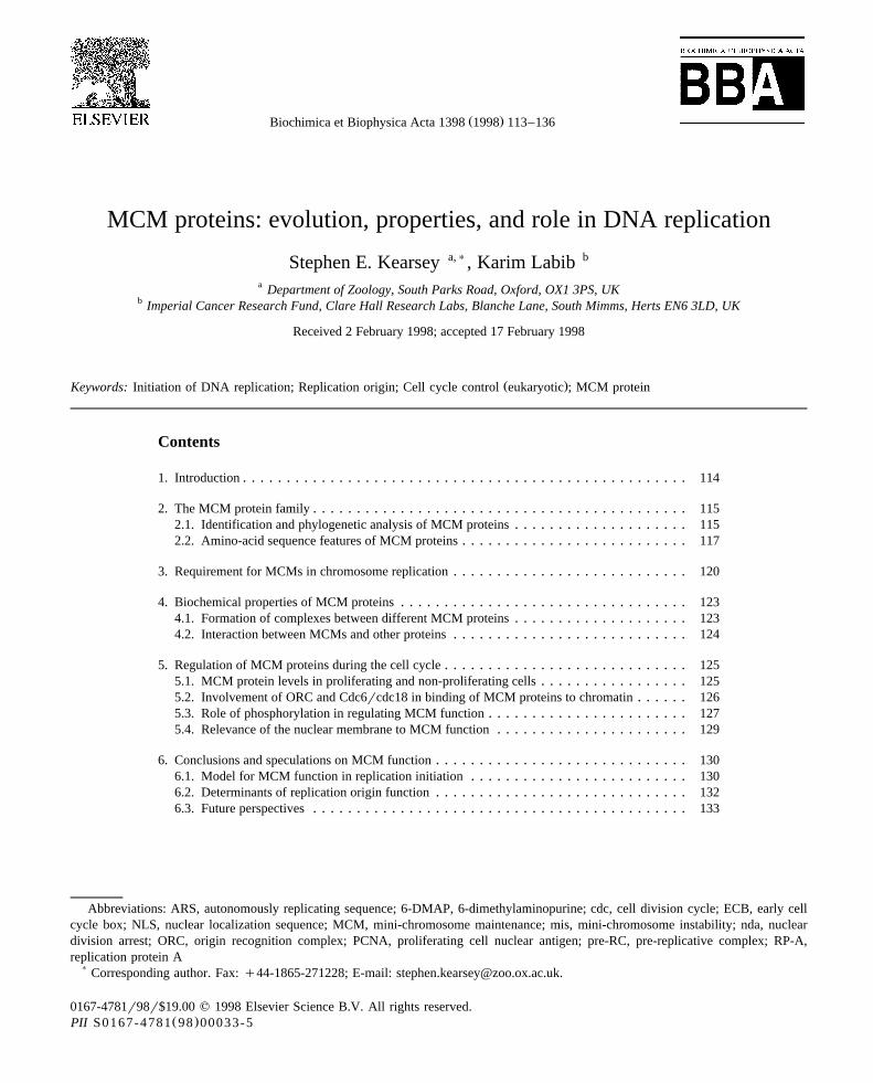
Fig. 1. Phylogenetic tree of MCM protein sequences. The treewas created from a CLUSTALW alignment, and the figure wasgenerated using PHYLODENDRON.
leave the nucleus before mitosis, thereby restrictingDNA replication to one round per cell cycle. A goodcandidate for such an inhibitor would be some formof cyclin-dependent kinase activity, and there is someevidence to suggest that Cdk2rcyclin E may fulfil
w x Žsuch a role in Xenopus egg extracts 77 Section.5.3 .Phylogenetic comparison of eukaryotic MCM se-
quences shows that the six classes of MCM proteinsŽ .are approximately equally related Fig. 1 . However,
we now know that MCM proteins are not confined toeukaryotes. Although they are not found in eubacte-ria, sequencing of the complete genomes of a varietyof Archaea has shown that MCM proteins are also
found in this domain of living organisms. Archaeapossess homologues of most of the key genes in-
w xvolved in eukaryotic DNA replication 78 , implyingthat the eukaryotic mechanism of chromosome repli-cation evolved in a common ancestor of eukaryotesand Archaea. Remarkably, whilst all eukaryotes ap-pear to have six MCM genes, this is not true forArchaea. The Methanococcus jannaschii genomecontains four genes that belong to the MCM familyw x79 , all of which encode proteins that are moreclosely related to each other than to eukaryotic MCMsŽ .Fig. 1 . The MCM genes of Methanobacterium
w xthermoautotrophicum 80 and Archaeoglobusw xfulgidus 81 are of exceptional interest, since both of
these archaeons have just a single MCM. The ar-chaeal MCMs form a sub-class that is marginallycloser to MCM4 than the other eukaryotic MCM
Ž .proteins Fig. 1 , and it is tempting to speculate thatMCM4 may represent the most ancient of eukaryoticMCMs. Whilst it remains to be shown that the pro-teins encoded by these genes are indeed involved inDNA replication, it seems likely that these organismsmay utilize a simplified version of the eukaryoticDNA replication apparatus. These organisms are thuspotentially of great interest as model systems withwhich to study the central elements of initiation. Asignificant drawback is that they are obligate anaer-obes requiring problematic growth conditions, butthey certainly represent a challenge that may havemuch to contribute to our understanding of the mech-anism of eukaryotic DNA replication.
2.2. Amino-acid sequence features of MCM proteins
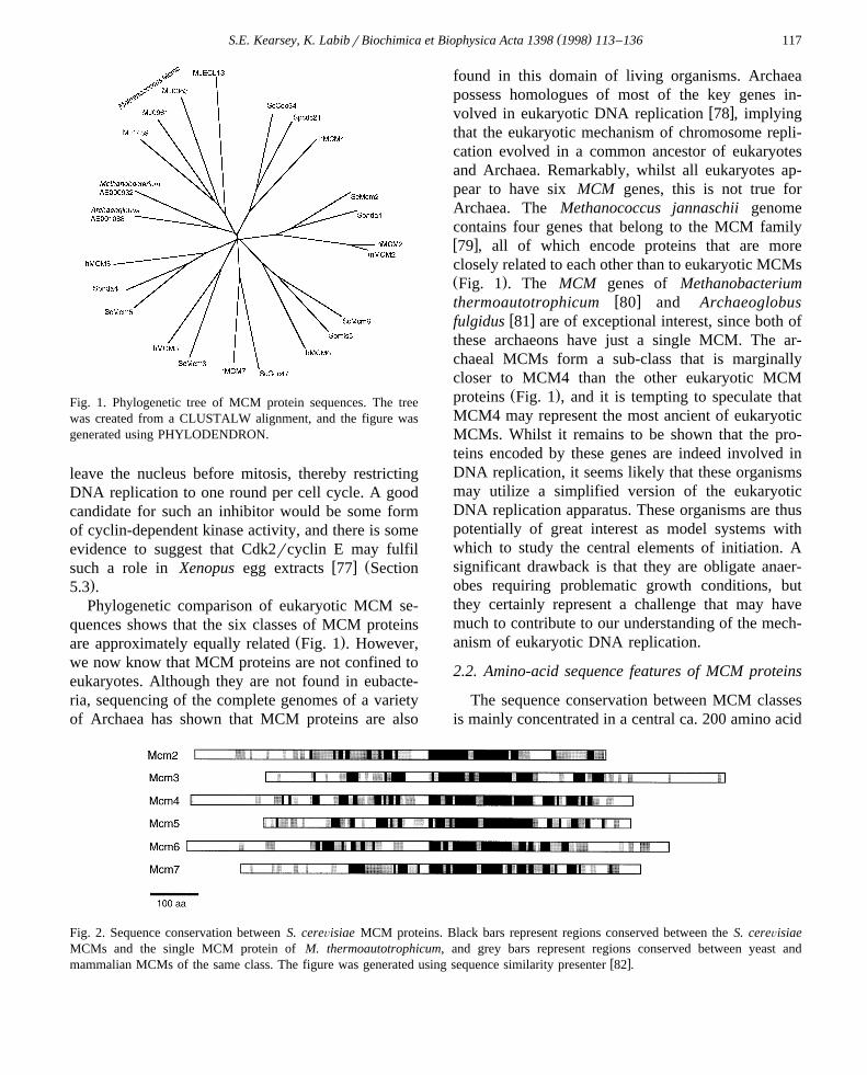
The sequence conservation between MCM classesis mainly concentrated in a central ca. 200 amino acid
Fig. 2. Sequence conservation between S. cereÕisiae MCM proteins. Black bars represent regions conserved between the S. cereÕisiaeMCMs and the single MCM protein of M. thermoautotrophicum, and grey bars represent regions conserved between yeast and
w xmammalian MCMs of the same class. The figure was generated using sequence similarity presenter 82 .

( )S.E. Kearsey, K. LabibrBiochimica et Biophysica Acta 1398 1998 113–136118

( )S.E. Kearsey, K. LabibrBiochimica et Biophysica Acta 1398 1998 113–136 119
Fig. 3. Sequence conservation between S. cereÕisiae and M. thermoautotrophicum MCM proteins. The figure was generated usingw xCLUSTALW 83 and Boxshade 3.2. ‘)’ indicates the cysteines conserved in the zinc-finger motif in MCM2, MCM4, MCM6 and
MCM7 and ‘q’ indicates conserved basic residues which occur as a heptad repeat. The thick line is above the region within the centralŽ .conserved domain or ‘MCM box’ that shows similarity to the NtrC family of transcription factors. In the consensus line, upper-case
letters indicate residues identical in all seven proteins, while lower-case letters indicate residues conserved in a subset of the proteins.
region, while MCMs in the same class show moreŽextensive similarity outside this region Figs. 2 and
.3 . The highly conserved central region contains anelement similar to the A motif of the Walker-typeNTP-binding sequence, though depending on theMCM either alanine or serine replaces the second orthird conserved glycine in the motif GXXGXGKSrT.
This region shows moderate sequence similarity withŽ . w xtwo groups of putative prokaryotic ATPases 84 .
These are the NtrC family of bacterial transcriptionfactors and a set of proteins related to magnesiumchelatases. The homology with NtrC transcriptionfactors is particularly intriguing, since these proteinsbind to specific promoter sites and stimulate the
( )S.E. Kearsey, K. LabibrBiochimica et Biophysica Acta 1398 1998 113–136120
formation of open complexes by the prebound s 54-ŽRNA polymerase complex rather than stimulating
polymerase binding in the conventional manner of.transcription factors , and thereby facilitate the initia-w xtion of transcription 85–87 . It is tempting to imag-
ine an analogous role for MCM proteins in theŽ .initiation step of DNA replication Section 6 .
In addition to the NTP-binding motif, four of theŽ .MCM proteins MCM2, MCM4, MCM6, and MCM7
contain a zinc-finger-type motif of the formCX CX CX C. The functional importance of2 18 –19 2 – 4
this motif has been suggested by mutagenesis studiesw x29 although it does not conform closely to standardDNA-binding zinc motifs. It is possible that it has astructural role or a role in mediating protein–protein
w xinteractions 88,89 . Other sequence motifs that havebeen noted suggest conservation of a-helical struc-ture in the C-terminal region of the MCM proteins;
w x Žthese comprise a conserved heptad repeat 46 Fig.. w x3 and a putative four-helix bundle 32 . Most of the
MCMs show acidic regions, or alternately repeatedclusters of acidic and basic amino acids, which mayhelp to explain the anomalous migration of certainMCMs on SDS-PAGE, which is lower than would beexpected from the predicted amino acid sequencesw x Ž .46 Table 2 .
3. Requirement for MCMs in chromosome repli-cation
The essential requirement for MCM proteins inchromosome replication has been emphasized by anumber of studies. Yeast MCM genes are essentialand certain conditional alleles have been shown toundergo cell cycle arrest in non-permissive condi-tions with predominantly unreplicated DNAw x24,40,73 ; other mcm mutants show a less severedefect in S phase, but incomplete replication or aslowed rate of DNA synthesis can be demonstratedw x24,26,28–30,37,40 . It is likely that MCM proteinsare involved in the initiation step of DNA replication,since alleles of the budding yeast MCM2 and MCM3genes show reduced efficiency in replication origin
w xfunction in two dimensional gel electrophoresis 34 .Furthermore, the plasmid instability phenotype of
Ž . Ž . Ž .cdc46 mcm5 , cdc47 mcm7 and cdc54 mcm4 mu-tants can be suppressed by including multiple tandem
copies of an ARS element in the plasmid, againindicating that the mutants are defective in the initia-
w xtion of DNA replication 5 . The extensive DNAreplication that occurs in many mcm mutants at therestrictive temperature may reflect the fact that Mcmproteins retain residual activity in these mutants,permitting some replication origins to function, butpresumably an insufficient number for completereplication of the genome. Cell cycle arrest in mcmmutants depends on intact checkpoint controlw x29,37,40,97 suggesting that some signal is gener-ated reflecting damaged DNA or incomplete replica-tion. In fact, some mcm mutants show an incomplete
w xblock to mitotic entry 40,73 and there is evidenceŽ .from Aspergillus that the nimQ MCM2 function is
required, probably via initiation of DNA replication,to trigger tyrosine phosphorylation of cdc2 and thus
w xprevent premature entry into mitosis 73 . In mam-malian cells, microinjection of anti-MCM2 antibodiesw x w x60 , anti-MCM3 antibodies 56 or antisense
w xoligomers against MCM7 mRNA 98 blocks DNAreplication. In Drosophila, mutations in the
w x Ž . w xDmMCM2 42 or dpa MCM4 43 genes inhibit cellproliferation and are lethal, with the mutants showingevidence of prolonged S phases.
In Xenopus, the availability of an in vitro systemfor DNA replication has facilitated a biochemicalexamination of the role of MCM proteins. Whendemembranated sperm nuclei are added to an inter-phase egg extract, MCM proteins are rapidly loadedonto chromatin, and this event precedes assembly ofthe nuclear envelope and the commencement of DNA
w xreplication 48–50 . Immunodepletion of MCM pro-teins from the extract before addition of sperm nucleiblocks DNA replication, showing that MCMs are
w xessential for S phase in this system 49,50 . Immun-odepletion using a single anti-MCM antibody candeplete all MCMs from Xenopus extracts, andrestoration of DNA replication only occurs if all theMCMs are added back in approximately equal
w xamounts 46,99 . Nuclei undergo a single round ofreplication in Xenopus extracts treated with cyclo-heximide to prevent the onset of mitosis, but can beinduced to undergo a second complete round ofreplication by permeabilising the nuclear envelope.This allows MCM proteins to become reloaded onto
w xchromatin 48 , and permeabilized G2 phase nucleido not replicate again when added to MCM-depleted

()
S.E.K
earsey,K.L
abibr
Biochim
icaet
Biophysica
Acta
13981998
113–
136121
Table 2Summary of MCM protein properties
MCM2 MCM3 MCM4 MCM5 MCM6 MCM7
Ž .MW kDaŽ . Ž . Ž . Ž .S. cereÕisiae calculated observed 99 120 107 125 105 86 95 113 94.8
Ž . Ž . Ž . Ž .S. pombe calculated observed 92.8 115 100 110 80 80 97Ž . w x Ž . Ž . Ž . Ž . Ž . Ž .Human calculated observed 68 101 125 91 105 97 97 82 90 93 105 81 85
w x w x w x w x w x w xHuman map location 3q21 61 6p12 63 8q12–q13 68 22q13.1–q13.2 90 2q14–q21 68 7q21.3–q22.1 91
Ž )pI S. cereÕisiae 4.95 5.2 6.0 5.6 4.95 5.05
4 5Ž . w xAbundance molecules per cell S. cereÕisiae 4=10 92 S. cereÕisiae 2=10 S. cereÕisiae S. cereÕisiae3 4 4 4w x w x w x w xS. pombe 3=10 92 1.8=10 93 1=10 93 3.0=0 93
6 5Ž w x w xcomplexes also containing Human 10 64 Human 10 94. w xMcm3-7 41
w x w x w x w x w xSubstrate in vitro for protein kinase Cdc7 95,96 Cdc7 95,96 cdc2 53 Cdc7 95 Cdc7 95

( )S.E. Kearsey, K. LabibrBiochimica et Biophysica Acta 1398 1998 113–136122
w xextracts 49 . MCMs are therefore required for theŽreestablishment of replication competence or licens-
.ing once S phase has been completed. It is not yetclear how many other replication proteins show simi-lar properties, or to what extent MCM proteins have aunique role in determining replication competence ofchromosomal DNA. We need to know whether otherproteins such as DNA polymerases also show a peri-odic pattern of chromatin association similar to MCMsand, above all, need to establish the order of events inthe initiation of chromosomal replication, and thedependency of the loading of each protein uponanother.
Analysis of MCM protein localization during thecell cycle suggests that these proteins associate withchromatin before S phase itself and function in theinitiation step of DNA replication, after which theproteins are displaced from chromatin. While initialobservations in S. cereÕisiae suggested that nucleartransport could be relevant to MCM function, subse-quent analysis of MCM proteins in mammalian cellshas shown that the proteins remain in the nucleus
w xduring interphase 54–56,60,63,67,100,101 . How-ever, detergent extraction before fixation reveals twopopulations of MCM proteins, one which is freelyextractable, the other which is bound to a nuclearstructure. Since the bound population shows a similarnuclear distribution to that seen with DNA stains andcan be released by DNase I digestion, the MCMproteins in this fraction are presumably bound to
w xchromatin 55,94,101 . During the G1 phase, MCMproteins are predominantly, but not entirely, associ-ated with chromatin since a small fraction is deter-
w xgent extractable 54,94,100 . During S phase, MCMproteins become increasingly extractable and residualMCM proteins appear to be bound to regions of
Žunreplicated DNA such as late replicating hete-. w xrochromatin 55,56,101 , suggesting that MCM pro-
teins are displaced by ongoing DNA replication. Dur-ing S phase, there is no observable colocalization ofMCM proteins with replication foci or proteins in-volved in the DNA synthesis at the replication fork
Ž .such as proliferating cell nuclear antigen PCNA ,which would have been expected if MCM proteinswere involved in the elongation step of DNA replica-
w xtion 54,56,94,101 . From the end of S phase untilmitosis, MCMs do not appear to be associated withchromatin since practically all the protein is detergent
extractable. During mitosis, MCMs remain excludedfrom condensed chromatin and rebind in telophase,
w xbefore reformation of the nuclear membrane 67,69 .This cell cycle variation in the nuclear binding ofMCMs potentially provides an explanation for earlycell fusion experiments, which demonstrated that G1phase but not G2 phase nuclei could replicate in the
w xenvironment of an S phase cell 102,103 .In Xenopus, similar results have been obtained
regarding MCM-chromatin association. After mitosis,Ž .MCM4 XCdc21 binds to chromatin before PCNA
Ž .or replication protein A RP-A , and before formationw xof the nuclear membrane 52 . When sites of RP-A
Žstaining appear later termed pre-replication centresw x.104 these do not colocalize with MCM4. As inmammalian cells, the chromatin displacement of
w xMCMs occurs in S phase 46,48–52,54 and this canw xbe prevented by aphidicolin 46,48,51,52 , suggesting
that the elongation step of DNA replication is re-quired for displacement of the proteins. MCMs donot show S phase co-localization with sites of ongo-ing DNA replication and are rapidly displaced after
w xthe synchronous start of S phase 51,52,54 , consis-tent with an exclusive role in initiation.
In both budding and fission yeasts, hydroxyureacan be used to block the elongation phase of DNAreplication, arresting cells after early origins of repli-cation have already fired. When temperature-sensitive
Ž . Ž .cdc46 mcm5 and cdc21 mcm4 mutants are inhib-ited with hydroxyurea at the permissive temperature,and then released from the hydroxyurea block at therestrictive temperature for the mutant, S phase is
w xcompleted and cells proceed with mitosis 26,31 .This would again suggest that MCMs function in theinitiation of DNA replication, but are not required forlater stages. However, the situation may not be quiteso straightforward, as MCM2 function appears to berequired after the hydroxyurea block in Aspergillusw x73 , and there is also some evidence that MCMproteins may in fact be associated with replication
w x Ž .forks after initiation 105 Section 5.2 . It remains tobe determined whether this does in fact reflect a rolefor MCM proteins after initiation.
The significance of the change in nuclear localiza-tion of budding yeast MCMs is also unclear at pre-sent. This does not seem to be related to the closedmitosis of fungi, since fission yeast MCMs showconstitutive nuclear localization as in mammalian

( )S.E. Kearsey, K. LabibrBiochimica et Biophysica Acta 1398 1998 113–136 123
w xcells 38,40 . One recent report, based on results fromcellular fractionation, suggests that MCM proteins inS. cereÕisiae may after all remain in the nucleus
w xthroughout the cell cycle as in other eukaryotes 106 ,though in either case the key point is that they show achange in chromatin binding through the cell cyclesimilar to that detected in mammalian cellsw x7,93,105–107 .
4. Biochemical properties of MCM proteins
Insight into the function of MCM proteins hascome from studies of how they interact with otherreplication and cell cycle control proteins. This sec-tion summarizes evidence that MCMs function to-gether as a complex and describes biochemical orgenetic data which suggest interactions with otherproteins. Functional evidence for interactions be-tween ORC, Cdc6rcdc18 and MCMs in effectingchromatin loading is summarized in Section 5.2, andSection 5.3 describes the evidence for regulation byprotein kinases. A summary of all the proteins in-volved is given in Table 3.
4.1. Formation of complexes between different MCMproteins
It is clear from a variety of experiments in differ-ent systems that MCMs associate with one another,perhaps forming a variety of complexes with differ-
ing stoichiometries. A large MCM protein complexof around 450–600 kDa has been detected in extracts
w x w xfrom budding yeast 92 , fission yeast 41 ,w x w xDrosophila 44 , Xenopus 46,53,99 and mammalian
w x55,116 cell extracts. Some studies support the sim-ple notion that this complex represents a hexamer ofMCM proteins, where each MCM type is present inequal stoichiometry. Immunoprecipitation with anti-bodies to a specific MCM protein in Xenopus ex-tracts precipitates all six MCM proteins in approxi-
w xmately equal amounts 46 , and similar results havew xbeen reported for HeLa cell extracts 116,117 . Glyc-
erol gradient centrifugation of mammalian cell ex-tracts also suggests that all six MCM proteins make
w xup the protein complex 55,116 . In fission yeast, a560 kDa MCM complex has been purified, contain-ing approximately equal amounts of each of the sixMCM protein types, again suggesting the proteins
w xmay form a heteromeric hexamer 41 . Ultrastructuralstudies of the purified complex indicate that it has aglobular shape with a central cavity.
High molecular weight MCM complexes tend tobe destabilized by high salt concentrations, oftenbreaking down into a subcomplex composed ofMCM3 and MCM5, and another consisting of three
Žor four of the zinc-finger containing MCMs MCM4,.MCM6, MCM7 or MCM2, MCM4, MCM6, MCM7
w x46,55,64,66,99,117 . MCM2 appears to be easilydisplaced in the MCM2, MCM4, MCM6, MCM7
w xcomplex 66 , contributing to the variable composi-tion of the larger complex.
Table 3Proteins implicated in MCM function
S. cereÕisiae S. pombe Vertebrate Relationship References
Genetic Biochemical Kinase Required forinteraction interaction substrate in chromatin
vitro binding
w xCdc28–Clb1–6 cdc2–cdc13, cig2, cig1 Cdc2–cyclin B U 52,53w xCdc45 U U 24,108–110w xCdc7–Dbf4 hsk1 Cdc7 U U U 95,96,111w xCdc6 cdc18 Cdc6rcdc18 U U 6,7,39,93,105w xHistone H3 U 112w xOrc1 orp1 ORC1 U U U 16,105,113–115
Ž . Ž . w xCdc2 Pol3 pol3 pold-ts1r cdc6 DNA polymerase d U 39large subunit
w xHys2 cdc1 DNA polymerase d U 39small subunit

( )S.E. Kearsey, K. LabibrBiochimica et Biophysica Acta 1398 1998 113–136124
Other reports suggest that MCM proteins mayinteract to form more heterogeneous complexes, con-sisting of tetramers or hexamers with different com-
w xposition 45,92,99 . Indeed, different MCM proteinsŽ .are not equally abundant Table 2 , which is consis-
tent with the possibility that MCMs may not bepresent in equal stoichiometries in complexes. Of
w xparticular interest is the observation by Ishimi 118that a subcomplex of MCM proteins from humancells has an associated DNA helicase or strand dis-placement activity, a property which had already
w xbeen suggested from sequence comparisons 84 . Thepurified MCM4, MCM6, MCM7 complex can dis-place an 18-mer oligonucleotide from ssDNA; onfractionation the activity is associated with fractionswhich have equal amounts of MCM4, MCM6 andMCM7, and no other co-purifying proteins are appar-ent. MCM2 protein weakly associated with this com-plex may possibly inhibit the activity of the helicase.Crosslinking studies suggest that a hexamer, consist-ing of two molecules each of MCM4, MCM6 andMCM7, may have helicase activity and this observa-tion is intriguing given that other helicases, such asSV40 T antigen, function as hexamers. The complexrequires hydrolysable ATP or dATP for activity, isstimulated by nucleic acids and either MCM4 orMCM6 can be affinity labelled with ATP. The rele-vance of this activity to replication remains unclear atpresent. The reported helicase activity is relativelyweak and unprocessive, and cannot displace a 34-meroligonucleotide. These are not the features predictedfor a replicative helicase moving with replicationforks. Other possible roles for such an activity exist,by analogy with current models for the initiation oftranscription. For example, the DNA helicase activityassociated with the transcription initiation factorTFIIH has been suggested to be required to allow thenewly initiated RNA polymerase complex to ‘escape’from the promoter region, without being required forsubsequent elongation of transcripts. The helicaseactivity of TFIIH may help RNA polymerase to breakaway from the extensive protein–protein interactions
w xestablished during initiation 119 . Perhaps a similaractivity is required during the initiation of DNAreplication, and MCMs could be involved in thisprocess? Another caveat to the suggestion of MCMhelicase activity is provided by the observation thatthe purified complex of MCMs from fission yeast,
containing all six proteins in stoichiometricallyequivalent amounts, has no detectable ATPase or
w xhelicase activity 41 . Whilst this may simply reflectconditions used during the purification, it is alsopossible that MCMs do not themselves have helicaseactivity, but are associated with a helicase that waspresent at very low amounts in the preparations iso-lated by Ishimi.
Another issue is whether the nature of MCMprotein complexes changes during the cell cycle; forinstance, do the same MCM complexes exist both onand off chromatin? In mammalian cells, soluble MCMproteins prepared either from interphase or mitoticcells occur in a ca. 600 kDa complex, perhaps con-sisting of one molecule of each of the six proteinsw x116 . In addition, the S phase fraction contains asmaller complex, probably organized as an MCM2,MCM4, MCM6, MCM7 tetramer. Solubilization ofchromatin-bound MCMs using DNase I releases anMCM complex where all six MCMs can be precipi-tated by a single specific anti-MCM antibody, similar
Žto results obtained with soluble detergent-extracta-.ble MCMs. No changes in the fission yeast 560 kDa
complex were detected comparing G2 phase cellsw xwith cells arrested in G1 or S phase 41 . Similarly, in
Drosophila, analysis of MCM complexes in singleearly embryos, where the cell cycles are synchronous,showed MCMs to be always present in 600 kDa
w xcomplexes 45 . Thus the MCM complexes are pre-sumably not sensitive to the cell cycle stage, since theembryos were randomly sampled and therefore indifferent stages of the cell cycle. Using Xenopus eggextracts, again no differences in multiprotein MCMcomplexes could be detected comparing samples taken
w xat different stages during the cell cycle 53 , andXMCM3 and XMCM7 show identical colocalization
w xpatterns on sperm chromatin 54 . Taken together,these observations suggest that putative hexamericMCM complexes may be the predominant form bothon and off chromatin.
4.2. Interaction between MCMs and other proteins
Interactions have been described between MCMsand components of the origin recognition complexŽ .ORC , which was identified by its ability to bind tothe conserved ARS consensus sequence of replication
w xorigins in S. cereÕisiae 1 . ORC binds replication

( )S.E. Kearsey, K. LabibrBiochimica et Biophysica Acta 1398 1998 113–136 125
w xorigins in vivo 120 and six components of theŽ .complex encoded by the ORC1–6 genes have been
w xidentified 2–5,113,121 . Mutations in ORC genescause defects in DNA replication, and reduce the
w xefficiency of initiation at origins 2–5 . The S. pombehomologue of Orc1 has been shown to interact with
Ž .cdc21 MCM4 both genetically and biochemicallyw x114 , and genetic interactions have also been shown
Ž . w xbetween the CDC46 MCM5 and ORC6 genes 113 .In addition to this ORC interaction, both
Ž . Ž .CDC46 MCM5 and CDC47 MCM7 were origi-nally identified as extragenic suppressors of a muta-
w xtion in the CDC45 gene 25 , which encodes anotherw xessential replication factor 24,108–110,122 , and this
interaction was both dominant and allele specific.This interaction is most easily explained if Cdc45physically associates with MCM proteins and indeedthe proteins have subsequently been shown to co-im-
w xmunoprecipitate 108 . The role of Cdc45 in DNAreplication is not known, but it also appears to inter-
w x Žact at least genetically with ORC 109,123 Section.5.3 . MCM proteins are still loaded onto chromatin in
a cdc45 cold-sensitive mutant, suggesting that thelink between Cdc45 and MCMs is not related toMCM chromatin binding, but may instead involve
w xsome other aspect of initiation or elongation 124 .In addition to interacting with ORC and other
initiation factors, which would potentially allow ori-gin-specific association with chromatin, MCMs mayalso associate less specifically with nucleosomes viaan interaction with histones. The tetrameric MCM
Ž .complex MCM2, MCM4, MCM6, MCM7 interactswith histone H3, which could be important for stabi-lizing the interaction between MCM proteins andchromatin, or changing the stability of nucleosomes
w xin the vicinity of replication origins 112 . TheMCM2, MCM4, MCM6, MCM7 tetramer does notinteract with histone H2ArH2B and the dimeric
Ž .MCM subcomplex MCM3, 5 appears to show nointeraction with histones. The stability of the interac-
Ž .tion between MCMs MCM3 and MCM7 and chro-matin in extracted cells has been shown to be in-creased in the presence of ATP or non-hydrolysable
w xanalogues of ATP 117 suggesting that ATP bindingŽby MCMs or other chromatin proteins e.g., compo-
w x.nents of ORC 125 may in some way enhance theinteraction with histones.
Human MCM3 was first described as a protein
w xassociated with DNA polymerase a-primase 62 ,although subsequent analysis failed to demonstrate a
w xdirect interaction between the two proteins 64 .However, genetic interactions have recently beendocumented between subunits of DNA polymerase d
Ž . w xand cdc19 mcm2 in fission yeast 39 .
5. Regulation of MCM proteins during the cellcycle
5.1. MCM protein leÕels in proliferating and non-proliferating cells
In S. cereÕisiae, at least two of the MCM genesŽ Ž . w x Ž . w x.CDC46 MCM5 31 , CDC47 MCM7 33 are pe-riodically transcribed in the cell cycle, during mitosisand early G1 phase, and this control is dependent on
Žan Mcm1-dependent promoter element termed anŽ . . w xearly cell cycle box ECB element 126 . In spite of
this periodic transcription, levels of MCM proteins doŽ w xnot vary with the cell cycle Mcm2 and Mcm3 106 ,
Ž . w x Ž . w x.Cdc46 Mcm5 31 , Cdc47 Mcm7 33 which pre-sumably reflects the stability of these proteins. Sincetranscription of the MCM genes is dependent onMcm1, this potentially could explain the replicationdefect shown by mcm1 mutants. However, periodictranscription of the CDC6 gene also requires a ECBelement, and since overexpression of CDC6 cansuppress the replication defect of an mcm1 mutant itseems likely that the unstable Cdc6 protein links the
w xreplication defect to Mcm1 function 126 .Analysis of MCMs in mammalian cells presents a
similar picture. mRNA levels are low in serum-starvedcells and, on serum addition, peak at the late G1
w xphase 56,62,67,75,100 . A similar peak of late G1phase transcription is seen in cells synchronized bymitotic or S-phase arrest suggesting that MCM tran-
w xscription is periodic in cycling cells 58,63,67,69 .E2F binding sites are found in the promoter regionsof a number of mammalian MCM genes, and adeletion analysis of the human MCM6 gene suggeststhat E2F is partly responsible for promoter activityw x67 . In spite of periodic transcription, levels of mostMCM proteins also appear to remain constant during
w xthe mammalian cell cycle 63,66,69,75,117,127 .More direct analysis of synthesis rates and turnoverof human MCM3 also indicates that this protein is
w xrelatively stable 63 . A comparison of human MCM6
( )S.E. Kearsey, K. LabibrBiochimica et Biophysica Acta 1398 1998 113–136126
protein levels with MCM2 and MCM3 levels insynchronized HeLa cells shows that, in contrast tomost MCMs, MCM6 levels are periodic, peaking
w xduring the late G1 phase 67 .MCM expression has also been compared in quies-
w xcent and proliferating cells. In plants 72,74 andw xDrosophila 42 , MCM mRNA levels go down dra-
matically in differentiated cells. MCM levels arepresent in high amounts during murine spermatogen-esis when meiotic DNA replication is occurring, but
w xare much reduced in pachytene spermatocytes 128 .Other studies on mammalian cells have shown that
w x w xMCM mRNAs 69,71 and protein levels 69,127 arereduced in serum-starved G0 cells or in cells inducedto differentiate. MCM3 and MCM5 appear to be evenless abundant than other MCMs in the G0 state,although the proteins remain predominantly localized
w xin the nucleus 127 . In this connection, antibodiesagainst MCM proteins may be useful reagents forassessing the index of cell proliferation in tissue
w xsamples 129 .Thus de novo synthesis of MCM proteins may be
important in the transition between the G0 to G1phases, but in actively proliferating cells it seemslikely that other mechanisms must be used for regu-lating MCM function. Consistent with this interpreta-tion is the finding that while overexpression of indi-vidual MCM proteins in fission yeast may be delete-rious, major disruption of replication control is not
w xseen 38,40 . Both secondary modification by phos-phorylation and interaction with unstable regulatoryfactors are probably relevant to understanding MCMfunction.
5.2. InÕolÕement of ORC and Cdc6rcdc18 in bind-ing of MCM proteins to chromatin
Purified MCM complexes do not appear to havew xDNA binding ability 41 and other proteins are
needed to allow their interaction with chromatin. InXenopus, immunodepletion of ORC blocks DNA
w xreplication 130 , and prevents the association ofw xMCM proteins with chromatin 115,131 . In contrast,
depletion of MCM proteins has no effect on ORCw xbinding 115 , and treatments with protein kinaseŽ .inhibitors e.g., 6-DMAP that prevent MCM associa-
tion with chromatin have no effect on ORC bindingw x130,131 .
Thus ORC is required for MCM binding, but sinceORC remains bound to chromatin throughout inter-
w xphase 107,115,130,131 , this finding alone does notexplain the periodic binding of MCMs. An additionalfactor, Cdc6rcdc18, may regulate this association.Cdc6rcdc18 is an unstable protein that is expressedin yeast during late mitosisrearly G1 phasew x126,132–134 and is essential for replication initia-
w xtion 135–137 . Cdc6rcdc18 seems to associatew xphysically with ORC 114,136,138 , and shares se-
quence similarity with the largest subunit of ORCw x121 . Analyzing the effects of Cdc6rcdc18 depletionin Xenopus extracts shows that, as with ORC, initia-tion of replication is inhibited, but Cdc6rcdc18 andORC proteins show a different pattern of chromatin
w xassociation 6 . Cdc6rcdc18 binds to chromatin inG1 phase but, unlike the situation with MCMs, thisbinding is not blocked by 6-dimethylaminopurineŽ .6-DMAP . Additionally, Cdc6rcdc18 is displacedfrom chromatin during interphase in a reaction that isnot prevented by aphidicolin, which does stop thedisplacement of most MCM proteins. Thus,Cdc6rcdc18 is apparently displaced either beforereplication, or during very early stages of replicationwhich may not be inhibited by aphidicolin. Depletionof Cdc6rcdc18 does not affect ORC binding, butprevents MCM binding, suggesting that the order ofchromatin association is first ORC, then Cdc6rcdc18
w xand finally MCMs 6 .Genomic footprinting in the vicinity of replication
origins in S. cereÕisiae demonstrates cell cyclechanges which are likely to reflect the functionalinteractions detected in Xenopus extracts. DuringM-G1 phase, an extended region of nuclease protec-
w xtion is seen over replication origins 139 . This struc-Ž .ture, termed the pre-replicative complex pre-RC ,
may indicate that additional proteins have been re-cruited to ORC. During S phase, the pre-RC footprintchanges to the standard post-replicative ORC-depen-dent footprint, in a reaction that requires initiation butnot elongation, suggesting that pre-RCs are necessaryfor the initiation of DNA replication and may signalreplication competence. Cdc6 is required for the for-mation of pre-RCs, and pre-RCs vanish in a condi-tional cdc6 strain shifted to the restrictive tempera-ture, suggesting that continued function of Cdc6 is
w xrequired to maintain pre-RCs 140 . In a chromatin-binding assay for S. cereÕisiae cells, Cdc6 has also

( )S.E. Kearsey, K. LabibrBiochimica et Biophysica Acta 1398 1998 113–136 127
been shown to be necessary for binding of MCMs tochromatin, suggesting that pre-RCs may recruit
w xMCMs 93 . Once MCMs have bound, however,continued function of Cdc6 is not required to main-tain binding of MCMs to chromatin, and both ORCand Cdc6 can be extracted from chromatin in vitro byincreased salt concentrations without removing MCM
w xproteins 93 .Chromatin cross-linking studies have recently pro-
vided direct evidence that MCMs associate withreplication origins in S. cereÕisiae. Reversible chro-matin crosslinking allows immunoprecipitation ofDNA sequences associated with chromatin bindingproteins, and the DNA enriched by this procedure can
w xthen be analyzed by PCR. In one study 7 , Mcm7was shown to be associated with chromatin in thevicinity of DNA sequences known to be functionalorigins, whereas no association was shown with DNAsequences only 2–4 kb away. The association washighest in G1 phase and is Cdc6 dependent, butMCMs were displaced from origin sequences in Sphase and remained unassociated until telophase. Atpresent it is not clear whether MCM binding con-tributes to the pre-replicative footprint observed atorigins, or whether this merely reflects Cdc6 associa-tion. Overexpression of Cdc6 in cells arrested inG2rM phase by nocodozole does not lead to refor-
w xmation of pre-RCs 141 , though Cdc6 can becrosslinked to DNA in the vicinity of origins in these
w xconditions 7 .A second crosslinking study of MCM chromatin
association produced broadly similar results to thoseabove. ORC and Cdc6 were shown to be importantfor the binding of Mcm4 and Mcm7 to origin DNAw x105 , consistent with the findings in Xenopus eggextracts. Intriguingly, it was observed that as S phaseprogresses, MCMs can be crosslinked to DNA furtheraway from origins of replication, suggesting that theymay associate with replication forks after initiation,and a similar conclusion was reached for Cdc45w x105 . This is difficult to reconcile with the results ofMCM localization in vertebrate cells, which arguesagainst a role after initiation, and also with the data
Ž .discussed above Section 3 showing that MCMs inat least some yeast mutants appear not to be requiredafter a hydroxyurea arrest. Whether MCMs really dohave a role after initiation remains to be clearlydemonstrated, and is a key point for our understand-
ing of the role of these proteins during chromosomalreplication.
In both budding and fission yeast, MCM proteinsw xare more abundant than ORC proteins 93 or esti-
w xmates of the total number of replication origins 41,92Ž .Table 2 . In Xenopus, MCMs do not appear to belimiting for replicon size in egg extracts, and eachorigin appears to recruit many MCM proteinsw x142,143 . Thus, there is the potential for large com-plexes of MCM proteins to form on chromatin atsites marked by ORC. Although this question has notbeen experimentally addressed, it is possible thatMCMs may accumulate at individual origins to dif-fering extents and this may affect the subsequentprobability or timing of origin firing, and could evenbe relevant to the location and distribution of initia-
Ž .tion sites in an origin region see Section 6 .
5.3. Role of phosphorylation in regulating MCMfunction
At least some MCM proteins are phosphorylated inw xa cell cycle specific manner. MCM2 94,106 , MCM3
w x w x w x56,69,106 , MCM4 52,53,66 and MCM7 127 havebeen shown to be phosphoproteins. MCM2, MCM3and MCM4 proteins show a similar cell cycle pattern
Žof phosphorylation, becoming dephosphorylated or.hypophosphorylated on exit from mitosis, and being
phosphorylated as cells enter S phase. MCM proteinsbound to chromatin are hypophosphorylated com-pared to the displaced proteins suggesting that phos-phorylation triggers or shortly precedes the displace-ment of MCMs, or alternatively that phosphorylationcan only occur on displaced MCM proteinsw x Ž53,56,66,94,116 . For at least two MCMs MCM2
.and MCM4 , the proteins are phosphorylated first inS phase, but become hyperphosphorylated during mi-
w x Ž .tosis 52,94 see below .In Xenopus, levels of Cdk2-cyclin E appear to be
important for regulating MCM-chromatin bindingw x77 . Although overall levels of Cdk2-cyclin E levelsare similar throughout the embryonic cell cycle, com-partmentalization of the kinase in newly-formed nu-clei gives rise to local concentrations around chro-matin that are much higher than those in the cytosol.Low levels of Cdk2-cyclin E or cyclin A are essentialto allow formation of replication-competent chro-matin, apparently because MCM3 can only associate

( )S.E. Kearsey, K. LabibrBiochimica et Biophysica Acta 1398 1998 113–136128
with chromatin under these conditions, but no effectis seen on ORC binding. Once MCM3 is chromatin
Ž .bound, increasing Cdk2-cyclin E or cyclin A doesnot displace the protein. Thus a window of low Cdk2activity is essential, perhaps because Cdk2-mediatedphosphorylation of MCM proteins themselves, or anecessary loading factor, such as Cdc6rcdc18, pre-
Žvents MCMs from binding to chromatin see Section.5.2 . In a related study, Cdc2-cyclin B has also been
shown to be an inhibitor of a component of replica-w xtion licensing 143 , and in Drosophila, mitotic
Ž .degradation of cyclin A but not cyclin B is requiredw xfor the rebinding of MCMs to chromatin 144 .
In S. cereÕisiae Cdks also seem to have a role incontrolling the association of MCMs with chromatin.
w xThe ability of Mcm7 to associate with origins 7 , orthe ability of origins to form pre-RCs as defined by
w xgenomic footprinting 141 requires expression ofŽ .Cdc6 during a period of low Cdk Cdc28-Clb activ-
ity. Cdk activity could in principle cause this effectby destabilising Cdc6 and thus preventing both itsassociation with chromatin and the subsequent bind-ing of MCMs. Overexpression of cdc18 in fissionyeast perhaps overwhelms this regulatory step, ac-counting for the multiple rounds of replication of the
w xgenome without mitosis 8,9 . The situation in S.cereÕisiae appears to be different, in that overexpres-
w xsion of Cdc6 does not cause rereplication 141 ;under these conditions, Cdc6 can bind to originsduring the G2 phase, i.e., after Cdk has been acti-vated, and still pre-RCs do not form and MCMs are
w xunable to bind 7 . Conceivably, there is some extraregulatory step in S. cereÕisiae that prevents recruit-ment of MCMs to origins in the G2 phase and thusrereplication. Such a control may be abrogated by arecently reported dominant mutation of CDC6, whichallows partial overreplication of the genome and pro-motes constant association of MCM proteins withchromatin through the cell cycle, despite high Cdk
w xactivity 107 . Although the details of replicationcontrol may differ in the two yeasts, it seems reason-able to infer that Cdk activity blocks the binding ofMCMs to origins via an effect on Cdc6rcdc18.
The ability of Cdk activity to prevent the associa-tion of MCMs with chromatin is likely to provide anexplanation for the observation that, during S and G2phases, active kinase must be maintained to preventadditional rounds of DNA replication. Lowering cdc2
kinase activity in S. pombe, using mutations thataffect the regulatory or catalytic subunits of the ki-
w xnase 145–147 , or by overexpression of the cdc2-in-w xhibitor rum1 148,149 can allow cells to undergo
multiple S phases without going through mitosis, andrelated observations have been made in budding yeastw x w x150 , and Drosophila 151 .
MCM4 may be phosphorylated by Cdk2rcdc2,since it contains consensus phosphorylation sites inthe N-terminal region of the protein that are con-served amongst eukaryotes, and there is direct experi-mental evidence that it is a substrate in vitro. Exami-nation of XMCM4 during the Xenopus cell cycleprovides evidence for Cdk phosphorylation. XMCM4is hypo- or unphosphorylated in G1 phase but chro-matin-bound XMCM4 is phosphorylated in S phase
w xbefore it is displaced by replication 52 . The identityof the kinase responsible for this step is uncertain, butcandidates would be Cdk2-cyclin E and Cdc7–Dbf4Ž .see below . Later, hyperphosphorylation of the entireXMCM4 pool occurs in mitosis. This is probablybrought about by the cdc2-cyclin B kinase and invitro phosphorylation of chromatin-bound XMCM4
Žby cdc2-cyclin B kinase can displace this MCM and.probably the whole MCM complex from nuclei iso-
w xlated from cycling Xenopus extracts 53 . The signif-icance of cdc2-mediated MCM displacement is notclear since replication alone can displace XMCM3from chromatin, under conditions where cdc2-cyclin
w xB activation is prevented 52 . Thus, replication wouldnormally have displaced XMCM4 before the mitoticincrease in cdc2-cyclin B activity. Perhaps this mi-totic phosphorylation has a role in preventing thepremature reassociation of MCM proteins with chro-matin? Alternatively, this could reflect part of aprogramme of cdc2 phosphorylation of chromatin-bound proteins during mitosis, that causes their dis-placement and thus aids chromosome condensationw x152 .
While MCM4 is the only MCM which containsconsensus cdc2 kinase sites that are conservedamongst eukaryotes, and for which in vitro phospho-rylation has been shown, there is evidence thatMCM2, MCM3, MCM4 and MCM6 may be phos-phorylated by a distinct kinase, Cdc7. Cdc7 is re-quired for initiation of replication, and functions incombination with a regulatory subunit, Dbf4, whichmay tether the kinase to ORC and stimulate the

( )S.E. Kearsey, K. LabibrBiochimica et Biophysica Acta 1398 1998 113–136 129
w xactivity of the catalytic subunit 153,154 . Cdc7 ki-w xnase is not required for MCM loading 93 and
instead may trigger activation of prebound MCMcomplexes. Evidence for MCMs being the key targetsof Cdc7 is provided by the fact that both the CDC7and DBF4 genes can be deleted without causing
Ž .lethality in cells carrying a particular mcm5 cdc46w xmutation 111 . Although S phase may be slightly
advanced in this suppressor strain, cells do not initi-ate S phase immediately after mitosis, showing thatsome other control must act somewhat redundantlywith Cdc7, with regard to triggering the initiation ofDNA replication. In addition, a dbf4 allele can sup-press an mcm2-1 mutation and there is some evi-dence that Cdc7–Dbf4 physically interacts with
w xMcm2 95 . The possibility that Cdc7 phosphorylatesMCM is supported by the in vitro observations thatyeast Cdc7–Dbf4 can phosphorylate Mcm2, Mcm3,
w xMcm4 and Mcm6 95 and human Cdc7 expressed inŽCOS7 cells perhaps complexed to a homologue of
. w xDbf4 can phosphorylate MCM2 and MCM3 96 .ŽThere is considerable evidence that the cdc2 in
.yeasts or Cdk2 kinase is a major activator of DNAreplication, which operates either independently or inthe same pathway as the Cdc7–Dbf4 switch. In S.
Ž .cereÕisiae, inactivation of the Cdk Cdc28–Clb in-hibitor Sic1 seems to be the last regulating step
w xleading to S phase onset 155 . Similarly in fissionyeast cdc2-cyclin B promotes entry into S phasew x156–158 . In higher eukaryotes, rather than cdc2fulfilling both S and M phase roles, a separate Cdk2kinase is required for S phase entry. Immunodeple-
w xtion of Cdk2 159 or inhibition of its kinase activityby p21 inhibits DNA replication in Xenopusw x160,161 . The key substrates of cdc2rCdk2 forreplication activation have yet to be identified, andthis represents a central problem still to be addressed.
5.4. ReleÕance of the nuclear membrane to MCMfunction
The original candidature of MCMs as licensingfactor has made it natural to consider the role of thenuclear membrane in their regulation. Given theirconstitutive nuclear localization in vertebrate and fis-sion yeast cells, it seems unlikely that periodic accessof MCMs to the nucleus is fundamentally importantin all eukaryotes. However, it could still be important
in embryonic situations, such as in Xenopus eggs,where large maternal stockpiles of replication compo-nents are present. Work in this area has produced
w xconflicting reports. Kubota et al. 46 have reportedthat nuclei assembled in an XMCM3-depleted extractcan only replicate when the XMCM protein complexis added back before nuclear membrane formation,since nuclear formation prevents the accumulation ofXMCM3 in the nuclei. If this result reflects thefailure of MCMs to bind chromatin, rather than aspecific barrier to nuclear entry per se, it could beexplained by accumulation of Cdk2-cyclin E in
Ž .newly-forming nuclei Section 5.3 . Madine et al.w x51 have shown that XMCM3 can cross the nuclearmembrane but cannot bind to G2-phase chromatinunless the membrane is permeabilized. Rather thanallowing access of a licensing factor, permeabiliza-tion of G2 phase-nuclei could allow release of Cdk2-cyclin E, and thus rebinding of MCMs. This explana-tion, though, fails to explain the report that XMCM3can cross the nuclear membrane during interphase
w xand still bind to G1-phase chromatin 49 ; under theseconditions intranuclear accumulation of Cdk2-cyclinE might be expected to prevent binding. Also, if G2phase-nuclei are permeabilized and repaired beforeadding to egg extracts, they do not rereplicate, whichwould have been expected if replication were pre-
w xvented by a diffusible inhibitor 162 . Although vari-ous possible explanations for these conflicting resultscan be envisaged, at present it is not possible todistinguish amongst them based on the available data.
Even if nuclear transport of MCMs is not generallyrelevant to their regulation, it is clear that transport ofsome MCM proteins into the nucleus requires forma-tion of MCM complexes. When individually overex-pressed, MCM4, MCM5, MCM6 and MCM7 arepredominantly cytoplasmic in mammalian cells, incontrast to MCM2 and MCM3 which are nuclearly
w xlocalized 55 . In mammalian cells, overexpression ofboth MCM2 and MCM6 results in nuclear localiza-tion of both proteins, and a similar result was foundfor the combination of MCM3 and MCM5. A nuclear
Ž .localization sequence NLS motif is present in theMCM2 and MCM3 sequences so it seems possiblethat these proteins are able to transport other MCMproteins into the nucleus after formation of com-
Žplexes for instance MCM3 with MCM5, and MCM2.with MCM4,MCM6 and MCM7 . In S. cereÕisiae,

( )S.E. Kearsey, K. LabibrBiochimica et Biophysica Acta 1398 1998 113–136130
the MCM3 NLS has been identified by deletionanalysis, and cells only expressing a version of MCM3
w xwhere the NLS is deleted are inviable 163 .
6. Conclusions and speculations on MCM function
6.1. Model for MCM function in replication initiation
A cartoon summarizing a detailed model for thepossible role of MCM proteins in the stages leadingto formation of a replication fork is shown in Fig. 4.The model breaks down into two basic steps: the firstinvolves the loading of MCM proteins onto chro-matin at replication origins to make them competentfor initiation; the second involves the firing ofMCM-associated origins, which is brought about bythe combined action of Cdc7 and Cdk kinases.
Ž .In the first step Fig. 4b , ORC and Cdc6rcdc18are needed to bring about the association of MCMproteins with chromatin. Cdc6rcdc18 can associatewith origins in the absence of MCMs and may func-tion to adapt origins so that MCMs can bind. Inaddition to the requirement for ORC and Cdc6rcdc18,
Ž .Cdk2 or cdc2 in yeasts activity must be low. SinceMCMs are considerably in excess over ORC in S.cereÕisiae, each origin may attract a large complex ofMCM proteins, which may form a larger target than
the ORC alone for subsequent events leading toreplication initiation. This first step corresponds tothe licensing reaction in Xenopus, and is reflected bythe formation of pre-RCs in S. cereÕisiae.
Ž .In the second step Fig. 4d , replication is initiatedŽ .via activation of the Cdk2 or cdc2 in yeasts and
Cdc7 protein kinases. The Cdc7 kinase associateswith replication origins via its Dbf4 regulatory sub-unit, and its key target may be the MCM complex,given the genetic and biochemical evidence for their
w xinteraction 95,111 . In addition to Cdc7, Cdk2rcdc2kinase activity is responsible for activating replica-tion and also brings a halt to further binding of MCM
Ž .proteins. The important substrate s of this kinase forreplication activation are unknown, but could include
w xORC components 138,164 , DNA polymerase a
w x w x165 , Cdc6rcdc18 166 , as well as MCM proteinsw x52 while the negative role of Cdk2rcdc2 may bemediated through Cdc6rcdc18. In principle, phos-phorylation of a key single factor could be responsi-ble for both functions. Thus for instance, phosphory-lation of Cdc6rcdc18 could activate the initiation ofreplication and subsequent local replication activitycould displace MCMs from the origin. Phosphoryla-tion of Cdc6rcdc18 may trigger its degradation, thuspreventing rebinding of MCMs to origins and block-ing reinitiation. In reality, the control could be farmore involved than this basic mechanism, with the
Fig. 4. Model for MCM function in the initiation of eukaryotic DNA replication. The events shown at origin 1 present an orthodox viewof the steps leading to initiation. We also speculate that origins may differ in their ability to recruit MCMs prior to replication, and this
Ž .may affect for instance the timing of origin firing during S phase compare events at origins 1 and 2 , although there is no direct evidenceŽ .for this aspect of the figure. a Chromatin with ORC bound, but otherwise in a ‘ground’ state, incompetent for replication. May
Ž . Žcorrespond to chromatin in the G0 state in mammalian cells. b Increase in Cdc6rcdc18 levels permits MCM proteins in. Ž .hyposphosphorylated state to bind to chromatin, at sites marked by ORC. c Later stage in G1 phase. Origin 1 is shown to be more
Ž . Ž .efficient in recruiting MCM proteins than origin 2 e.g., due to differences in local chromatin structure . d Start of S phase. Increase inŽ .the activity of Cdc7–Dbf4 kinase and the cdc2 in yeasts or Cdk2 kinase allows initiation of replication at sites on chromatin marked by
MCMs. Initiation involves the association of the Cdc7–Dbf4 kinase at origins; it seems plausible that MCMs are the key substrate ofŽ .Cdc7–Dbf4. The key substrate s of Cdc2rCdk2 have not been identified but these could be Cdc6rcdc18, MCMs, other components of
the pre-RC such as ORC, or factors involved in the elongation step of replication. As replication proceeds, MCM proteins disassociatefrom chromatin at origins probably as a result of elongation of replication forks, although there is one report that MCMs associate with
w xreplication forks 105 . Cdc2rCdk2 phosphorylation also has a negative role in that it blocks reinitiation on already replicated DNA bypreventing the rebinding of MCMs to chromatin. This may be via phosphorylation-induced inactivation of Cdc6rcdc18, but phosphoryla-tion of other proteins, such as MCMs themselves, may also be relevant. Although the details of phosphorylation of individual MCMsprobably differ, the MCMs are here shown to be phosphorylated shortly before displacement from chromatin. For simplicity, theactivation of replication is shown to be triggered by a simultaneous rise in the activity of the two protein kinases, but in reality their
Ž . Ž .activities may not be so coordinated. e Origin 2, marked with less MCM than origin 1, fires later in S phase. f G2 phase. MCMproteins have been displaced from chromatin by replication fork elongation and cannot rebind due to low Cdc6rcdc18 levels and perhapsalso due to cdc2rCdk2 phosphorylation.

( )S.E. Kearsey, K. LabibrBiochimica et Biophysica Acta 1398 1998 113–136 131
phosphorylation of multiple substrates being relevantto replication activation and prevention of rereplica-tion. Multiplicity of substrates for both activation ofreplication and blocking reinitiation could have theadvantage of reducing the probability of inappropri-ate activation of replication origins, which would beexpected to be very deleterious to the cell, even ifoccurring at a very low frequency.
Following this phosphorylation step, local unwind-ing of the DNA may occur in the vicinity of theorigin, analogous to the formation of the ‘open com-
plex’ during the initiation of transcription. This couldbe directly effected by MCM proteins themselves, oralternatively MCMs could use ATP hydrolysis tostimulate open complex formation by neighbouringproteins such as DNA polymerase arprimase, in amanner analogous to the action of NtrC-like tran-
Ž .scription factors in prokaryotes see Section 2.2 .This initial unwinding would then allow the loadingof other factors, and the formation of a replicationfork. At present, this is a particularly obscure part ofthe initiation process. Origin-bound proteins may as-

( )S.E. Kearsey, K. LabibrBiochimica et Biophysica Acta 1398 1998 113–136132
sist this process by interacting with elongation factorsand thus encouraging their binding, or by having achaperonin-like function and aiding the disassemblyof stable protein complexes to allow elongation com-ponents to move from the origin. During the elonga-tion step of DNA replication MCM proteins aredisplaced from chromatin, and in the continuing pres-ence of Cdk2 kinase activity, the MCMs are unableto rebind to chromatin, thus limiting DNA replicationto a single round.
There are similarities between the role of MCMproteins in establishing domains of replication-com-petent chromatin and SIR proteins which establish adomain of chromatin that is transcriptionally re-
Žpressed. Sir proteins probably a polymer of a com-.plex of Sir3 and Sir4 assemble onto chromatin from
ORC via Sir1. The only function of ORC in thisprocess appears to be as an assembly point for Sir1,since Sir1 that is tethered to DNA by fusing it to aDNA-binding domain can also establish a domain of
w xsilenced chromatin 167–169 ; Thus the binding ofSir3 and Sir4 proteins to Sir1–ORC may be analo-gous to the binding of MCM proteins toCdc6rcdc18–ORC. Perhaps the only purpose of ORCand Cdc6rcdc18 in DNA replication is to allowMCM proteins to associate with chromatin, with thekey unwinding event in initiation being carried out byMCMs? In other words, ORC would be acting as aflag, marking out sites on the chromosomes at whichMCMs can associate. If a large zone of bound MCMswere seeded by ORC, conceivably replication activa-tion could occur anywhere in this zone. This interpre-tation might help to explain the pattern of initiationevents seen in metazoan origins of DNA replication.
ŽMetazoan origins show a relatively discrete 0.5–2.kb origin of bidirectional replication where most
initiation events occur, surrounded by a larger regionŽ .6–55 kb which in which some initiation events may
Ž w x.occur for review, see Ref. 170 . However, in bud-ding yeast it has recently been shown that initiation atARS1 occurs in a highly localized region coincidentwith DNase I hypersensitive sites induced by ORC,which is bound immediately adjacent, suggesting thatperhaps ORC binding does indeed specify the site of
w xinitiation 171 . In higher eukaryotes we do not yetknow the sequences that specify ORC binding, ifsuch specific sequences do indeed exist, and therelatively broad zones of initiation observed at meta-
zoan origins could reflect the fact that ORC binds atdiffering sites within such a zone, in different cells ina given population.
6.2. Determinants of replication origin function
In S. cereÕisiae the sequence requirements forautonomous replication have been well characterized,and it is clear that origin function is not assured bythe location in the chromosome of appropriate con-
w xsensus sequences 172–174 or even by ORC bindingw xor pre-RC formation 175 . Perhaps some sites in the
genome assemble ORC, and even Cdc6rcdc18, butdo not assemble MCM complexes and so do notbecome competent for initiation? In addition, originsare subject to other types of control. While someorigins fire every S phase, others may have lowerprobabilities of functioning and initiation events donot occur synchronously at the start of S phase. In S.cereÕisiae, origins seem to function early in S phaseby default, and late replication seems to be a propertyimposed by cis-acting elements, such as telomericsequences, which are distinct from genetic determi-
w xnants required for origin function 176–178 . It ispossible that the stepwise conversion of an ORC-as-sociated origin to a replication-competent complexcould be relevant to understanding some of theseprocesses. Thus, for example one determinant oforigin efficiency could be the ability of MCM pro-teins to associate with origins before S phase and avariety of factors, such as chromatin structure or theproximity of ancillary sequences which improve theefficiency of MCM binding to ORC, could affect thisinteraction. If the amount of MCM recruited by ORCvaries at different origins, this might affect the effi-ciency or timing of replication activation during S
Ž .phase compare origins 1 and 2 in Fig. 4 . There is nodirect evidence for such a quantitative model ofreplication competence, but one recent observationsuggests that ability of the telomere to effect latereplication is imposed not during S phase but be-tween mitosis and START, in other words whenchromatin is converted into a replication-competent
w xstate 179 . Perhaps the efficiency of chromatin bind-ing of MCM proteins at ORC-bound sites could beinhibited by the special chromatin structure propa-gated from the telomere, and MCM-poor origins

( )S.E. Kearsey, K. LabibrBiochimica et Biophysica Acta 1398 1998 113–136 133
might be activated later in S phase than MCM-richones?
6.3. Future perspectiÕes
There are clearly a large number of uncertaintiesconnected with the involvement of MCM proteins inreplication initiation and control. Details of the inter-actions between ORC, Cdc6rcdc18 and MCMs needto be worked out, as do the precise biochemicalcontributions of each of these components to initia-tion. Other genetically identified initiation factors,
w xsuch as Cdc45 and Mcm10 180 , need to be fittedinto the picture, and one very unclear area at themoment concerns how factors involved in the elonga-tion step of DNA replication, such as DNA poly-merases, associate with initiation sites. Concerningthe overall regulation of replication, a fundamentaluncertainty relates to the substrates of Cdc7 andCdk2rcdc2 kinases, that must be phosphorylated forinitiation to occur. In addition, the Cdk-inhibitedsubstrates that are responsible for the block to reiniti-ation remain to be clearly determined. In fissionyeast, perhaps cdc18 alone is inhibited by cdc2 to asignificant level, since overexpression of cdc18 issufficient to induce multiple rounds of S phase in the
w xabsence of mitosis 8,9 . Finally, as suggested in thissummary, a better understanding of the biochemicalevents leading to initiation should help to shed lightnot just upon once per cell cycle replication, but alsoon more subtle processes that determine the timing oforigin firing, and the distribution of initiation eventsin replication origins.
The recent discovery of archaeons with just asingle MCM gene not only points to the antiquity ofthe initiation mechanism used in eukaryotic chromo-somes, but also may provide a system for biochemi-cal analysis of MCM function, stripped of some ofthe regulatory complexity found in eukaryotic cells.Comparing MCM function in Archaea and eukary-otes should give insight into the additional controlmechanisms that eukaryotes have acquired, allowingthe ordered replication of their large genomes.
Acknowledgements
We are grateful to John Diffley for discussions,particularly relating to NtrC function and archaeal
MCMs. Work in SEK’s group is supported by theŽCancer Research Campaign and the EU contract
.ERB-MRX-CT970125 .
References
w x Ž .1 S.P. Bell, B. Stillman, Nature 357 1992 128–134.w x Ž .2 S.P. Bell, R. Kobayashi, B. Stillman, Science 262 1993
1844–1849.w x3 M. Foss, F.J. McNally, P. Laurenson, J. Rine, Science 262
Ž .1993 1838–1844.w x4 G. Micklem, A. Rowley, J. Harwood, K. Nasmyth, J.F.
Ž .Diffley, Nature 366 1993 87–89.w x5 S. Loo, C.A. Fox, J. Rine, R. Kobayashi, B. Stillman, S.
Ž .Bell, Mol. Biol. Cell. 6 1995 741–756.w x6 T.R. Coleman, P.B. Carpenter, W.G. Dunphy, Cell 87
Ž .1996 53–63.w x Ž .7 T. Tanaka, D. Knapp, K. Nasmyth, Cell 90 1997 649–660.w x Ž .8 H. Nishitani, P. Nurse, Cell 83 1995 397–405.w x9 M. Muzifalconi, G.W. Brown, T.J. Kelly, Proc. Natl. Acad.
Ž .Sci. U.S.A. 93 1996 1566–1570.w x10 J.P. Chong, P. Thommes, J.J. Blow, Trends Biochem. Sci.
Ž .21 1996 102–106.w x Ž .11 G.T. Maine, P. Sinha, B.K. Tye, Genetics 106 1984
365–385.w x Ž .12 J. Wuarin, P. Nurse, Cell 85 1996 785–787.w x Ž .13 B.K. Tye, Trends Cell Biol. 4 1994 160–166.w x14 S.E. Kearsey, D. Maiorano, E.C. Holmes, I.T. Todorov,
Ž .Bioessays 18 1996 183–190.w x Ž .15 P. Romanowski, M.A. Madine, Trends Cell Biol. 6 1996
184–188.w x Ž .16 A. Rowles, J.J. Blow, Curr. Opin. Genet. Dev. 7 1997
152–157.w x Ž .17 T. Su, P. Follette, P. O’ Farrell, Cell 81 1995 825–828.w x Ž .18 B. Stillman, Science 274 1996 1659–1664.w x Ž .19 K. Nasmyth, Trends Genet. 12 1996 405–412.w x20 S.E. Kearsey, K. Labib, D. Maiorano, Curr. Opin. Genet.
Ž .Dev. 6 1996 208–214.w x Ž .21 C. Ford, S. Chevalier, Curr. Biol. 5 1995 1009–1012.w x Ž .22 J. Diffley, Genes Dev. 10 1996 2819–2830.w x23 W.M. Toone, B.L. Aerne, B.A. Morgan, L.H. Johnston,
Ž .Annu. Rev. Microbiol. 51 1997 125–149.w x24 K. Hennessy, A. Lee, E. Chen, D. Botstein, Genes Dev. 5
Ž .1991 959–969.w x25 D. Moir, S. Stewart, B. Osmond, D. Botstein, Genetics 100
Ž .1982 547–563.w x Ž .26 K.A. Nasmyth, P. Nurse, Mol. Gen. Genet. 182 1981
119–124.w x27 S. Miyake, N. Okishio, I. Samejima, Y. Hiraoka, T. Toda,
Ž .I. Saitoh, M. Yanagida, Mol. Biol. Cell 4 1993 1003–1015.
w x28 K. Takahashi, H. Yamada, M. Yanagida, Mol. Biol. Cell 5Ž .1994 1145–1158.

( )S.E. Kearsey, K. LabibrBiochimica et Biophysica Acta 1398 1998 113–136134
w x Ž .29 H. Yan, S. Gibson, B.K. Tye, Genes Dev. 5 1991 944–957.
w x30 S. Gibson, R.T. Surosky, B.K. Tye, Mol. Cell Biol. 10Ž .1990 5707–5720.
w x Ž .31 K. Hennessy, C. Clark, D. Botstein, Genes Dev. 4 19902252–2263.
w x32 A. Coxon, K. Maundrell, S. Kearsey, Nucleic Acids Res.Ž .20 1992 5571–5577.
w x33 S. Dalton, L. Whitbread, Proc. Natl. Acad. Sci. U.S.A. 92Ž .1995 2514–2518.
w x Ž .34 H. Yan, A.M. Merchant, B.K. Tye, Genes Dev. 7 19932149–2160.
w x Ž .35 L. Whitbread, S. Dalton, Gene 155 1995 113–117.w x36 Y. Chen, K. Hennessy, D. Botstein, B.K. Tye, Proc. Natl.
Ž .Acad. Sci. U.S.A. 89 1992 10459–10463.w x Ž .37 S.L. Forsburg, P. Nurse, J. Cell Sci. 107 1994 2779–2788.w x38 N. Okishio, Y. Adachi, M. Yanagida, J. Cell Sci. 109
Ž .1996 319–326.w x39 S. Forsburg, D. Sherman, S. Ottilie, J. Yasuda, J. Hodson,
Ž .Genetics 147 1997 1025–1041.w x40 D. Maiorano, G. Blom van Assendelft, S. Kearsey, EMBO
Ž .J. 15 1996 861–872.w x Ž .41 Y. Adachi, J. Usukura, M. Yanagida, Genes Cells 2 1997
467–479.w x42 J.E. Treisman, P.J. Follette, P.H. O’Farrell, G.M. Rubin,
Ž .Genes Dev. 9 1995 1709–1715.w x43 G. Feger, H. Vaessin, T.T. Su, E. Wolff, L.Y. Jan, Y.N.
Ž .Jan, EMBO J. 14 1995 5387–5398.w x Ž .44 T.T. Su, N. Yakubovich, P.H. OFarrell, Gene 192 1997
283–289.w x Ž .45 T.T. Su, G. Feger, P.H. Ofarrell, Mol. Biol. Cell. 7 1996
319–329.w x46 Y. Kubota, S. Mimura, S. Nishimoto, T. Masuda, H.
Ž .Nojima, H. Takisawa, EMBO J. 16 1997 3320–3331.w x47 S. Miyake, I. Saito, H. Kobayashi, S. Yamashita, Gene 175
Ž .1996 71–75.w x48 J. Chong, H. Mahbubani, C. Khoo, J. Blow, Nature 375
Ž .1995 418–421.w x49 M. Madine, C. Khoo, A. Mills, R. Laskey, Nature 375
Ž .1995 421–424.w x50 Y. Kubota, S. Mimura, S. Nishimoto, H. Takisawa, H.
Ž .Nojima, Cell 81 1995 601–609.w x51 M.A. Madine, C.Y. Khoo, A.D. Mills, C. Mushal, R.A.
Ž .Laskey, Curr. Biol. 5 1995 1270–1279.w x Ž .52 M. Coue, S.E. Kearsey, M. Mechali, EMBO J. 15 1996´ ´
1085–1097.w x53 M. Hendrickson, M. Madine, S. Dalton, J. Gautier, Proc.
Ž .Natl. Acad. Sci. U.S.A. 93 1996 12223–12228.w x54 P. Romanowski, M.A. Madine, R.A. Laskey, Proc. Natl.
Ž .Acad. Sci. U.S.A. 93 1996 10189–10194.w x55 H. Kimura, T. Ohtomo, M. Yamaguchi, A. Ishii, K. Sugi-
Ž .moto, Genes Cells 1 1996 977–993.w x Ž .56 H. Kimura, N. Nozaki, K. Sugimoto, EMBO J. 13 1994
4311–4320.w x Ž .57 I. Yoshida, H. Kimura, N. Takagi, Genomics 32 1996
483–484.
w x58 H. Kimura, N. Takizawa, N. Nozaki, K. Sugimoto, NucleicŽ .Acids Res. 21 1995 5289–5293.
w x Ž .59 N. Takizawa, H. Kimura, K. Sugimoto, Gene 167 1995343–344.
w x60 I.T. Todorov, R. Pepperkok, R.N. Philipova, S.E. Kearsey,Ž .W. Ansorge, D. Werner, J. Cell Sci. 107 1994 253–265.
w x61 A. Mincheva, I. Todorov, D. Werner, T.M. Fink, P. Lichter,Ž .Cytogenet. Cell Genet. 65 1994 276–277.
w x62 P. Thommes, R. Fett, B. Schray, R. Burkhart, M. Barnes,¨C. Kennedy, N.C. Brown, R. Knippers, Nucleic Acids Res.
Ž .20 1992 1069–1074.w x63 D. Schulte, R. Burkhart, C. Musahl, B. Hu, C. Schlatterer,
Ž .H. Hameister, R. Knippers, J. Cell Sci. 108 1995 1381–1389.
w x64 R. Burkhart, D. Schulte, D. Hu, C. Musahl, F. Gohring, R.Ž .Knippers, Eur. J. Biochem. 228 1995 431–438.
w x65 B. Hu, R. Burkhart, D. Schulte, C. Masahl, R. Knippers,Ž .Nucleic Acids Res. 21 1993 5289–5293.
w x66 C. Musahl, D. Schulte, R. Burkhart, R. Knippers, Eur. J.Ž .Biochem. 230 1995 1096–1101.
w x67 H. Tsuruga, N. Yabuta, S. Hosoya, K. Tamura, Y. Endo,Ž .H. Nojima, Genes Cell 2 1997 381–399.
w x68 H.P. Holthoff, H. Hameister, R. Knippers, Genomics 37Ž .1996 131–134.
w x69 D. Schulte, A. Richter, R. Burkhart, C. Musahl, R. Knip-Ž .pers, Eur. J. Biochem. 235 1996 144–151.
w x70 S. Bucci, M. Ragghianti, I. Nardi, M. Bellini, G. Mancino,Ž .J.C. Lacroix, Int. J. Dev. Biol. 37 1993 509–517.
w x Ž .71 D. Sykes, M. Weiser, Gene 163 1995 243–247.w x72 P. Springer, W. McCombie, V. Sundaresan, R. Mar-
Ž .tienssen, Science 268 1995 877–880.w x73 X.S. Ye, R.R. Fincher, A. Tang, K.K. McNeal, S.E. Gy-
gax, A.N. Wexler, K.B. Ryan, S.W. James, S.A. Osmani,Ž .J. Biol. Chem. 272 1997 33384–33393.
w x74 P.A. Sabelli, S.R. Burgess, A.K. Kush, M.R. Young, P.R.Ž .Shewry, Mol. Gen. Genet. 252 1996 125–136.
w x75 M. Starborg, E. Brundell, K. Gell, C. Larsson, I. White, B.Ž .Daneholt, C. Hoog, J. Cell Sci. 108 1995 927–934.¨¨
w x Ž .76 J. Blow, R.A. Laskey, Nature 332 1988 546–548.w x Ž .77 X.H. Hua, H. Yan, J. Newport, J. Cell Biol. 137 1997
183–192.w x Ž .78 D. Edgell, W. Doolittle, Cell 89 1997 995–998.w x79 C.J. Bult, O. White, G.J. Olsen, L.X. Zhou, R.D. Fleis-
chmann, G.G. Sutton, J.A. Blake, L.M. Fitzgerald, R.A.Clayton, J.D. Gocayne, A.R. Kerlavage, B.A. Dougherty,J.F. Tomb, M.D. Adams, C.I. Reich, R. Overbeek, E.F.Kirkness, K.G. Weinstock, J.M. Merrick, A. Glodek, J.L.Scott, N.S.M. Geoghagen, J.F. Weidman, J.L. Fuhrmann,D. Nguyen, T.R. Utterback, J.M. Kelley, J.D. Peterson,P.W. Sadow, M.C. Hanna, M.D. Cotton, K.M. Roberts,M.A. Hurst, B.P. Kaine, M. Borodovsky, H.P. Klenk, C.M.Fraser, H.O. Smith, C.R. Woese, J.C. Venter, Science 273Ž .1996 1058–1073.
w x80 D.R. Smith, L.A. DoucetteStamm, C. Deloughery, H.M.Lee, J. Dubois, T. Aldredge, R. Bashirzadeh, D. Blakely,R. Cook, K. Gilbert, D. Harrison, L. Hoang, P. Keagle, W.

( )S.E. Kearsey, K. LabibrBiochimica et Biophysica Acta 1398 1998 113–136 135
Lumm, B. Pothier, D.Y. Qiu, R. Spadafora, R. Vicaire, Y.Wang, J. Wierzbowski, R. Gibson, N. Jiwani, A. Caruso,D. Bush, H. Safer, D. Patwell, S. Prabhakar, S. Mc-Dougall, G. Shimer, A. Goyal, S. Pietrokovski, G.M.Church, C.J. Daniels, J.I. Mao, P. Rice, J. Nolling, J.N.
Ž .Reeve, J. Bacteriol. 179 1997 7135–7155.w x81 H.P. Klenk, R.A. Clayton, J.F. Tomb, O. White, K.E.
Nelson, K.A. Ketchum, R.J. Dodson, M. Gwinn, E.K.Hickey, J.D. Peterson, D.L. Richardson, A.R. Kerlavage,D.E. Graham, N.C. Kyrpides, R.D. Fleischmann, J. Quack-enbush, N.H. Lee, G.G. Sutton, S. Gill, E.F. Kirkness,B.A. Dougherty, K. McKenney, M.D. Adams, B. Loftus,S. Peterson, C.I. Reich, L.K. McNeil, J.H. Badger, A.Glodek, L.X. Zhou, R. Overbeek, J.D. Gocayne, J.F. Wei-dman, L. McDonald, T. Utterback, M.D. Cotton, T. Spriggs,P. Artiach, B.P. Kaine, S.M. Sykes, P.W. Sadow, K.P.DAndrea, C. Bowman, C. Fujii, S.A. Garland, T.M. Ma-son, G.J. Olsen, C.M. Fraser, H.O. Smith, C.R. Woese,
Ž .J.C. Venter, Nature 390 1997 364.w x Ž .82 K. Frohlich, Comput. Appl. Biosci. 10 1994 179–183.¨w x83 J.D. Thompson, D.G. Higgins, T.J. Gibson, Nucleic Acids
Ž .Res. 22 1994 4673–4680.w x Ž .84 E. Koonin, Nucleic Acids Res. 21 1993 2541–2547.w x Ž .85 A. Wedel, S. Kustu, Genes Dev. 9 1995 2042–2052.w x Ž .86 A. North, S. Kustu, J. Mol. Biol. 267 1997 17–36.w x Ž .87 M. Ptashne, A. Gann, Nature 386 1997 569–577.w x88 A. Szabo, R. Korszun, F.U. Hartl, J. Flanagan, EMBO J.
Ž .15 1996 408–417.w x Ž .89 J. MacKay, M. Crossley, Trends Biochem. Sci. 23 1998
1–4.w x90 R. Paul, B. Hu, C. Musahl, H. Hameister, R. Knippers,
Ž .Cytogenet. Cell Genet. 73 1996 317–321.w x91 S. Nakatsuru, K. Sudo, Y. Nakamura, Cytogenet. Cell
Ž .Genet. 68 1995 226–230.w x Ž .92 M. Lei, Y. Kawasaki, B.K. Tye, Mol. Cell Biol. 16 1996
5081–5090.w x93 S. Donovan, H. J, L. Drury, J. Diffley, Proc. Natl. Acad.
Ž .Sci. U.S.A. 94 1997 5611–5616.w x94 I. Todorov, A. Attaran, S. Kearsey, J. Cell Biol. 129
Ž .1995 1433–1446.w x95 M. Lei, Y. Kawasaki, M.R. Young, M. Kihara, A. Sugino,
Ž .B.K. Tye, Genes Dev. 11 1997 3365–3374.w x Ž .96 N. Sato, K. Arai, H. Masai, EMBO J. 16 1997 4340–4351.w x Ž .97 A. Ray, P. Shina, Curr. Genet. 27 1995 95–101.w x98 M. Fujita, T. Kiyono, Y. Hayashi, M. Ishibashi, Biochem.
Ž .Biophys. Res. Commun. 219 1996 604–607.w x99 P. Thommes, Y. Kubota, H. Takisawa, J. Blow, EMBO J.¨
Ž .16 1997 3312–3319.w x100 M. Fujita, T. Kiyono, Y. Hayashi, M. Ishibashi, J. Biol.
Ž .Chem. 271 1996 4349–4354.w x101 T. Krude, C. Musahl, R.A. Laskey, R. Knippers, J. Cell
Ž .Sci. 109 1996 309–318.w x Ž .102 S. Guttes, E. Guttes, J. Cell Biol. 37 1968 761–772.w x Ž .103 P.N. Rao, R.T. Johnson, Nature 225 1970 159–164.w x Ž .104 Y. Adachi, U.K. Laemmli, J. Cell Biol. 119 1992 1–15.w x Ž .105 O. Aparicio, D. Weinstein, S. Bell, Cell 91 1997 59–69.
w x Ž .106 M. Young, B. Tye, Mol. Biol. Cell. 8 1997 1587–1590.w x Ž .107 C. Liang, B. Stillman, Genes Dev. 11 1997 3375–3386.w x108 B. Hopwood, S. Dalton, Proc. Natl. Acad. Sci. U.S.A. 93
Ž .1996 12309–12314.w x Ž .109 L. Zou, J. Mitchell, B. Stillman, Mol. Cell Biol. 17 1997
553–563.w x Ž .110 C.F. Hardy, Mol. Cell Biol. 16 1996 1832–1841.w x111 C. Hardy, O. Dryga, S. Seematter, P. Pahl, R.A. Sclafani,
Ž .Proc. Natl. Acad. Sci. U.S.A. 94 1997 3151–3155.w x112 Y. Ishimi, S. Ichinose, A. Omori, K. Sato, H. Kimura, J.
Ž .Biol. Chem. 271 1996 24115–24122.w x Ž .113 J.J. Li, I. Herskowitz, Science 262 1993 1870–1874.w x Ž .114 B. Grallert, P. Nurse, Genes Dev. 10 1996 2644–2654.w x115 P. Romanowski, M.A. Madine, A. Rowles, J.J. Blow, R.A.
Ž .Laskey, Curr. Biol. 6 1996 1416–1425.w x Ž .116 A. Richter, R. Knippers, Eur. J. Biochem. 247 1997
136–141.w x117 M. Fujita, T. Kiyono, Y. Hayashi, M. Ishibashi, J. Biol.
Ž .Chem. 272 1997 10928–10935.w x Ž .118 Y. Ishimi, J. Biol. Chem. 272 1997 24508–24513.w x119 A. Dvir, R.C. Conaway, J.W. Conaway, Proc. Natl. Acad.
Ž .Sci. U.S.A. 94 1997 9006–9010.w x Ž .120 J.F. Diffley, J.H. Cocker, Nature 357 1992 169–172.w x121 S.P. Bell, J. Mitchell, J. Leber, R. Kobayashi, B. Stillman,
Ž .Cell 83 1995 563–568.w x122 J.C. Owens, C.S. Detweiler, J.J. Li, Proc. Natl. Acad. Sci.
Ž .U.S.A. 94 1997 12521–12526.w x Ž .123 C. Hardy, Gene 187 1997 239–246.w x124 S. Donovan, PhD thesis, University of London, 1997.w x Ž .125 R.D. Klemm, R.J. Austin, S.P. Bell, Cell 88 1997 493–
502.w x126 C. McInerny, J. Partridge, G. Mikesell, D. Creemer, L.
Ž .Breeden, Genes Dev. 11 1997 1277–1288.w x127 H. Tsuruga, N. Yabuta, K. Hashizume, M. Ikeda, Y. Endo,
Ž .H. Nojima, Biochem. Biophys. Res. Commun. 236 1997118–125.
w x Ž .128 Starborg, C. Hoog, Eur. J. Cell Biol. 68 1995 206–210.¨¨w x129 A. Hiraiwa, M. Fujita, T. Nagasaka, A. Adachi, M. Ohashi,
Ž .M. Ishibashi, Int. J. Cancer 74 1997 180–184.w x130 P.B. Carpenter, P.R. Mueller, W.G. Dunphy, Nature 379
Ž .1996 357–360.w x131 A. Rowles, J.P. Chong, L. Brown, M. Howell, G.I. Evan,
Ž .J.J. Blow, Cell 87 1996 287–296.w x Ž .132 S. Piatti, C. Lengauer, K. Nasmyth, EMBO J. 14 1995
3788–3799.w x Ž .133 C.S. Detweiler, J.J. Li, J. Cell Sci. 110 1997 753–763.w x Ž .134 B. Baum, J. Wuarin, P. Nurse, EMBO J. 16 1997 4676–
4688.w x135 T. Kelly, G. Martin, S. Forsburg, R. Stephen, A. Russo, P.
Ž .Nurse, Cell 74 1993 371–382.w x Ž .136 C. Liang, M. Weinreich, B. Stillman, Cell 81 1995
667–676.w x137 E. Hogan, D. Koshland, Proc. Natl. Acad. Sci. U.S.A. 89
Ž .1992 3098–3102.w x Ž .138 J. Leatherwood, G.A. Lopez, P. Russell, Nature 379 1996
360–363.

( )S.E. Kearsey, K. LabibrBiochimica et Biophysica Acta 1398 1998 113–136136
w x139 J.F.X. Diffley, J.H. Cocker, S.J. Dowell, A. Rowley, CellŽ .78 1994 303–316.
w x140 J.H. Cocker, S. Piatti, C. Santocanale, K. Nasmyth, J.F.Ž .Diffley, Nature 379 1996 180–182.
w x141 S. Piatti, T. Bohm, J.H. Cocker, J.F. Diffley, K. Nasmyth,Ž .Genes Dev. 10 1996 1516–1531.
w x Ž .142 J. Walter, J.W. Newport, Science 275 1997 993–995.w x143 H.M. Mahbubani, J.P. Chong, S. Chevalier, P. Thommes,
Ž .J.J. Blow, J. Cell Biol. 136 1997 125–135.w x Ž .144 T.T. Su, P.H. OFarrell, J. Cell Biol. 139 1997 13–21.w x Ž .145 J. Hayles, D. Fisher, A. Wollard, P. Nurse, Cell 78 1994
813–822.w x146 D. Broek, R. Bartlett, K. Crawford, P. Nurse, Nature 349
Ž .1991 388–393.w x Ž .147 K. Labib, S. Moreno, P. Nurse, J. Cell Sci. 108 1995
3285–3294.w x Ž .148 J. Correa Bordes, P. Nurse, Cell 83 1995 1001–1009.w x Ž .149 S. Moreno, P. Nurse, Nature 367 1994 236–242.w x Ž .150 C. Dahmann, J. Diffley, K. Nasmyth, Curr. Biol. 5 1995
1257–1269.w x151 K. Sauer, J.A. Knoblich, H. Richardson, C.F. Lehner,
Ž .Genes Dev. 9 1995 1327–1339.w x Ž .152 S. Moreno, P. Nurse, Cell 61 1990 549–551.w x153 A.L. Jackson, P. Pahl, K. Harrison, J. Rosamond, R.A.
Ž .Sclafani, Mol. Cell Biol. 13 1993 2899–2908.w x Ž .154 S.J. Dowell, P. Romanowski, J. Diffley, Science 265 1994
1243–1246.w x155 E. Schwob, T. Bohm, M.D. Mendenhall, K. Nasmyth, Cell
Ž .79 1994 233–244.w x Ž .156 D. Fisher, P. Nurse, EMBO J. 15 1995 850–860.w x157 C. Martin Castellanos, K. Labib, S. Moreno, EMBO J. 15
Ž .1995 839–849.w x Ž .158 P. Nurse, Y. Bissett, Nature 292 1981 558–560.w x Ž .159 F. Fang, J. Newport, Cell 66 1991 731–742.w x160 U.P. Strausfeld, M. Howell, R. Rempel, J.L. Maller, T.
Ž .Hunt, J.J. Blow, Curr. Biol. 4 1994 876–883.w x Ž .161 H. Yan, J. Newport, J. Cell Biol. 129 1995 1–15.
w x162 D. Coverley, C. Downes, P. Romanowski, R. Laskey, J.Ž .Cell Biol. 122 1993 985–992.
w x163 M.R. Young, K. Suzuki, H. Yan, S. Gibson, B.K. Tye,Ž .Genes Cells 2 1997 631–643.
w x Ž .164 D.A. Wolf, D. Wu, F. McKeon, J. Biol. Chem. 271 199632503–32506.
w x165 C. Voitenleitner, E. Fanning, H. Nasheuer, Oncogene 14,pp. 1611–1615.
w x166 S. Elsasser, F. Lou, B. Wang, J.L. Campbell, A. Jong,Ž .Mol. Biol. Cell 7 1996 1723–1735.
w x Ž .167 C. Chien, S. Buck, R. Sternglanz, D. Shore, Cell 75 1993531–541.
w x Ž .168 T. Triolo, R. Sternglanz, Nature 381 1996 251–253.w x169 C.A. Fox, M.A. Ehrenhofer, S. Loo, J. Rine, Science 276
Ž .1997 1547–1551.w x Ž .170 M. DePamphilis, in: M. DePamphilis Ed. , DNA replica-
tion in eukaryotic cells, Cold Spring Harbor, New York,1997, pp. 45–86.
w x Ž .171 A. Bielinsky, S. Gerbi, Science 279 1998 95–98.w x172 B.J. Brewer, W.L. Fangman, Proc. Natl. Acad. Sci. U.S.A.
Ž .91 1994 3418–34122.w x Ž .173 B.J. Brewer, W.L. Fangman, Science 262 1993 1728–
1731.w x Ž .174 Y. Marahrens, B. Stillman, EMBO J. 13 1994 3395–3400.w x Ž .175 C. Santocanale, J.F. Diffley, EMBO J. 15 1996 6671–
6679.w x Ž .176 B.M. Ferguson, W.L. Fangman, Cell 68 1992 333–339.w x177 B.M. Ferguson, B.J. Brewer, A.E. Reynolds, W.L. Fang-
Ž .man, Cell 65 1991 507–515.w x178 K.L. Friedman, J.D. Diller, B.M. Ferguson, S.V. Nyland,
Ž .B.J. Brewer, W.L. Fangman, Genes Dev. 10 1996 1595–1607.
w x179 M. Raghuraman, B. Brewer, W. Fangman, Science 276Ž .1997 806–809.
w x180 A. Merchant, Y. Kawasaki, Y. Chen, M. Lei, B. Tye, Mol.Ž .Cell Biol. 17 1997 3261–3271.